

Biology Direct 2006, 1:29
Open peer review
This article was reviewed by W. Ford Doolittle, J. Peter Gogarten, and Arcady Mushegian.
For the full reviews, please go to the Reviewers' comments section.
Background
The extraordinary diversity of viruses
Viruses are ubiquitous companions of cellular life forms: it appears that every cellular organism studied has its own viruses or, at least, virus-like selfish genetic elements [1]. Recent environmental studies have shown that viruses, primarily, bacteriophages, are "most abundant biological entities on the planet" [2], with the total number of virus particles exceeding the number of cells by at least an order of magnitude [3,4]. Viruses actively move between biomes and are thought to be major agents of evolution by virtue of their capacity to operate as vehicles of horizontal gene transfer (HGT) [5].
A remarkable feature of viruses is the diversity of their genetic cycles, in a sharp contrast to the uniformity of the cellular genetic cycle [6-9] (Fig. 1). Viruses with different genome strategies span a vast range of genome sizes (the genomes of the largest known virus, the mimivirus, and the smallest viruses, e.g., circoviruses, differ by three orders of magnitude) and show a non-uniform and nontrivial distribution among the host taxa (Fig. 1). For example, the extraordinary diversity of double-stranded (ds) DNA bacteriophages is in a stark contrast to the absence of bona fide dsDNA viruses in plants. Conversely, RNA viruses are extremely abundant and diverse in plants and animals but are currently represented by only two compact families in bacteria, and so far have not been detected in archaea (Fig. 1).
Given the variety of genetic strategies, genome complexity, and global ecology of viruses, the problem of virus evolution inevitably digresses into a web of interlocking questions. What qualifies as a virus? Are viruses as a whole monophyletic, i.e., ultimately descend from a single primordial virus or polyphyletic, i.e., have multiple origins? If viruses are polyphyletic, how many independent lineages are there? What is the age distribution of different groups of viruses – are they ancient or have they been emerging continuously throughout life's evolution? And, perhaps, the most fundamental questions of all: what is the ultimate origin of viruses and what are the relationships between evolution of viruses and cellular life forms? The recent advances of comparative genomics create the unprecedented opportunity to start tackling these issues by inferring some of the likely answers from sequence and structure data analysis. Here, we address several of these questions but, primarily, the last two, most general ones,
http://www.biology-direct.com/content/1/1/29
in an attempt to outline a coherent scenario of virus origin and evolution and delineate the connections between the evolution of viruses and cellular life forms.
Results and discussion
Polyphyly versus monophyly in virus evolution
Comparative genomics provides no evidence of a monophyletic origin of all viruses. Many virus groups simply share no common genes, effectively, ruling out any conventional notion of common origin. When applied to viruses, the notion of "common genes" is not a simple one because commonality is not necessarily limited to clear-cut orthologous relationships between genes that translate into highly significant sequence similarity. Instead, as discussed in the next sections, distant homologous relationships among viral proteins and between viral proteins and their homologs from cellular life forms could convey more complex but important messages on evolution of viruses. This complexity notwithstanding, cases of major virus groups abound that either share no homologous genes under any definition or have in common only distantly related domains with obviously distinct evolutionary trajectories. For example, most of the viruses of hyperthermophilic crenarchaea have literally no genes in common with any other viruses [10,11], whereas RNA viruses share with DNA viruses and plasmids that replicate via the rolling circle mechanism only extremely distant domains in their respective replication proteins.
By contrast, monophyly of several large classes of viruses, including vast assemblages of RNA viruses and complex DNA viruses, can be demonstrated with confidence (Table 1). Some of these monophyletic classes of viruses cross the boundaries set by genome strategies: thus, the retroid class includes both RNA viruses and viruses, mobile elements, and plasmids with DNA genomes, and the rolling circle replication (RCR) class combines ssDNA and dsDNA viruses and plasmids. Furthermore, based on similarities in the structure of RNA replication complexes, along with the presence of homologous, even if distant, replication enzymes, it has been suggested that positive-strand RNA viruses, double-stranded RNA viruses, and retroid elements all have a common origin [9]. On the whole, however, the conclusion seems inevitable that viruses comprise several distinct lines of descent (Table 1).
A brief natural history of viral genes
Sequence analysis of viral proteins reveals several categories of virus genes that markedly differ in their provenance. The optimal granularity of classification might be subject to debate but at least 5 classes that can be assorted into three larger categories seem to be readily distinguishable.
Genes with readily detectable homologs in cellular life forms
Page 2 of 27
(page number not for citation purposes)
Biology Direct 2006, 1:29 |
http://www.biology-direct.com/content/1/1/29 |
|
Class |
|
|
|
|
|
Replication cycle |
|
|
|
Host range |
||||||||||||||||
|
Positive- |
|
RdRp |
|
|
+ |
|
T |
JRC |
|
|
|
|||||||||||||||
|
|
|
|
|
|
|
|
||||||||||||||||||||
|
|
T |
|
|
|
|
|
|
|
|
|
|
|
|
|
|
|
|
|
||||||||
|
strand |
|
|
|
|
|
|
|
|
|
R |
|
|
|
|
|
|
|
|
P |
|
||||||
|
|
|
|
|
|
|
|
|
|
|
|
R, E |
|
|
|
|
|||||||||||
|
RNA |
+ |
|
R |
|
|
|
|
- |
|
|
+ |
|
|
|
|
|
||||||||||
|
3-30 kb |
|
|
|
|
|
|
|
|
|
|
|
|
|
|
|
|
UE |
Mz |
||||||||
|
|
|
|
|
|
|
|
|
|
|
|
|
|
|
|
|
|
||||||||||
|
Double- |
|
|
|
|
RdRp |
|
|
|
|
|
|
|
|
|
JRC |
|
|
|
|
|
|
|||||
|
|
|
|
|
|
|
|
|
|
|
|
|
|
|
|
|
|
|
|
||||||||
|
strand |
|
|
|
|
|
|
|
|
|
|
T |
|
|
|
|
|
|
|
|
|
|
|
P |
|
|
|
|
RNA |
|
|
|
R |
|
|
|
R, E |
± |
|
|
|
|
|
||||||||||||
|
± |
|
+ |
|
|
|
|
|
UE |
|
|||||||||||||||||
|
4-25 kb |
|
|
|
|
|
|
|
|
|
|
|
|
|
|
|
|
|
F |
B |
Mz |
||||||
|
|
|
|
|
|
|
|
|
|
|
|
|
|
|
|
|
|
|
|||||||||
|
Negative- |
|
|
+ |
|
|
T |
|
RdRp |
|
|
|
|
|
|
|
|
|
|
||||||||
|
|
|
|
|
|
|
|
|
|
|
|
|
P |
|
|
||||||||||||
|
strand |
|
|
|
|
|
|
|
|
|
|
|
|
|
|
|
|
||||||||||
|
|
Tr |
|
|
|
|
|
|
|
|
|
|
|
|
|
|
|
|
|
|
|
|
|||||
|
RNA |
- |
|
|
R |
|
|
|
+ |
|
|
R, E |
|
|
|
|
|
|
|
|
|||||||
|
11-20 kb |
|
|
|
|
|
|
|
|
|
|
|
|
|
|
|
|
|
|
|
|
|
Mz |
||||
|
|
|
|
|
|
|
|
|
|
|
|
|
+ |
|
|
T |
RT |
|
|
|
|
|
|
||||
|
|
|
|
|
|
|
|
|
|
|
|
|
|
|
|
|
|
|
|
|
|||||||
|
Retroid |
|
|
|
|
|
|
|
|
Tr |
|
|
|
|
|
|
|
|
|
|
|
|
|
|
|||
|
RNA |
|
|
|
|
|
|
|
|
|
|
|
Tr, E |
|
|
|
|
|
|
||||||||
|
|
RT |
|
|
|
|
+ |
|
|
|
|
Mz |
|||||||||||||||
|
7-12 kb |
+ |
± |
|
|
|
|
|
|
|
|
|
|
|
|
F |
|
||||||||||
Retroid DNA |
|
|
|
|
|
|
|
|
|
|
T |
|
|
RT |
|
|
|
|
|
|
|
|
|
|
|||
|
|
|
|
|
|
|
|
|
|
|
|
|
|
|
|
|
|
|
|
|
|
||||||
|
|
Tr + |
|
|
|
|
|
|
|
|
|
|
P |
UE |
|
||||||||||||
|
viruses, |
|
|
|
|
|
|
|
|
|
|
|
|
|
|
||||||||||||
|
|
|
|
|
|
|
|
|
E, RT |
|
|
|
|
|
|||||||||||||
|
|
|
|
|
|
|
|
|
± |
|
|
|
A |
|
|||||||||||||
|
elements |
|
|
|
|
|
|
Tr |
|
|
|
|
|
|
|
|
|
|
|
|
|
|
|||||
|
± |
|
|
|
|
|
|
+ |
|
|
|
|
|
|
|
|
|
Mz |
|||||||||
|
2-10 kb |
|
|
|
|
|
|
|
|
|
|
|
|
|
|
|
|
|
F |
|
|||||||
|
|
|
|
|
|
|
|
|
|
|
|
|
|
|
|
|
|
|
|
|
|
|
|
|
|||
|
ssDNA |
|
|
|
|
|
|
|
|
|
|
|
+ |
|
T |
|
|
|
|
|
|
||||||
|
|
|
|
|
|
|
|
|
|
|
|
|
|
|
|
|
|
|
|||||||||
|
|
|
|
|
|
|
|
|
|
|
|
|
|
|
|
|
|
|
|
|
|
|
|||||
|
viruses, |
|
|
|
|
|
|
|
|
|
|
Tr |
|
|
|
|
RCE S3H |
P |
|
|
|||||||
|
plasmids |
|
|
RCR |
|
|
|
|
RCR, E |
|
|
|
|
|
Mz |
||||||||||||
|
2-11 kb |
+ |
|
± |
|
|
|
|
|
|
|
||||||||||||||||
|
|
|
|
|
|
|
|
|
|
|
|
|
|
|
|
|
|
|
|
|
|
||||||
|
|
|
|
|
|
|
|
|
|
|
|
|
|
|
|
|
+ |
|
|
|
A |
||||||
|
|
|
|
|
|
|
|
|
|
|
|
|
|
|
|
|
|
|
|
|
|
||||||
|
|
|
|
|
|
|
|
|
|
|
|
|
|
|
|
|
|
|
|
|
|
|
|||||
|
|
|
|
|
|
|
|
|
|
|
|
|
|
|
|
|
|
|
|
|
|
|
|
|
|
||
|
|
|
|
|
|
|
|
|
T |
|
|
|
|
|
|
|
|
|
|
|
|
|
|
|
|
||
|
dsDNA |
|
Tr |
+ |
|
|
|
|
|
|
|
|
|
|
|
|
|
Pr-Pol |
|
|
|
|
|
|
|||
|
viruses, |
|
|
|
|
|
|
|
|
|
|
|
|
|
|
|
|
|
|
|
|
|
|
|
|
||
|
|
|
|
|
|
|
|
|
|
|
|
|
|
|
|
|
|
|
|
|
|
|
|
|
|
||
|
plasmids |
|
|
|
|
|
|
|
|
|
R, E |
|
|
|
|
|
|
|
|
|
|
|
|
|
|
||
|
5-1,200 kb |
± |
|
|
|
|
|
|
|
|
|
|
|
|
|
± |
|
|
|
A |
UE |
Mz |
|||||
|
|
|
|
|
|
|
|
|
|
|
|
|
|
|
|
|
|
|
|||||||||
|
|
|
|
|
|
|
|
|
|
|
|
|
|
|
|
|
|
|
|
|
|
|
|
||||
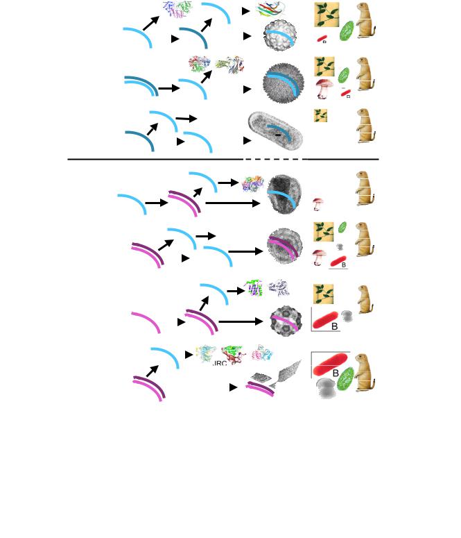
Figure 1
Viruses and other selfish elements: the replication strategies, genome size distribution, global ecology, and hallmark proteins. For each class of viruses and related elements, the approximate range of genome sizes is indicated (kb, kilobases). '+' denotes positive strand (same polarity as mRNA) and '-' denotes negative strand. Tr, transcription; T, translation; R, replication; E, encapsidation; A, archaea; B, bacteria; F, fungi; Mz, Metazoa; P, plants; UE, unicellular eukaryotes. For each class of viruses (elements), characteristic structures of hallmark proteins and characteristic electron-microscopic images of viruses are shown. RdRp, RNA-dependent RNA polymerase; JRC, jelly-roll capsid protein; RT, reverse transcriptase; RCRE, rolling-circle replication (initiating) endonuclease. The rightmost panel shows the host range, with the size of the respective image and acronym roughly proportionate to the abundance of the given virus class in the respective taxon.
Page 3 of 27
(page number not for citation purposes)

Biology Direct 2006, 1:29 |
|
|
http://www.biology-direct.com/content/1/1/29 |
|
Table 1: The major monophyletic classes of viruses and selfish genetic elements |
|
|
||
|
|
|
|
|
Class of viruses |
Constituent virus lineages |
Hosts |
Support for monophyly |
Refs |
|
|
|
|
|
Positive-strand RNA |
Superfamily I: picorna-like; |
Animals, plants, protists, |
Conserved RdRp; JRC in |
[87] |
viruses |
superfamily II: alpha-like; |
bacteria (one family of |
most superfamily 1 viruses, |
|
|
superfamily III: flavi-like; the |
bacteriophages) |
and subsets of |
|
|
exact affinity of RNA |
|
superfamilies 2 and 3 |
|
|
bacteriophages within this class |
|
viruses. Reconstructed |
|
|
of viruses remains uncertain |
|
ancestor with RdRp and |
|
|
(possibly, a fourth lineage) |
|
JRC |
|
Retroid viruses and |
Retroviruses, hepadnaviruses, |
Animals, fungi, plants, |
Conserved RT |
[103, 104] |
elements |
caulimoviruses, badnaviruses; |
protists, bacteria, archaea |
|
|
|
LTRand nonLTR retroelements; |
|
|
|
|
retrons; group II self-splicing |
|
|
|
|
introns – the progenitors of |
|
|
|
|
eukaryotic spliceosomal introns |
|
|
|
Small DNA viruses, |
Gemini-, circo-, parvo-, |
Animals, plants, archaea, |
Conserved RCRE, JRC, |
[17, 18, 20] |
plasmids, and transposons |
papovaviruses, phages (e.g., |
bacteria |
S3H (in eukaryotic viruses) |
|
with rolling circle |
φX174), archaeal and bacterial |
|
|
|
replication |
plasmids, eukaryotic helitron |
|
|
|
|
transposons |
|
|
|
Tailed bacteriophages |
Families: Myoviridae (e.g., T4), |
Bacteria, euryarchaea |
Complex, overlapping |
[11, 93, 94, 105, 106] |
(Caudovirales) |
Podoviridae (e.g., T7), |
|
arrays of genes conserved |
|
|
Siphoviridae (e.g., λ) |
|
in subsets of tailed phages; |
|
|
|
|
genes of all tailed phages |
|
|
|
|
thought to comprise a |
|
|
|
|
single pool |
|
Nucleo-cytoplasmic large |
Poxviruses, asfarviruses, |
Animals, algae, protests |
Core set of 11 conserved |
[50–53, 107] |
DNA viruses (NCLDV) |
iridoviruses, phycodnaviruses, |
|
genes, including JRC, S3H, |
|
|
mimiviruses |
|
and a FtsK-like packaging |
|
|
|
|
ATPase, found in all |
|
|
|
|
NCLDVs; reconstructed |
|
|
|
|
ancestor with ~40 genes |
|
Abbreviations: JRC, Jelly-Roll Capsid protein; LTR, Long Terminal Repeat; RdRp, RNA-dependent RNA polymerase; RCRE, Rolling Circle Replication (initiation) Endonuclease; RT, Reverse Transcriptase; S3H, Superfamily 3 Helicase.
1.Genes with closely related homologs in cellular organisms (typically, the host of the given virus) present in a narrow group of viruses.
2.Genes that are conserved within a major group of viruses or even several groups and have relatively distant cellular homologs.
Virus-specific genes
3.ORFans, i.e., genes without detectable homologs except, possibly, in closely related viruses.
4.Virus-specific genes that are conserved in a (relatively) broad group of viruses but have no detectable homologs in cellular life forms.
Viral hallmark genes
5. Genes shared by many diverse groups of viruses, with only distant homologs in cellular organisms, and with strong indications of monophyly of all viral members of the respective gene families, – we would like to coin the
phrase "viral hallmark genes" to denote these genes that can be viewed as distinguishing characters of the "virus state".
The relative contributions of each of these classes of genes to the gene sets of different viruses strongly depend on the viral genome size which differs by more than three orders of magnitude. Viruses with small genomes, such as most of the RNA viruses, often have only a few genes, the majority of which belong to the hallmark class. By contrast, in viruses with large genomes, e.g., poxviruses, all 5 classes are broadly represented. In order to illustrate the diversity of viral "genomescapes" more concretely, we show in Table 2 the decomposition of the gene sets of three viruses with a small, an intermediate-sized, and a large genomes, respectively, into the 5 classes. Notably, moderate-sized and large genomes of bacteriophages and archaeal viruses are dominated by ORFans that often comprise >80% of the genes in these viruses. Rapidly evolving phage ORFans are thought to supply many, if not most, of the ORFans found in prokaryotic genomes (the lack of detectable sequence conservation notwithstanding), hence playing an important role in evolution of prokaryotes [12].
Page 4 of 27
(page number not for citation purposes)

Biology Direct 2006, 1:29 |
|
|
http://www.biology-direct.com/content/1/1/29 |
|||
Table 2: Representation of the five classes of viral genes in three selected viruses with small, medium-size and large genomes |
||||||
|
|
|
||||
|
Genome size |
Representation of the 5 classes of viral genes (number and brief description) |
||||
|
(kb)/number of |
|
|
|
|
|
|
annotated genes |
|
|
|
|
|
|
|
|
|
|
|
|
|
|
1. Recent |
2. Ancient |
3. ORFans |
4. Conserved |
5. Hallmark |
|
|
acquisitions |
acquisitions |
|
virus-specific |
genes |
|
|
from cells |
from cells |
|
genes |
|
|
|
|
|
|
|
|
Virus |
|
|
|
|
|
|
Poliovirusa |
7.4/10 |
None |
2: a duplication of |
1: uncharacterized |
1: genome-linked |
6: 4 diverged |
|
|
|
a chymotrypsin- |
protein (3A) |
protein (VPg) |
copies of JRC |
|
|
|
like protease (3C, |
|
|
(VP1-4), S3H (3C), |
|
|
|
2A) |
|
|
RdRp (3D) |
Sulfolobus |
17.6/36 |
3: two predicted |
5: four predicted |
26: |
None |
2: JRC, packaging |
turreted |
|
transcription |
transcription |
uncharacterized |
|
ATPase |
icosahedral |
|
regulators and an |
regulators and an |
proteins |
|
|
virus (STIV)b |
|
uncharacterized |
uncharacterized |
|
|
|
|
|
protein |
protein |
|
|
|
Vaccinia virusc |
194.7/~200 |
~48: primarily, |
~36: primarily, |
~24: poorly |
~84: primarily, |
5: JRC, S3H/ |
|
|
proteins involved |
proteins involved |
characterized |
structural |
primase, packaging |
|
|
in virus-host |
in genome |
proteins, possibly, |
components of the |
ATPase, DNA |
|
|
interaction |
replication and |
involved in virus- |
virion and some |
polymerase(?) |
|
|
|
expression |
host interactions |
proteins involved |
|
|
|
|
|
|
in genome |
|
|
|
|
|
|
expression |
|
aThe classification is based on the analysis described in [87]. bThe classification is based on the analysis described in [11].
cThe classification is based on the analysis described in [108] and EVK, unpublished observations; the uncertainty in the number of genes is due to the pseudogenization of varying subsets of genes in different strains of vaccinia virus.
The evolutionary origins of the 5 classes of viral genes are likely to be very different. The least controversial are the two classes of genes with readily detectable homologs from cellular life forms that appear to represent, respectively, relatively recent (class 1) and ancient (class 2) acquisitions from the genomes of the cellular hosts. Where do virus-specific genes come from is a much harder question. In the absence of direct evidence, the default hypothesis, probably, should be that these genes evolved from cellular genes as a result of dramatic acceleration of evolution linked to the emergence of new, virus-specific functions, such that all traces of the ancestral relationships are obliterated. This notion is compatible with the fact that many, probably, most class 4 genes (virus-specific genes conserved within a group of viruses) are virion components (e.g., see the vaccinia virus case in Table 2), a quintessential viral function. The hallmark genes that cross the barriers between extremely diverse virus lineages seem to be of the greatest interest and relevance for the problem of the ultimate origins of viruses, at least, in the context of the long argument we attempt to develop here. Thus, we discuss the distribution among viruses, evolution and significance of these genes in a separate section.
Viral hallmark genes: beacons of the ancient virus world
Although there are no traceable vertical relationships between large groups of viruses outside the major classes listed in Table 1, a considerable number of genes that
encode proteins with key roles in genome replication, expression, and encapsidation are shared by overlapping arrays of seemingly unrelated groups of viruses. As already noted above, some of these widespread viral genes are genuine viral hallmarks that are found in a variety of diverse viruses (although never in all viruses) but not in cellular life forms except as easily recognizable proviruses or mobile elements (Table 3, Fig. 1). The two proteins that are most widely dispersed among viruses are the jelly-roll capsid protein (JRC) [13-15] and the superfamily 3 helicase (S3H) [16]. Each of these proteins crosses the boundary between RNA and DNA viruses and spans an astonishing range of virus groups, from some of the smallest positive-strand RNA viruses to the nucleo-cytoplasmic large DNA viruses (NCLDV), the class of viruses that includes the giant mimivirus (Table 3). Other proteins listed in Table 3 are not as common as JRC or S3H but still form multiple, unexpected connections between groups of viruses that otherwise appear to be unrelated. A case in point is the rolling circle replication (RCR) initiation endonuclease (RCRE) which unites a great variety of small ss and dsDNA replicons, including viruses, plasmids, and transposable elements that reproduce in animals, plants, bacteria, and archaea [17-20]. Notably, a more recent and more careful analysis has shown that the DNA-binding domain of the replication protein of polyoma and papilloma viruses (e.g., the T antigen of SV40) is a derived form of the RCRE that has lost the catalytic amino acid residues
Page 5 of 27
(page number not for citation purposes)
Biology Direct 2006, 1:29 |
http://www.biology-direct.com/content/1/1/29 |
[18]. Thus, through this detailed analysis of one of the hallmark proteins, the well-known connection between a variety of small ssDNA-replicons is extended to a group of similar-sized dsDNA-replicons. A similar expansion of the set of viral groups covered by a particular hallmark gene resulted from the detailed analyses of the packaging ATPase and the archaeo-eukaryotic primase (Table 3).
Replication of positive-strand RNA viruses, dsRNA viruses, negative-strand RNA viruses, and retroid viruses/ elements is catalyzed by another idiosyncratic class of viral enzymes, RNA-dependent RNA polymerases (RdRp) and reverse transcriptases (RT). The positive-strand RNA virus RdRp and the RT form a monophyletic cluster within the vast class of the so-called palm-domains that are characteristic of numerous polymerases [21-24]. The RdRps of dsRNA viruses and negative-strand RNA viruses are likely to be highly diverged derivatives of the same polymerase domain, an old conjecture [24-26] that has been vindicated by the recent determination of the structure of a dsRNA bacteriophage RdRp [27,28].
The palm domain is likely to be the primordial protein polymerase that emerged from the RNA world where nucleotide polymerization was catalyzed by ribozymes [29]. This is supported not only by the wide spread of this domain in modern life forms but also by the structural and, by inference, evolutionary link between the palm domain and the RNA-recognition-motif (RRM) domain, an ancient RNA-binding domain that might have, initially, facilitated replication of ribozymes [30]. The palmdomain RdRps and RTs are excluded from the regular life cycles of cellular life forms, although most eukaryotic genomes encompass numerous copies of RT-containing retroelements, and prokaryotes have some such elements as well [31,32]. These elements, however, are selfish and, from the evolutionary standpoint, virus-like. The most notable incursion of an RT into the cellular domain is the catalytic subunit of the eukaryotic telomerase, the essential enzyme that is involved in the replication of chromosome ends [33,34].
The list of viral hallmark genes given in Table 3 is a conservative one. There well might be other genes that merit the hallmark status but for which clear evidence is hard to come up with. An important case in point is the B-family DNA polymerase that is the main replication enzyme of numerous dsDNA viruses of bacteria and eukaryotes. Homologs of these DNA polymerases are found in all archaeal and eukaryotic genomes, so that monophyly of all viral polymerases does not seem to be demonstrable in phylogenetic analyses [35,36]. However, this potentially could be explained by relatively (with respect to cellular homologs) fast evolution of the polymerases in various viral lineages, which would obscure their common origin.
Furthermore, monophyly of the polymerases of all viruses that employ a protein-primed mechanism of dsDNA replication (animal adenoviruses and tailed phages like PRD1 or φ29) has been claimed [37]. Thus, although this currently cannot be shown convincingly, it seems possible (and, as discussed below, even likely) that the DNA polymerase is a viral hallmark gene in disguise. More generally, further sequencing of viral genomes, combined with comprehensive comparative analysis, might reveal additional genes that, despite relatively limited spread among viral lineages, will qualify as hallmark genes.
The combination of features of viral hallmark proteins is highly unusual and demands an evolutionary explanation. Indeed, the hallmark genes are, without exception, responsible for essential, central aspects of the viral life cycles, including genome replication, virion formation, and packaging of the genome DNA into the virion (Table 3). These genes span sets of extremely diverse classes of viruses, often possessing different reproduction strategies and differing by three orders of magnitude in genome size. Finally, all viral hallmark genes have remote homologs in cellular life forms (Table 3) but the viral versions appear to be monophyletic.
Three hypotheses on the origin of viral hallmark genes immediately come to mind. The first possibility is that the notion of hallmark virus proteins is based on an artifact. The argument, that is commonly invoked in discussions of unexpected patterns of homologous relationships and could well be waged against the notion of viral hallmark proteins, is that genuine orthologs of these proteins (direct evolutionary counterparts, typically, with the same function) actually do exist in cellular life forms but are not detectable due to rapid sequence divergence between viral and cellular proteins. However, this reasoning does not seem to survive closer scrutiny. Firstly, the conservation of the hallmark proteins in extremely diverse classes of viruses with widely different replication/expression strategies (Table 3) but not in cellular life forms is hardly compatible with the rapid divergence interpretation. Indeed, for this to be the case, acceleration of evolution of the hallmark genes in diverse classes of viruses should occur in such a manner that the similarity between viral proteins survived, whereas the similarity between viral proteins and their hypothetical cellular orthologs vanished. Parallel conservation of hallmark protein sequences might be perceived in the case of structural protein, such as JRC, but is hardly imaginable for proteins involved in replication of structurally very different genomes, such as S3H that is conserved among viruses with RNA, ssDNA, and dsDNA genomes. Furthermore, for most of the hallmark proteins, distant and functionally distinct homologs from cellular organisms are detectable (S3H and viral RNA-dependent
Page 6 of 27
(page number not for citation purposes)

Biology Direct 2006, 1:29 |
|
http://www.biology-direct.com/content/1/1/29 |
|||
Table 3: Proteins encoded by hallmark viral genes |
|
|
|
||
|
|
|
|
|
|
Protein |
Function |
Virus groups |
Homologs in |
Comments |
References |
|
|
|
cellular life forms |
|
|
Jelly-roll capsid |
Main capsid subunit of |
Picornaviruses, comoviruses, |
protein (JRC) |
icosahedral virions |
carmoviruses, dsRNA phage, |
|
|
NCLDV, herpesviruses, |
|
|
adenoviruses, papovaviruses, |
|
|
parvoviruses, icosahedral |
|
|
DNA phages and archaeal |
|
|
viruses |
Superfamily 3 |
Initiation and |
Picornaviruses, comoviruses, |
helicase (S3H) |
elongation of genome |
eukaryotic RCR viruses, |
|
replication |
NCLDV, baculoviruses, some |
|
|
phages (e.g., P4), plasmids, |
|
|
particularly, archaeal ones |
Archaeo- |
Initiation of genome |
NCLDV, herpesviruses, |
eukaryotic DNA |
replication |
baculoviruses, some phages |
primase |
|
|
UL9-like |
Initiation and |
Herpesviruses, some |
superfamily 2 |
elongation of genome |
NCLDV, some phages |
helicase |
replication |
|
Rolling-circle |
Initiation of genome |
Small eukaryotic DNA viruses |
replication |
replication |
(parvo-, gemini-, circo-, |
initiation |
|
papova), phages, plasmids, and |
endonuclease |
|
eukaryotic helitron |
(RCRE)/origin- |
|
transposons |
binding protein |
|
|
Packaging ATPase |
DNA packaging into |
NCLDV, adenoviruses, |
of the FtsK family |
the virion |
polydnaviruses, some phages |
|
|
(e.g., P9, M13), nematode |
|
|
transposons |
ATPase subunit of |
DNA packaging into |
Herpesviruses, tailed phages |
terminase |
the virion |
|
Distinct jelly-roll |
Certain icosahedral |
[13–15, 53, 54, |
domains are seen in |
viruses, such as |
109–111] |
eukaryotic |
ssRNA phages and |
|
nucleoplasmins and in |
alphaviruses, have |
|
protein-protein |
unrelated capsid |
|
interaction domains of |
proteins. In |
|
certain enzymes |
poxviruses, the JRC is |
|
|
not a virion protein |
|
|
but forms |
|
|
intermediate |
|
|
structures during |
|
|
virion morphogenesis |
|
S3H is a distinct, |
Characteristic fusion |
[16, 112] |
deep-branching family |
with primase in DNA |
|
of the AAA+ ATPase |
viruses and plasmids |
|
class |
|
|
All viral primases |
Characteristic fusion |
[18] |
appear to form a clade |
with S3H in most |
|
within the archaeo- |
NCLDV, some |
|
eukaryotic primase |
phages, and archaeal |
|
family |
plasmids |
|
Viral UL9-like |
Fusion with primase in |
[53] |
helicases form a |
asfarviruses, |
|
distinct branch in the |
mimiviruses |
|
vast superfamily of |
|
|
DNA and RNA |
|
|
helicases |
|
|
No cellular RCRE or |
Papovaviruses have an |
[17–20] |
papovavirus-type |
inactivated form of |
|
origin-binding protein; |
RCRE that functions |
|
however, these |
as origin-binding |
|
proteins have a |
protein |
|
derived form of the |
|
|
palm domain that is |
|
|
found in the majority |
|
|
of cellular DNA |
|
|
polymerases |
|
|
A distinct clade in the |
|
[113] |
FtsK/HerA |
|
|
superfamily of P-loop |
|
|
NTPases that includes |
|
|
DNA-pumping |
|
|
ATPases of bacteria |
|
|
and archaea |
|
|
The terminases |
|
[109, 114] |
comprise a derived |
|
|
family of P-loop |
|
|
NTPases that is |
|
|
distantly related to |
|
|
Superfamily I/II helicases and AAA+ ATPases
Page 7 of 27
(page number not for citation purposes)