

Nanomaterials and Nanosystems for Biomedical Applications - M. Reza Mozafari
.pdf36 |
ZHDANOV ET AL. |
mixture was incubated 10 min at 37°C. 50 μL of 60 mM p-nitrophenylphosphate solution (Sigma) (37°C) was added to every probe, and mixture was incubated for 20–30 min. Optical density at 405 nm was measured with rider “Titertek” (Flow).
3.RESULTS AND DISCUSSION
3.1.Cholenims
3.1.1.DNA encapsulation
To determine the relationship between the structure of cholenims and cholenimbased lipoplexes and their effectiveness in gene transfer, it was necessary to study the interaction between these compounds and nucleic acids, as well as their effect on DNA structure. For this purpose, we used the following physicochemical methods: fluorescence probes, spectrophotometry, circular dichroism spectroscopy, and electron microscopy. The hydrophilic moiety of cholenims includes the groups which are characteristic of the structure of natural polyamines spermine and spermidine, which exhibit affinity to and stabilize DNA helix [42], as well as polyethyleneimine, which display activity in gene transfer [41]. Due to complexity of the melting curves of plasmid DNA, we studied the effect of cholenims on the melting curves of genomic DNA.
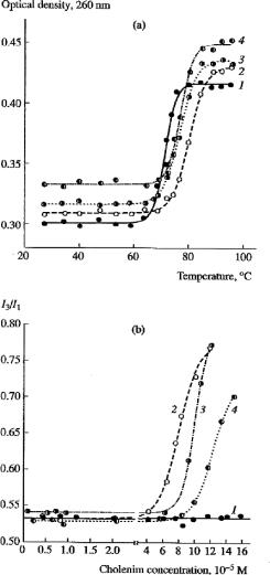
Figure 1 (upper field) shows the melting curves of fragments of genomic DNA and its complexes with cholenims. Analysis of these curves showed that the complexes formed by DNA and compounds I, II, or III have a higher melting temperature (by 8, 5, and 4°C, respectively) compared to pure DNA fragments. Thus, these compounds stabilize the DNA helix, with their stabilizing effect decreasing in the following order: compound I > compound II > compound III. The affinity of colenims for the double helix of DNA is different due to different positive charges of their hydrophilic groups and different hydrophobicity/hydrophilicity ratios. Apparently, electrostatic interactions between the amino groups of compounds I and II and the negatively charged phosphate groups of the polynucleotide chain are important of stabilizing complexes. There is a good correlation between the Tmelt value and the charge of cholenim: the greater the charge, the greater the stabilizing effect (Table 1). Analysis of circular dichroism spectra of the pCMV-SPORT- -Gal plasmid and its complexes with compounds I–III led us to conclude that they are practically identical and that these compounds do not affect the structure of double helix of DNA, which retains B-conformation (spectra not shown). As a fluorescent probe we used pyrene, whose oscillatory structure of emission spectra is highly sensitive to polarity of its microenvironment. Due to this property, pyrene is widely used in studies of membranes, micelles, and hydrophobic clusters [22, 23].
As seen from the results, the value of this ratio almost did not depend on the concentrationofcholenimuptothethresholdvalue;furtherincreaseincholenimconcentration results in a sharp increase in the I3/I1 ratio (in the absence of DNA, this parameter did not depend on the concentration of cholenims within the concentration range analyzed). These values, different for compounds I 6 0 × 10−5 M), II 8 6 × 10⁻⁵ M), and III
LIPIDAND GLYCOLIPID-BASED NANOSYSTEMS |
37 |
Figure 1. Physicochemical characteristics of the complexes (lipoplexes) DNA-CHOLENIMS. (a) UV melting curves of salmon roe DNA in the buffer containing 10 mM NaCl and 1 mM Tris-HCl (pH 7.2) (1) in the control and in the presence of (2) monocholenim, (3) dicholenim, and (4) tricholenim (1.0±0.2). 10−4 M. (b) Dependence of pyrene emission spectrum (the I3/I1 index) on the concentration of (2) monocholenim, (3) dicholenim, and (4) tricholenim. Curve 1 shows DNA spectrum in the absence of cholenims.
Designations: I1 and I3, amplitudes of oscillatory lines of emission spectra of monomeric pyrene at 383 and 372 nm, respectively, in the presence of salmon sperm DNA (45. μM by phosphate)
38 |
|
|
ZHDANOV ET AL. |
|
|
|
||
Table 1. Properties of hydrophobic oligocation CHOLENIMS and their lipoplexes |
|
|
||||||
|
|
|
|
|
|
|
|
|
Cholesterol |
T melt., |
T |
CMC, M |
Charge* |
EM, diameter |
Transfection efficacy |
||
derivatives |
(°C) |
|
|
|
(nm) |
against PC-12 cells |
||
|
|
|
|
|
|
|
|
|
|
|
|
|
|
|
DNA/ |
Picog |
|
|
|
|
|
|
|
cholenim |
protein per |
|
|
|
|
|
|
|
ratio |
105 cells |
|
|
|
|
|
|
|
|
|
|
Monocholenim |
80 |
+8 |
6.0*10−5 |
+2 |
100–130 |
3:1 |
105 |
|
|
|
|
|
|
|
2:1 |
187 |
|
|
|
|
|
|
|
0,7:1 |
36 |
|
Dicholenim |
77 |
+5 |
8.6*10−5 |
+1 |
200–250 |
3:1 |
100 |
|
|
|
|
|
|
|
2:1 |
113 |
|
|
|
|
|
|
|
0,7:1 |
14 |
|
Tricholenim |
76 |
+4 |
1.0*10−4 |
0 |
300–340 |
3:1 |
56,5 |
|
|
|
|
|
|
|
2:1 |
31,3 |
|
|
|
|
|
|
|
0,7:1 |
7,5 |
|
|
|
|
|
|
|
|
|
|
Note: T designates an increase in melting temperature of DNA samples in the complex with an oligocation; CMC, critical micelle concentration; EM, diameter of particles of the corresponding complexes with plasmid DNA or DNA fragments (electron microscopy data).
Calculated for the amino groups at pH 7.0.
1 0 × 10−4 M), correspond to formation of complexes between these compounds and DNA, which contain hydrophobic clusters where pyrene molecules are inserted, and may be regarded as critical micelle concentrations (CMC). There is a good correlation between the CMC value and the decrease in the total positive charge of polar groups of cholenims. Thus, it can be postulates that cholenims bind with DNA to form a hydrophobic coat around the helix, and that the disadvantageous (in terms of energy) contact between hydrophobic cholesterol residues with aqueous environment at certain concentration results in a decrease in solubility of complexes.
Electron-microscopic study showed that the complexes between genomic DNA and the plasmid with cholenims represent spherical particles with a diameter of 100 to 300 nm. Condensation of 4–6 kb DNA fragments and compound II showed that the size of particles significantly varies. The fact that the size of DNA/cholenim particles is large and almost does not depend on the molecular weight of DNA is unusual for a simple micellar structure. Figure 1 (lower field) shows the dependence of the spectral parameter I3/I1, which is the most sensitive to hydrophobicity of microenvironment, on the concentration of cholenims at a constant DNA concentration (I1 and I3 are the amplitudes of oscillatory lines of emission spectra of the monomeric form of pyrene at 383 and 372 nm).
3.1.2.Gene transfer and delivery
The results of transfection of PC-12 cells with the complexes containing the pCMVSPORT- -Gal plasmid and cholenims are summarized in Table 1. The greatest effectiveness of transfection of PC-12 cells was reached when DNA/cholenim

LIPIDAND GLYCOLIPID-BASED NANOSYSTEMS |
39 |
genosomes were used at a ratio of 2:1. However, significant effectiveness of transfection was also observed at DNA/cholenim ratio of 3:1. Similar results were obtained for the dicholenim-based complex. The effectiveness of transfection in the case of DNA/dicholenim genosomes at ratios of 2:1 and 3:1 was considerably higher than at the ratio 0.7:1 and comparable with the effectiveness of transfection for the DNA/cholenim ratio at the ratio 3:1. Tricholenim was much less effective in gene delivery compared to the other two compounds. In this case, the effectiveness of transfection markedly decreased as the proportion of tricholenim in genosomes increased. The effectiveness of transfection of RGGN-1 cells was 30 and 32 pg protein per 105 cells for the DNA/monocholenim complex and 14 and 23 pg protein per 105 cells for the DNA/dicholenim complex (ratio, 2:1 and 1:1, respectively). Although this index for RGGN-1 cells in general was considerably lower compared to the effectiveness of transfection of PC-12 cells, this finding also supports the fact that monocholenim and dicholenim may be used as gene carriers in vitro. However, it should be noted that, in the case of in vivo transfection, there might be another relationship between the effectiveness of gene transfer and qualitative and quantitative composition of cholenim-based complexes. Amphiphilic liposomes consisting of phosphatidylcholine and dicholenim at the ratio 1:1 (w/w) were used to transfer the -galactosidase gene using intravenous injection at the lipid/DNA ratio 1.6:1 (w/w). Sections of organs were incubated with the substrate X-Gal, which in the presence of -galactosidase is degraded, yielding the bright blue dye indigo. In preparations analyzed, the reporter DNA was expressed predominantly in endothelial cells of pulmonary vessels and in neighboring cells, which provides evidence that vascular endothelial cells are permeable for our complexes (Figure 2).
Figure 2. Histochemical preparation of ICR mouse lung after injection into the portal vein of the liver of lipoplexes formed by the pCMV-SPORT- -Gal plasmid and liposomes PC/DICHOLENIM (1:1). Staining around the blood vessel is the result of degradation of the substrate X-Gal by bacterial-galactosidase. Magnification, 200; computer processing; AXIOSKOP 20 Carl Zeiss
40 |
ZHDANOV ET AL. |
This distribution pattern is characteristic of cationic liposomes injected intravenously. Thus, the introduction of the cholesterol fragment into the structure of oligoethylene imines improves the characteristics of the corresponding complexes: increases the hydrophobicity/hydrophilicity ratio, stabilizes the lipoplex, and ensures optimal CMC values. Our data confirm the existence of stable DNA/cholenim complexes and electrostatic interaction in them of positively charged groups with negatively charged phosphate groups of DNA, with the deoxyribose phosphate backbone being apparently involved in the stabilization of genosomes. Compounds I–III interact with DNA to form a hydrophobic coat around its double helix. The high effectiveness of DNA/cholenim lipoplexes in gene transfer in vitro is probably determined by their complete dissociation in the cytosol before the nuclear membrane, because this ability of lipoplexes is a key characteristic required for transfection [43].
3.2.Glycoclip
3.2.1.Cytoand genotoxicity
Potential cytoand genotoxicity of GLYCOCLIP/DOPE and CLIP/PC liposomes were estimated in experiments with a cultured glyal cell line [44], which is very sensitive to any influence, as described earlier [4]. The influence of the former liposomal preparation on the growth (24hrs) of RGNN cells and the DNA synthesis in these cells was studied. The preparations have almost no effect on cell growth at both concentrations used: 6μg/ml (number of cells survived after 24 hrs incubation was 110.5 +/−3.1% (M+/−s) comparing to the control one) and 60μg/ml (98.6+/−9.3%). The influence on DNA synthesis was evaluated as the extent of incorporation of 14C-thymidine into RGNN cell genomic DNA. It had equally essential effect on the DNA synthesis at both concentrations (6 or 60 μg/ml): the values of the DNA synthesis were 58.5+/−7.8% and 66.3+/−9.2% comparing to the control ones, correspondingly. The influence of CLIP liposomes on DNA synthesis in RGGN cells was not so pronounced (in the range of experimental error), as found for the GLYCOCLIP ones. The CLIP/PC liposomal preparation has no effect on either the cell survival, or the DNA synthesis in RGGN cells at 6μg/ml level. The number of cells survived after 24 hrs incubation was 100.0 +/−5.0% comparing to the control, and the value of the DNA synthesis in the cells was 101.8 +/−7.0% comparing to the control one. Only 10-fold dose of CLIP/PC liposomes (60μg/ml) had an effect on the DNA synthesis: 55.5 +/−11.7% (p < 0.05) comparing to the control one. CHOLENIM preparation itself appeared to be completely non-toxic at the range of concentrations used [18].
3.2.2.Gene encapsulation and delivery in vitro
Gene transfer activity of the liposomes based on GLYCOCLIP was studied with the commonly used reporter gene transfer system: transfection of pCMV-Luc plasmid into CHO cells followed by gene transfer efficiency testing using luminometer assay [45]. Figure 3 represents data on reporter gene (pLuc) transfer efficiency with liposomal preparations of compounds I and II into CHO cells in comparison with

LIPIDAND GLYCOLIPID-BASED NANOSYSTEMS |
41 |
Figure 3. Transfection efficiency of lipoplex and glycolipoplex preparations formed of various cationic lipids, GLYCOCLIP and pCMV-Luc reporter plasmid against CHO cells: 1-CHOLENIM I/GLYCOCLIP V; 2-CLIP IV; 3-GLYCOCLIP VI; 4-Lipofectin; 5-Dosper
corresponding data for commercial gene transfer agent DOSPER. As follows from the results the introduction of a triacetyl-glucose moiety into the structure of a cationic lipid enhances remarkably the transfection: RLU value of GLYCOCLIP/DOPE liposomes equals to 7.106 (compare GLYCOCLIP/DOPE and CLIP/PC values).
The GLYCOCLIP-based liposomes’ RLU values are only slightly less than those of DOSPER mediated gene transfer. Our data on the inhibition of DNA synthesis in RGGN cells after 24hrs incubation with GLYCOCLIP/DOPE liposomes corresponds to the data testifying to the toxicity of many cationic liposomes during in vitro experiments [14]. However, it was demonstrated that the efficiency of gene transfer with cationic liposomes is not directly connected with the degree of their toxicity [46], so one may get high transfection efficiency with the use of gene transfer agents demonstrating a certain toxicity in vitro. It is possible that lowering the concentration of GLYCOCLIP/DOPE liposomes used for transfection will help to avoid their influence on DNA synthesis. It cannot be excluded that this effect will not appear during in vivo transfection. The fact that GLYCOCLIP/DOPE liposomes don’t influence the cell growth at least during the first 24hrs is also promising. Thus, partial glyconylation of polylysine has been shown to increase the efficiency of transfection with its participation, and the conjugation of modified polylysine with a few lactose moieties causes appearance of genosome’s specificity to cell surface lectin [47, 48]. A series of amphiphilic dendritic galactosides were synthesized to be used for selective targeting of liposomes to the hepatic asialoglycoprotein receptor [49]. Introduction of carbohydrate moieties into the structures involved in lipoplex formation increases the efficacy and the specificity (hepatocytes) of transfection. Lipoplexes composed of galactosylated peptides demonstrate tropicity to hepatocytes [50].
42 |
ZHDANOV ET AL. |
DOPE and PC represent helper lipids, which enhance transfection efficiency being included into liposomes and lipoplex composition [24,51]. The presence of a helper lipid and the difference between the helper lipids (DOPE or PC) in GLYCOCLIP and CLIP liposomal formulations used can give no strong influence on the gene transfer efficiency in the case of CHO cells, because of the endocytotic way of the lipoplex internalization into this cell line [51]. Therefore the enhanced transfection efficiency of GLYCOCLIP liposomes compare to CLIP liposomes can be explained by the presence of carbohydrate (glucose) moiety in the first one. Introduction of CHOLENIM preparation into glycolipoplex composition facilitates the elaboration of DNA from a lipoplex in perinuclear space. That is the main reason for increasing transfection efficiency of GLYCOCLIP/ CHOLENIM/DOPE liposomes comparing to GLYCOCLIP/DOPE ones. Another reason is the higher value (3.2) of +/− ratio. It appeares that mechanism of gene transfer with the glycolipoplex includes both adsorbic endocytosis usual for lipoplex formulations, and receptor-mediated gene transfer characteristic for carbohydrate ligand-mediated gene transfer. Our results represent one of the first examples of the use of a cationic glycolipid, its liposomal formulations, and genosomes/lipoplexes composed of GLYCOCLIP as gene transfer agents. We believe that glycocationic lipids of this type will be effective especially for in vivo studies due to the affinity of carbohydrate structures to the cell surface and the vessell’s endothelium as well.
3.3.Glycolipid
3.3.1.Gene delivery in vivo
The first stage in the study of the effectiveness of gene transfer using liposomes containing phosphatidylcholine and GLYCOLIPID VI included the determination of the pattern of distribution of 14C-adenosine-labeled eukaryotic DNA in mouse organs. The maximal DNA level (recalculated per gram tissue) was detected in the kidneys (6000–8000 cpm per gram) and liver (4000 cpm per gram). Note that the content of 14C–DNA in the liver was three times greater than in the lungs (Figure 4). It is known that intravenous injections of the complexes of cationic liposomes with DNA are usually characterized by “the effect of the first passage,” i.e., the majority of injected liposome with bloodstream get from the heart to the lungs [52]. When using liposomes containing GLYCOLIPID VI, this effect was not observed. In our experiments, we observed certain affinity of the complex of these 14C-DNA-containing liposomes for the liver and kidneys.
The maximal level of 14C-DNA in the kidneys is probably due to the fact that it might have been eliminated as early as 24 h after injection, because kidneys are excretory organs. Then, we studied the expression of the –galactosidase gene in mouse organs in the case of delivery of the pCMV-SPORT- -Gal plasmid (100 μg) in the complex with mixed liposomes consisting of phosphatidylcholine, GLYCOLIPID, and dicholenim (160 μg).
When this lipoplex was injected into the portal vein, the Lac Z gene was expressed predominantly in hepatocytes. However, despite the presence on the surface of

LIPIDAND GLYCOLIPID-BASED NANOSYSTEMS |
43 |
Figure 4. Distribution of the lipoplex formed by 14C-adenosine-labeled DNA and liposomes comprised of phosphatidylcholine, lactosolipid, and dicholenim in mouse organs after injection into the portal vein of the liver (cpm/min per gram organ; n = 4)
liposomes of a lactose residue, which exhibits affinity for the lectin located on the surface of hepatocytes, the degree of expression was low, and expression was observed mostly in epithelium of blood vessels and in the immediate vicinity of them. This fact is indicative of a low permeability of tissues for such complexes. A more long-term incubation with the substrate led to appearance of the dye indigo in the form of small (less than 1 μm) bright blue granules both in the control and experimental liver section. It can be assumed that this phenomenon may be accounted for by location of the endogenous enzyme in lysosomes or other compartments of the cytoplasm of hepatocytes. In the lungs and spleen, the level of expression of the Lac Z gene (reaction with X-Gal) was high (Figure 5).
Figure 5. Histochemical assessment of expression of the LacZ gene (the pCMV-SPORT- -Gal plasmid) in the ICR mouse spleen after injection of the lipoplex based on the pCMV-SPORT- -Gal plasmid and liposomes composed of phosphatidylcholine/GLYCOLIPID/dicholenim (ratio 1:1.6, w/w) into the portal vein of the liver. Dark areas indicate the sites of the highest expression (magnification ×200)
44 ZHDANOV ET AL.
A high endogenous activity of -galactosidase was detected in the kidneys, which hampered the assessment of the effectiveness of the exogenous enzyme. For quantitative estimation of expression of the -galactosidase gene in mouse organs after injecting the complex of the plasmid with the liposomes consisting of phosphatidylcholine, lactosolipid, and dicholenim, the activity of this enzyme in tissues was determined spectrophotometrically.
The maximal activity of the enzyme was observed in the spleen (data are not shown), and equal activity was detected in the lungs and liver. A high level of endogenous activity of -galactosidase in some organs hampers quantitative assessment of expression. Thus, the results of this study showed that GLYCOLIPID VI containing a lactose residue, which was used in the form of liposomes to transfer 14C-adenosine-labeled or plasmid DNA, determined the affinity of lipoplexes for kidney, liver, and spleen tissues. The effect of the first passage, characteristic of cationic complexes, was not observed when 14C-DNA was injected in the complex with liposomes comprised of phosphatidylcholine, lactosolipid, and dicholenim, was considerably decreased when the plasmid was injected in the complex with liposomes comprised of phosphatidylcholine, lactosolipid, and dicholenim. In the last case, the expression of -galactosidase was maximum in the spleen. GLYCOLIPID VI, which determines the affinity of lipoplexes for tissues, as well as glycolipids on the whole, is a prospective tool for designing on its basis of nonviral vectors of a new generation for targeted gene delivery to tissues.
3.4.Modified Chitosan (mCHIT)
We used natural polycationic polysaccharide, chitosan, which can usually be prepared by deacetylation of chitin – linear poly– (N-acetyl-glucosamine) to gene transfer against cultured cell lines. Chitosan macromolecule represents linear polymer of glucosamine, part of whose primary amino-groups (normally 5–20%) are still acetylated. It is well–known that chitosan being one of the most widespead biomass represents non-toxic, biocompatible biopolymer [53, 54], which is suitable for gene delivery purpose [14–16],[55–57]. However, in our preliminary study we also worked to get reporter gene transfer of transformed cells using non-modified chitosan preparations. After the data on efficient transfection of 3T3 and HepG2 cells with complexes of plasmid DNA and polyethylenimine (PEI) were published [41], Dr. G.G. Krivtsov decided to introduce the secondary and tertiary aminogroups into chitosan structure to use modified chitosan preparations (mCHIT) for gene transfer. The matter is that PEI contains the secondary amino groups along with the primary and tertiary ones. He synthesized the chitosan preparation, containing N-ethyl- (the secondary one) and N,N-dimethyl amino (the tertiary one) groups to facilitate ionic interaction of chitosan with the DNA and to increase transfection efficiency against different transformed cell lines, especially the suspension blood cell ones. The latter topic is an acquit area of research now, and is also very important for development of non-viral delivery systems for ex vivo gene therapy of variety of genetic diseases and cancer pathologies [58–60].
LIPIDAND GLYCOLIPID-BASED NANOSYSTEMS |
45 |
A number of papers on the usage of different chitosan preparations and nanospheres as transfection agents have been published [14–16],[55–57]. It was reported that unmodified chitosan has ability to condense DNA and form small discrete particles [57]. They can transfect HeLa cells ( -gal [14] or Luc [57] genes) independently of the presence of 10% fetal serum. Gene expression gradually increased with time, being at 96 hours 10 times more efficient, than polyethylenimine [57]. It was suggested that non-ionic interactions between chitosan macromolecule and cell surface might play an important role in chitosan-mediated transfection [56]. pH-sensitive endosomolytic peptide enhanced gene expression in COS-1 cells by factor 4, but during in vivo experiments on rabbits (intestine and colon) gene expression appeared to be still low [15]. Hydophobically modified chitosan (containing five deoxycholic acyl moieties per 100 anhydroglucose units) was prepared, its aggregates being 162 +/−18 nm in diameter [14]. Transfection of COS-1 cells using self-aggregates/plasmid DNA complexes at +/− charge ratio 4 was reported. Nanospheres composed of cDNA and gelatin or chitosan (200–750 nm) were used for in vitro transfection, efficiency being lower than in the case of lipofectamine-mediated and Ca–phosphate ones [55]. Method for oral DNA delivery with N-acetylated chitosan was reported [16].
All groups that have been working with chitosan preparations as gene delivery systems used non-N-alkylated chitosan samples containing only primary aminogroups along with N-acetyl moiety. These preparations usually represent particles of small size (80 nm) as measured by variety of techniques [14–16],[55–57]. Chitosan preparation hydrophobized with deoxycholeic acyl moieties (5%) forms self-aggregates of medium size (200 nm) itself. Nanosheres formed of chitosan are even bigger: 200–700 nm [14,15],[55–57]. These chitosan preparations are characterized with ability to form DNA aggergates with supercoiled plasmid like cationic liposomes and other polycations usually do [39]. The size of these aggregates is even bigger. All known chitosan preparations tested for gene delivery in vitro and/or in vivo are far from being as effective as any commercial gene transfer ones, e.g. PEITM. We usually obtained low transfection efficiency values with non-modified chitosan preparations. The reason for these, by our opinion, is unsufficient ability of polysaccharide bearing only primary glucosamine moieties and forming big aggregates to be as stable as to survive in endosome-lyzosomal complex. There are very few reasons to add any hydrophobic moieties (like choleic acid) into glucosamine residue, as chitosan biomacromolecule having well-known hydrophobic properties is able to bind 10-fold amount (w/w) of fat molecules [61].
Transfection was carried out with two various reporter gene plasmids: pEQ176 ( -galactosidase) and pCSEAP (secreted alkaline phosphatase) (under IE CMV promoters) against transformed cell lines with different ethiology: three adherent cell lines (MeWo, HeLa, and HOS-1) and two suspension cell cultures (U937 and Jurkat) as well. Transformed blood cell lines had been cultured by conventional methods. A number of transfection techniques (Ca-phosphate; pH-sensitive amphiphylic liposomes/Ca2+- and PEI-mediated gene transfer) were used for comparing results of mCHIT glycoplex transfection. Glycoplex composition was choosen with mCHIT