

SURGICAL ANATOMY by Joseph Maclise
.pdf52 |
COMMENTARY ON PLATE 22. |
At the hypochondriac angles formed between the points F, L, N, on either side the lungs are absent both in inspiration and expiration. Percussion, when made over the surface of the angle of the right side, discovers the presence of the liver, G G*. When made over the median line, and on either side of it above the umbilicus, N, we ascertain the presence of the stomach, M M*. In the left hypochondriac angle, the stomach may also be found to occupy this place wholly.
Beneath the umbilicus, N, and on either side of it as far outwards as the lower asternal ribs, K L, thus ranging the abdominal parietes transversely, percussion discovers the transverse colon, O, P, O*. The small intestines, S S*, covered by the omentum, P*, occupy the hypogastric and iliac regions.
The organs situated within the thorax give evidence that they are developed in accordance to the law of symmetry. The lungs form a pair, one placed on either side of the median line. The heart is a double organ, formed of the right and left heart. The right lung differs from the left, inasmuch as we find the former divided into three lobes, while the latter has only two. That place which the heart now occupies in the left thoracic side is the place where the third or middle lobe of the left lung is wanting. In the abdomen we find that most of its organs are single. The liver, stomach, spleen, colon, and small intestine form a series of single organs: each of these may be cleft symmetrically. The kidneys are a pair.
The extent to which the ribs are bared in the figure Plate 22, marks exactly the form and transverse capacity of the thoracic walls. The diaphragm, H H*, has had a portion of its forepart cut off, to show how it separates the thin edges of both lungs above from the liver, G, and the stomach, M, below. These latter organs, although occupying abdominal space, rise to a considerable height behind K L, the asternal ribs, a fact which should be borne in mind when percussing the walls of the thorax and abdomen at this region.
DESCRIPTION OF PLATE 22.
A. Upper bone of the sternum.
B B*. Two first ribs.
C C*. Second pair of ribs.
D D*. Right and left lungs.
E.Pericardium, enveloping the heart--the right ventricle.
F.Lower end of the sternum.
G G*. Lobes of the liver.
H H*. Right and left halves of the diaphragm in section. The right half separating the right lung from the liver; the left half separating the left lung from the broad cardiac end of the stomach.
I I*. Eighth pair of ribs.
K K*. Ninth pair of ribs.
L L*. Tenth pair of ribs.
M M*. The stomach; M, its cardiac bulge; M*, its pyloric extremity.
N. The umbilicus.
OO*. The transverse colon.
P P*. The omentum, covering the transverse colon and small intestines.
Q. The gall bladder.
R R*. The right and left pectoral prominences.
S S*. Small intestines.

Plate 22
COMMENTARY ON PLATE 23.
THE RELATIVE POSITION OF THE DEEPER ORGANS OF THE THORAX AND
THOSE OF THE ABDOMEN.
The size or capacity of the thorax in relation to that of the abdomen varies in the individual at different periods of life. At an early age, the thorax, compared to the abdomen, is less in proportion than it is at adult age. The digestive organs in early age preponderate considerably over the respiratory organs; whereas, on the contrary, in the healthy and well-formed adult, the thoracic cavity and organs of respiration manifest a greater relative proportion to the ventral cavity and organs. At the adult age, when sexual peculiarities have become fully marked, the thoracic organs of the male body predominate over those of the abdomen, whilst in the female form the ventral organs take precedence as to development and proportions. This diversity in the relative capacity of the thorax and abdomen at different stages of development, and also in persons of different sexes, stamps each individual with characteristic traits of physical conformation; and it is required that we should take into our consideration this normal diversity of character, while conducting our examinations of individuals in reference to the existence of disease.
The heart varies in some measure, not only as to size and weight, but also as to position, even in healthy individuals of the same age and sex. The level at which the heart is in general found to be situated in the thorax is that represented in PLATE 23, where the apex points to the sixth intercostal space on the left side above K, while the arch of the aorta rises to a level with C, the second costal cartilage. In some instances, the heart may be found to occupy a much lower position in the thorax than the one above mentioned, or even a much higher level. The impulse of the right ventricle, F, has been noticed occasionally as corresponding to a point somewhat above the middle of the sternum and the intercostal space between the fourth and fifth left costal cartilages; while in other instances its beating was observable as low down as an inch or more below the xiphoid cartilage, and these variations have existed in a state of health.
Percussion over the region of the heart yields a dull flat sound. The sound is dullest opposite the right ventricle, F; whilst above and to either side of this point, where the heart is overlapped by the anterior shelving edges of both lungs, the sound is modified in consequence of the lung's resonant qualities. The heart-sounds, as heard through the stethoscope, in valvular disease, will, of course, be more distinctly ascertained at the locality of F, the right ventricle, which is immediately substernal. While the body lies supine, the heart recedes from the forepart of the chest; and the lungs during inspiration expanding around the heart will render its sounds less distinct. In the erect posture, the heart inclines forwards and approaches the anterior wall of the thorax. When the heart is hypertrophied, the lungs do not overlap it to the same extent as when it is of its ordinary size. In the latter state, the elastic cushion of the lung muffles the heart's impulse. In the former state, the lung is pushed aside by the overgrown heart, the strong muscular walls of which strike forcibly against the ribs and sternum.
(Page 53 )
54 |
COMMENTARY ON PLATE 23. |
The thorax is separated from the abdomen by the moveable diaphragm. The heart, F E, lies upon the diaphragm, L L*. The liver, M, lies immediately beneath the right side of this muscular septum, L*, while the bulging cardiac end of the stomach, O, is in close contact with it on the left side, L. As these three organs are attached to the diaphragm-- the heart by its pericardium, the stomach by the tube of the oesophagus, and the liver by its suspensory ligaments--it must happen that the diaphragm while descending and ascending in the motions of inspiration and expiration will communicate the same alternate motions to the organs which are connected with it.
In ordinary respiration the capacity of the thorax is chiefly affected by the motions of the diaphragm; and the relative position which this septum holds with regard to the thoracic and abdominal chambers will cause its motions of ascent and descent to influence the capacity of both chambers at the same time. When the lungs expand, they follow the descent of the diaphragm, which forces the abdominal contents downwards, and thus what the thorax gains in space the abdomen loses. When the lungs contract, the diaphragm ascends, and by this act the abdomen gains that space which the thorax loses. But the organs of the thoracic cavity perform a different office in the economy from those of the abdomen. The air which fills the lungs is soon again expired, whilst the ingesta of the abdominal viscera are for a longer period retained; and as the space, which by every inspiration the thorax gains from the abdomen, would cause inconvenient pressure on the distended organs of this latter cavity, so we find that to obviate this inconvenience, nature has constructed the anterior parietes of the abdomen of yielding material. The muscular parietes of the abdomen relax during every inspiration, and thus this cavity gains that space which it loses by the encroachment of the dilating lungs.
The mechanical principle upon which the abdominal chamber is constructed, enables it to adjust its capacity to such exigence or pressing necessity as its own visceral organs impose on it, from time to time; and the relation which the abdominal cavity bears to the thoracic chamber, enables it also to be compensatory to this latter. When the inspiratory thorax gains space from the abdomen, or when space is demanded for the increasing bulk of the alimentary canal, or for the enlarging pregnant uterus; or when, in consequence of disease, such as dropsical accumulation, more room is wanted, then the abdominal chamber supplies the demand by the anterior bulge or swell of its expansile muscular parietes.
The position of the heart itself is affected by the expansion of the lungs on either side of it. As the expanding lungs force the diaphragm downwards, the heart follows it, and all the abdominal viscera yield place to the descending thoracic contents. In strong muscular efforts the diaphragm plays an important part, for, previously to making forced efforts, the lungs are distended with air, so as to swell and render fixed the thoracic walls into which so many powerful muscles of the shoulders, the neck, back, and abdomen, are inserted; at the same time the muscular diaphragm L L*, becomes tense and unbent from its arched form, thereby contracting abdominal space, which now has no compensation for this loss of space, since the abdominal parietes are also rendered firm and unyielding. It is at this crisis of muscular effort that the abdominal viscera become impacted together; and, acting by their own elasticity against the muscular force, make an exit for themselves through the weakest parts of the abdominal walls, and thus herniae of various kinds are produced. The most common situations of abdominal herniae are at the inguinal regions, towards which the intestines, T T, naturally gravitate; and at these situations the abdominal parietes are weak and membranous.
The contents of a hernial protrusion through the abdominal parietes, correspond in general with those divisions of the intestinal tube, which naturally lie adjacent to the part where the rupture has taken place. In the umbilical hernia it is either the transverse colon S*, or some part of the small intestine occupying the median line, or both together, with some folds of the omentum, which will be found to form the contents of this swelling.
COMMENTARY ON PLATE 23. |
55 |
When the diaphragm itself sustains a rupture in its left half, the upper portion of the descending colon, S, protrudes through the opening. A diaphragmatic hernia has not, so far as I am aware, been seen to occur in the right side; and this exemption from rupture of the right half of the diaphragm may be accounted for anatomically, by the fact that the liver, M, defends the diaphragm at this situation. The liver occupies the whole depth of the right hypochondrium; and intervenes between the diaphragm L*, and the right extremity of the transverse colon, S**.
The contents of a right inguinal hernia consist of the small intestine, T. The contents of the right crural hernia are formed by either the small intestine, T, or the intestinum caecum, S***. I have seen a few cases in which the caecum formed the right crural hernia. Examples are recorded in which the intestine caecum formed the contents of a right inguinal hernia. The left inguinal and crural herniae contain most generally the small intestine, T, of the left side.
The right lung, I*, is shorter than the left; for the liver, M, raises the diaphragm, L, to a higher level within the thorax, on the right side, than it does on the left. When the liver happens to be diseased and enlarged, it encroaches still more on thoracic space; but, doubtless, judging from the anatomical connexions of the liver, we may conclude that when it becomes increased in volume it will accommodate itself as much at the expense of abdominal space. The liver, in its healthy state and normal proportions, protrudes for an inch (more or less) below the margins of the right asternal ribs. The upper or convex surface of the liver rises beneath the diaphragm to a level corresponding with the seventh or sixth rib, but this position will vary according to the descent and ascent of the diaphragm in the respiratory movements. The ligaments by which the liver is suspended do not prevent its full obedience to these motions.
The left lung, I, descends to a lower level than the right; and the left diaphragm upon which it rests is itself supported by the cardiac end of the stomach. When the stomach is distended, it does not even then materially obstruct the expansion of the left lung, or the descent of the left diaphragm, for the abdominal walls relax and allow of the increasing volume of the stomach to accommodate itself. The spleen, R, is occasionally subject to an extraordinary increase of bulk; and this organ, like the enlarged liver and the distended stomach, will, to some extent, obstruct the movements of the diaphragm in the act of respiration, but owing to its free attachments it admits of a change of place. The abdominal viscera, one and all, admit of a change of place; the peculiar forms of those mesenteric bonds by which they are suspended, allow them to glide freely over each other; and this circumstance, together with the yielding nature of the abdominal parietes, allows the thoracic organs to have full and easy play in the respiratory movements performed by agency of the diaphragm.
The muscles of respiration perform with ease so long as the air has access to the lungs through the normal passage, viz., the trachea. While the principle of the thoracic pneumatic apparatus remains underanged, the motor powers perform their functions capably. The physical or pneumatic power acts in obedience to the vital or muscular power, while both stand in equilibrium; but the ascendancy of the one over the other deranges the whole thoracic machine. When the glottis closes by muscular spasm and excludes the external air, the respiratory muscles cease to exert a motor power upon the pulmonary cavity; their united efforts cannot cause a vacuum in thoracic space in opposition to the pressure of the external air. When, in addition to the natural opening of the glottis, a false opening is made in the side at the point K, the air within the lung at I, and external to it in the now open pleural cavity, will stand in equilibrio; the lung will collapse as having no muscular power by which to dilate itself, and the thoracic dilator muscles will cease to affect the capacity of the lung, so long as by their action in expanding the thoracic walls, the air gains access through the side to the pleural sac external to the lung.
56 |
COMMENTARY ON PLATE 23. |
Whether the air be admitted into the pleural sac, by an opening made in the side from without, or by an opening in the lung itself, the mechanical principle of the respiratory apparatus will be equally deranged. Pneumo-thorax will be the result of either lesion; and by the accumulation of air in the pleura the lung will suffer pressure. This pressure will be permanent so long as the air has no egress from the cavity of the pleura.
The permanent distention of the thoracic cavity, caused by the accumulation of air in the pleural sac, or by the diffusion of air through the interlobular cellular tissue consequent on a wound of the lung itself, will equally obstruct the breathing; and though the situation of the accumulated air is in fact anatomically different in both cases, yet the effect produced is similar. Interlobular pressure and interpleural pressure result in the same thing, viz., the permanent retention of the air external to the pulmonary cells, which, in the former case, are collapsed individually; and, in the latter case, in the mass. Though the emphysematous lung is distended to a size equal to the healthy lung in deep inspiration, yet we know that emphysematous distention, being produced by extrabronchial air accumulation, is, in fact, obstructive to the respiratory act. The emphysematous lung will, in the same manner as the distended pleural sac, depress the diaphragm and render the thoracic muscles inoperative. The foregoing observations have been made in reference to the effect of wounds of the thorax, the proper treatment of which will be obviously suggested by our knowledge of the state of the contained organs which have suffered lesion.
DESCRIPTION OF PLATE 23.
A. Upper end of the sternum.
B B.* First pair of ribs.
C C.* Second pair of ribs.
D.Aorta, with left vagus and phrenic nerves crossing its transverse arch.
E.Root of pulmonary artery.
F.Right ventricle.
G.Right auricle.
H.Vena cava superior, with right phrenic nerve on its outer border.
I I*. Right and left lungs collapsed, and turned outwards, to show the heart's outline.
K K*. Seventh pair of ribs.
L L*. The diaphragm in section.
M.The liver in section.
N.The gall bladder with its duct joining the hepatic duct to form the common bile duct. The hepatic artery is seen superficial to the common duct; the vena portae is seen beneath it. The patent orifices of the hepatic veins are seen on the cut surface of the liver.
O.The stomach.
P.The coeliac axis dividing into the coronary, splenic and hepatic arteries.
Q.Inferior vena cava.
R.The spleen.
S S* S**. The transverse colon, between which and the lower border of the stomach is seen the gastro-epiploic artery, formed by the splenic and hepatic arteries.
S***. Ascending colon in the right iliac region.
T. Convolutions of the small intestines distended with air.
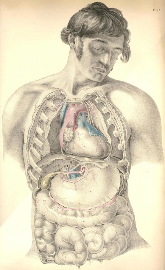
Plate 23
COMMENTARY ON PLATE 24.
THE RELATIONS OF THE PRINCIPAL BLOODVESSELS
TO THE VISCERA OF THE THORACICO-ABDOMINAL CAVITY.
The median line of the body is occupied by the centres of the four great systems of organs which serve in the processes of circulation, respiration, innervation, and nutrition. These organs being fashioned in accordance with the law of symmetry, we find them arranged in close connexion with the vertebrate centre of the osseous fabric, which is itself symmetrical. In this symmetrical arrangement of the main organs of the trunk of the body, a mechanical principle is prominently apparent; for as the centre is the least moveable and most protected region of the form, so have these vitally important structures the full benefit of this situation. The aortal trunk, G, of the arterial system is disposed along the median line, as well for its own safety as for the fitting distribution of those branches which spring symmetrically from either side of it to supply the lateral regions of the body.
The visceral system of bloodvessels is moulded upon the organs which they supply. As the thoracic viscera differ in form and functional character from those of the abdomen, so we find that the arterial branches which are supplied by the aorta to each set, differ likewise in some degree. In the accompanying figure, which represents the thoracic and abdominal visceral branches of the aorta taken in their entirety, this difference in their arrangement may be readily recognised. In the thorax, compared with the abdomen, we find that not only do the aortic branches differ in form according to the variety of those organs contained in either region, but that they differ numerically according to the number of organs situated in each. The main vessel itself, however, is common to both regions. It is the one thoracico-abdominal vessel, and this circumstance calls for the comparison, not only of the several parts of the great vessel itself, but of all the branches which spring from it, and of the various organs which lie in its vicinity in the thorax and abdomen, and hence we are invited to the study of these regions themselves connectedly.
In the thorax, the aorta, G G*, is wholly concealed by the lungs in their states both of inspiration and expiration. The first part of the aortic arch, as it springs from the left ventricle of the heart, is the most superficial, being almost immediately sub-sternal, and on a level with the sternal junctions of the fourth ribs. By applying the ear at this locality, the play of the aortic valves may be distinctly heard. From this point the aorta, G, rises and arches from before, backwards, to the left side of the spine, G*. The arch of the vessel lies more deeply between the two lungs than does its ventricular origin. The descending thoracic aorta lies still more deeply situated at the left side of the dorsal spine. At this latter situation it is in immediate contact with the posterior thick part of the left lung; whilst on its right are placed, L, the thoracic duct; I, the oesophagus; K, the vena azygos, and the vertebral column. In Plate 26 may be seen the relation which the superior vena cava, H, bears to the aortic arch, A.
In the span of the aortic arch will be found, H*, the left bronchus, together with the right branch of the pulmonary artery, and the right pulmonary veins. The pneumo-gastric and phrenic nerves descend on either side of the arch. The left pneumo-gastric nerve winds round beneath the arch at the point where the obliterated ductus arteriosus joins it. See Plates 12 & 26.
(Page 57 )
58 |
COMMENTARY ON PLATE 24. |
The pulmonary artery, B, Plates 1 & 2, lies close upon the fore part, and conceals the origin, of the systemic aorta. Whenever, therefore, the semilunar valves of either the pulmonary artery or the systemic aorta become diseased, it must be extremely difficult to distinguish by the sounds alone, during life, in which of the two the derangement exists. The origins of both vessels being at the fore part of the chest, it is in this situation, of course, that the state of their valves is to be examined. The descending part of the thoracic aorta, G*, being at the posterior part of the chest, and lying on the vertebral ends of the left thoracic ribs, will therefore require that we should examine its condition in the living body at the dorsal aspect of the thorax. As the arch of the aorta is directed from before backwards--that is, from the sternum to the spine, it follows that when an aneurism implicates this region of the vessel, the exact situation of the tumour must be determined by antero-posterior examination; and we should recollect, that though on the fore part of the chest the cartilages of the second ribs, where these join the sternum, mark the level of the aortic arch, on the back of the chest its level is to be taken from the vertebral ends of the third or fourth ribs. This difference is caused by the oblique descent of the ribs from the spine to the sternum. The first and second dorsal vertebrae, with which the first and second ribs articulate, are considerably above the level of the first and second pieces of the sternum.
In a practical point of view, the pulmonary artery possesses but small interest for us; and in truth the trunk of the systemic aorta itself may be regarded in the same disheartening consideration, forasmuch as when serious disease attacks either vessel, the "tree of life" may be said to be lopped at its root.
When an aneurism arises from the aortic arch it implicates those important organs which are gathered together in contact with itself. The aneurismal tumour may press upon and obstruct the bronchi, H H*; the thoracic duct, L; the oesophagus, I; the superior vena cava, H, Plate 26, or wholly obliterate either of the vagi nerves. The aneurism of the arch of the aorta may cause suffocation in two ways--viz., either by pressing directly on the tracheal tube, or by compressing and irritating the vagus nerve, whose recurrent branch will convey the stimulus to the laryngeal muscles, and cause spasmodic closure of the glottis. This anatomical fact also fully accounts for the constant cough which attends some forms of aortic aneurism. The pulmonary arteries and veins are also liable to obstruction from the tumour. This will occur the more certainly if the aneurism spring from the right or the inferior side of the arch, and if the tumour should not break at an early period, slow absorption, caused by pressure of the tumour, may destroy even the vertebral column, and endanger the spinal nervous centre. If the tumour spring from the left side or the fore part of the arch, it may in time force a passage through the anterior wall of the thorax.
The principal branches of the thoracic aorta spring from the upper part of its arch. The innominate artery, 2, is the first to arise from it; the left common carotid, 6, and the left subclavian artery, 5, spring in succession. These vessels being destined for the head and upper limbs, we find that the remaining branches of the thoracic aorta are comparatively diminutive, and of little surgical interest. The intercostal arteries occasionally, when wounded, call for the aid of the surgeon; these arteries, like all other branches of the aorta, are largest at their origin. Where these vessels spring from G, the descending thoracic aorta, they present considerable caliber; but at this inaccessible situation, they seldom or never call for surgical interference. As the intercostal arteries pass outwards, traversing the intercostal spaces with their accompanying nerves, they diminish in size. Each vessel divides at a distance of about two inches, more or less, from the spine; and the upper larger branch lies under cover of the inferior border of the adjacent rib. When it is required to perform the operation of paracentesis thoracis, this distribution of the vessel should be borne in mind; and also, that the farther from the spine this operation is performed, the less in size will the vessels be found.
COMMENTARY ON PLATE 24. |
50 |
The intercostal artery is sometimes wounded by the fractured end of the rib, in which case, if the pleura be lacerated, an effusion of blood takes place within the thorax, compresses the lung, and obstructs respiration.
The thoracic aorta descends along the left side of the spine, as far as the last dorsal vertebra, at which situation the pillars of the diaphragm overarch the vessel. From this place the aorta passes obliquely in front of the five lumbar vertebrae, and on arriving opposite the fourth, it divides into the two common iliac branches. The aorta, for an extent included between these latter boundaries, is named the abdominal aorta, and from its fore part arise those branches, which supply the viscera of the abdomen.
The branches which spring from the abdominal aorta to supply the viscera of this region, are considerable, both as to their number and size. They are, however, of comparatively little interest in practice. To the anatomist they present many peculiarities of distribution and form worthy of notice, as, for example, their frequent anastomosis, their looping arrangement, and their large size and number compared with the actual bulk of the organs which they supply. As to this latter peculiarity, we interpret it according to the fact that here the vessels serve other purposes in the economy besides that of the support and repair of structure. The vessels are large in proportion to the great quantity of fluid matter secreted from the whole extent of the inner surface of this glandular apparatus--the gastro-intestinal canal, the liver, pancreas, and kidneys.
As anatomists, we are enabled, from a knowledge of the relative position of the various organs and bloodvessels of both the thorax and abdomen, to account for certain pathological phenomena which, as practitioners, we possess as yet but little skill to remedy. Thus it would appear most probable that many cases of anasarca of the lower limbs, and of dropsy of the belly, are frequently caused by diseased growths of the liver, P, obstructing the inferior vena cava, R, and vena portae, rather than by what we are taught to be the "want of balance between secreting and absorbing surfaces." The like occurrence may obstruct the gall-ducts, and occasion jaundice. Over-distention of any of those organs situated beneath the right hypochondrium, will obstruct neighbouring organs and vessels. Mechanical obstruction is doubtless so frequent a source of derangement, that we need not on many occasions essay a deeper search for explaining the mystery of disease.
In the right hypochondriac region there exists a greater variety of organs than in the left; and disease is also more frequent on the right side. Affections of the liver will consequently implicate a greater number of organs than affections of the spleen on the left side, for the spleen is comparatively isolated from the more important blood vessels and other organs.
The external surface of the liver, P, lies in contact with the diaphragm, N, the costal cartilages, M, and the upper and lateral parts of the abdominal parietes; and when the liver becomes the seat of abscess, this, according to its situation, will point and burst either into the thorax above, or through the side between or beneath the false ribs, M. The hepatic abscess has been known to discharge itself through the stomach, the duodenum, T, and the transverse colon, facts which are readily explained on seeing the close relationship which these parts hold to the under surface of the liver. When the liver is inflamed, we account for the gastric irritation, either from the inflammation having extended to the neighbouring stomach, or by this latter organ being affected by "reflex action." The hepatic cough is caused by the like phenomena disturbing the diaphragm, N, with which the liver, P, lies in close contact.
When large biliary concretions form in S, the gallbladder, or in the hepatic duct, Nature, failing in her efforts to discharge them through the common bile-duct, into the duodenum, T, sets up inflammation and ulcerative absorption, by aid of which processes they make a passage for themselves through some adjacent part of the intestine, either the duodenum or the transverse colon. In these processes the gall-bladder, which contains the calculus, becomes soldered by effused lymph to the neighbouring part of the intestinal tube, into which the stone is to be discharged, and thus its escape into the peritoneal sac is prevented. When the hepatic abscess points externally towards M, the like process isolates the matter from the cavities of the chest and abdomen.