

SURGICAL ANATOMY by Joseph Maclise
.pdf76 |
COMMENTARY ON PLATES 28 & 29. |
The femoral vessels, K I, contained in their proper sheath, lie immediately beneath the iliac part of the fascia lata, in that angle which is expressed by Poupart's ligament, along the line C D above; by the sartorius muscle in the line C M externally; and by a line drawn from D to N, corresponding to the pectineus muscle internally. The femoral vein, I, lies close to the outer margin of the saphenous opening. The artery, K, lies close to the outer side of the vein; and external to the artery is seen, L, the anterior crural nerve, sending off its superficial and deep branches.
When a femoral hernia protrudes at the saphenous space L h, Pl. 28, the dense falciform process, h, embraces its outer side, while the pubic portion of the fascia, L, lies beneath it. The cord, K, is placed on the inner side of the hernia; the cribriform fascia covers it; and the upper end of the saphena vein, M, passes beneath its lower border. The upper cornu, h, Pl. 29, of the falciform process would seem, by its situation, to be one of the parts which constrict a crural hernia. An inguinal hernia, which descends the cord, K, Pl. 28, provided it passes no further than the point indicated at K, and a crural hernia turning upwards from the saphenous interval over the cord at K, are very likely to present some difficulty in distinctive diagnosis.
DESCRIPTION OF THE FIGURES OF PLATES 28 & 29.
PLATE 28.
A.The fleshy part of the external oblique muscle; a, its tendon covering the rectus muscle.
B.The umbilicus.
C.The anterior superior spinous process of the ilium.
D.The spinous process of the os pubis.
E.The point where in this instance the fibres of the aponeurotic tendon of the external oblique muscle begin to separate and form the pillars of the external ring.
F G. See Plate 29.
H.The fascia lata--its iliac portion. The letter indicates the situation of the common femoral artery; h, the falciform edge of the saphenous opening.
I.The sartorius muscle covered by a process of the fascia lata.
K.The spermatic fascia derived from the external oblique tendon.
L.The pubic part of the fascia lata forming the inner and posterior boundary of the saphenous opening.
M.The saphenous vein.
N.A tributary vein coming from the fore part of the thigh.
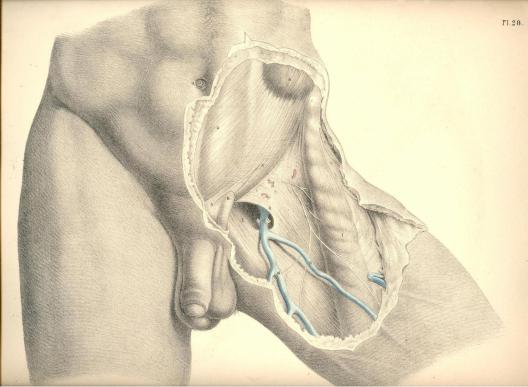
Plate 28
PLATE 29.
A.The muscular part of the external oblique; a, its tendon.
B.The umbilicus.
C.The anterior superior iliac spine.
D.The spine of the os pubis.
E.The cremasteric fibres, within the external ring, surrounding the cord; e, the cremasteric fibres looping over the cord outside the ring.
F.The muscular part of the internal oblique giving off, E, the cremaster; its tendon sheathing the rectus muscle.
G.The linea alba; f, g, the linea semilunaris.
H.The iliac part of the fascia lata; h, the upper cornu of its falciform process.
I.The femoral vein.
K.The femoral artery.
L.The anterior crural nerve.
M.The sartorius muscle.
N.The sheath of the femoral vessels; n, its upper part.
O.The saphena vein.
P.The pubic part of the fascia lata.
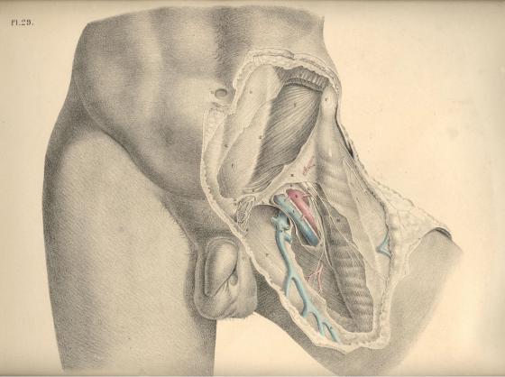
Plate 29
COMMENTARY ON PLATES 30 & 31.
THE SURGICAL DISSECTION OF THE FIFTH, SIXTH, SEVENTH, AND EIGHTH LAYERS OF THE INGUINAL REGION, AND THEIR CONNEXION WITH THOSE OF THE THIGH.
When we remove the internal oblique and cremaster muscles, we expose the transverse muscle, which may be regarded as the fifth inguinal layer, F, Pl. 30. This muscle is similar in shape and dimensions to the internal oblique. The connexions of both are also similar, inasmuch as they arise from the inner edge of the crista ilii, and from the outer half of, V, Poupart's ligament. The fleshy fibres of these two muscles vary but little in direction, and terminate at the same place--viz., the linea semilunaris, which marks the outer border of the rectus muscle. But whilst the fleshy parts of these three abdominal muscles, D E F, form successive strata in the groin, their aponeurotic tendons present the following peculiarities of arrangement in respect to the rectus muscle. The tendon of the external oblique, d, passes altogether in front of the rectus; that of the internal oblique is split opposite the linea semilunaris into two layers, which enclose the rectus between them as they pass to be inserted into the linea alba. But midway between the navel and pubes, at the point marked G, both layers of the tendon are found to pass in front of the rectus. The tendon of the transverse muscle passes behind the rectus; but opposite the point G, it joins both layers of the internal oblique tendon, and with this passes in front of the rectus. The fibrous structure thus constituted by the union of the tendons of the internal oblique and transverse muscles, e f, is named the "conjoined tendon."
The conjoined tendon, f, Plates 30 and 31, appears as a continuation of the linea semilunaris, for this latter is in itself a result of the union of the tendons of the abdominal muscles at the external border of the rectus. As the conjoined tendon curves so far outwards to its insertion into the pectineal ridge of the pubic bone, as to occupy a situation immediately behind the external ring, it thereby fortifies this part against the occurrence of a direct protrusion of the bowel. But the breadth, as well as the density, of this tendon varies in several individuals, and these will accordingly be more or less liable to the occurrence of hernia.
The arched inferior border of the transverse muscle, F, Plate 30, expresses by its abrupt termination that some part is wanting to it; and this appearance, together with the fact that the fibres of this part of the muscle blend with those of the internal oblique and cremaster, and cannot be separated except by severing the connexion, at once suggests the idea that the cremaster is a derivation from both these muscles.
Assuming this to be the case, therefore, it follows that when the dissector removes the cremaster from the space L h, he himself causes this vacancy in the muscular parietes of the groin to occur, and at the same time gives unnatural definition to the lower border of the transverse and oblique muscles. In a dissection so conducted, the cord is made to assume the variable positions which anatomists report it to have in respect to the neighbouring muscles. But when we view nature as she is, and not as fashioned by the scalpel, we never fail to find an easy explanation of her form.
In the foetus, prior to the descent of the testicle, the cremaster muscle does not exist. (Cloquet, op cit.) From this we infer, that those parts of the muscles, E F, Plate 30, which at a subsequent period are converted into a cremaster, entirely occupy the space L h. In the adult body, where one of the testicles has been arrested in the inguinal canal, the muscles, E F, do not present a defined arched margin, above the vacant space L h, but are continued (as in the foetus) as low down as the external abdominal ring.
(Page 77)
78 |
COMMENTARY ON PLATES 30 & 31. |
In the adult, where the testicle has descended to the scrotum, the cremaster exists, and is serially continuous with the muscles, E F, covering the space L h; the meaning of which is, that the cremasteric parts of the muscles, E F, cover this space. The name cremaster therefore must not cancel the fact that the fibres so named are parts of the muscles, E F. Again, in the female devoid of a cremaster, the muscles, E F, present of their full quantities, having sustained no diminution of their bulk by the formation of a cremaster. But when an external inguinal hernia occurs in the female body, the bowel during its descent carries before it a cremasteric covering at the expense of the muscles E F, just in the same way as the testicle does in the foetus. (Cloquet.)
From the above-mentioned facts, viewed comparatively, it seems that the following inferences may be legitimately drawn:--1st, that the space L h does not naturally exist devoid of a muscular covering; for, in fact, the cremaster overlies this situation; 2nd, that the name cremaster is one given to the lower fibres of the internal oblique and transverse muscles which cover this space; and 3rd, that to separate the cremasteric elongation of these muscles, and then describe them as presenting a defined arched margin, an inch or two above Poupart's ligament, is an act as arbitrary on the part of the dissector as if he were to subdivide these muscles still more, and, while regarding the subdivisions as different structures, to give them names of different signification. When once we consent to regard the cremaster as constituted of the fibres originally proper to the muscles, E F, we then are led to the discovery of the true relations of the cord in respect to these muscles.
On removing the transverse muscle, we expose the inguinal part of the transversalis fascia--the sixth inguinal layer, L h, Plate 30--K k, Plate 31. This fascia or membrane affords a general lining to the abdominal walls, in some parts of which it presents of a denser and stronger texture than in others. It is stretched over the abdomen between the muscles and the peritonaeum. The fascia iliaca, the fascia pelvica, and the fascia transversalis, are only regional divisions of the one general membrane. On viewing this fascia in its totality, I find it to exhibit many features in common with those other fibrous structures which envelope serous cavities. The transversalis fascia supports externally the peritonaeum, in the same way as the dura mater supports the arachnoid membrane, or as the pleural fascia supports the serous pleura. While the serous membranes form completely shut sacs, the fibrous membranes which lie external to those sacs are pierced by the vessels which course between them and the serous membranes, and afford sheaths or envelopes for these vessels in their passage from the interior to the external parts. The sheath, H h, Plates 30 and 31, which surrounds the spermatic vessels, and the sheath, R, Plate 31, which envelopes the crural vessels, are elongations of the fascia transversalis.
In the groin, the transversalis fascia, K k, Plate 31, presents, in general, so dense a texture as to offer considerable resistance to visceral pressure. Here it is stretched between the transverse muscle, F, Plate 31, and the peritonaeum, I. It adheres to the external surface of the peritonaeum, and to the internal surface of the transverse muscle, by means of an intervening cellular tissue. It is connected below to Poupart's ligament, along the line of which it joins the fascia iliaca. It lines the lower posterior aspect of the rectus muscle, where this is devoid of its sheath; and it is incorporated with f, the conjoined tendon, thereby fencing the external abdominal ring. Immediately above the middle of Poupart's ligament, this membrane, at the point marked h, Plate 30, is pouched into a canal-shaped elongation, which invests the spermatic vessels as far as the testicle in the scrotum; and to this elongation is given the names "fascia spermatica interna" (Cooper), "fascia infundibuliform" (Cloquet). The same part, when it encloses an external oblique hernia, is named "fascia propria." The neck or inlet of this funnelshaped canal is oval, and named the "internal abdominal ring." As this ring looks towards the interior of the abdomen, and forms the entrance of the funnel-shaped canal, it cannot of course be seen from before until we slit open this canal. Compare the parts marked H h in Plates 30 and 31.
COMMENTARY ON PLATES 30 & 31. |
79 |
The inguinal and iliac portions of the fascia transversalis join along the line of Poupart's ligament, A C. The iliac vessels, in their passage to the thigh, encounter the fascia at the middle third of the crural arch formed by the ligament, and take an investment (the sheath, R) from the fascia. The fore part of this sheath is mentioned as formed by the fascia transversalis--the back part by the fascia iliaca; but these distinctions are merely nominal, and it is therefore unnecessary to dwell upon them. The sheath of the femoral vessels is also funnel-shaped, and surrounds them on all sides. Its broad entrance lies beneath the middle of Poupart's ligament. Several septa are met with in its interior. These serve to separate the femoral vessels from each other. The femoral vein, O, Plate 30, is separated from the falciform margin, S s, of the saphenous opening by one of these septa. Between this septum and the falx an interval occurs, and through it the crural hernia usually descends. These parts will be more particularly noticed when considering the anatomy of crural hernia.
Beneath the fascia transversalis is found the subserous cellular membrane, which serves as a connecting medium between the fascia and the peritonaeum. This cellular membrane may be considered as the seventh inguinal layer. It is described by Scarpa (sull' Ernie) as forming an investment for the spermatic vessels inside the sheath, where it is copious, especially in old inguinal hernia. It is also sometimes mixed with fatty tissue. In it is found embedded the infantile cord--the remains of the upper part of the peritoneal tunica vaginalis--a structure which will be considered in connexion with congenital herniae.
By removing the subserous cellular tissue, we lay bare the peritonaeum, which forms the eighth layer of the inguinal region. Upon it the epigastric and spermatic vessels are seen to rest. These vessels course between the fascia transversalis and the peritonaeum. The internal ring which is formed in the fascia, K h, may be now seen to be closed by the peritonaeum, I. The inguinal canal, therefore, does not, in the normal state of these parts, communicate with the general serous cavity; and here it must be evident that before the bowel, which is situated immediately behind the peritonaeum, I, can be received into the canal, H h, it must either rupture that membrane, or elongate it in the form of a sac.
The exact position which the epigastric, L, Plate 31, and spermatic vessels, M, bear in respect to the internal ring, is a point of chief importance in the surgical anatomy of the groin; for the various forms of herniae which protrude through this part have an intimate relation to these vessels. The epigastric artery, in general, arises from the external iliac, close above the middle of Poupart's ligament, and ascends the inguinal wall in an oblique course towards the navel. It applies itself to the inner border of the internal ring, and here it is crossed on its outer side by the spermatic vessels, as these are about to enter the inguinal canal.
The inguinal canal is the natural channel through which the spermatic vessels traverse the groin on their way to the testicle in the scrotum. In the remarks which have been already made respecting the several layers of structures found in the groin, I endeavoured to realize the idea of an inguinal canal as consisting of elongations of these layers invaginated the one within the other, the outermost layer being the integument of the groin elongated into the scrotal skin, whilst the innermost layer consisted of the transversalis fascia elongated into the fascia spermatica interna, or sheath. The peritonaeum, which forms the eighth layer of the groin, was seen to be drawn across the internal ring of this canal above in such a way as to close it completely, whilst all the other layers, seven in number, were described as being continued over the spermatic vessels in the form of funnel-shaped investments, as far down as the testicle.
80 |
COMMENTARY ON PLATES 30 & 31. |
With the ideas of an inguinal canal thus naturally constituted, I need not hesitate to assert that the form, the extent, and the boundaries of the inguinal canal, as given by the descriptive anatomist, are purely conventional, and do not exist until after dissection; for which reason, and also because the form and condition of these parts so described and dissected do not appear absolutely to correspond in any two individuals, I omit to mention the scale of measurements drawn up by some eminent surgeons, with the object of determining the precise relative position of the several parts of the inguinal region.
The existence of an inguinal canal consisting, as I have described it, of funnel-shaped elongations from the several inguinal layers continued over the cord as far as the testicle, renders the adult male especially liable to hernial protrusions at this part. The oblique direction of the canal is, in some measure, a safeguard against these accidents; but this obliquity is not of the same degree in all bodies, and hence some are naturally more prone to herniae than others.
DESCRIPTION OF THE FIGURES OF PLATES 30 & 31.
PLATE 30.
A.The anterior superior iliac spine.
B.The umbilicus.
C.The spine of the pubis.
D.The external oblique muscle; d, its tendon. .
E.The internal oblique muscle; e, its tendon.
F.The transverse muscle; f, its tendon, forming, with e, the conjoined tendon.
G.The rectus muscle enclosed in its sheath.
H.The fascia spermatica interna covering the cord; h, its funnel-shaped extremity. I, K, L, M. See Plate 31.
N.The femoral artery; n, its profunda branch.
O.The femoral vein.
P.The saphena vein.
Q.The sartorius muscle.
R.The sheath of the femoral vessels.
S.The falciform margin of the saphenous opening.
T.The anterior crural nerve.
U.The pubic portion of the fascia lata.
V.The iliac portion attached to Poupart's ligament.
W.The lower part of the iliacus muscle.

PLATE 30
PLATE 31.
A.The anterior superior iliac spine.
B.The umbilicus.
C.The spine of the pubis.
D.The external oblique muscle; d, its tendon; d*, the external ring.
E.The internal oblique muscle.
F.The transverse muscle; f, its tendon; forming, with e, the conjoined tendon.
G.The rectus muscle laid bare.
H h. The fascia spermatica interna laid open above and below d*, the external ring.
I. The peritonaeum closing the internal ring.
K.The fascia transversalis; k, its pubic part.
L.The epigastric artery and veins.
M.The spermatic artery, veins, and vas deferens bending round the epigastric artery at the internal ring; m, the same vessels below the external ring.
N.The femoral artery; n, its profunda branch.
O.The femoral vein, joined by--
P.The saphena vein.
Q.The sartorius muscle.
R.The sheath of the femoral vessels.
S S. The falciform margin of the saphenous opening,
T.The anterior crural nerve.
U.The pubic part of the fascia lata.
V.The iliac part of the fascia lata.
W.The lower part of the iliacus muscle.