

SURGICAL ANATOMY by Joseph Maclise
.pdf90 |
COMMENTARY ON PLATES 39 & 40. |
PLATE 39, Fig. 2.--The testicle in the scrotum.--When the testicle, 5, descends into the scrotum, 7, which happens in general at the time of birth, the abdomino-scrotal fibroserous membrane, 6 a, 6 d, is still continuous at the internal ring, 6 b. From this point downwards, to a level with the upper border of the testicle, the canal of communication between the scrotal cavity and the abdomen becomes elongated and somewhat constricted. At this part, the canal itself consists, like the abdominal membrane above and the scrotal membrane below, of a fibrous and serous layer, the latter enclosed within the former. The serous lining of this canal is destined to be obliterated, while the outer fibrous membrane is designed to remain in its primitive condition. When the serous canal contracts and degenerates to the form of a simple cord, it leaves the fibrous canal still continuous above with the fibrous membrane (transversalis fascia) of the abdomen, and below with the fibrous envelope (tunica albuginea) of the testis; and at the adult period, this fibrous canal is known as the internal spermatic sheath, or infundibuliform fascia enclosing the remains of the serous canal, together with the spermatic vessels, &c.
Plate 39--Figure 2

PLATE 39, Fig. 3.--The serous tunica vaginalis is separated from the peritonaeum.--
When the testicle, 7, has descended to the scrotum, the serous tube or lining of the inguinal canal and cord, 6 b, 6 c, closes and degenerates into a simple cord, (infantile spermatic cord,) and thereby the peritonaeal sac, 6 a, becomes distinct from the serous tunica vaginalis, 6 d. But the fibrous tube, or outer envelope of the inguinal canal, remains still pervious, and continues in this condition throughout life. In the adult, we recognise this fibrous tube as the infundibuliform fascia of the cord, or as forming the fascia propria of an external inguinal hernia. The anterior part of the fibrous spermatic tube descends from the fascia transversalis; the posterior part is continuous with the fascia iliaca. In relation to the testicle, the posterior part will be seen to be reflected over the body of the gland as the tunica albuginea, while the anterior part blends with the cellular tissue of the front wall of the scrotum. The tunica vaginalis, 6 d, is now traceable as a distinct sac,[Footnote] closed on all sides, and reflected from the fore part of the testicle, above and below, to the posterior aspect of the front wall of the scrotum.
[Footnote: Mr. Owen states that the Chimpanzee alone, amongst brute animals, has the tunica vaginalis as a distinct sac.]
Plate 39--Figure 3
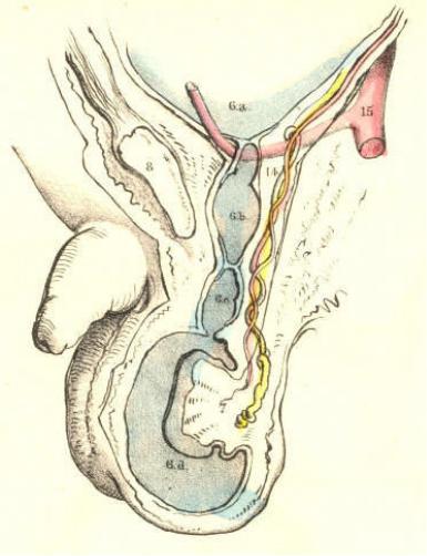
PLATE 40, Fig. 1.--The abdomino-scrotal serous lining remains continuous at the internal ring, and a congenital hydrocele is formed.--When the serous spermatic tube, 6 b, 6 c, remains pervious and continuous above with the peritonaeum, 6 a, and below with the serous tunica vaginalis, 6 d, the serous fluid of the abdomen will naturally gravitate to the most depending part--viz., the tunica vaginalis; and thus a hydrocele is formed. This kind of hydrocele is named congenital, owing to the circumstance that the natural process of obliteration, by which the peritonaeum becomes separated from the tunica vaginalis, has been, from some cause, arrested. [Footnote 1] As long as the canal of communication, 6 b, 6 c, between the tunica vaginalis, 6 d, and the peritonaeum 6 a, remains pervious, which it may be throughout life, this form of hydrocele is, of course, liable to occur. It may be diagnosed from diseased enlargements of the testicle, by its transparency, its fluctuation, and its smooth, uniform fulness and shape, besides its being of less weight than a diseased testis of the same size would be. It may be distinguished from the common form of hydrocele of the isolated tunica vaginalis by the fact, that pressure made on the scrotum will cause the fluid to pass freely into the general cavity of the peritonaeum. As the fluid distends the tunica vaginalis, 6 c, 6 d, in front of the testis, this organ will of course lie towards the back of the scrotum, and therefore, if it be found necessary to evacuate the fluid, the puncture may be made with most safety in front of the scrotum. If ascites should form in an adult in whom the tunica vaginalis still communicates with the peritonaeal sac, the fluid which accumulates in the latter membrane will also distend the former, and all the collected fluid may be evacuated by tapping the scrotum. When a hydrocele is found to be congenital, it must be at once obvious that to inject irritating fluids into the tunica vaginalis (the radical cure) is inadmissible. In an adult, free from all structural disease, and in whom a congenital hydrocele is occasioned by the gravitation of the ordinary serous secretion of the peritonaeum, a cure may be effected by causing the obliteration of the serous spermatic canal by the pressure of a truss. When a congenital hydrocele happens in an infant in whom the testicle, 5, Fig. 1, Plate 39, is arrested in the inguinal canal, [Footnote 2] if pressure be made on this passage with a view of causing its closure, the testicle will be prevented from descending.
[Footnote 1: The serous spermatic tube remains open in all quadrupeds; but their natural prone position renders them secure against hydrocele or hernial protrusion. It is interesting to notice how in man, and the most anthropo-morphous animals, where the erect position would subject these to the frequent accident of hydrocele or hernia, nature causes the serous spermatic tube to close.]
[Footnote 2: In many quadrupeds (the Rodentia and Monotremes) the testes remain within the abdomen. In the Elephant, the testes always occupy their original position beneath the kidneys, in the loins. Human adults are occasionally found to be "testiconde;" the testes being situated below the kidneys, or at some part between this position and the internal inguinal ring. Sometimes only one of the testes descends to the scrotum.]

Plate 40--Figure 1.
COMMENTARY ON PLATES 39 & 40. |
91 |
PLATE 40, Fig. 2.--The serous spermatic canal closes imperfectly, so as to become sacculated, and thus a hydrocele of the cord is formed.--After the testicle, 7, has descended to the scrotum, the sides of the serous tube, or lining of the inguinal canal and cord, 6 b, 6 c, may become adherent at intervals; and the intervening sacs of serous membrane continuing to secrete their proper fluid, will occasion a hydrocele of the cord. This form of hydrocele will differ according to the varieties in the manner of closure; and these may take place in the following modes:--1st, if the serous tube close only at the internal ring, 6 a, while the lower part of it, 6 b, 6 c, remains pervious, and communicating with the tunica vaginalis, 6 d, a hydrocele will be formed of a corresponding shape; 2nd, if the tube close at the upper part of the testicle, 6 c, thus isolating the tunica vaginalis, 6 d, while the upper part, 6 b, remains pervious, and the internal ring, 6 a, open, and communicating with the peritonaeal sac, a hydrocele of the cord will happen distinct from the tunica vaginalis; or this latter may be, at the same time, distended with fluid, if the disposition of the subject be favourable to the formation of dropsy; 3rd, the serous tube may close at the internal ring, form sacculi along the cord, and close again at the top of the testicle, thus separating the tunica vaginalis from the abdomen, and thereby several isolated hydroceles may be formed. If in this condition of the parts we puncture one of the sacs for the evacuation of its contents, the others, owing to their separation, will remain distended.
Plate 40--Figure 2.

PLATE 40, Fig. 3.--Hydrocele of the isolated tunica vaginalis.--When the serous spermatic tube, 6 b, 6 c, becomes obliterated, according to the normal rule, after the descent of the testicle, 7, the tunica vaginalis, 6 d, is then a distinct serous sac. If a hydrocele form in this sac, it may be distinguished from the congenital variety by its remaining undiminished in bulk when the subject assumes the horizontal position, or when pressure is made on the tumour, for its contents cannot now be forced into the abdomen. The testicle, 7, holds the same position in this as it does in the congenital hydrocele. [Footnote] The radical cure may be performed here without endangering the peritonaeal sac. Congenital hydrocele is of a cylindrical shape; and this is mentioned as distinguishing it from isolated hydrocele of the tunica vaginalis, which is pyriform; but this mark will fail when the cord is at the same time distended, as it may be, in the latter form of the complaint.
[Footnote: When a hydrocele is interposed between the eye and a strong light, the testis appears as an opaque body at the back of the tunica vaginalis. But this position of the organ is, from several causes, liable to vary. The testis may have become morbidly adherent to the front wall of the serous sac, in which case the hydrocele will distend the sac laterally. Or the testis may be so transposed in the scrotum, that, whilst the gland occupies its front part, the distended tunica vaginalis is turned behind. The tunica vaginalis, like the serous spermatic tube, may, in consequence of inflammatory fibrinous effusion, become sacculated-multilocular, in which case, if a hydrocele form, the position of the testis will vary accordingly.--See Sir Astley Cooper's work, ("Anatomy and Diseases of the Testis;") Morton's "Surgical Anatomy;" Mr. Curling's "Treatise on Diseases of the Testis;" and also his article "Testicle," in the Cyclopaedia of Anatomy and Physiology.]
Plate 40--Figure 3.

92 |
COMMENTARY ON PLATES 39 & 40. |
PLATE 40, Fig. 4.--The serous spermatic tube remaining pervious, a congenital hernia is formed.--When the testicle, 7, has descended to the scrotum, if the communication between the peritonaeum, 6 a, and the tunica vaginalis, 6 c, be not obliterated, a fold of the intestine, 13, will follow the testicle, and occupy the cavity of the tunica vaginalis, 6 d. In this form of hernia (hernia tunicae vaginalis, Cooper), the intestine is in front of, and in immediate contact with, the testicle. The intestine may descend lower than the testicle, and envelope this organ so completely as to render its position very obscure to the touch. This form of hernia is named congenital, since it occurs in the same condition of the parts as is found in congenital hydrocele--viz., the inguinal ring remaining unclosed. It may occur at any period of life, so long as the original congenital defect remains. It may be distinguished from hydrocele by its want of transparency and fluctuation. The impulse which is communicated to the hand applied to the scrotum of a person affected with scrotal hernia, when he is made to cough, is also felt in the case of congenital hydrocele. But in hydrocele of the separate tunica vaginalis, such impulse is not perceived. Congenital hernia and hydrocele may co-exist; and, in this case, the diagnostic signs which are proper to each, when occurring separately, will be so mingled as to render the precise nature of the case obscure.
Plate 40--Figure 4.

PLATE 40, Fig. 5.--Infantile hernia.--When the serous spermatic tube becomes merely closed, or obliterated at the inguinal ring, 6 b, the lower part of it, 6 c, is pervious, and communicating with the tunica vaginalis, 6 d. In consequence of the closure of the tube at the inguinal ring, if a hernia now occur, it cannot enter the tunica vaginalis, and come into actual contact with the testicle. The hernia, 13, therefore, when about to force the peritonaeum, 6 a, near the closed ring, 6 b, takes a distinct sac or investment from this membrane. This hernial sac, 6 e, will vary as to its position in regard to the tunica vaginalis, 6 d, according to the place whereat it dilates the peritonaeum at the ring. The peculiarity of this hernia, as distinguished from the congenital form, is owing to the scrotum containing two sacs,--the tunica vaginalis and the proper sac of the hernia; whereas, in the congenital variety, the tunica vaginalis itself becomes the hernial sac by a direct reception of the naked intestine. If in infantile hernia a hydrocele should form in the tunica vaginalis, the fluid will also distend the pervious serous spermatic tube, 6 c, as far up as the closed internal ring, 6 b, and will thus invest and obscure the descending herniary sac, 13. This form of hernia is named infantile (Hey), owing to the congenital defect in that process, whereby the serous tube lining the cord is normally obliterated. Such a form of hernia may occur at the adult age for the first time, but it is still the consequence of original default.
Plate 40--Figure 5.
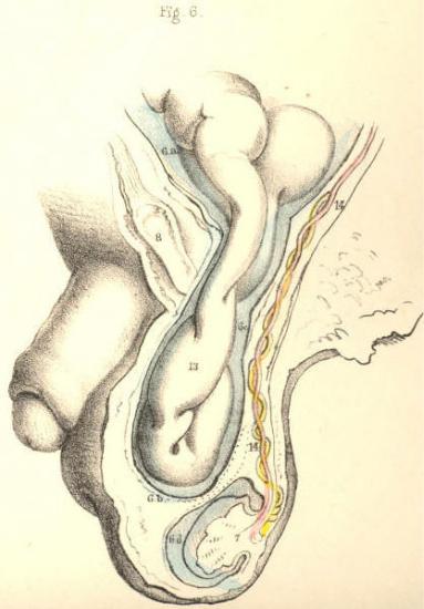
PLATE 40, Fig. 6.--Oblique inguinal hernia in the adult.--This variety of hernia occurs not in consequence of any congenital defect, except inasmuch as the natural weakness of the inguinal wall opposite the internal ring may be attributed to this cause. The serous spermatic tube has been normally obliterated for its whole length between the internal ring and the tunica vaginalis; but the fibrous tube, or spermatic fascia, is open at the internal ring where it joins the transversalis fascia, and remains pervious as far down as the testicle. The intestine, 13, forces and distends the upper end of the closed serous tube; and as this is now wholly obliterated, the herniary sac, 6 c, derived anew from the inguinal peritonaeum, enters the fibrous tube, or sheath of the cord, and descends it as far as the tunica vaginalis, 6 d, but does not enter this sac, as it is already closed. When we compare this hernia, Fig. 6, Plate 40, with the infantile variety, Fig. 5, Plate 40, we find that they agree in so far as the intestinal sac is distinct from the tunica vaginalis; whereas the difference between them is caused by the fact of the serous cord remaining in part pervious in the infantile hernia; and on comparing Fig. 6, Plate 40, with the congenital variety, Fig. 4, Plate 40, we see that the intestine has acquired a new sac in the former, whereas, in the latter, the intestine has entered the tunica vaginalis. The variable position of the testicle in Figs. 4, 5, & 6, Plate 40, is owing to the variety in the anatomical circumstances under which these herniae have happened.
Plate 40--Figure 6.

COMMENTARY ON PLATES 41 & 42.
DEMONSTRATIONS OF THE ORIGIN AND PROGRESS OF INGUINAL HERNIAE
IN GENERAL.
PLATE 41, Fig. 1.--When the serous spermatic tube is obliterated for its whole length between the internal ring, 1, and the top of the testicle, 13, a hernia, in order to enter the inguinal canal, 1, 4, must either rupture the peritonaeum at the point 1, or dilate this membrane before it in the form of a sac. [Footnote] If the peritonaeum at the point 1 be ruptured by the intestine, this latter will enter the fibrous spermatic tube, 2, 3, and will pass along this tube devoid of the serous sac. If, on the other hand, the intestine dilates the serous membrane at the point, 1, where it stretches across the internal ring, it will, on entering the fibrous tube, (infundibuliform fascia,) be found invested by a sac of the peritonaeum, which it dilates and pouches before itself. As the epigastric artery, 9, bends in general along the internal border of the ring of the fibrous tube, 2, 2, the neck of the hernial sac which enters the ring at a point external to the artery must be external to it, and remain so despite all further changes in the form, position, and dimensions of the hernia. And as this hernia enters the ring at a point anterior to the spermatic vessels, its neck must be anterior to them. Again, if the bowel be invested by a serous sac, formed of the peritonaeum at the point 1, the neck of such sac must intervene between the protruding bowel and the epigastric and spermatic vessels. But if the intestine enter the ring of the fibrous tube, 2, 2, by having ruptured the peritonaeum at the point 1, then the naked intestine will lie in immediate contact with these vessels.
[Footnote: Mr. Lawrence (op. cit.) remarks, "When we consider the texture of the peritonaeum, and the mode of its connexion to the abdominal parietes, we cannot fancy the possibility of tearing the membrane by any attitude or motion." Cloquet and Scarpa have also expressed themselves to the effect, that the peritonaeum suffers a gradual distention before the protruding bowel.]
Plate 41--Figure 1