

1Paul M Dewick Medicalc Natural / booktext@id88013693placeboie
.pdfPEPTIDE HORMONES |
415 |
(Continued )
result, ovulation does not usually occur during breast feeding, preventing further conception. No prolactin derivatives are currently used in medicine, but the ergot alkaloid derivatives bromocriptine and cabergoline (see page 375) are dopamine agonists employed to inhibit prolactin release by pituitary tumours. They are not now recommended for routine suppression of lactation.
Gonadotrophins
Follicle-stimulating hormone (FSH) and luteinizing hormone (LH) are involved in controlling both male and female reproduction (see LH-RH, page 412). These are glycoproteins both composed of two polypeptide chains of 89 and 115 amino acid residues. The shorter chains are essentially identical, and differences in activity are caused by differences in the longer chain. Each chain has asparagine-linked oligosaccharide residues: the short chains each have one, the long chains two in the case of FSH or one for LH. FSH and LH together (human menopausal gonadotrophins; menotrophin) are purified from the urine of post-menopausal women and used in the treatment of female infertility. FSH alone is also available for this purpose, either natural material from urine termed urofollitropin (urofollitrophin), or the recombinant proteins follitropin alfa and follitropin beta. Chorionic gonadotrophin (human chorionic gonadotrophin; HCG) is a gonad-stimulating glycoprotein hormone which is obtained for drug use from the urine of pregnant women. It may be used to stimulate testosterone production in males with delayed puberty.
the antidiuretic hormone. These nonapeptides containing a disulphide bridge are structurally very similar, differing in only two amino acid residues (Figure 7.11). Structurally related peptides are
classified as belonging to the vasopressin family when the amino acid residue at position 8 is basic, e.g. Arg or Lys, or to the oxytocin family when this amino acid is neutral.
Cys–Tyr–Ile–Gln–Asn–Cys–Pro–Leu–Gly–NH2 |
(desamino)Cys–Tyr–Phe–Gln–Asn–Cys–Pro–D-Arg–Gly–NH2 |
oxytocin |
desmopressin (1-desamino-8-D-Arg-VP) |
Cys–Tyr–Phe–Gln–Asn–Cys–Pro–Arg–Gly–NH2 |
Gly–Gly–Gly–Cys–Tyr–Phe–Gln–Asn–Cys–Pro–Lys–Gly–NH2 |
argipressin (arginine vasopressin) (human/bovine) |
terlipressin (Gly-Gly-Gly-8-L-Lys-VP) |
Cys–Tyr–Phe–Gln–Asn–Cys–Pro–Lys–Gly–NH2
lypressin (lysine vasopressin) (porcine)
Figure 7.11
Oxytocin
Oxytocin (Figure 7.11) stimulates the pregnant uterus, causing contractions, and also brings about ejection of milk from the breasts. It thus plays a major role in the normal onset of labour at the end of pregnancy. Oxytocin for drug use is produced by synthesis, and is employed to induce or augment labour, as well as to minimize subsequent blood loss.
(Continues)

416 |
PEPTIDES, PROTEINS, AND OTHER AMINO ACID DERIVATIVES |
(Continued )
Vasopressin
Vasopressin (antidiuretic hormone, ADH) is a hormone that has an antidiuretic action on the kidney, regulating the reabsorption of water. A deficiency in this hormone leads to diabetes insipidus, where the patient suffers increased urine output and intense thirst, typically consuming enormous quantities of fluid. Vasopressin is used to treat this condition. At high dosage, vasopressin promotes contraction of arterioles and capillaries, and brings about an increase in blood pressure. The structure of human and bovine vasopressin (arginine vasopressin; argipressin) (Figure 7.11) differs from that of oxytocin only in two amino acid residues. Lysine vasopressin (lypressin) (Figure 7.11) from pigs differs from arginine vasopressin in the second amino acid from the C-terminus. Both bovine and porcine peptides have been used medicinally, but these have been replaced by synthetic materials. The 1- desamino-8-D-Arg-vasopressin analogue desmopressin (Figure 7.11) has a longer duration of action than vasopressin and may also be administered orally. In contrast to the natural hormone, desmopressin has no vasoconstrictor effect. Terlipressin (Figure 7.11) is a lypressin pro-drug in which the polypeptide chain has been extended by three glycine residues. Enzymic hydrolysis liberates lypressin. It is mainly used for control of oesophageal bleeding.
Pancreatic Hormones
The hormone insulin (Figure 7.12) plays a key role in the regulation of carbohydrate, fat, and protein metabolism. In particular, it has a hypoglycaemic effect, lowering the levels of glucose in the blood. A deficiency in insulin synthesis leads to the condition diabetes, treatment of which requires daily injections of insulin. Insulin is composed of two straight chain polypeptides joined by disulphide bridges. This structure is known to arise from a straight chain
polypeptide preproinsulin containing 100 amino acid residues. This loses a 16-residue portion of its chain, and forms proinsulin with disulphide bridges connecting the terminal portions of the chain in a loop (Figure 7.13). A central portion of the loop (the C chain) is then cleaved out, leaving the A chain (21 residues) bonded to the B chain (30 residues) by two disulphide bridges. This is the resultant insulin. Mammalian insulins (Figure 7.12) from different sources are very similar, showing variations in the sequence
A chain
Gly–Ile–Val–Glu–Gln–Cys–Cys–Thr–Ser–Ile–Cys–Ser–Leu–Tyr–Gln–Leu–Glu–Asn–Tyr–Cys–Asn
8 9 10 21
B chain Phe–Val–Asn–Gln–His–Leu–Cys–Gly–Ser–His–Leu–Val–Glu–Ala–Leu–Tyr–Leu–Val–Cys–Gly–Glu–Arg
|
|
|
|
|
|
Thr–Lys–Pro–Thr–Tyr–Phe–Phe–Gly |
|||
|
|
|
|
|
|
30 |
29 |
28 |
|
|
|
|
|
|
|
human insulin |
|
|
|
|
|
A |
|
|
B |
|
|
|
|
|
8 |
9 |
10 |
28 |
29 |
30 |
|
|
|
insulin (human) |
Thr |
Ser |
Ile |
Pro |
Lys |
Thr |
His–Ser–Gln–Gly–Thr–Phe–Thr–Ser–Asp–Tyr– |
||
insulin (porcine) |
Thr |
Ser |
Ile |
Pro |
Lys |
Ala |
|||
Ser–Lys–Tyr–Leu–Asp–Ser–Arg–Arg–Ala–Gln– |
|||||||||
insulin (bovine) |
Ala |
Ser |
Val |
Pro |
Lys |
Ala |
Asp–Phe–Val–Gln–Trp–Leu–Met–Asn–Thr |
||
|
|
||||||||
insulin lispro |
Thr |
Ser |
Ile |
Lys |
Pro |
Thr |
|
glucagon |
|
|
|
||||||||
insulin aspart |
Thr |
Ser |
Ile |
Asp |
Lys |
Thr |
|
|
|
Figure 7.12

PEPTIDE HORMONES |
417 |
B |
A |
|
|
|
S S |
|
A |
|
|
|
S |
|
S |
|
A |
|
|
|
|
|
|
|
|
|
|||||||
|
|
|
|
|
|
|
|||||||||
|
B |
|
|
|
|
B |
|
|
|
||||||
|
C |
|
|
|
S S |
|
C |
|
|
|
S |
|
S |
|
|
|
|
|
|
|
|
|
|
|
|
|
|||||
|
|
|
|
|
|
|
|
||||||||
|
|
|
|
|
|
|
|
|
|
|
|
|
|
|
|
preproinsulin |
proinsulin |
insulin |
Figure 7.13
of amino acid residues 8–10 in chain A, and at amino acid 30 in chain B. Glucagon (Figure 7.12) is a straight-chain polypeptide hormone containing 29 amino acids that is secreted by the pancreas when blood sugar levels are low, thus stimulating breakdown of glycogen in the liver. Unlike insulin, its structure is identical in all animals.
Interferons
The interferons are a family of proteins secreted by animal cells in response to viral and parasitic infections, and are part of the host’s defence mechanism. They display multiple activities, affecting the functioning of the immune system, cell proliferation, and cell differentiation, primarily by inducing the synthesis of other proteins. Accordingly, they have potential as antiviral, antiprotozoal, immunomodulatory, and cell growth regulatory agents.
Opioid Peptides
Although the pain-killing properties of morphine and related compounds have been known for a considerable time (see page 329), the existence of endogenous peptide ligands for the receptors to which these compounds bind is a more recent discovery. It is now appreciated that the body
Insulin
Insulin is a hormone produced by the pancreas that plays a key role in the regulation of carbohydrate, fat, and protein metabolism. In particular, it has a hypoglycaemic effect, lowering the levels of glucose in the blood. If a malfunctioning pancreas results in a deficiency in insulin synthesis or secretion, the condition known as diabetes mellitus ensues. This results in increased amounts of glucose in the blood and urine, diuresis, depletion of carbohydrate stores, and subsequent breakdown of fat and protein. Incomplete breakdown of fat leads to the accumulation of ketones in the blood, severe acidosis, coma, and death. Where the pancreas is still functioning, albeit less efficiently, the condition is known as type 2 diabetes (non-insulin-dependent diabetes, NIDDM) and can be controlled by a controlled diet or oral antidiabetic drugs. In type 1 diabetes (insulin-dependent diabetes, IDDM) pancreatic cells no longer function, and injections of insulin are necessary, one to four times daily, depending on the severity of the condition. These need to be combined with a controlled diet and regular monitoring of glucose levels, but do not cure the disease, so treatment is lifelong. Mammalian insulins from different sources are very similar and may be used to treat diabetes. Porcine insulin and bovine insulin (Figure 7.12) are extracted from the pancreas of pigs and cattle respectively. Human insulin (Figure 7.12) is produced by the use of recombinant DNA technology in Escherichia coli to obtain the two polypeptide chains, and linking these chemically to form the disulphide bridges (such material is coded ‘crb’), or by modification of proinsulin produced in genetically modified E. coli (coded ‘prb’). Human insulin may also be obtained from porcine insulin by semi-synthesis, replacing the terminal alanine in chain B with threonine by enzymic methods (coded ‘emp’). Human insulin does not appear to be less immunogenic than animal insulin, but genetic engineering offers significant advantages over animal sources for obtaining highly purified material. Insulin may be provided in a rapid-acting soluble form, as suspensions of the zinc complex which have longer duration, or as suspensions with protamine
(Continues)

418 |
PEPTIDES, PROTEINS, AND OTHER AMINO ACID DERIVATIVES |
(Continued )
(as isophane insulin). Protamine is a basic protein from the testes of fish of the salmon family, e.g. Salmo and Onchorhynchus species, which complexes with insulin, thereby reducing absorption and providing a longer period of action. Recently introduced recombinant human insulin analogues insulin lispro and insulin aspart have a faster onset and a shorter duration of action than soluble insulin. Insulin lispro has the reverse sequence for the 28 and 29 amino acids in the B chain, i.e. B28-Lys–B29-Pro, whilst insulin aspart has a single substitution of aspartic acid for proline at position 28 in the B chain (Figure 7.12). These changes in the primary structure affect the tendency of the molecule to associate into dimers and larger oligomers, thus increasing the availability of absorbable monomers. Insulin aspart is produced by expression in Saccharomyces cerevisiae (baker’s yeast). Insulin glargine is a new ultra- long-acting analogue that differs from human insulin by replacing the terminal asparagine at position 21 in chain A with glycine, and also adding two arginines to the end of the B chain, i.e. positions B31 and B32. These changes result in enhanced basicity, causing precipitation at neutral pH post-injection, and consequently a delayed, very gradual and prolonged activity profile (up to 24–48 hour duration of action) and allowing once-daily dosing.
Glucagon
Glucagon (Figure 7.12) is a straight-chain polypeptide hormone containing 29 amino acids that is secreted by the pancreas when blood sugar levels are low, thus stimulating breakdown of glycogen in the liver. It may be isolated from animal pancreas, or be produced by recombinant DNA processes using Saccharomyces cerevisiae. It may be administered for the emergency treatment of diabetes patients suffering from hypoglycaemia as a result of building up a dangerously high insulin level. Normally, a patient would counter this by eating some glucose or sucrose, but hypoglycaemia can rapidly cause unconsciousness, requiring very prompt action.
produces a family of endogenous opioid peptides, which bind to a series of receptors in different locations. These peptides include enkephalins, endorphins, and dynorphins, and are produced primarily, but not exclusively, in the pituitary gland. The pentapeptides Met-enkephalin and Leu-enkephalin (Figure 7.14) were the first to be characterized. The largest peptide is β-endorphin
(‘endogenous morphine’ ) (Figure 7.14), which is several times more potent than morphine in relieving pain. Although β-endorphin contains the sequence for Met-enkephalin, the latter peptide and Leu-enkephalin are derived from a larger peptide proenkephalin A, whilst β-endorphin itself is formed by cleavage of pro-opiomelanocortin. The proenkephalin A structure contains four
Interferons
Interferons were originally discovered as proteins that interfered with virus replication. When mice were injected with antibodies to interferons, they became markedly susceptible to virus-mediated disease, including virus-related tumour induction. Interferons can be detected at low levels in most human tissues, but amounts increase upon infection with viruses, bacteria, protozoa, and exposure to certain growth factors. Interferons were initially classified according to the cellular source, but recent nomenclature is based primarily on sequencing data. Thus leukocyte interferon (a mixture of proteins) is now known as interferon alfa, fibroblast interferon as interferon beta, and immune interferon as interferon gamma.
(Continues)

PEPTIDE HORMONES |
419 |
(Continued )
These typically range in size from 165 to 172 amino acid residues. The interferon system can often impair several steps in viral replication, and can modulate the immune system, and affect cell growth and differentiation. They have potential in treating many human diseases, including leukaemias and solid tumours, and viral conditions such as chronic infection with hepatitis B and C. Much of their current drug use is still research based. Interferon alfa is a protein containing 166 amino acid residues with two disulphide bridges and is produced by recombinant DNA techniques using Escherichia coli, or by stimulation of specific human cell lines. Variants with minor differences in sequence may be obtained according to the gene used, and are designated as alfa-2a, alfa-2b, etc. Interferon alfa is employed as an antitumour drug against certain lymphomas and solid tumours, and in the management of chronic hepatitis. Interferon beta is of value in the treatment of multiple sclerosis patients, though not all patients respond. Variants are designated beta-1a (a glycoprotein with 165 amino acid residues and one disulphide bridge) or beta-1b (a non-glycosylated protein with 164 amino acid residues and one disulphide bridge). Interferon gamma produced by recombinant DNA methods contains an unbridged polypeptide chain, and is designated gamma-1a (146 residues) or gamma-1b (140 residues) according to sequence. The immune interferon interferon gamma-1b is used to reduce the incidence of serious infection in patients with chronic granulomatous disease.
Met-enkephalin sequences and one of Leuenkephalin. The dynorphins, e.g. dynorphin A (Figure 7.14) are also produced by cleavage of a larger precursor, namely proenkephalin B (prodynorphin), and all contain the Leu-enkephalin sequence. Some 20 opioid ligands have now been characterized. When released, these endogenous opioids act upon specific receptors, inducing analgesia, and depressing respiratory function and several other processes. The individual peptides have relatively high specificity towards different receptors. It is known that morphine, β-endorphin, and Met-enkephalin are agonists for the same site. The opioid peptides are implicated in analgesia brought about by acupuncture, since opiate antagonists can reverse the effects. The hope of exploiting similar peptides as ideal, non-addictive analgesics has yet to be attained; repeated doses of endorphin or enkephalin produce addiction and withdrawal symptoms.
Enzymes
Enzymes are proteins that act as biological catalysts. They facilitate chemical modification of substrate molecules by virtue of their specific binding properties, which arise from particular combinations of functional groups in the constituent amino acids at the so-called active site. In many cases, an essential cofactor, e.g. NAD+, PLP, or TPP, may also be bound to participate in the transformation. The involvement of enzymes in biochemical reactions has been a major theme throughout this book. The ability of enzymes to carry out quite complex chemical reactions, rapidly, at room temperature, and under essentially neutral conditions is viewed with envy by synthetic chemists, who are making rapid progress in harnessing this ability for their own uses. Several enzymes are currently of importance commercially, or for medical use, and
Tyr–Gly–Gly–Phe–Met–Thr–Ser–Glu–Lys–Ser– |
Tyr–Gly–Gly–Phe–Met |
Tyr–Gly–Gly–Phe–Leu |
|
Gln–Thr–Pro–Leu–Val–Thr–Leu–Phe–Lys–Asn– |
Met-enkephalin |
Leu-enkephalin |
|
Ala–Ile–Val–Lys–Asn–Ala–His–Lys–Lys–Gly– |
|||
|
|
Gln
β-endorphin
Tyr–Gly–Gly–Phe–Leu–Arg–Arg–Ile–Arg–Pro–Lys–Leu–Lys–Trp–Asp–Asn–Gln
dynorphin A
Figure 7.14
420 |
PEPTIDES, PROTEINS, AND OTHER AMINO ACID DERIVATIVES |
||
|
Table 7.2 Pharmaceutically important enzymes |
|
|
|
|
|
|
Enzyme |
Action |
Source |
Use |
|
|
|
|
Hydrolytic enzymes |
|
|
|
Pancreatin |
Hydrolysis of starch |
Porcine pancreas |
Digestive aid |
|
(amylase), fat (lipase), and |
|
|
|
protein (protease) |
|
|
Papain |
Hydrolysis of proteins |
Papaya fruit (Carica |
Meat tenderizer; |
|
|
papaya; Caricaceae) |
cleaning of contact |
|
|
|
lenses |
Chymotrypsin |
Hydrolysis of proteins |
Bovine pancreas |
Zonal lysis in cataract |
|
|
|
removal |
Hyaluronidase |
Hydrolysis of |
Mammalian testes |
Renders tissues more |
|
mucopolysaccharides |
|
permeable for |
|
|
|
subcutaneous or |
|
|
|
intramuscular |
|
|
|
injections |
Pepsin |
Hydrolysis of proteins |
Porcine stomach |
Digestive aid |
Trypsin |
Hydrolysis of proteins |
Bovine pancreas |
Wound and ulcer |
|
|
|
cleansing |
Fibrinolytic enzymes |
|
|
|
Streptokinase |
No enzymic activity, until it |
Streptococcus |
Treatment of venous |
|
complexes with and activates |
haemolyticus |
thrombosis and |
|
plasminogen in blood plasma |
|
pulmonary embolism |
|
to produce the proteolytic |
|
|
|
enzyme plasmin, which |
|
|
|
hydrolyses fibrin clots |
|
|
Urokinase |
A protease which activates |
Human urine, or |
Treatment of venous |
|
plasminogen in blood plasma |
human kidney tissue |
thrombosis and |
|
to form plasmin, which |
cultures |
pulmonary embolism; |
|
hydrolyses fibrin clots |
|
thrombolysis in the |
|
|
|
eye |
Alteplase (recombinant tissue-type plasminogen activator; rt-PA)
Anistreplase (acylated plasminogen-streptokinase activator complex; APSAC)
A protease which binds to |
Recombinant genetic |
Treatment of acute |
fibrin converting it to a potent |
engineering: human |
myocardial infarction |
plasminogen activator; only |
gene expressed in |
|
active at the surface of the |
Chinese hamster |
|
blood clot |
ovary cells |
|
An inactive acylated form of |
Semi-synthesis from |
Treatment of acute |
the plasminogen–strepto- |
urokinase |
myocardial infarction |
kinase activator complex; the |
|
|
acyl group (p-anisoyl) is |
|
|
slowly hydrolysed in the |
|
|
blood to give the active agent |
|
|
Reteplase |
A fibrinolytic protease; a |
Recombinant genetic |
Treatment of acute |
|
genetically-engineered human |
engineering |
myocardial infarction |
|
tissue-type plasminogen |
|
|
|
activator differing from |
|
|
|
alteplase at four amino acid |
|
|
|
residues |
|
|
|
|
|
|
|
|
|
(Continues) |
|
NONRIBOSOMAL PEPTIDE BIOSYNTHESIS |
421 |
|
|
Table 7.2 (Continued) |
|
|
|
|
|
|
Enzyme |
Action |
Source |
Use |
|
|
|
|
Others |
|
|
|
Asparaginase |
Degradation of L-asparagine |
Erwinia chrysanthemi |
Treatment of acute |
(crisantaspase) |
|
|
lymphoblastic |
|
|
|
leukaemia; results in |
|
|
|
death of those tumour |
|
|
|
cells which require |
|
|
|
increased levels of |
|
|
|
exogenous |
|
|
|
L-asparagine; |
|
|
|
side-effects nausea |
|
|
|
and vomiting, allergic |
|
|
|
reactions and |
|
|
|
anaphylaxis |
Streptodornase |
Depolymerization of |
Streptococcus |
In combination with |
|
polymerized |
haemolyticus |
streptokinase as |
|
deoxyribonucleo-proteins |
|
desloughing agent to |
|
|
|
cleanse ulcers and |
|
|
|
promote healing |
|
|
|
|
these are described in Table 7.2. Enzymes are typically larger than most of the polypeptides discussed above, and are thus extracted from natural sources. Recombinant DNA procedures are likely to make a very significant contribution in the future.
NONRIBOSOMAL PEPTIDE
BIOSYNTHESIS
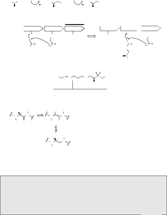
In marked contrast to the ribosomal biosynthesis of peptides and proteins, where a biological production line interprets the genetic code, many natural peptides are known to be synthesized by a more individualistic sequence of enzyme-controlled processes, in which each amino acid is added as a result of the specificity of the enzyme involved. The many stages of the whole process appear to be carried out by a multi-functional enzyme (nonribosomal peptide synthase, NRPS) with a modular arrangement comparable to that seen with type I polyketide synthases (see page 114). The linear sequence of modules in the enzyme usually corresponds to the generated amino acid sequence in the peptide product. The amino acids are first activated by conversion into AMP esters, which then bind to the enzyme through thioester linkages. The residues are held so as to allow a sequential series of peptide bond formations (Figure 7.15
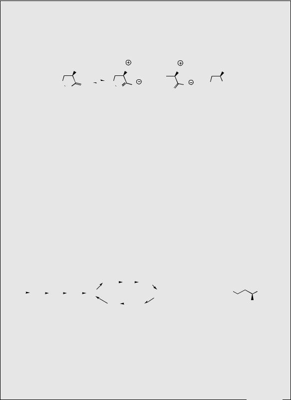
gives a simplified representation), until the peptide is finally released from the enzyme. A typical module consists of an adenylation (A) domain, a peptidyl carrier protein (PCP) domain, and a condensation (C) or elongation domain. The A domain activates a specific amino acid as an aminoacyl adenylate, which is then transferred to the PCP domain forming an aminoacyl thioester. Pantothenic acid (vitamin B5, see page 31) bound to the enzyme as pantatheine is used to carry the growing peptide chain through its thiol group (Figure 7.15). The significance of this is that the long ‘pantatheinyl arm’ allows different active sites on the enzyme to be reached in the chain assembly process (compare biosynthesis of fatty acids, page 36, and polyketides, page 62). Nucleophilic attack by the amino group of the neighbouring aminoacyl thioester is catalysed by the C domain and results in amide (peptide) bond formation. Enzyme-controlled biosynthesis in this manner is a feature of many microbial peptides, especially those containing unusual amino acids not encoded by DNA and where post-translational modification is unlikely, and also for the cyclic structures which are frequently encountered. As well as activating the amino acids and catalysing formation of the peptide linkages, the enzyme may possess other domains that are responsible for epimerizing L- amino acids to D-amino acids, probably through
422 |
|
PEPTIDES, PROTEINS, AND OTHER AMINO ACID DERIVATIVES |
|||||||||||
activation of amino acid by |
|
|
formation of thioester |
||||||||||
formation of mixed anhydride |
|
|
linkage to enzyme |
||||||||||
|
|
AMP |
O |
|
|
|
O |
||||||
ATP |
PP |
Enz–SH |
|
||||||||||
|
|
AMP |
|
|
|
|
|||||||
|
|
|
|
||||||||||
R CO2H |
|
|
R |
|
|
AMP |
|
|
R |
|
|
|
SEnz |
|
|
|
|
|
|||||||||
NH2 |
|
|
NH2 |
|
|
|
NH2 |
||||||
|
|
|
aminoacyl–AMP |
|
|
|
|
|
|
||||
|
Module 1 |
|
Module 2 |
|||
A PCP C |
|
A PCP C |
||||
|
|
S |
|
|
|
S |
O |
|
R1 |
O |
|
R2 |
|
|
|
|||||
|
|
|||||
|
|
|
|
|
||
H2N |
|
H2N |
||||
A: adenylation domain
PCP: peptidyl carrier protein domain
C: condensation domain
Module 3 |
Module 1 |
|
Module 2 |
|
Module 3 |
A PCP C |
A PCP C |
SH |
SH |
growing peptide chain generated by sequentially forming amide bond linkages
'pantotheinyl arm'
A PCP C  A PCP C
|
|
S |
|
|
S |
||
O |
|
|
|
R2 |
O |
|
R3 |
|
|
|
|
||||
|
|
|
|
||||
|
|
|
|
|
|
||
HN |
H2N |
||||||
|
|
|
|
O |
|
|
|
|
|
|
|
|
|
|
|
|
|
|
|
|
|
|
|
R2 |
|
|
|
||||
|
|
NH2 |
|
|
|
||
attached to growing |
O |
|
O |
|
bound to enzyme |
||
HS |
|
|
|
|
OH |
||
peptide chain |
|
N |
|
|
through phosphate |
||
N |
|
|
|
||||
|
H |
H |
|
OH |
|
|
|
|
|
|
|
|
|
|
|
|
cysteamine |
pantothenic acid |
|
|
|||
pantotheine
Figure 7.15
R |
H H |
R |
H |
|
N |
L |
N |
N |
N |
|
|
|||
H |
|
O |
H |
OH |
epimerization through intermediate enol-like
tautomer
R H H
 N N D
N N D 

H O
Figure 7.16
enol-like tautomers in the peptide (Figure 7.16). A terminal thioesterase domain is responsible for
terminating chain extension and releasing the peptide from the enzyme. Many of the medicinally useful peptides have cyclic structures. Cyclization may result if the amino acids at the two termini of a linear peptide link up to form another peptide bond, but very often, ring formation can be the result of ester or amide linkages, which utilize side-chain functionalities in the constituent amino acids. As with polyketide synthases, genetic manipulation of nonribosomal peptide synthases allows production of peptide derivatives in which rational modifications may be programmed according to the genes encoded.
PEPTIDE ANTIBIOTICS
Cycloserine
D-Cycloserine (Figure 7.17) is produced by cultures of Streptomyces orchidaceus, or may be prepared synthetically, and is probably the simplest substance with useful antibiotic
(Continues)
PEPTIDE ANTIBODIES |
423 |
activity. Cycloserine is water soluble and has a broad spectrum of antibacterial activity, but it is primarily employed for its activity against Mycobacterium tuberculosis. It behaves as a structural analogue of D-alanine, and inhibits the incorporation of D-alanine into bacterial cell walls. Since it can produce neurotoxicity in patients, it is reserved for infections resistant to first-line drugs.
|
|
NH2 |
|
|
|
|
NH3 |
NH3 |
NH2 |
|
O |
|
|
|
|
|
O |
|
O |
O |
HO CO2H |
N |
O |
|
|
N |
||||||
|
|
|||||||||
|
|
|
|
|
|
|
O |
|
||
|
H |
|
|
|
|
|
|
|
|
|
|
|
|
|
|
|
|
|
D-Ala |
D-Ser |
|
D-cycloserine |
|
|
|
|
|
|||||
Figure 7.17
Polymyxins
The polymyxins are a group of cyclic polypeptide antibiotics produced by species of Bacillus. Polymyxins A–E were isolated from Bacillus polymyxa, though polymyxin B and polymyxin E were both subsequently shown to be mixtures of two components. A polypeptide mixture called colistin isolated from Bacillus colistinus was then found to be identical to polymyxin E. Polymyxin B and colistin (polymyxin E) are both used clinically. These antibiotic mixtures respectively contain principally polymyxin B1 with small amounts of polymyxin B2, or predominantly polymyxin E1 (≡ colistin A) with small amounts of polymyxin E2 (≡ colistin B) (Figure 7.18). These molecules contain ten amino acids, six of which are L-α,γ- diaminobutyric acid (L-Dab), with a fatty acid (6-methyloctanoic acid or 6-methylheptanoic acid) bonded to the N-terminus, and a cyclic peptide portion constructed via an amide bond between the carboxyl terminus and the γ-amino of one of the Dab residues. The γ-amino groups of the remaining Dab residues confer a strongly basic character to the antibiotics. This results in detergent-like properties and allows them to bind to and damage bacterial membranes. These peptides have been used for the treatment of infections with Gramnegative bacteria such as Pseudomonas aeruginosa, but are seldom used now because of
|
|
|
|
|
|
|
|
Dab |
|
|
Y |
|
|
|
|
|
|
|
|
|
|||
X |
|
Dab |
|
Thr |
|
Dab |
|
Dab |
|
||
|
|
|
|||||||||
|
|
|
|
||||||||
|
|
|
|
|
|
|
|
Thr |
|
||
|
|
|
|
|
|
|
|
|
|||
|
|
|
|
|
|
|
|
X |
|
||
|
|
polymyxin B1 |
|
6-methyloctanoic acid |
|
||||||
|
|
polymyxin B2 |
|
6-methylheptanoic acid |
|
||||||
|
|
polymyxin E1 |
|
6-methyloctanoic acid |
|
||||||
|
|
(colistin A) |
|
|
|
|
|
|
|
||
|
|
polymyxin E2 |
|
6-methylheptanoic acid |
|
||||||
|
|
(colistin B) |
|
|
|
|
|
|
|
||
|
Leu |
H2N |
CO2H |
|
|||
|
Dab |
||
|
Dab = |
|
|
|
|
|
|
Dab |
|
NH2 |
|
L-α,γ-diaminobutyric acid
Y
D-Phe
D-Phe
D-Leu
D-Leu
Figure 7.18
(Continues)

424 |
PEPTIDES, PROTEINS, AND OTHER AMINO ACID DERIVATIVES |
(Continued )
neurotoxic and nephrotoxic effects. However, they are included in some topical preparations such as ointments, eye drops, and ear drops, frequently in combination with other antibiotics.
Bacitracins
Bacitracin is a mixture of at least nine peptides produced by cultures of Bacillus subtilis, with the principal component being bacitracin A (Figure 7.19). The structure contains a cyclic peptide portion, involving the carboxyl terminus and the ε-amino of lysine, and at the N-terminus an unusual thiazolinecarboxylic acid, which is a condensation product from isoleucine and cysteine residues (compare epothilones, page 105, and bleomycin, page 429). Bacitracin is active against a wide range of Gram-positive bacteria, and appears to affect biosynthesis of the bacterial cell wall by binding to and sequestering a polyprenyl diphosphate carrier of intermediates; this binding also requires a divalent metal ion, with zinc being especially active. It is rarely used systemically because some bacitracin components are nephrotoxic, but as zinc bacitracin, it is a component of ointment formulations for topical application. The vast majority of bacitracin manufactured is used at subtherapeutic doses as an animal feed additive, to increase feed efficiency, and at therapeutic dosage to control a variety of disorders in poultry and animals.
|
|
|
|
|
|
|
|
D-Orn |
|
|
Ile |
|
|
D-Phe |
|
|
|
CO2H |
|||
|
|
|
|
|
|
|
|
|
|
|
|
|
|
||||||||
X |
|
Leu |
|
D-Glu |
|
Ile |
|
Lys |
|
|
|
|
|
|
|
|
X = |
N |
Cys |
||
|
|
|
|
|
|
|
|
|
|
||||||||||||
|
|
|
|
|
|
|
|
|
|
|
|
||||||||||
|
|
|
|
|
|
|
|
Asn |
|
|
|
|
|
|
|
|
|
|
|
S |
|
|
|
|
|
|
|
|
|
|
|
|
D-Asp |
|
|
His |
|
|
|||||
|
|
|
|
|
|
|
|
|
|
|
|
|
NH2 |
||||||||
|
|
|
|
|
|
|
|
|
|
|
|
|
|
||||||||
|
|
|
|
|
|
bacitracin A |
|
|
|
|
|
|
|
|
Ile |
|
|||||
|
|
|
|
|
|
|
|
|
|
|
|
|
|
|
|
|
|||||
Figure 7.19
Tyrothricin and Gramicidins
Tyrothricin is a mixture of polypeptide antibiotics produced by cultures of Bacillus brevis. The mixture contains about 20–30% linear polypeptides called gramicidins (Figure 7.20), and 70–80% of cyclic structures called tyrocidines (Figure 7.20). Tyrothricin is active against many Gram-positive bacteria, with the linear gramicidins being more active than the cyclic tyrocidines. The two groups are readily separated by solvent fractionation, and the gramicidin fraction, also termed gramicidin D, a mixture of at least eight closely related compounds, is used principally in ophthalmic preparations. The gramicidins are neutral polypeptides having the N-terminal amino group formylated, and the carboxy group linked to ethanolamine. Most of the gramicidin mixture is composed of valine-gramicidin A (about 80%) (Figure 7.20). Apart from the glycine residue, these compounds have a sequence of alternating D- and L-amino acids. Gramicidins act by producing ion channels in bacterial membranes. The tyrocidines are too toxic for therapeutic use on their own, but the tyrothricin mixture is incorporated into lozenges for relief of throat infections.
Gramicidin S is a mixture of cyclic peptides obtained from another strain of Bacillus brevis. Its main component is gramicidin S1 (Figure 7.20), a symmetrical
(Continues)