

1Paul M Dewick Medicalc Natural / booktext@id88013693placeboie
.pdfPEPTIDE ANTIBODIES |
425 |
(Continued )
OHC–X–Gly–Ala–D-Leu–Ala–D-Val–Val–D-Val–Trp–D-Leu–Y–D-Leu–Trp–D-Leu–Trp–NH(CH2)2OH
D-Phe |
|
Pro |
|
|
|
X |
|
|
Y |
|
|
Asn |
D-Phe |
|
|
|
Pro |
|
|
|
Val |
|
|
Orn |
|
|
|
Leu |
||||||
|
|
|
|
|
|
|
|
|
|
|
|
|
|
|
|
|
|
|
||||||||||||||||
|
|
|
|
|
|
|
|
|
|
|
|
|
|
|
|
|
|
|
|
|
|
|
|
|
|
|
|
|
|
|
|
|
||
|
|
|
|
|
|
|
|
|
|
|
|
|
|
|
|
|
|
|
|
|
|
|
|
|
|
|
|
|
|
|
|
|
|
|
|
|
|
|
|
|
|
|
|
|
|
|
|
|
|
|
|
|
|
|
|
|
|
|
|
|
|
|
|
|
|
|
|
|
|
|
|
|
|
|
|
|
|
|
|
|
|
|
|
|
|
|
|
|
|
|
|
|
|
|
|
|
|
|
|
|
|
|
|
|
Leu |
|
Orn |
|
|
|
Val |
|
Tyr |
|
|
|
Gln |
Leu |
|
Orn |
|
Val |
|
Pro |
|
D-Phe |
|||||||||||||
|
|
|
|
|
|
|
|
|
|
|
|
|||||||||||||||||||||||
|
|
|
|
|
|
|
X |
|
Y |
|
|
|
|
|
gramicidin S1 |
|
|
|
|
|
|
|||||||||||||
|
tyrocidine A |
|
|
|
Phe |
|
D-Phe |
|
|
|
|
|
|
|
|
|
|
|
|
|
|
|
|
|
|
|
||||||||
|
tyrocidine B |
|
|
|
Tyr |
|
D-Phe |
|
|
|
|
|
|
|
|
|
|
|
|
|
|
|
|
|
|
|
||||||||
|
tyrocidine C |
|
|
|
Tyr |
|
D-Tyr |
|
|
|
|
|
|
|
|
|
|
|
|
|
|
|
|
|
|
|
||||||||
gramicidin A |
X |
Y |
|
|
|
Val-gramicidin A |
Val |
Trp |
Ile-gramicidin A |
Ile |
Trp |
gramicidin B |
|
|
Val-gramicidin B |
Val |
Phe |
Ile-gramicidin B |
Ile |
Phe |
gramicidin C |
|
|
Val-gramicidin C |
Val |
Tyr |
Ile-gramicidin C |
Ile |
Tyr |
Figure 7.20
decapeptide known to be formed by the joining together of two separate chains. Gramicidin S is fairly toxic, and its use is restricted to topical preparations. Gramicidin S also acts on bacterial membranes, increasing permeability and loss of barrier function.
Capreomycin
Capreomycin is a mixture of cyclic polypeptides obtained from cultures of Streptomyces capreolus, and contains about 90% of capreomycins I, principally capreomycin IB (Figure 7.21). The capreomycin structures incorporate an L-capreomycidine moiety derived by cyclization of L-arginine, and three molecules of 2,3-diaminopropionic acid (Dap), which originate from serine via dehydroalanine. This antibiotic is given intramuscularly to treat tuberculosis patients who do not respond to first-line drugs, or where patients are sensitive to
|
|
|
|
|
Ser |
|
|
Dap |
|
|
β-lysine |
|
|
|
|
|
or Ala |
|
|
||||
|
|
O |
R |
|
|
O NH2 |
|||||
|
|
|
H |
|
|
||||||
|
Dap |
|
|
|
|
|
|
|
|||
|
H2N |
|
|
|
|
N |
|
|
|
|
|
|
|
N |
N |
||||||||
|
|
|
|
|
|
|
|||||
|
|
|
|
|
H |
|
|
H |
|||
|
|
|
|
|
O |
||||||
|
|
NH |
O |
|
|
|
|||||
|
|
|
HN |
|
|
|
|||||
|
|
|
|
|
H |
|
|
|
|
|
|
|
|
|
|
|
N |
|
|
NH |
|
|
NH2 |
|
|
O |
|
|
|
|
|
|
|||
|
|
|
|
|
|
|
|
||||
|
L-capreomycidine |
O |
Dap |
O |
|||||||
HO2C |
NH2 |
|
|
|
NH |
|
|
|
|
||
|
|
|
|
|
|
|
|
|
|
||
|
|
|
|
|
N |
NH |
|
|
|
|
|
|
NH2 |
|
|
|
H |
|
|
|
|
|
|
|
|
|
|
R = OH, capreomycin 1A |
|||||||
|
|
|
|
|
|||||||
N |
NH |
|
|
|
R = H, capreomycin 1B |
||||||
H |
|
|
|
|
|
|
|
|
|
|
|
L-Arg |
|
|
|
|
|
|
|
|
|
|
|
NH2
Dap = 2,3-diaminopropionic acid
CO2H
H2N
NH2 |
|
|
|
|
CO2H |
|
CO2H |
|
|
||
|
|
||
|
|
|
HO |
|
|
|
|
NH2 |
|
NH2 |
|
dehydroalanine |
|
L-Ser |
|
Figure 7.21
(Continues)

426 |
PEPTIDES, PROTEINS, AND OTHER AMINO ACID DERIVATIVES |
(Continued )
streptomycin. It can cause irreversible hearing loss and impair kidney function. Capreomycin inhibits protein biosynthesis at the translocation step in sensitive bacteria.
Vancomycin and Teicoplanin
Vancomycin (Figure 7.22) is a glycopeptide antibiotic produced in cultures of Amycolatopsis orientalis (formerly Streptomyces orientalis), and has activity against Gram-positive bacteria, especially resistant strains of staphylococci, streptococci, and enterococci. It is an important agent in the control of methicillin-resistant Staphylococcus aureus (MRSA), with some strains now being sensitive only to vancomycin or teicoplanin (below). Vancomycin is not absorbed orally, and must be administered by intravenous injection. However, it can be given orally in the treatment of pseudomembranous colitis caused by Clostridium difficile, which may occur after administration of other antibiotics. Vancomycin acts by its ability to form a complex with terminal N-acyl–D-Ala–D-Ala residues of growing peptidoglycan chains (see Figure 7.36 and page 444), preventing their cross-linking to adjacent strands and thus inhibiting bacterial cell wall biosynthesis. The –D-Ala–D-Ala residues are accommodated in a ‘carboxylate-binding pocket’ in the vancomycin structure. By preventing peptidoglycan polymerization and cross-linking, it weakens the bacterial cell wall and ultimately causes cell lysis.
The novel feature of vancomycin, and several other related antibiotics, is the tricyclic structure generated by three phenolic oxidative coupling reactions. The β- hydroxychlorotyrosine and 4-hydroxyphenylglycine residues in vancomycin originate from L-tyrosine, but the 3,5-dihydroxyphenylglycine ring is actually acetate derived. These modified aromatic rings are presumably present in the heptapeptide before coupling occurs (Figure 7.22).
The teicoplanins (Figure 7.23) possess the same basic structure as vancomycin, but the N-terminal (4-hydroxyphenylglycine) and third (3,5-dihydroxyphenylglycine) amino acids are also aromatic, and this allows further phenolic oxidative coupling and generation of yet another ring system. Teicoplanin for drug use is a mixture of five teicoplanins produced by cultures of Actinoplanes teichomyceticus, which differ only in the nature and length of the fatty acid chain attached to the sugar residue. Teicoplanin has similar antibacterial activity to vancomycin, but has a longer duration of action, and may be administered by intramuscular as well as by intravenous injection. It is also used against Gram-positive pathogens resistant to established antibiotics.
Vancomycin, teicoplanin, and structurally related glycopeptides are often referred to as dalbaheptides (from D-alanyl–D-alanine-binding heptapeptide), reflecting their mechanism of action and their chemical nature. Unfortunately, with increasing use of vancomycin and teicoplanin, there have even been reports of these agents becoming ineffective because resistant bacterial strains have emerged, particularly in enterococci. In resistant strains, the terminal –D-Ala–D-Ala residues, to which the antibiotic normally binds, have become replaced by –D-Ala–D-lactate. The incorporation of D-lactate into the peptide intermediates results in loss of crucial hydrogen-bonding interactions and a thousand-fold lowering in binding efficiency.
(Continues)
PEPTIDE ANTIBODIES |
427 |
(Continued )
NH2
L-vancosamine
|
|
|
|
|
|
|
|
|
O |
|
|
|
|
|
|
HO |
|
|
|
|
|
O |
|
|
|
|
|
|
|
|
|
|
|
|
|
|
|
|
|
|
|
|
|
|
|
|
|
|
|
HO |
|
|
|
O |
|||||
D-glucose |
HO |
O |
|
|
|
|
|
|||||||
|
|
|
|
|
|
|
|
|
|
|
||||
|
|
|
|
|
HO |
|
|
|
|
|
|
|||
|
|
|
|
|
|
|
|
|
O |
|
|
O |
||
|
HO |
|
|
|
|
Cl |
|
|
|
|
|
|||
|
|
|
|
|
|
|
|
|
|
|||||
|
|
|
|
|
|
|
O |
|||||||
|
|
|
|
|
|
|
|
|
|
|
||||
|
|
|
|
|
|
|
O |
H |
|
|
||||
|
|
|
|
|
|
|
|
|
|
|
|
|||
|
O |
|
|
|
|
|
|
N |
|
|
|
|
|
|
|
|
|
|
N |
|
|
N |
|||||||
|
|
|
|
|
|
|
|
|
||||||
|
|
|
|
|
|
H |
|
|
|
H |
||||
|
|
|
|
|
|
|
O |
|||||||
|
HN |
|
CO2H |
|
O |
|||||||||
|
|
|
|
|
||||||||||
|
HO |
|
|
|
|
|
|
OH |
|
|
NH2 |
|||
|
|
|
|
|
|
|
|
|
||||||
|
|
|
|
|
|
|
|
|
|
|
|
|||
|
|
|
|
|
|
|
|
|
|
|
|
|||
|
|
|
|
|
|
|
|
|
|
|
|
|||
|
|
|
|
|
OH |
|
|
|
|
|
||||
|
|
|
|
|
|
|
|
|
vancomycin |
|||||
Tyr |
|
|
|
|
|
|
|
|
|
|
|
OH |
||
|
|
|
|
|
|
|
|
OH |
|
|
|
HO |
||
|
|
|
|
|
|
|
|
|
|
|
|
|||
|
HO |
|
|
|
|
|
|
* |
|
|
* |
|
|
|
|
|
|
|
|
|
|
|
|
|
|
||||
β-hydroxychloro- |
|
|
|
|
O |
Cl |
|
|
O |
|||||
|
|
|
|
|
|
|
|
|||||||
tyrosine |
|
|
|
|
|
|
H |
|
|
|||||
O |
|
|
|
|
|
|
|
|
|
|||||
|
|
|
N |
N |
|
|
|
|
|
|||||
|
|
|
|
|
N |
|||||||||
|
|
|
|
|
|
|
|
|||||||
|
|
|
|
|
H |
|
|
|
H |
|||||
|
HN |
|
|
|
O |
|||||||||
|
|
|
CO2H |
|
O |
|||||||||
|
|
|
|
|
|
|||||||||
3,5-dihydroxyphenyl- |
|
|
* |
|
|
|
|
|
NH2 |
|||||
|
|
|
|
|
|
|
||||||||
glycine |
|
|
|
|
|
|
|
|
|
|||||
|
|
|
|
|
|
|
|
OH |
|
|
|
Asn |
||
|
|
|
|
|
|
|
|
|
|
|
|
|||
HO |
|
|
|
OH |
|
|
|
|
|
|||||
|
|
|
|
|
|
|
|
|
||||||
|
|
|
|
|
|
|
|
|
||||||
|
|
|
|
|
|
|
|
|
|
|
|
|
|
|
acetate
4-hydroxyphenyl- glycine
Tyr
Cl |
|
|
|
|
|
|
|
|
|
OH |
|||
H |
|
|
O |
|||
|
|
|
|
|
|
|
N |
|
|
|
|
|
NHMe |
|
|
|
|
|||
|
|
|
N |
|||
|
|
|
||||
|
|
|
H |
|||
|
O |
|||||
|
|
|
|
|
||
Cl |
|
|
* oxidative coupling reactions |
|||
|
|
|
OH |
|||
H |
|
|
O |
|||
|
|
|
|
|
|
|
N |
|
|
|
|
|
NHMe |
|
|
|
|
|||
|
|
|
N |
|||
|
|
|
||||
|
|
|
H |
|||
|
O |
|||||
|
|
|
|
|
||
Leu
β-hydroxychloro- tyrosine
Tyr
Figure 7.22
Bleomycin
Bleomycin is a mixture of glycopeptide antibiotics isolated from cultures of Streptomyces verticillus, used for its anticancer activity. The major component (55–70%) of the mixture
(Continues)
428 |
|
|
|
PEPTIDES, PROTEINS, AND OTHER AMINO ACID DERIVATIVES |
||||||||||||||||||||||||||||
|
(Continued ) |
|
|
|
|
|
|
|
|
|
|
|
|
|
|
|
|
|
|
|
|
|
|
|
|
|
|
|
|
|
|
|
|
|
|
|
|
|
|
|
|
|
|
|
|
|
teicoplanin T-A2-1, |
R = |
|
|
|||||||||||||||
|
|
|
|
|
|
|
|
|
|
|
|
|
|
|
|
|
|
|||||||||||||||
|
|
|
|
|
|
|
|
|
|
|
|
|
|
|
|
|
||||||||||||||||
|
|
|
|
|
|
|
|
|
|
|
|
|
|
|
|
|
|
|
|
|
|
|
|
|
|
|||||||
|
|
|
|
|
|
|
|
|
|
|
|
|
|
|
|
|
|
|
|
|
|
O |
||||||||||
|
|
|
|
|
|
|
|
|
|
|
|
|
|
|
|
teicoplanin T-A2-2, |
R = |
|||||||||||||||
|
|
|
|
|
|
|
|
|
|
|
|
|
|
|
|
|
|
|
|
|
|
|
|
|
|
|
|
|
|
|||
|
|
|
|
|
|
|
|
|
|
|
|
|
|
|
|
|
|
|
|
|
|
O |
||||||||||
|
|
D-glucosamine |
NHR |
|
|
|
|
|
|
teicoplanin T-A2-3, |
R = |
|
|
|
|
|
|
|
|
|||||||||||||
|
|
|
|
|
|
|
|
|
|
|
|
|
|
|||||||||||||||||||
|
|
|
|
|
|
|
|
|
|
|
|
|
|
|
|
|
|
|
|
|
|
|
|
|
|
|
|
|
|
|
||
|
|
|
|
|
HO |
|
|
O |
|
|
|
|
|
|
|
|
|
|
O |
|||||||||||||
|
|
|
|
|
|
|
|
|
|
|
teicoplanin T-A2-4, |
R = |
|
|
|
|
|
|
|
|
||||||||||||
|
|
|
HO |
O |
|
|
Cl |
|
|
|
|
|
|
|
|
|
|
|||||||||||||||
|
|
|
|
|
|
|
|
|
||||||||||||||||||||||||
|
|
|
|
|
|
|
|
|
|
|
|
|
|
|
|
|
|
|
|
|
|
|
|
|
|
|
|
|
||||
|
|
|
|
|
HO |
|
|
|
|
|
|
|
|
|
|
|
|
O |
||||||||||||||
|
|
|
|
|
|
|
|
|
|
|
|
|
|
|
|
|
|
|
||||||||||||||
|
NHAc |
|
|
|
O |
O |
|
|
|
|
teicoplanin T-A2-4, |
R = |
||||||||||||||||||||
|
|
|
|
|
|
|
|
|
|
|
|
|
|
|
|
|
|
|
|
|
|
|
|
|
|
|
|
|
||||
HO |
|
O |
|
|
|
|
|
|
|
|
|
|
|
|
|
|
|
|
|
O |
||||||||||||
|
|
|
|
Cl |
O |
|
|
|
|
O |
||||||||||||||||||||||
HO |
O |
|
|
|
|
|
|
|
|
|
|
|
|
|
|
|
|
|
|
|||||||||||||
|
O |
H |
H |
|
|
|
|
|
|
|
|
|
|
|
|
|
||||||||||||||||
|
|
|
|
|
|
|
|
|
|
|
|
|
|
|
|
|
|
|
|
|
|
|
|
|||||||||
|
HO |
O |
|
|
|
N |
|
|
N |
|
|
|
|
|
|
NHMe |
|
|
|
|
|
|
|
|
|
|
|
|||||
|
N |
|
|
|
|
|
|
|
|
|
|
|
|
|
|
|
|
|
||||||||||||||
|
N-acetyl- |
|
|
|
|
|
N |
|
|
|
|
N |
|
|
|
|
|
|
|
|
|
|
|
|||||||||
|
|
|
|
|
|
|
|
|
|
|
|
|
|
|
|
|
|
|
|
|
||||||||||||
|
|
|
|
H |
|
|
H |
|
|
|
|
H |
|
|
|
|
|
|
|
|
|
|
|
|||||||||
|
|
|
|
O |
|
|
O |
|
|
|
|
|
|
|
|
|
|
|
||||||||||||||
D-glucosamine |
|
HN |
CO2H |
|
|
|
|
|
|
|
|
|
|
|
|
|
|
|
|
|
|
|
|
|
||||||||
|
|
|
HO |
|
|
|
|
|
|
|
|
|
|
|
|
|
|
|
|
|
|
|
|
|
||||||||
|
|
|
|
|
|
|
|
|
|
|
|
|
|
|
|
|
|
|
|
|
|
|
|
|||||||||
|
|
|
|
|
|
|
|
|
|
|
|
|
|
|
|
|
|
|
|
|
|
|
|
|
|
|
|
|
|
|
||
|
|
|
|
|
|
|
|
|
|
|
|
|
|
O |
|
|
|
|
|
|
|
|
|
|
|
|||||||
|
|
|
|
|
|
|
|
OH |
|
|
|
|
|
|
|
|
|
|
OH |
|
|
|
|
|
|
|
|
|
|
|
||
|
|
HO |
|
O |
|
|
|
|
|
|
|
|
|
|
|
|
|
|
|
|
|
|||||||||||
|
|
|
|
|
|
|
|
|
|
|
|
|
|
|
|
|
|
|
|
|
|
|
|
|
|
|||||||
|
|
|
|
|
|
|
|
|
|
|
|
|
|
|
4-hydroxyphenyl- |
|||||||||||||||||
|
|
|
|
|
|
|
|
|
|
OH |
|
|
|
|
|
|
|
|
||||||||||||||
|
|
|
|
|
|
|
|
O |
|
|
|
|
|
|
|
|
|
glycine |
||||||||||||||
|
|
|
|
|
|
|
|
OH |
3,5-dihydroxyphenyl- |
|
|
|
|
|
|
|
|
|
|
|
||||||||||||
|
|
|
|
|
|
|
|
OH |
|
|
|
|
|
|
|
|
|
|
|
|||||||||||||
|
|
|
|
|
|
|
|
|
|
|
|
|
|
glycine |
|
|
|
|
|
|
|
|
|
|
|
|||||||
|
|
|
|
|
|
|
|
OH |
|
|
|
|
|
|
|
|
|
|
|
|
|
|
|
|
|
|
|
|
|
|
|
|
|
|
|
|
|
|
|
|
D-mannose |
|
|
|
|
|
|
|
|
|
|
|
|
|
|
|
|
|
|
|
|
|
|||
Figure 7.23
is bleomycin A2 (Figure 7.24), with bleomycin B2 constituting about 30%. The various bleomycins differ only in their terminal amine functions, the parent compound bleomycinic acid (Figure 7.24) being inactive. The molecules contain several unusual amino acids and sugars, an asparagine-derived pyrimidine ring, and a planar dithiazole ring system which has its origins in two cysteines (compare epothilones, page 105, and bacitracin, page 424). A C2 unit supplied by malonyl-CoA also forms part of the main chain as a component of the amino acid 4-amino-3-hydroxy-2-methylvaleric acid, which in addition features a methionine-derived methyl group. Bleomycin is a DNA-cleaving drug, causing single and double-strand breaks in DNA. The dithiazole system is involved in binding to DNA, probably by intercalation, whilst other parts of the molecule near the N-terminus are involved in chelating a metal ion, usually Fe2+, and oxygen, which are necessary for the DNA degradation reaction. More recently, bleomycin A2 has been shown to cleave RNA as well as DNA.
Bleomycin is used alone, or in combination with other anticancer drugs, to treat squamous cell carcinomas of various organs, lymphomas, and some solid tumours. It is unusual amongst antitumour antibiotics in producing very little bone-marrow suppression, making it particularly useful in combination therapies with other drugs that do cause this response. However, there is some lung toxicity associated with bleomycin treatment. Various bleomycin analogues have been made by adding different precursor amines to the culture medium, or by semi-synthesis from bleomycinic acid. Peplomycin (Figure 7.24) is an example with some promise, in that it is more resistant to enzymes that cause in vivo hydrolysis of bleomycin at the N-terminal β-aminoalanine group.
(Continues)
PEPTIDE ANTIBODIES |
429 |
(Continued )
|
|
|
|
|
|
|
|
|
Ala + malonate + Me |
|
|
|
|
|
|
|
|
|
|
|||
|
|
|
|
|
Ser |
|
|
|
4-amino-3-hydroxy- |
|
|
|
|
|
|
|
|
|
|
|||
O |
NH2 |
|
|
|
|
|
|
|
|
|
|
|
|
|
|
|||||||
NH2 |
2-methylvaleric acid |
|
|
|
O R |
|
|
|
|
|||||||||||||
|
|
|
H |
|
|
|
|
|
|
|
||||||||||||
Asn |
|
N |
|
|
NH2 |
|
|
Thr |
|
|
|
|
|
|
|
|
|
|
||||
|
|
|
|
|
|
|
|
|
|
|
|
|
|
|
||||||||
|
|
|
|
|
|
|
|
|
|
|
|
|
|
|
|
|
Cys |
|
|
|
||
N |
|
N |
|
O |
|
|
|
|
O |
|
|
|
N |
|
|
|
|
|||||
|
H |
|
O |
|
|
|
H |
|
|
|
S |
|
|
|
|
|||||||
Asn |
|
|
|
|
|
|
|
|
|
|
|
|
|
|
|
|
|
|
|
|
||
|
|
|
|
|
|
|
|
|
|
|
|
|
|
|
|
|
|
|||||
|
|
|
|
N |
|
|
|
|
|
|
N |
|
|
|
|
|
|
|
|
|
|
|
H2N |
|
|
|
|
|
|
N |
|
|
|
|
NH |
N |
Cys |
|
|
|
|
||||
|
|
|
|
|
|
|
|
|
|
|
|
|
|
|
|
|||||||
|
|
|
|
|
|
|
|
|
|
|
|
|
|
|
|
|||||||
|
|
|
|
|
|
|
|
|
H |
|
|
|
|
|
|
|
|
|
|
|
|
|
Me |
|
O |
|
|
|
|
OH O |
|
|
|
|
|
|
|
|
|
|
|||||
|
|
|
|
|
N |
|
|
|
|
|
S |
|
|
|
|
|||||||
HO |
|
OH |
O |
|
|
|
|
|
|
HO |
|
|
|
|
|
|
|
|
|
|
||
|
|
|
|
|
|
|
|
|
|
|
|
|
|
|
||||||||
|
|
|
|
|
N |
|
β-OH-His |
|
|
|
|
|
|
|
|
|
|
|||||
|
O |
O |
|
|
|
|
|
|
β-Ala |
|
|
|
|
|
||||||||
|
|
|
|
|
|
|
|
|
|
|
|
|
|
|||||||||
L-gulose |
|
|
|
|
|
|
H |
|
|
|
|
|
|
|
|
|
|
|||||
|
|
|
|
|
|
|
|
|
OH |
|
|
|
|
|
bleomycin A2 |
R = NH |
S |
|||||
OH |
|
|
|
|
O |
|
|
|
|
|
|
|
||||||||||
|
|
|
|
|
|
|
D-mannose |
|
|
|
|
|
|
H |
||||||||
|
|
|
|
|
|
|
|
|
|
|
OH |
|
|
|
|
|
|
|
|
|||
|
|
|
|
|
|
|
|
|
|
|
|
|
|
|
|
|
|
|
N NH2 |
|||
|
|
|
|
|
|
OH |
O |
|
|
|
|
|
|
|
|
bleomycin B2 |
R = NH |
|
||||
|
|
|
|
|
|
|
|
|
|
|
|
|
|
|
|
|
|
|||||
|
|
|
|
|
|
|
|
O |
|
NH2 |
|
|
|
|
|
|
|
|
NH |
|||
|
|
|
|
|
|
|
|
|
|
|
|
|
|
bleomycinic acid |
R = OH |
|
|
|
||||
|
|
|
|
|
|
|
|
|
|
|
|
|
|
|
|
|
|
|
|
|
||
β-Ala |
= |
H2N |
|
|
CO2H |
|
|
|
|
|
|
|
|
peplomycin |
R = NH |
N |
|
|||||
|
|
|
|
|
|
|
|
|
|
|
|
|
|
|||||||||
|
|
|
|
|
|
|
|
|
|
|
|
|
|
|
|
|
|
|
|
H |
|
|
Figure 7.24
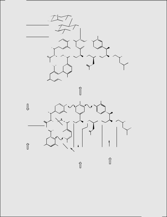
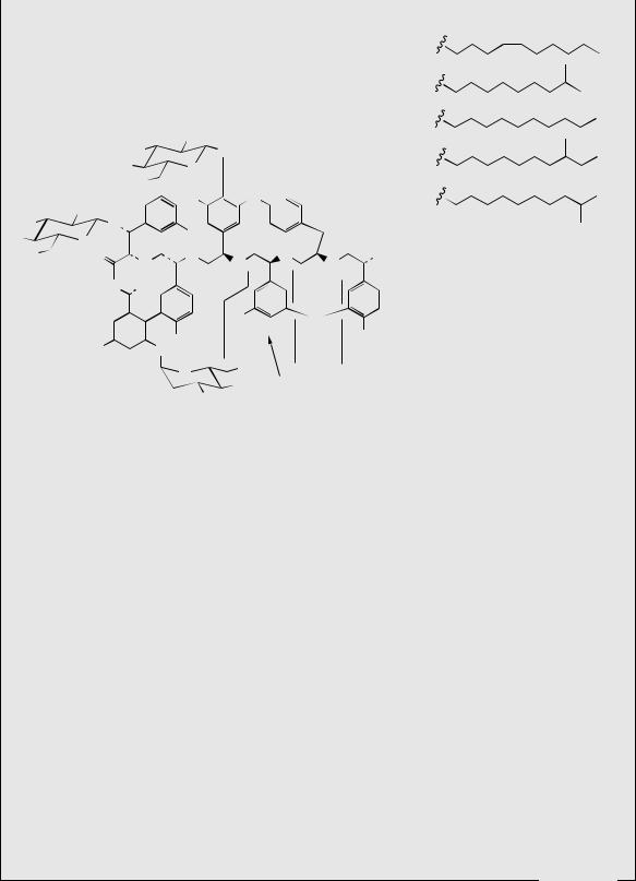
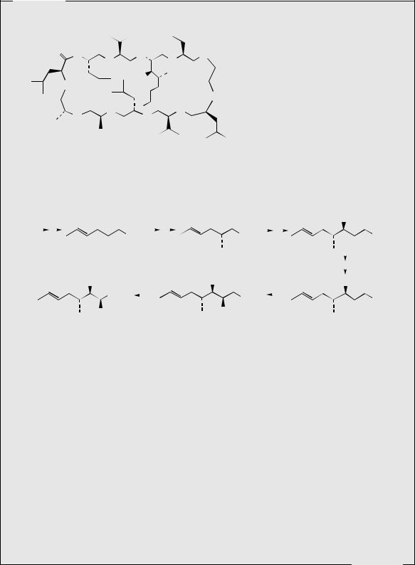
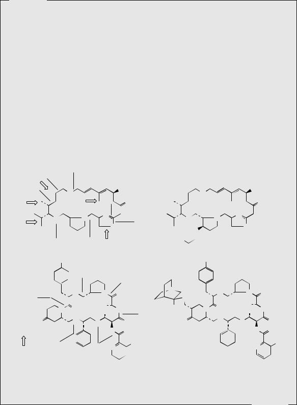
Cyclosporins
The cyclosporins are a group of cyclic peptides produced by fungi such as Cylindrocarpon lucidum and Tolypocladium inflatum. These agents showed a narrow range of antifungal activity, but high levels of immunosuppressive and anti-inflammatory activities. The main component from the culture extracts is cyclosporin A (ciclosporin, cyclosporin) (Figure 7.25), but some 25 naturally occurring cyclosporins have been characterized. Cyclosporin A contains several N-methylated amino acid residues, together with the less common L-α-aminobutyric acid and an N-methylated butenylmethylthreonine. This latter amino acid has been shown to originate via the acetate pathway, and it effectively comprises a C8 polyketide chain, plus a methyl group from SAM. The sequence shown in Figure 7.26 is consistent with experimental data, and is analogous to other acetate-derived compounds (see Chapter 3). The assembly of the polypeptide chain is known to start from the D-alanine residue. Many of the other natural cyclosporin structures differ only with respect to a single amino acid (the α-aminobutyric acid residue) or the amount of N-methylation. Of all the natural analogues, and many synthetic ones produced, cyclosporin A is the most valuable for drug use. It is now widely exploited in organ and tissue transplant surgery, to prevent rejection following bone-marrow, kidney, liver, and heart transplants. It has revolutionized organ transplant surgery, substantially increasing survival rates in transplant patients. It may be administered orally or by intravenous injection, and the primary side-effect is nephrotoxicity, necessitating careful monitoring of kidney function. Cyclosporin A has the same mode of action as the macrolide FK-506 (tacrolimus) (see page 104) and is believed to inhibit T-cell activation in the immunosuppressive mechanism
(Continues)
430 PEPTIDES, PROTEINS, AND OTHER AMINO ACID DERIVATIVES
(Continued )
(Me)Leu (Me)Val (Me)Bmt Abu
|
|
|
|
Me |
O |
|
|
|
|
Me |
|
O |
|
|||||||
|
O |
|
|
|
|
|
|
|
|
|
|
|
|
|
|
|
|
|
|
|
|
N |
|
|
N |
|
N |
|
|
N |
|
||||||||||
|
|
|
|
|
|
|
|
|
|
|||||||||||
|
|
|
|
|
|
|
|
|
|
|
|
|
|
|
|
|
||||
(Me)Leu |
|
|
|
|
|
|
|
|
|
|
|
|
|
|
|
|
H |
|
||
|
|
|
|
|
|
Me |
O HO |
|
|
|
||||||||||
|
|
N |
|
Me |
|
|
|
|
|
|
||||||||||
|
|
|
|
|
|
|
|
|
|
|
|
|
|
|
|
|
||||
O |
|
|
|
|
O |
|
|
H |
|
|
|
|
|
O |
H |
|||||
|
|
|
|
|
|
|
|
|
|
|
||||||||||
|
|
|
|
|
|
|
|
|
|
|
||||||||||
|
|
|
|
|
|
|
|
|
|
|
||||||||||
|
|
|
|
|
|
|
|
|
N |
|
|
|
|
|
|
|
|
N |
||
D-Ala |
|
N |
|
|
|
|
N |
|
|
|
||||||||||
|
|
|
|
|
|
|
|
|
|
|
|
|||||||||
|
|
|
|
|
|
|
|
|
|
|
||||||||||
|
H |
|
|
|
|
|
|
|
|
|
|
|
|
|
|
|||||
|
|
|
|
|
|
|
O |
|
|
Me |
|
|
|
|
||||||
|
|
|
|
|
|
|
|
|
|
|
|
|
|
|
|
|||||
|
|
|
|
|
|
|
Ala |
|
|
(Me)Leu |
|
|
Val |
|
||||||
|
|
|
|
|
|
|
|
|
|
|
|
|
|
|||||||
|
Me |
|
|
|
|
|
|
|
N |
|
|
|
|
Sar |
(Me)Bmt = 4-(2-butenyl)-4,N-dimethyl-L-threonine |
|
|||
O |
|
||
|
|
O |
|
|
|
|
Abu = L-α-aminobutyric acid |
|
|
|
|
||||||
|
|
|
|
|
|
|
||
|
|
|
|
|
N |
|
Me |
|
|
|
|
|
|
|
|||
|
|
|
|
|
|
|
Sar = sarcosine (N-methylglycine) |
|
|
|
|
|
|
|
|
(Me)Leu |
|
|
|
|
|
|
|
|
||
O |
||||||||
|
|
|
|
|
|
|||
|
|
|
|
ciclosporin |
(cyclosporin A) |
|
|
|
|
|
|
|
|
|
|
|
|
||||||||
|
|
|
|
|
|
|
|
|
|
|
|
|
Figure 7.25 |
|
|
|
|
|
|
|
|
|
|
||
|
|
|
|
|
|
|
C-methylation before |
|
|
chain extension |
|
|
|
|
|
||||||||||
|
|
|
|
O O |
reduction of carbonyl |
O |
then reduction |
OH |
O |
||||||||||||||||
|
|
|
|
|
|
|
|
|
|
|
|
|
|
|
|
||||||||||
acetate / |
|
|
|
|
|
|
|
|
|
|
|
|
|
|
|
|
|
|
|
|
|
|
|
|
|
malonate |
|
|
|
|
|
|
SEnz |
|
|
SEnz |
|
|
|
|
SEnz |
||||||||||
|
|
|
|
|
|
|
|
|
|
|
|
|
|
|
|
|
|
|
|
|
|
hydroxylation |
|||
|
|
|
|
|
|
|
|
|
|
|
|
|
|
|
|
|
|
|
|
|
|
||||
|
|
|
|
|
|
|
|
|
|
|
|
|
|
|
|
|
|
|
|
|
|
||||
|
|
|
|
OH |
N-methylation |
OH O |
transamination |
|
oxidation |
||||||||||||||||
|
|
|
|
|
|||||||||||||||||||||
|
|
|
|
OH |
O |
||||||||||||||||||||
|
|
|
|
|
CO2H |
|
|
|
|
|
|
|
|
|
|
|
|
|
|
||||||
|
|
|
|
|
NH2 |
SEnz |
|
|
|
|
SEnz |
||||||||||||||
|
|
|
|
|
|
|
|
|
|
||||||||||||||||
|
|
|
|
|
NHMe |
|
|
|
|
|
|
|
|
|
|
|
|||||||||
|
|
|
|
|
|
|
|
|
|
|
|
|
|
|
|
|
|
|
|
|
|
||||
|
|
|
|
|
|
|
|
|
|
|
|
|
|
|
|
|
|
O |
|
|
|||||
4-(2-butenyl)-4,N-dimethyl-L-threonine |
|
|
|
|
|
|
|
|
|
|
|
|
|||||||||||||
Figure 7.26
by first binding to a receptor protein, giving a complex that then inhibits a phosphatase enzyme called calcineurin. The resultant aberrant phosphorylation reactions prevent appropriate gene transcription and subsequent T-cell activation. Cyclosporin A also finds use in the specialist treatment of severe resistant psoriasis.
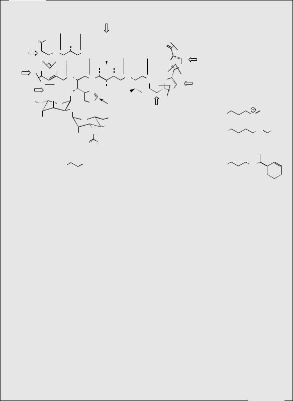
Streptogramins
The names streptogramin and virginiamycin have been applied to antibiotic mixtures isolated from strains of Streptomyces virginiae, and individual components have thus acquired multiple synonyms; as a family these antibiotics have now been termed streptogramin antibiotics. These compounds fall into two distinct groups, group A, containing a 23-membered unsaturated ring with peptide and lactone bonds, and group B, which are depsipeptides (essentially peptides cyclized via a lactone). These structures contain many nonprotein amino acids (Figure 7.27). Until recently, most commercial production of these antibiotics was directed towards animal feed additives, but the growing emergence of antibiotic-resistant
(Continues)
PEPTIDE ANTIBODIES |
431 |
(Continued )
bacterial strains has led to the drug use of some streptogramin antibiotics. Thus, dalfopristin and quinupristin (Figure 7.27) are water-soluble drugs that may be used in combination for treating infections caused by Gram-positive bacteria that have failed to respond to other antibiotics, including methicillin-resistant Staphylococcus aureus (MRSA), vancomycinresistant enterococci and staphylococci, and drug-resistant Streptococcus pneumoniae. They may need to be combined also with other agents where mixed infections involve Gramnegative organisms. Dalfopristin is a semi-synthetic sulphonyl derivative of streptogramin A (also termed virginiamycin M1, mikamycin A, pristinamycin IIA, and other names), and quinupristin is a modified form of streptogramin B (also mikamycin B, pristinamycin IA, and other names). Members of the A group tend to be less powerful antibiotics than those of the B group, but together they act synergistically, providing greater activity than the combined activity expected from the separate components. The dalfopristin and quinupristin combination is supplied in a 70:30 ratio, which provides maximum synergy (a 100-fold increase in activity compared to the single agents), and also corresponds to the natural proportion of group A to group B antibiotics in the producer organism. The streptogramins bind to the peptidyl transferase domain of the 50S ribosomal subunit; the remarkable synergism arises because initial binding of the group A derivative causes a conformational change to the ribosome, increasing affinity for the group B derivative and formation of an extremely stable
|
|
Gly |
|
|
|
|
|
|
|
6 x acetate |
|
|
|
|
|
|
|
|
|
|
|
|
|
|
|||||
acetate |
|
|
|
O |
|
|
|
|
|
|
|
|
O |
|
|
|
|
|
|
|
|
|
|
|
|||||
|
|
|
|
|
|
|
|
|
|
|
|
|
|
|
|
|
|
|
|
|
|
|
|
|
|
||||
|
|
|
|
|
|
|
N |
|
|
|
|
|
OH |
|
|
|
N |
|
|
|
|
|
|
|
OH |
||||
|
|
|
|
|
|
|
|
|
|
|
|
|
|
|
|
|
|
|
|
|
|
||||||||
|
|
|
|
|
|
|
|
|
|
|
|
|
|
|
|
|
|
|
|
|
|
|
|
|
|
|
|||
|
|
|
|
|
|
|
|
|
|
|
|
|
|
|
|
|
|
|
|
|
|
|
|
|
|
||||
Met |
|
|
|
O |
|
H Met |
O |
|
|
O |
|
O |
H |
|
|
O |
|
|
|
O |
|||||||||
|
|
|
|
|
|
|
|
|
|
|
|||||||||||||||||||
|
|
|
|
|
|
|
|
|
|
|
|
|
|
|
|
|
|
|
|
|
|||||||||
|
|
|
|
|
|
|
|
|
|
|
|
|
N |
|
|
|
|
|
|
|
|
|
|
|
|
N |
|
|
|
Val |
|
O |
|
|
|
|
N |
|
|
|
|
O |
|
|
N |
|
|
|
|
|
|
||||||||
|
|
|
|
|
|
|
O |
|
|
|
|
|
|
|
|
|
O |
|
|
||||||||||
|
|
|
|
|
|
|
|
|
|
|
|
|
|
|
|
||||||||||||||
|
|
|
|
|
|
|
|
|
|
|
|
|
|
|
|
|
|
|
|
|
|
|
|
|
|
|
|||
|
|
|
|
|
|
|
|
|
|
|
|
|
|
|
O2S |
|
|
|
|
|
|
|
|
|
|
||||
|
|
|
|
|
|
|
|
|
|
|
|
|
|
|
|
|
|
|
|
|
|
|
|
|
|
|
|||
|
|
|
|
|
|
|
Pro |
|
|
|
|
Ser |
|
|
Et2N |
|
dalfopristin |
|
|
|
|||||||||
|
|
|
|
|
streptogramin A |
|
|
|
|
|
|
|
|
|
|
||||||||||||||
|
|
|
|
NMe2 |
|
|
|
|
|
|
|
|
|
|
NMe2 |
|
|
|
|
|
|
|
|
||||||
α-N-methyl- |
|
|
|
|
|
|
Me |
|
|
|
Pro |
|
|
|
N |
|
|
Me |
|
|
|
|
|
||||||
|
|
|
|
|
|
|
|
|
|
|
|
|
|
|
|
|
|
|
|||||||||||
|
|
|
|
|
|
|
|
|
|
|
|
|
|
|
|
|
|
|
|
||||||||||
4-dimethylamino-L-Phe |
|
|
|
|
|
|
|
|
|
|
|
|
|
|
|
|
|||||||||||||
|
|
N |
|
|
|
|
N |
O |
D-amino- |
|
|
N |
|
|
|
|
|
N |
O |
||||||||||
|
|
|
|
|
|
S |
|
|
|
|
|
|
|
||||||||||||||||
|
|
|
|
|
|
|
|
|
|
|
|
|
|
|
butyric acid |
|
|
|
|
|
|
|
|
Et |
|
|
|||
|
|
|
|
|
|
|
|
O |
O |
|
|
Et |
|
H |
N |
|
O |
|
O |
O |
NH |
||||||||
|
|
|
|
N |
|
|
O |
NH |
|
|
|||||||||||||||||||
|
|
|
|
|
|
|
|
Me |
|
|
|
|
Me |
||||||||||||||||
|
|
|
|
|
|
|
|
H |
|
|
|
|
|
|
|
|
|
|
|
H |
|
|
|
|
|
|
|
||
4-oxo- |
O |
|
|
|
|
|
|
N |
|
|
|
O |
|
|
O Thr |
O |
|
|
N |
|
|
|
|
O |
|
O |
|||
|
|
|
|
|
|
|
|
|
|
|
|
|
|
|
|
|
|||||||||||||
|
|
|
|
|
|
|
|
|
|
|
|
|
|
|
|
|
|
|
|
||||||||||
L-pipecolic acid |
|
|
|
|
O |
|
|
|
|
HN |
|
O |
|
|
|
|
O |
|
|
|
|
|
HN |
O |
|||||
L-pipecolic acid |
|
|
|
|
|
|
|
|
|
|
|
N |
|
OH |
|
|
|
|
|
|
|
|
|
|
|
N |
|
OH |
|
|
|
|
|
|
|
|
|
|
|
|
|
|
|
|
|
|
|
|
|
|
|
|
|||||||
|
phenylglycine |
|
|
|
3-hydroxy- |
|
|
|
|
|
|
|
|
|
|
|
|
|
|||||||||||
|
|
|
|
|
|
|
|
|
|
|
|
|
|
|
|
|
|||||||||||||
|
|
|
|
|
|
|
|
|
|
|
|
|
|
|
|
|
|
|
|||||||||||
|
|
|
|
|
|
|
picolinic acid |
|
|
|
|
|
quinupristin |
|
|
|
|||||||||||||
|
|
|
|
|
|
|
|
|
|
|
|
|
|
|
|
|
|
|
|
|
|
|
|||||||
streptogramin B
Figure 7.27
(Continues)
432 |
PEPTIDES, PROTEINS, AND OTHER AMINO ACID DERIVATIVES |
(Continued )
ternary complex. This makes the streptogramin combination bactericidal, whereas the single agents provide only bacteriostatic activity.
Streptogramin A is known to be biosynthesized from four amino acids, namely valine, glycine, serine, and proline, a polyketide-like chain containing six acetate units, a further isolated acetate unit, and two methionine-derived methyl groups. The oxazole ring is formed from serine by incorporating the carboxyl terminus of the polyketide chain (compare the thiazole rings in bleomycin, page 429). Streptogramin B is formed by a typical nonribosomal peptide synthase, utilizing several rare modified amino acid precursors. All except one are modified before assembly; oxidation of the pipecolic acid residue (see page 310) to 4-oxopipecolic acid is carried out post-cyclization. The starter unit is 3-hydroxypicolinic acid, which arises via picolinic acid in the kynurenine pathway (see page 312); p- aminophenylalanine has been met previously in chloramphenicol biosynthesis (page 129).
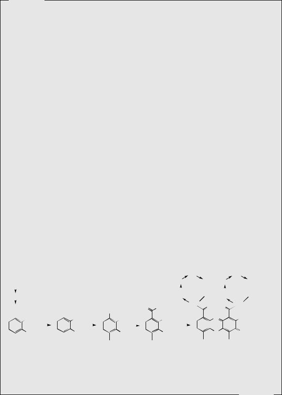
Dactinomycin
Dactinomycin (actinomycin D) (Figure 7.28) is an antibiotic produced by Streptomyces parvullus (formerly S. antibioticus), which has antibacterial and antifungal activity, but whose high toxicity limits its use to anticancer therapy. Several related natural actinomycins are known, but only dactinomycin is used medicinally. Dactinomycin has a planar phenoxazinone dicarboxylic acid system in its structure, to which are attached two identical cyclic pentapeptides via amide bonds to threonine. The peptides are cyclized by lactone linkages utilizing the hydroxyl group of this threonine. The peptide portions of dactinomycin contain N-methylvaline and N-methylglycine (sarcosine) residues. In other actinomycins, the two peptides are not necessarily identical. The phenoxazinone ring system is known to be formed by fusing together two molecules of 3-hydroxy-4-methylanthranilic acid (Figure 7.28), which arises by C-methylation of 3-hydroxyanthranilic acid, a metabolite of tryptophan by the kynurenine pathway (see page 312). This planar phenoxazinone ring intercalates with double-stranded DNA inhibiting DNA-dependent RNA polymerases, but can also cause single-strand breaks in DNA. It is principally used to treat paediatric cancers, including Wilms’ tumour of the kidney, but produces several serious and painful side-effects. However, as a selective inhibitor of DNA-dependent RNA synthesis (transcription), it has become an important research tool in molecular biology.
|
L-Trp |
|
|
|
|
|
|
|
|
|
|
|
Sar |
|
|
|
|
Sar |
|||||
|
|
|
|
|
|
|
|
|
|
Pro |
|
|
Pro |
|
|
||||||||
|
|
|
|
|
|
|
|
|
|
|
|
|
|
(Me)Val |
(Me)Val |
||||||||
|
|
|
|
|
|
|
|
|
|
|
|
|
|
|
|
|
|
|
|
|
|
|
|
|
|
|
|
|
|
|
|
|
|
|
|
|
D-Val |
O |
D-Val |
O |
|||||||
|
|
|
|
|
|
|
|
|
|
|
|
|
|||||||||||
|
|
|
|
|
|
|
|
|
|
|
O |
|
|
|
Thr |
|
O |
O |
Thr |
||||
|
|
|
|
|
|
|
|
|
|
|
|
|
|
||||||||||
|
CO2H |
|
CO2H |
|
|
peptide |
|
|
|
||||||||||||||
|
|
COAMP |
|
|
|
|
|
|
|
|
|
|
|
||||||||||
|
|
NH2 SAM |
|
NH2 ATP |
NH2 |
NH2 x 2 |
|
N |
|
|
NH2 |
||||||||||||
|
|
|
|
|
|
OH |
|
|
|
|
|
|
OH |
|
|
|
|
O |
|
|
O |
||
|
|
OH |
|
OH |
|
|
|
||||||||||||||||
3-hydroxyanthranilic 3-hydroxy-4-methyl- |
|
|
|
|
|
|
|
|
|
dactinomycin |
|
|
|||||||||||
|
|
acid |
anthranilic acid |
|
|
|
|
|
|
|
|
|
(actinomycin D) |
||||||||||
Figure 7.28
(Continues)

PEPTIDE TOXINS |
433 |
(Continued )
PEPTIDE TOXINS
Death Cap (Amanita phalloides)
The death cap, Amanita phalloides, is a highly poisonous European fungus with a mushroom-like fruiting body. The death cap has a whitish-green cap, and white gills. It has a superficial similarity to the common mushroom, Agaricus campestris, and may sometimes be collected in error. Some 90% of human fatalities due to mushroom poisoning are attributed to the death cap. Identification of the death cap as a member of the genus Amanita, which includes other less poisonous species, is easily achieved by the presence of a volva at the base of the stem. This cuplike membranous structure is the remains of the universal veil in which the immature fruiting body was enclosed. Ingestion of the death cap produces vomiting and diarrhoea during the first 24 hours, followed after 3–5 days by coma and death. Some recoveries do occur, but the fatality rate is probably from 30 to 60%. There is no guaranteed treatment for death cap poisoning, though removal of material from the gastrointestinal tract, replacement of lost fluids, blood dialysis, and blood transfusions may be undertaken. The antihepatotoxic agent silybin (see page 153) has been used successfully. The toxic principles are cyclic polypeptides, which bring about major degeneration of the liver and kidneys. At least ten toxins have been identified, which may be subdivided into two groups, the phallotoxins and the amatoxins. The most extensively studied compounds are phalloin, a phallotoxin, and α-amanitin, an amatoxin (Figure 7.29). The phallotoxins are much less toxic than the amatoxins since after ingestion they are not well absorbed into the blood stream. When injected, they can cause severe damage to the membranes of liver cells, releasing potassium ions and enzymes. The amatoxins are extremely toxic when ingested,
with a lethal dose of 5–7 mg |
for an adult human, and an average specimen of the |
|||||||||||||||||||||||||||||||||||||||||||
death |
cap |
containing about 7 mg. The |
amatoxins |
cause |
lesions |
to the |
stomach and |
|||||||||||||||||||||||||||||||||||||
|
|
|
|
|
|
|
|
|
|
|
|
|
|
|
H |
|
|
O |
|
|
|
|
|
|
|
|
|
|
|
|
|
|
|
|
|
|
|
|
|
|
|
|
|
|
|
|
|
|
|
|
|
|
|
|
|
|
|
|
|
|
|
|
|
|
|
|
|
|
|
|
|
|
|
|
|
|
|
|
|
|
|
|
|
|
|
|
|
|
|
Ala |
|
|
Trp |
|
|
|
(γ-OH)Ile |
|
|
N |
|
|
|
|
|
|
|
|
|
|
|
Pro |
|
|
|
Phe |
|
|
|
Phe |
|
|
|
Val |
|
|
Pro |
|||||||
|
|
|
|
|
|
|
|
|
|
|
|
|
|
|
|
|
|
|
|
|
|
|
|
|
|
|
|
|
||||||||||||||||
|
|
|
|
|
|
|
|
|
|
|
|
|
|
|
|
|
|
|
|
|
|
|
|
|
|
|
|
|
||||||||||||||||
|
|
|
|
|
|
|
|
|
|
|
|
|
|
|
|
|
|
|
|
|
|
|
|
|
|
|
|
|
||||||||||||||||
|
|
|
|
|
|
|
|
|
|
|
|
|
|
|
|
|
|
|
|
|
|
|
|
|
|
|
|
|
|
|
|
|
|
|
|
|
|
|
|
|
|
|
|
|
|
|
|
|
|
|
|
|
|
|
|
|
|
|
|
|
|
|
|
|
|
|
|
|
|
|
|
|
|
|
|
|
|
|
|
|
|
|
|
|
|
|
|
|
|
HPro |
S |
|
Ala |
|
|
|
|
|
|
|
|
|
|
|
|
|
|
|
|
|
|
|
|
|
|
|
|
|
|
|
|
|
|
|
|
|||||||||
|
|
|
|
|
|
|
|
|
|
|
|
|
|
|
Pro |
|
Phe |
|
|
|
Phe |
|
|
|
Ala |
|
Pro |
|||||||||||||||||
|
|
|
|
|
|
|
|
|
|
|
|
|
|
|
|
|
|
|
|
|
|
|
|
|
|
|
|
|||||||||||||||||
|
|
Cys |
|
|
|
Thr |
O |
S |
|
N |
|
|
|
|
|
|
|
|
|
|
|
|
|
|
|
|||||||||||||||||||
|
|
|
|
|
|
|
|
|
|
|
|
|
|
|
|
|
|
|
|
antamanide |
|
|
|
|||||||||||||||||||||
|
|
|
|
|
|
H |
Trp |
|
|
|
|
|
|
|
|
|
|
|
|
|
|
|
||||||||||||||||||||||
|
|
|
|
phalloin |
|
|
|
|
|
|
|
|
|
|
|
|
|
|
|
|
|
|
|
|
|
|
|
|
|
|||||||||||||||
|
|
|
|
|
|
|
|
|
HN |
|
|
|
Cys |
|
|
|
|
|
|
|
|
|
|
|
|
|
|
|
|
|
|
|
|
|
|
|
||||||||
|
|
|
|
|
|
|
|
|
|
|
|
|
|
|
|
|
|
|
|
|
|
|
|
|
|
|
|
|
|
|
|
|
|
|
||||||||||
|
|
|
|
|
|
|
|
|
|
|
|
|
|
|
|
|
|
|
|
|
|
|
|
|
|
|
|
|
|
|
|
|
|
|
|
|
|
|
|
|
|
|||
|
|
|
|
|
|
|
|
|
|
|
|
|
|
|
|
|
|
|
|
|
|
|
|
H |
O |
|
|
|
|
|
|
|
|
|
|
|
|
|
|
|
|
|
|
|
|
|
|
|
|
|
|
|
|
|
|
|
|
|
|
|
|
|
|
|
|
|
|
|
|
|
|
|
|
|
|
|
|
|
|
|
|
|
|
|
|
|
|
|
|
|
|
|
|
|
|
|
|
|
|
[γ,γ-(OH)2]Ile |
|
|
(6-OH)Trp |
Gly |
|
|
N |
|
|
|
|
|
|
|
|
OH |
|
|
|
|
|
|
|
|
||||||||||
|
|
|
|
|
|
|
|
|
|
|
|
|
|
|
|
|
|
|
|
|
|
|
|
|
|
|
|
|
|
|||||||||||||||
|
|
|
|
|
|
|
|
|
|
|
|
|
|
|
|
|
|
|
|
|
|
|
|
|
|
|
|
|
|
|
||||||||||||||
|
|
|
|
|
|
|
|
|
O |
|
|
|
|
|
|
|
|
|
|
|
|
|
|
|
|
|
|
|
||||||||||||||||
|
|
|
|
|
|
|
|
|
|
|
|
|
|
|
|
|
|
|
|
|
|
|
|
|
|
|
|
|
|
|
|
|
|
|
|
|
||||||||
|
|
|
|
|
|
|
|
|
|
|
|
|
|
|
|
|
|
|
|
|
|
|
|
|
|
|
|
|
|
|
|
|
|
|
|
|
||||||||
|
|
|
|
|
|
|
|
|
|
|
|
|
|
|
|
|
|
|
|
|
|
|
|
|
|
|
|
|
|
|
|
|
|
|
|
|
|
|
|
|
||||
|
|
|
|
|
|
|
|
|
|
|
HPro |
|
S |
|
|
O |
O |
S |
N |
|
|
|
|
|
|
|
|
|
|
|
|
|
|
|||||||||||
|
|
|
|
|
|
|
|
|
|
|
|
|
|
|
|
|
|
|
|
|
|
|
|
|
|
|
|
|
|
|||||||||||||||
|
|
|
|
|
|
|
|
|
|
|
|
|
|
|
|
|
|
|
|
|
|
|
|
|
|
|
|
|||||||||||||||||
|
|
|
|
|
|
|
|
|
|
|
|
|
|
|
|
|
|
|
|
Ile |
|
|
|
|
|
|
|
|
|
|
|
|
|
|
||||||||||
|
|
|
|
|
|
|
|
|
|
|
|
|
|
|
|
|
|
|
|
|
H |
|
|
|
|
|
|
|
|
|
|
|
|
|
|
|||||||||
|
|
|
|
|
|
|
|
|
|
|
|
|
|
Cys |
|
|
|
|
|
|
|
|
|
(6-OH)Trp |
|
|
|
|
|
|
|
|
||||||||||||
|
|
|
|
|
|
|
|
|
|
|
Asn |
|
|
|
|
Gly |
|
|
|
|
|
|
|
|
|
|
|
|
|
|||||||||||||||
|
|
|
|
|
|
|
|
|
|
|
|
|
|
|
|
|
|
|
|
|
|
|
|
|
|
|
|
|
|
|
|
|
|
|||||||||||
|
|
|
|
|
|
|
|
|
|
|
|
|
|
|
|
|
|
|
|
|
|
|
|
|
|
|
|
|
|
|
||||||||||||||
|
|
|
|
|
|
|
|
|
|
|
|
|
α-amanitin |
|
|
|
|
|
HN |
|
|
Cys |
|
|
|
|
|
|
|
|
|
|
|
|
|
|
||||||||
Figure 7.29
(Continues)

434 |
PEPTIDES, PROTEINS, AND OTHER AMINO ACID DERIVATIVES |
(Continued )
intestine, and then irreversible loss of liver and kidney function. α-Amanitin has been shown to be a powerful inhibitor of RNA polymerase, blocking the elongation step. Structurally, the two groups of toxins appear similar. They both contain a γ-hydroxylated amino acid essential for toxic action, and have a sulphur bridge between a cysteine residue and the 2-position of a tryptophan or 6-hydroxytryptophan residue (Figure 7.29). In the case of the phallotoxins, this is a simple sulphide bridge, but in the amatoxins it is in the form of a sulphoxide. Interestingly, a rather more simple cyclic peptide antamanide (anti-amanita peptide) has also been isolated from Amanita phalloides. When pre-administered to laboratory animals, antamanide (Figure 7.29) provided prophylactic protection from the lethal affects of the phallotoxins.
Ricin
The distinctive mottled-brown seeds of the castor oil plant Ricinus communis (Euphorbiaceae) are crushed to produce castor oil, which is predominantly composed of glycerides of ricinoleic acid (see page 47 and Table 3.2). The seed itself and the seed cake remaining after expression of the oil are highly toxic, due to the presence of polypeptide toxins, termed ricins. One or more forms of ricin are present according to the strain of plant. Seeds typically contain about 1mg g−1 ricin, representing about 5% of the protein content. The toxicity of ricins to mammals is so high that the ricin content of one seed (about 250 g) is sufficient to kill an adult human, though, because of considerable variations in absorption and metabolism, consumption of a single seed might not be fatal, but would certainly lead to severe poisoning. The toxic symptoms include irritation and haemorrhage in the digestive tract, leading to vomiting and bloody diarrhoea, with subsequent convulsions and circulatory collapse. If death does not occur within 3–5 days, the patient may recover.
Ricin is probably the best studied of a group of polypeptide toxins, known as ribosomeinactivating proteins (the acronym RIP seems most appropriate!). These toxins are potent inhibitors of eukaryotic protein biosynthesis by virtue of their cleavage of an N-glycosidic bond at a specific nucleotide residue in the 28S molecule of ribosomal RNA, which is part of the larger (60S) subunit of the eukaryotic ribosome. RIPs fall into two types. Type I proteins comprise a single polypeptide chain, sometimes glycosylated. Type II proteins have two chains linked by a disulphide bond: an A chain is essentially equivalent to type I proteins, whilst the B chain functions as a lectin, which means it has high affinity for specific sugar groups, in this case sugars (especially galactose) in glycolipids or glycoproteins on a cell membrane. Thus the B chain binds to the cell membrane, and, in so doing, facilitates entry of the A chain into the cytosol, where it inactivates the 60S ribosomal subunit and rapidly stops protein biosynthesis. The A chain is thus the toxic principle, but it is nontoxic to intact cells, and requires the B chain for its action.
Ricin is a type II toxin. The A chain (ricin A) contains 267 amino acid residues, and the B chain (ricin B) 262 residues. Ricin A is exceptionally toxic, and it has been estimated that a single molecule is sufficient to kill an individual cell. This peptide can be prepared by genetic engineering using Escherichia coli. The potent action of this material on eukaryotic cells has been investigated in anticancer therapy. Ricin A has been coupled to monoclonal antibodies and successfully delivered specifically to the tumour cells. However, in vitro toxicity of ricin A-based immunotoxins is enhanced significantly if ricin B is also present.
(Continues)