

Interactions of Cannabinoids With Membranes. The Role of Cannabinoid Stereochemistry and Absolute Configuration and the Orientation of Delta-9-THC in the Membrane Bilayer
Alexandros Makriyannis, Ph.D.; Ali Banijamali, Ph.D.; Cornelis Van Der Schyf, Ph.D.; and Harold Jarrell, Ph.D.
INTRODUCTION
Cannabinoid Membrane Effects
Many pharmacological actions of the cannabinoids, such as psychotropic effects, bronchodilatation, increased heart rate, reduced intraocular pressure, analgesia, alteration in body temperature, and anticonvulsant activity, can be related to their known effects on a variety of cellular membranes. Indeed, cannabinoids are known to affect a variety of membrane functions (Martin 1986; Mechoulam et al. 1985; Hillard et al. 1985, 1986). Among these we can list their effects on numerous membrane enzymes (e.g., adenylate cyclase; Na+, K+-, Mg2+-, Ca2+- ATPases; NADH dehydrogenase; LPC acyltransferase; phospholipase A2; monoamine oxidase; cholesterol esterase; 5'-nucleotidase), brain synaptosomes, blood platelets, human fibroblasts, neuroblastoma cells, rabbit neutrophils (Naccache et al. 1982), excitable membranes (Martin 1986), and membrane receptors (e.g., beta-adrenergic, cholinergic, opioid, dopaminergic) (Bloom 1984; Bloom and Hillard 1985). The membrane functions affected by the cannabinoids can be related to a wide variety of membrane-bound proteins. A common feature with all of these effects is that the cannabinoids do not seem to interact directly with the protein catalytic sites and do not compete with those ligands known to interact with these sites.
Cannabiniod Activity Through Interactions with Phospholipids
It is generally accepted that the lipid microenvironment surrounding membraneassociated enzymes (Sandermann 1978), receptors, or other functional proteins (Loh and Law 1980) can influence protein function. On this basis, the view was advanced that cannabinoids may exert their effects by interacting with the lipid component of the membrane. The following evidence is offered in support of this view: (I) Cannabinoids are highly lipophilic molecules and partition favorably in membrane lipids. (2) Cannabinoids have been shown to affect the thermotropic properties of phospholipids. They thus decrease the phase transition temperature of pure (Bruggemann and Melchior 1983; Makriyannis et al. 1987) and brain membrane (Bach and Sela 1981) phospholipids. Also, they eliminate the discontinuity in the Arrhenious plot for microsomal O-demethylase (Bach et al. 1977), an effect attributed to a change in the phase transition properties of the lipids associated with the enzyme. (3) Cannabinoids affect the "microviscosity" of model and biological membranes as determined by electron
123
spin resonance (ESR) (Lawrence and Gill 1975), fluorescence (Hillard et al. 1986), and NMR methods (Makriyannis et al. 1987; Kriwacki 1986). The lipid hypothesis is bolstered by the fact that some of these studies were carried out using cannabinoid concentrations considered to be "physiological" (Hillard et al. 1985).
The Role of Drug Geometry in Drug: Phospholipid Interactions
Traditionally, the potency of drugs that are believed to act on the lipid component of the membrane has been correlated with their lipid solubility and no consideration was made for variations in their conformational properties or their absolute configurations. This assumption was shown to be true with the low molecular weight general anesthetics (Kendig et al. 1973), but does not seem to hold with the larger membrane-active drugs. It was thus reported that enantiomeric pairs of barbiturates showed marked differences in potency (Wahlstrom et al. 1970). This has also been well documented in the case of anesthetic sterioids where small structural variations in the steroid molecule, having no effect on its lipid solubility, produce dramatic changes in anesthetic activity (Atkinson et al. 1965). When tested for their effectiveness in inhibiting the function of a membrane-bound protein (Makriyannis and Fesik 1980) and for their abilities to perturb model phospholipid membranes (Makriyannis and Fesik 1983; Makriyannis et al. 1986), the anesthetic steroids showed variations that paralleled those of their respective potencies. We explained the observed differences in steroid membrane interactions on the basis of observed conformational differences between the drug molecules (Fesik and Makriyannis 1985).
Interestingly, an analogous effect was demonstrated by Bloch and coworkers, who found that small variations in sterol structure influence in a dramatic manner their effects on model membranes, including solute permeation, microviscosity, and steroid mobility in the membrane (Lala et al. 1978; Dahl et al. 1980), as well as the growth rates of microorganisms (Lala et al. 1978).
Analogous evidence is also available for cannabinoids, pointing to the role of structure and geometry in their interactions with phospholipids. Earlier work using ESR (Lawrence and Gill 1975) showed that cannabinoid analogs with only slight structural differences, or having the same structure but differing in absolute configuration, exhibited relatively large differences in their effects on model membrane preparations. As reported in a recent publication, fluorescence polarization was used to study the effects of cannabinoids on synaptosomal plasma membranes (Hillard et al. 1985). The study found significant changes in membrane "fluidity" when "physiological" concentrations of cannabinoids were used. Another study compared the thermotropic properties of model membrane preparations containing each of the two delta-9-THC enantiomers and found differences between them (Burstein et al. 1986). We would like here to report on more detailed work from our laboratories using solid-state 2H-NMR spectroscopy.
RESULTS AND DISCUSSION
Model Membranes and the Use of Solid-State NMR Techniques
Because of their demonstrable applicability to the natural systems, studies with model membranes have contributed a great deal toward understanding the properties of biological membranes. Model membranes have also been used
124
successfully to obtain information on drug:membrane interactions. Many of these studies have made use of fluorescence, E.S.R., and high resolution NMR techniques. However, the availability of solid-state NMR has opened new horizons in this field of research. During the past few years, these methods have led to previously unavailable molecular information on the conformational and dynamical properties of model and biological membranes (semisolids) (Griffin 1981; Davis 1983). The use of solid-state NMR has also been extended to study drug:membrane interactions and showed it to be the method of choice (Boulanger et al. 1981; Makriyannis et al. 1986). The model membranes used for the solidstate NMR experiments are multilamellar bilayer phospholipid dispersions that have proven to be excellent models, sharing to a surprisingly large degree many of the properties of the lipid component in biological membranes (Davis 1983).
The Role of Cannabinoid Conformation and Absolute Configuration During its Interaction with Membranes as Studied by Solid-State 2H-NMR
In systems undergoing anisotropic motions, as is the case with phospholipid multilamellar bilayers, the deuterium nucleus gives rise to a "powder spectrum" in which the doublet with the maximum intensity is the 2H quadrupolar splitting
(  ) and equal to:
 = (3/4) (e2qQ/h) SCD (Davis 1983)
where e2qQ/h) is the static quadrupole coupling constant which for, paraffinic C-D bonds, is approximately 170 KHz. SCD is the order parameter which can be used to describe the amount of motional averaging of the C-D bond vector with respect to a fixed symmetry axis and has been used as a measure of membrane "fluidity."
In its liquid crystalline phase, the 2H spectrum of DPPC labeled in a single site has the powder pattern features observed in the 2H spectra of solids. However, the quadrupolar splitting is reduced due to motional averaging, including axial diffusion of the chains and gauche:trans isomerization in the methylene chain segments.
To compare the effects of cannabinoid analogs on membranes, we used DPPC model membranes containing 0.15M cholesterol into which 0.15M cannabinoid was incorporated. From earlier studies with anesthetic steroids (Makriyannis, unpublished), we have found that model membranes of such composition are very suitable for discriminating between membrane-acting drug analogs and that the relative effects of these molecules on the model membrane parallel their effects on biological membranes.
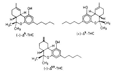
We compared the effects of (-)-delta-9-THC to those of its inactive (-)-delta- 9,11-THC isomer on the model membrane by obtaining solid-state 2H-NMR spectra as a function of temperature for each of the two preparations as well as spectra for the membrane preparation without cannabinoid. We also compared in a similar manner the effects of the two delta-9-THC enantiomers.
Figure 1 shows representative spectra from the model membrane preparations with (-)-delta-9-THC, (-)-delta-9,11-THC, and no drug, obtained at two different temperatures. The spectra are of the liquid crystalline type and allow us to extract directly values for the residual 2H quadrupolar splittings (  ). As
). As
125
expected, the splittings decrease as the temperature is increased, reflecting the higher ratio of gauche:trans chain conformers in the chain at higher temperatures.
In figure 2, we compare the  values for the above three preparations as a function of temperature, while figure 3 compares the effects of the two delta-9- THC enantiomers on the same membrane preparation. As can be seen, the presence of each of the three cannabinoids in the membrane results in a decrease in the respective 2H quadrupolar splittings. Also, membrane preparations containing the pharmacologically active (-)-delta-9-THC analog give 2H spectra with consistently smaller splittings than those from its inactive or less active isomers, (-)-delta-9,11-THC or (+)-delta-9-THC. This is evidence that, although all three cannabinoids increase the ratio of gauche:trans conformers in the membrane chain, the more active analog produces the larger increase in gauche:trans methylene ratio and the smaller order parameter (SCD). This, in
turn, may be interpreted to mean that the active (-)-delta-9-THC analog "perturbs" the model membrane more effectively than its two less active isomers.
The above data show that the interaction of cannabinoids with the phospholipid component of the membrane is governed by specific stereoelectronic requirements. Our results also provide support for the view that at least some of the cannabinoid effects result from their interactions with the lipid component of the membrane. The above observations on the stereoselectivity of cannabinoid: phospholipid interactions are understandable if the defined geometric parameters of the phospholipid bilayer are considered. These include the natural asymmetry of the lecithin and cholesterol molecules as well as the conformational
preferences and dynamic properties of the lipid hydrocarbon chains and/or polar head groups.
126
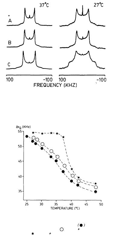
FIGURE 1. Solid-state 2H-NMR spectra due to the 2(7’,7’-C2H) segment of DPPC obtained using a 90°x -  -90° quadrupolar echo sequence at 45.3 MHz with a 90° pulse width of 2.1-2.4 µs. To obtain each spectrum, 8,000-16,000 ethos were signal averaged. (A) DPPC + 0.15m cholesterol + 0.15M (-)-delta-9- THC; (B) DPPC + 0.15M cholesterol + 0.15M (-)- delta-9,11-THC; (C) DPPC + 0.15M cholesterol.
FIGURE 2. Temperature dependence of the 2H quadrupolar splittings in the 2H-
NMR spectra of the 2(7’,7’-C2H2) segment DPPC. |
DPPC + 0.15M |
|
cholesterol + 0.15M (-)-delta-9-THC: ( )DPPC |
0.15M cholesterol + 0.15M |
|
(-)-delta-9'11-THC; ( |
) DPPC 0.15M cholesterol. |
|
127
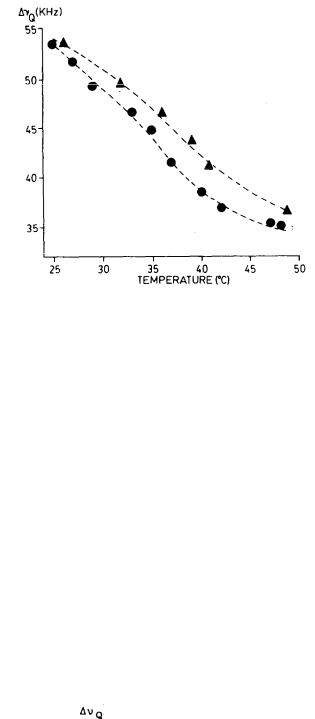
FIGURE 3. Temperature dependence of the 2H quadrupolar splittings in the 2H- NMR spectra of the 2(7’, 7’-C2H2) segment of DPPC. ( ) DPPC + 0.15M cholesterol + 0.15M (-)-delta-9-THC; (
  ) DPPC + 0.15M cholesterol + 0.15M
(+)-delta-9-THC.
Since the drug concentrations used in our experiments are much higher than "pharmacological," the results presented here cannot be viewed as a quantitative description of the membrane perturbation produced by cannabinoids in vivo. However, the data give us a qualitative description of the type of conformational and dynamical changes produced by cannabinoids on the membrane bilayer. Moreoever, such experiments provide us with a method for comparing the effects of structurally related cannabinoid analogs on membranes.
The Orientation of (-)-delta-9-THC in DPPC Bilayers
In our effort to understand the geometric parameters involved in the interaction of cannabinoids with membrane bilayers. we have made use of solid-state 2H- NMR to study the orientation of (-)-delta-9-THC in the membrane bilayer. This experiment required the strategic introduction of 2H labels in different
positions of the tricyclic cannabinoid ring system. To that effect, we synthesized (-)delta-9-THC with specific 2H labels in each of the 2, 4, 8 alpha, 8 beta, 11CH3, 10, and 10a positions (Banijamali et al. 1987). The labeled drug molecule was then introduced in DPPC bilayers. 2H spectra were generally obtained for membrane preparations containing (-)-delta-9-THC with one 2H label or with up to three 2H labels if these could be unambiguosly assigned in the solid-state spectra. Spectra from the 2H-labeled THC in the membrane in the liquid crystalline phase were axially symmetrical or nearly so and had different quadrupolar splittings ( ) depending on the position of the 2H label in the molecule (figure 4). The value of these splittings was then used to calculate the orientation of (-)-delta-9-THC in DPPC bilayers.
128
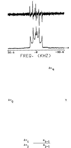
FIGURE 4. Below: Representative solid-state 2H-NMR spectrum of 2,4-d2-(-)- delta-9-THC (.3M) in hydrated DPPC bilayers at 42°C;  (2H4 ) = 43.0 KHz,
  (2H2) = 20.8 KHZ. The central doublet ( =6.6 KHz) is due to the phenolic deuteron. Above: Firs& derivative of the same spectrum from which 2H splittings could be obtained more accurately.
Briefly, the principle of this determination is as follows (Taylor 1981): Deuterons attached to a rigid structure will give quadrupolar splittings described by the equation:
= 3/4 e2qQ |
(3cos 2 a - 1) |
[ 3cos2 - 1] |
h |
2 |
2 |
where is the angle between the individual C-2H bond and the rotation axis. The last term, which is known as the molecular order parameter (Smol), defines the anisotropic motion of the entire molecule. The angles between the rotation axis and the C-2H bonds cannot be calculated directly because Smol is not known. However, Smol is expected to be identical for all 2H atoms attached to the cannabinoid ring system in which overall tumbling is expected to dominate the dynamics of this part of the molecule. Thus, the ratios (R ) between two
quadrupolar splittings for two different deuterium atoms (2H |
and |
2H ) are equal |
|||||
to: |
|
|
i |
|
j |
||
|
|
|
|
|
|||
= |
3cos2 |
|
|
||||
Ra |
|
|
3cos |
2 |
|
|
|
|
|
|
|
||||
|
|
|
|
|
|
|
|
Our calculations showed that (-)-delta-9-THC orients with its axis of rotation almost in the plane of the aromatic ring. However, the molecule does not orient with its long axis parallel to the bilayer chains, as is the case with cholesterol, but almost orthogonal to the chains. Details of the above calculations will be given elsewhere.
129
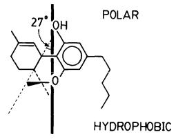
The above-described orientation allows the cannabinoid phenolic hydroxy group to direct itself toward the polar side of the bilayer, indicating that this group serves as the orienting anchor in the molecule. We refer to this cannabinoid: bilayer interaction as "amphipathic interaction" in which its primary incentive is to direct the polar and hydrophobic components of the cannabinoid molecule toward the respective components in the amphipathic bilayer. This anchoring effect of the THC phenolic OH on the membrane is intriguing and deserves more extensive investigation.
Indeed, the requirement of a free OH for activity in THC is well established. Also, differences in activities have been reported between cannabinoid isomers, differing only in the presence and/or orientation of hydroxy and keto groups in the C ring. Since it is reasonable to assume that these hydrogen bonding groups may also be involved in the orientation of the cannabinoid molecule, one may speculate that cannabinoid orientation in the membrane may have an important effect on its biological activity. The drug orientation in the amphipathic membrane may, thus, play an important (although not unique) role in determining the nature of the cannabinoid:membrane interaction and its ability to produce "productive membrane perturbations." We are currently investigating the above hypothesis.
FIGURE 5. The orientation of (-)-delta-9-THC in DPPC bilayers as determined from solid-state 2H-NMR data.
We combined our results on the orientation of delta-9-THC in the bilayer with previously obtained data on the conformation of delta-9-THC (Kriwacki 1986) to model its interaction with a DPPC bilayer using computer graphics. The parameters for modeling the phospholipid bilayer were obtained from available crystallographic data. As can be seen, the THC molecule inserts between the two fatty acid chains of the bilayer while maintaining its phenolic hydroxy group at the level of the bilayer interphase. Also, it can be noted that the plane of the C ring in delta-9-THC deviates from those of the A and B rings. This conformational property of delta-9-THC may explain its ability to perturb membranes.
130
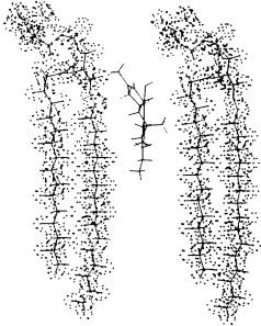
FIGURE 6. Modeling of (-)-delta-9-THC in a DPPC bilayer from experimental data using a E&S PS 300 system and ChemX graphics software (developed and distributed by Chemical Design Ltd., Oxford, England).
REFERENCES
Atkinson, R.M.; Davis, B.; Pratt, M.A.; Sharpe, H.M.; and Tomich, E.G. Action of some steroids on the central nervous system of the mouse. II Pharmacology. J Med Chem 8:426-432, 1965.
Bach, D.; Bursuker, L.; and Goldman, R. Differential scanning calorimetry and enzymic activity of rat liver microsomes in the presence and absence of delta-1-tetrahydrocannabinol. Biochim Biophys Acta 469:171-179, 1977.
Bach, D., and Sela, B. Thermotropic properties of calf brain lipids interacting with drugs. Biochem Pharmacol 30:1777-1780, 1981.
Banijamali, A.; Abou Taleb, N.; Charalambous, A.; Van der Schyf, C.J.; and Makriyannis, A. Synthesis of deuterium labeled cannabinoids. Submitted for publication, J Label Comp, 1987.
Bloom, A.S. Effects of cannabinoids on neurotransmitter receptors in the brain. In: Agurell, S.; Dewey, W.L.; and Willette, R.E., eds. The Cannabinoids; Chemical, Pharmacologic, and Therapeutic Aspects. New York: Academic Press, 1984. pp. 575-590.
Bloom, A.S., and Hillard, C.J. Cannabinoids, neurotransmitter receptors and brain membranes. In: Harvey, D.J., ed. Marihuana ’84. Proceedings of the Oxford Symposium on Cannabis. Oxford: IRL Press, 1985. pp. 217-231.
131
Boulanger, Y.; Schreier, S.; and Smith, I.C.P. Molecular details of anestheticlipid interactions as seen by deuterium and phosphorus-31 nuclear magnetic resonance. Biochemistry 20:6824-6830, 1981.
Bruggemann, E.P., and Melchior, D.L. Alterations in the organization of phosphatidylcholine/cholesterol by tetrahydrocannabinol. J Biol Chem 258:8293-8303, 1983.
Burstein, S.; Hunter, S.A.; Latham, V.; Mechoulam, R.; Melchoir, D.; and Renzuli, L. Prostaglandins and cannabis. XV. Comparison of enantiomeric cannabinoids in stimulating prostaglandin synthesis on fibroblasts. Life Sci 39:1813-1823, 1986.
Dahl, C.E.; Dahl, J.S.; and Bloch, K. Effect of alkyl-substituted precursors of cholesterol on artificial and natural membranes and on the viability of Mycoplasma capricolum Biomchemistry 19:1462-1467, 1980.
Davis, J.H. The description of membrane lipid conformation, order and dynamics using 2H NMR. Biochim Biophys Acta 737:117-171, 1983.
Fesik, S.W., and Makriyannis, A. Geometric requirements for membrane perturbation and anesthetic activity. Conformational analysis of alphaxalone and delta-16-alphaxalone and 2H NMR studies on their interactions with model membranes. Mol Pharmacol 27:624-629, 1985.
Griffin, R.G. Solid state nuclear magnetic resonance in lipid bilayers. Methods Enzymol 72:108-174, 1981.
Hillard, C.J.; Harris, R.A.; and Bloom, A.S. Effects of cannabinoids on physical properties of brain membranes and phospholipid vesicles: Fluorescence studies. J Pharmacol EXp Ther 232:579-588, 1985.
Hillard, C.J.; Bloom, A.S.; and Houslay, M.D. Effects of delta-9-tetrahydro- cannabinol on glucagon receptor coupling to adenylate cyclase in rat liver plasma membranes. Biochem Pharmacol 35:2797-2803, 1986.
Kendig, J.J.; Trudell, J.R.; and Cohen, E.N. Halothane stereoisomers: Lack of stereospecificity in two model systems. Anesthesiology 39:518-524, 1973.
Kriwacki, R. Studies on the conformations of two cannabinoids and their interactions with model membranes using high resolution NMR techniques. Dissertation Abstr 1986.
Lala, A.K.; Lin, H.; and Bloch, K. The effect of some alkyl derivatives of cholesterol on the permeability properties and microviscosities of model membranes. Biorg Chem 7:437-445, 1978.
Lawrence, D.K., and Gill, E.W. The effects of delta-1-tetrahydrocannabinol and other cannabinoids on spin-labeled liposomes and their relationship to mechanisms of general anesthesia. Mol Pharmacol 11:595-602, 1975.
Loh, H.H., and Law, P.Y. The role of membrane lipids in receptor mechanisms. Annu Rev Pharmacol Toxicol 20:201-234, 1980.
Makriyannis, A., and Fesik, S.W. Effects of anesthetics on sulfate transport in the red cell. J Neurosci Res 5:25-33, 1980.
Makriyannis, A., and Fesik, S.W. Mechanism of steroid anesthetic action. Interactions of alphaxalone and delta- 16-alphaxalone with bilayer vesicles. J Med Chem 26:463-465, 1983.
Makriyannis, A.; Siminovitch, D.; Das Gupta, SK.; and Griffin, R.G. Studies on the interaction of anesthetic steroids with phosphatidylcholine using 2H and 13C solid state NMR. Biochim Biophys Acta 859:49-55, 1986.
Makriyannis, A.; Siminovitch, D.; Das Gupta, S.K.; and Griffin, R.G. The perturbation of model membranes by (-)-delta-9-tetrahydrocannabinol. Studies using 2H and 13C-solid state NMR. In preparation, 1987.
Martin, B.R. Cellular effects of cannabinoids. Pharmacol Rev 38:45-75, 1986.
132
Mechoulam, R.; Srebnik, M.; and Burstein, S. Cannabis chemistry, biochemistry and therapeutic applications - An overview. In: Harvey, D.J., ed. Marihuana ’84. Proceedings of the Oxford Svmposium on Cannabis. Oxford: IRL Press, 1985. pp. 1-12.
Naccache, P.H.; Volpi, M.; Becker, E.L.; Makriyannis, A.; and Sha’afi, R.I. Cannabinoid induced degranulation of rabbit neutrophils. Biochem Biophys Res Commun 106:1286-1290, 1982.
Sandermann, H., Jr. Regulation of membrane enzymes in lipids. Biochim Biophys Acta 515:209-237, 1978.
Taylor, M.G.; Alkiyama, T.; and Smith, I.C.P. The molecular dynamics of cholesterol in bilayer membranes: A deuterium NMR study. Chem Phys Lipids 29:327-338, 1981.
Wahlstrom, G.; Buch, H.; and Buzello, W. Unequal anaesthetic activity despite equal brain concentration of hexobarbital antipodes. Acta Pharmacol Toxicol 28:493-498, 1970.
ACKNOWLEDGMENT
This research was supported by grant number 3801 from the National Institute on Drug Abuse.
AUTHORS
Alexandros Makriyannis, Ph.D.
School of Pharmacy and Institute of Materials Science
University of Connecticut
Storrs, Connecticut 06268
and
F. Bitter National Magnet Laboratory
Massachusetts Institute of Technology
Cambridge, Massachusetts 02139
Ali Banijamali, Ph.D.
Cornelis Van der Schyf, Ph.D.
School of Pharmacy and Institute of Materials Science
University of Connecticut
Storrs, Connecticut 06268
Harold Jarrell, Ph.D.
Division of Biological Sciences
National Research Council of Canada
Ottawa, Canada K1A 0R6
133