

The High Affinity Cannabinoid Binding Site in Brain: Regulation by Guanine Nucleotides and Isolation of an Endogenous Inhibitor
Jeffrey S. Nye and Solomon H. Snyder, M.D.
INTRODUCTION
The molecular mechanism by which delta-9-tetrahydrocannabinol (delta-9-THC) produces its characteristic psychotropic effect in man and animals remains a mystery. The very high potency of cannabinoids and their stereochemical and structural requirements for eliciting characteristic psychotropic effects suggest that a neuronal receptor exists for delta-9-THC. Neuronal receptors have been identified for a wide range of psychotropic agents; these sites have been characterized by studying the binding of radiolabeled analogues of high potency drugs.
The biochemical labeling of a drug receptor helps illuminate the drug’s mechanism of action. The identification of receptor subtypes in different organs or regions of the brain has helped clarify the pharmacological effects of certain drugs. Sometimes the site of action of a drug may in fact be the receptor for a well known neurotransmitter, as was the case for the antipsychotic drugs which act at dopamine and serotonin receptors. Alternatively, a drug receptor may physiologically interact with a previously unknown neurotransmitter, as in the case of opiate receptors and the enkephalins.
DEVELOPMENT AND CHARACTERIZATION OF THE RADIOLIGAND [3H]5'-TRIMETHYLAMMONIUM-DELTA-8-THC ([3H]TMA)
To identify a receptor for cannabinoids, we developed a cationic analogue of THC which was water-soluble and biologically active. Because of the extensive membrane partioning of cannabinoids, studies with 3H-delta-9-THC have not revealed saturable binding sites. TMA (figure 1) (Seltzman et al. 1985) possesses a quaternary ammonium moiety at the 5’ position. Pharmacological studies reveal that modification of the 5’ position does not result in loss of psychotropic activity (Ohlsson et al. 1979). A tritiated version of TMA was prepared from 5'dimethylammonium delta-8-THC and [3H]CH3I.
Cannabimimetic Activity of TMA
To evaluate the biological activity of TMA, we studied its effects on the fieldstimulated contractions of the guinea pig ileum (Nye et al. 1985, table 1). The ileum serves as an excellent model of cannabinoid action since its contractions
134

FIGURE 1. 5’-Trimethylammonium delta-8-tetrahydrocannabinol (TMA)
are inhibited by cannabinoids in a stereospecific manner which accurately parallels their psychotropic effects (Rosell et al. 1979). Active cannabinoids suppress the contractions of the ileum when stimulated by low voltage fields or serotonin application, but not those elicited by acetylcholine. Contractions are suppressed by an inhibition of release of acetylcholine from the presynaptic nerves.
TMA and 5’-dimethylammonium delta-8-THC inhibit contractions of the ileum with IC50s of 1000 and 900 nM, respectively. TMA also inhibits serotonininduced contractions, but not those elicited by acetylcholine. These studies suggest that TMA is an active cannabinoid. Because of its permanent positive charge, TMA may not cross the blood-brain barrier and, thus, be inactive when administered i.v.
Binding of [3H]TMA to Neuronal Membranes
Binding of [3H]TMA to homogenates of membranes from whole rat brain is saturable (Nye et al. 1985). Typically, we use 75 pM [3H]TMA with 5 to 10 ug protein of brain membranes. Inhibition of binding by unlabeled TMA reveals a half-maximal inhibition at 100 nM TMA (figure 2). The dissociation constant (KD) is 89 nM, calculated from Scatchard analysis of the data. The maximal number of binding sites is calculated as 1.1 nmol mg protein. The Hill coefficient is 1.1, indicating a single class of binding sites and the absence of significant cooperative interactions.
Detailed kinetic analysis of the binding of [3H]TMA to brain membranes indicates very slow association and dissociation rates (figure 3). The association rate constant (k1) is 4.5 x 10⁹ M-1h-l. Dissociation of radioligand was initiated with 10 uM unlabeled TMA yielding a dissociation rate constant (k-1) of
0.24 h-1. The dissociation constant (KD) calculated as the quotient of the rate constants (k-1/k1) is 5.7 x 10⁻¹¹ M, which is more than 1000-fold lower than the KD determined above, calculated from inhibition of equilibrium binding. This discrepancy may reflect a nonspecific interaction of [3H] TMA with membranes, resulting in a low free concentraction of TMA.
135
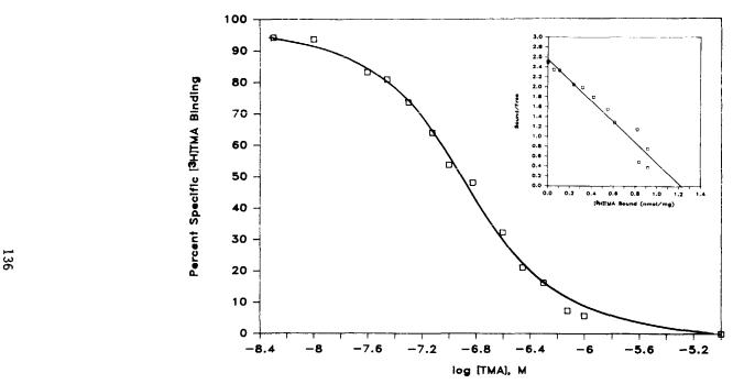
FIGURE 2. [3H]TMA binding to rat cortical membrane homogenates. Triplicate tubes containing [ 3H]TMA (75 PM) were incubated with crude homogenates (20 ug protein) at 37°C for 3 hr with varying concentrations of unlabelled TMA. Nonspecific binding in the presence of 10 uM TMA is subtracted and the result is compared to binding in the absence of unlabelled TMA. Data presented are from a representative experiment which was repeated four times with results varying less than 15%. Inset: Scatchard analysis of [ 3H]TMA
binding. Bmax, maximum binding. From Nye et al. 1985. Copyright 1985 by American Society for Pharmacology and Experimental Therapeutics (Baltimore).
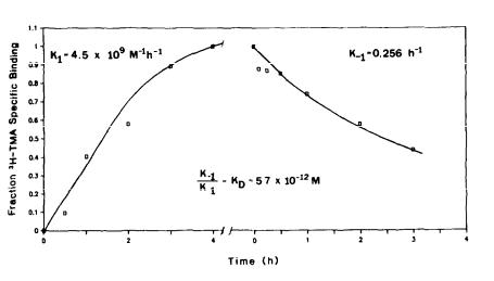
FIGURE 3. Kinetic analysis of [3H]TMA binding. Left: [3H]TMA (100 PM) was incubated with crude rat brain membrane homogenates at 37°C for varying periods of time before rapid filtration and washing. Right: [3H]TMA (100 PM) and crude membrane homogenates were preincubated for 4 hr. Ten micromolar TMA was added to aliquots of the mixture to initiate dissociation for varying times before rapid filtration and washing. Fraction specific binding is calculated as the total binding minus the binding in the presence of 10 uM TMA divided by the maximal specific binding (at 4 hr). The rate constants are calculated assuming first-order dissociation (r=.99) and pseudo first-order association kinetics (r=.96).
Cannabinoid Specificity of [3H]TMA Binding
The binding of [3H]TMA is not inhibited at concentrations less than 1 uM by a wide range of drugs and compounds spanning numerous drug-receptor classes, active peptides, and highly hydrophobic compounds (legend to table 1).
However, cannabinoids show high affinity for the [3H]TMA site. Delta-9-THC, the major naturally occurring psychotropic cannabinoid, inhibits [3H]TMA binding with an inhibitory constant (Ki) of 27.3±5.0 nM, and delta-8-THC has a Ki of 32.9±0.8 nM. These concentrations represent the calculated concentration of compound in the assay volume. Because of extensive nonspecific membrane partitioning, the actual free concentrations are substantially lower. Similar concentrations of delta-9-THC are found in the blood of human subjects and rhesus monkeys after threshold doses (Agurell et al. 1985).
Binding studies of a series of cannabinoid analogues reveal a well-defined structure-potency relationship for the [3H]TMA site in brain (Nye et al. 1985, table 1). Taken together, the potencies for all of the cannabinoids at the [3H]TMA site do not correlate with their potencies observed in the psychotropic test or in any other known behavioral assay. However, among groups of
137

TABLE 1
Comparison of potencies for cannabinoids
aInhibitory constant (Ki) for [3H]TMA site. Compounds with negligible effects at 1 uM are: amphetamine, atropine, capsaicin, captopril, carbamazepine, chloral hydrate, cholesterol, choline, cocaine, cortisone, cyclohexyladenosine, diazepam, dihydro-ergotamine, doxepin, 17-beta-estradiol, GABA, haloperidol, imipramine, 2-isobutyl-3-methoxypyrazine, lysergic acid diethylamide, mazindol, meprobamate, mianserin, muscimol, naloxone, norepinephrine, nitrendipine, nortriptyline, orcinol, ouabain, paraldehyde, pargyline, phencyclidine, phenobarbitol, phentolamine, phenytoin, phorbol dibutyrate, picrotoxin, progesterone, resorcinol, SKF525a. strychnine, testosterone, thiothixene, thyroxine, tyrosine, valproic acid, verapamil, and zopiclone. bPotency of cannabinoids on guinea-pig ileum. cThreshold for rhesus psychotropic effects from Mechoulam and Edery (1973) and references therein. Codes are: H, high,
<.1 mg/kg; M, medium, .1-.5 mg/kg; L, low, .5-2 mg/kg; I,inactive, >2 mg/kg. dDifferent from levo isomer (P<.05). eDifferent from levo isomer (P<.1). fntNot tested. jSee Lee et al. 1983. From Nye et al. 1985. Copyright 1985 by American Society for Pharmacology and Experimental Therapeutics (Baltimore).
138
cannabinoids which are active in eliciting a psychotropic response, the order of affinity for the [3H]TMA site parallels the order of potency in behavioral studies and in inhibiting contractions of the ileum. The features of [3H]TMA pharmacology which parallel behavioral potencies are as follows:
1. The [3H]TMA site shows a stereochemical preference for the natural isomer of THC. Thus, for both delta-8- and delta-9-THC, the levo stereoisomer is significantly more potent than the dextro isomer.
2.The pentyl side chain is essential for [3H]TMA binding. Substitution of the
side chain with a carboxylic acid abolishes potency, while the addition of 1’ and 2’ methyl groups enhances affinity for the [3H]TMA site. The order of inhibitory potencies for side-chain hydroxylated analogues of delta-9-THC accurately reflects their order of potency in both psychotropic tests and physiological assays.
3.The phenolic hydroxyl (position 1) must be unobstructed for significant
[3H]TMA binding potency. Thus, desacetyl-levonantradol is 10-fold more potent than levonantradol (figure 4). This deacetylation occurs in vivo and is essential for psychotropic activity (McIlhenny et al. 1981). Cannabinol acetate is also significantly weaker than cannabinol.
4. The addition of a carboxylic acid moiety inactivates delta-9-THC in inhibiting [3H]TMA binding and in behavioral assays. The addition of a hydrox to the 11 position of delta-9- or delta-8-THC does not impair affinity for the[3 H]TMA site, whereas 8-hydroxyl derivatives have significantly reduced affinity.
A number of natural cannabinoids and synthetic analogues display significant affinity for the [3H]TMA site despite their apparent inactivity in numerous tests for psychotropic efficacy. Thus cannabinol, cannabidiol, cannabigerol, cannabicyclol and delta-9,11-THC lack psychoactivity but show affinities for the [3H]TMA site of 34.4, 73.1 8.3, 73.1, and 13.6 nM, respectively. These discrepancies suggest that [3H]TMA may not be labeling the site of action of delta-9-THC for producing psychotropic effects.
An alternative possibility suggested by analogy to other drug-receptor systems is that structural isomers that lack bioactivity may be antagonists. Although the pharmacological data are incomplete for many of the inactive cannabinoids, a growing body of evidence indicates that antagonists do exist. The best studied is cannabidiol, which antagonizes the effects of delta-9-THC in humans, rodents (Karniol and Carlini 1973; Karniol et al. 1974) and seizure-sensitive animals (Consroe et al. 1982, this volume). More recently, Martin et al. (this volume) have demonstrated that delta-9, 11-THC suppresses the psychoactivity of delta- 9-THC in rhesus monkeys. Burstein et al. (this volume) have shown that 9- catboxy,11-nor-delta-9-THC reduces the catalepsy in mice produced by THC.
Since most measures of the psychotropic effects of cannabinoids in man and monkey are not quantitative, it is difficult to observe the shifts in dose-response relationships that result from antagonism. Moreover, several of these compounds may be weak agonists or partial agonists. This is supported by the observation that cannabinol in high doses elicits psychotropic effects indistinguishable from delta-9-THC in humans (Perez-Reyes et al. 1973).
139

FIGURE 4. Inhibition of [3H]TMA binding by nantradol isomers. [3H]TMA (100 pM) was incubated with crude rat brain membranes in the presence of varying concentrations of nantradol isomers under standard assay conditions. Nonspecific binding is determined in incubations with 10 uM TMA and is subtracted from all data.
Localization and Physical Properties of the High Affinity Cannabinoid Site
[3H]TMA binding is found in brain as well as a number of peripheral organs. The density of binding sites is greatest in brain compared to other organs, and the highest binding levels occur in the hippocampus. The relative potencies for several cannabinoids at the [3H]TMA site are similar in all brain regions. Of peripheral organs, the kidney shows the highest density of sites, while lung and muscle show the lowest densities.
The [3H]TMA site is an integral membrane component which is fully extracted by detergents, such as CHAPS and Triton X-100. Molecular sieve chromatography of the [3H]TMA binding site in CHAPS-extracted membranes reveals a bimodal peak of activity with an Mr of 60,000 daltons. This molecular size probably includes an undetermined number of bound detergent molecules.
REGULATION OF THE HIGH AFFINITY CANNABINOID SITE BY PURINE NUCLEOTIDES
We evaluated the effect on [3H]TMA binding of guanine and adenine nucleotides (figure 5). The specific binding of [3H]TMA is enhanced about 3-fold by the nonhydrolyzable analogue GTP-gamma-S. This enhancement occurs between 0.1 and 10 micromolar; at higher concentrations the enhancement is abolished, with small inhibition observed at >200 uM. GTP does not produce an increase in [3H]TMA binding; instead, it shows a similar inhibition at high concentrations. GTP is probably hydrolyzed to GDP and GMP by nonspecific hydrolases during
140

FIGURE 5. Regulation of [3H]TMA binding by purine nucleotides. [3H]TMA was incubated with washed crude brain membranes under standard assay conditions in the presence of added nucleotides. Control binding is defined as the specific [3H]TMA binding measured in the absence of added nucleotides minus nonspecific binding (10 uM TMA). Each data point is the average of duplicate assays and is representative of three experiments.
141
the long incubations required for measuring [3H]TMA binding. The nonhydrolyzable analogue GDP-beta-S produces a similar enhancement of binding, but at somewhat higher concentrations. The ED200 (concentration that doubles binding) is about 1 uM for GTP-gamma-S and about 10 uM for GDP-beta-S. GMP is without either stimulatory or inhibitory effect. These concentrations are in the relevant range for an effect which is mediated by a GTP binding protein (Lefkowitz et al. 1983).
Adenine nucleotides fail to display high potency stimulation of specific [3H]TMA binding. ATP inhibits binding, but only at high concentrations (IC50 = 100 uM) (figure 5). The nonhydrolyzable analogue of ATP, ADP-imidophosphate (AMPPNP), inhibits [3H]TMA binding with an IC50 between 5 and 10 uM. These concentrations of AMP-PNP are sufficient to inhibit ATP-dependent processes, such as adenylate cyclase or GDP-phosphorylation. Forskolin, a stimulator of adenylate cyclase, has no effect on [3H]TMA binding at the concentrations tested.
The enhancement of [3H]TMA binding by nonhydrolyzable GTP and GDP analogues suggests that the site may be regulated by a guanine nucleotide binding protein or G-protein. G-proteins are coupled to a large number of neurotransmitter and hormone receptors, and they regulate a variety of intracellular processes, including adenylate cyclase and phospholipase C (Stryer 1986). Paradoxically, both GTP and GDP analogues produce an apparent increase in [3H]TMA binding. In the case of another receptor linked to a G- protein, the beta-adrenergic receptor, the affinity of agonists is decreased by GTP analogues leading to a reduction in binding. Antagonists, on the other hand, are unaffected by GTP analogues (Lefkowitz et al. 1976).
Recent work by Howlett et al. (1986) suggests that cannabinoids act via a G- protein in the neuroblastoma cell line N18tg2. Psychotropically active annabinoids inhibit the adenylate cyclase of these cells at submicromolar concentrations (Howlett and Fleming 1984; Howlett, this volume). Interestingly, stimulation of adenyl cyclase via a number of different receptors is inhibited by cannabinoids. Moreover, Howlett et al. (1986) have demonstrated that the inhibition is reversed by ADP-ribosylation with pertussis toxin, suggesting that the cannabinoids are acting via the inhibitory G-protein or Gi.
ISOLATION OF AN ENDOGENOUS INHIBITOR OF CANNABINOID BINDING
The presence of a neuronal receptor for a psychoactive drug implies that some endogenous neural mechanism is perturbed by that drug. One possible neurochemical mechanism is the interaction of a neurotransmitter or neuromodulator and its receptor. Accordingly, we decided to search in brain extracts for an endogenous inhibitor of [3H]TMA binding.
Extracts of rat brain were prepared according to the method of Bennett et al. (1978). Such extracts preserve peptides from proteolysis and precipitate most proteins. These preparations inhibit [3H]TMA binding in a dose-dependent manner when added to standard assays. Because inhibitory activity was stable to acid and acetonitrile, we partially purified the active component using a combination of conventional chromatography and HPLC. The extracts were loaded on a C18 reverse-phase Sep Pak (Waters), washed with 0.1% trifluoracetic acid, and eluted with 80% acetonitrile 0.1% aqueous TFA. This material was chromatographed on a Biogel P10 column (Biorad) and eluted with 10% formic acid 0.1% 2-mercaptoethanol. The fractions were evaporated and assayed for
142

inhibition of [3H]TMA binding (figure 6). A single peak of inhibitory activity is observed in fractions corresponding to the void volume of the column. HPLC gel filtration chromatography of the pooled fractions on a TSK column (Biorad) reveals a single peak of [3H]TMA inhibitory activity corresponding to a calculated molecular size of 15-20,000 daltons (figure 7).
FIGURE 6. Gel filtration chromatography of the endogenous inhibitor of [3H]TMA binding. Rat brain extracts. prepared as described in the text, were loaded onto a Biogel P10 (Biorad) column (1x25 cm) equilibrated in 10% formic acid/0.1% 2-mercapto-ethanol running at 5 ml/hr. Fractions (0.35 mL) were evaporated and resuspended in 25 mM TES buffer (pH 6.8) before being added to standard [3H]TMA binding assays in the presence of 75 pM [3H]TMA. Fractions 26-32 correspond to the void volume of the column. Assays were performed in singlicate and represent two similar experiments.
Partially purified fractions of the endogenous inhibitor reduce [3H]TMA binding
(figure 8) in a concentration-dependent manner. Although the preparation |
tested |
is only partially purified, it shows an inhibitory potency (IC ) of 1.5-2.0 x |
10-4 |
50 |
|
g protein/L. Assuming that a 15-20,000 dalton protein accounts for the |
|
inhibitory activity, the maximal molar concentration of the preparation at the IC50 is 10 nM. When fully purified, the actual IC50 of the endogenous inhibitor of [3H]TMA binding may therefore be substantially lower than 10 nM. Many possibilities exist for the role of the endogenous inhibitor of [3H]TMA binding. It is enticing to speculate that this protein is a neurotransmitter or neuromodulator whose normal functioning is perturbed by cannabinoids. Thus, further characterization of the endogenous inhibitor may lead to additional insights into the effects of cannabinoids upon the brain. Alternatively, the page endogenous inhibitor may be an intracellular regulatory protein which interacts with the cannabinoid site to alter certain cellular processes.
143
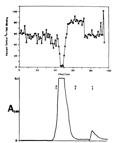
FIGURE 7. Molecular sizing of the endogenous inhibitor of [3H]TMA binding by high pressure liquid chromatography. Partially purified rat brain extracts (see text) were loaded onto a Biosil TSK column triplet (TSK-400, TSK 250, and TSK-125) connected in series. The columns were eluted with 32% acetonitrile 0.1% aqueous trifluoroacetic acid at a flow rate of mL/min. Fractions (0.5 mL) were evaporated, resuspended in 0.1 N HCl, and added to standard [3H]TMA binding assays. The data presented (upper tracing) are from single determinations on each fraction. Equivalent quantities of HCl reduced binding to 60% to 70% of maximal levels. The eluate was monitored for absorbance at 280 nm (lower tracing). Bovine serum albumin (a, Mr=66,000), alpha-MSH (b, Mr=1572), and 2-mercaptoethanol (c, Mr=78) were used as standards to calibrate the column.
144

FIGURE 8. Inhibitory potency of a partially purified preparation of the endogenous inhibitor of [3H]TMA binding. Dilutions of partially purified extracts obtained from gel filtration chromatography were added to triplicate standard [3H]TMA binding assays with crude bruin membranes. Maximal binding is defined as the total binding minus the binding in the presence of 10 uM TMA. The abscissa represents the logarithm of the concentration of added protein in the final volume of the [3H]TMA binding assay. Protein values were determined from protein assay (BCA, Pierce) of the partially purified endogenous inhibitor preparation.
MODEL OF CANNABINOID ACTIONS
One of the most perplexing phenomena in studying the cellular actions of cannabinoids is their profound effects on a host of enzymes, transport mechanisms, and neurochemical processes (Martin 1986). One possible explanation for these diverse effects is that cannabinoids modulate the metabolism of intracellular second messengers, such as cyclic AMP or the phosphoinositide phospholipids (PIP,). These intracellular mediators in turn regulate the activity of multiple cellular processes by stimulating the phosphorylation of proteins, by the release of calcium ion stores, and by other mechanisms.
A mechanism for the regulation of intracellular second messengers by cannabinoids is suggested by the finding that the high affinity cannabinoid site as labelled by [3H]TMA appears to be coupled to a G-protein. Moreover, the effect of cannabinoids on adenylate cyclase is mediated via Gi (Howlett, this volume). Since G-proteins are the regulatory proteins which modulate the activities of adenylate cyclase, phospholipase, and other second messengermetabolizing enzymes, an effect by cannabinoids upon these critical proteins would in turn affect multiple cellular processes. Although the evidence is incomplete, the hypothesis that all of the cellular actions of cannabinoids are
145
mediated via G-proteins offers a unified explanation for the diverse and multiple effects of cannabinoids in Vitro and in vivo.
REFERENCES
Agurell, S; Halldin, M.; Lindgren, J.-E.; Ohlsson, A.; Widman, M. Hollister, L.E.;
and 1 |
Gillespie, H.K. Recent |
studies on |
the |
pharmacokinetics and |
metabolism |
of d |
tetrahydrocannabinol, |
cannabidiol |
and |
cannabinol in man. |
In: Harvey, |
D.J., ed. Marijuana ’84: Proceedings of the Oxford Svmposium on Cannabis. Oxford: IRL Press, 1985. pp. 49-62.
Bennett, H.P.J.; Hudson, A.M.; Kelly, L.; McMartin, C.; and Purdon, G.E. A rapid method using octadecasilyl silica for the extraction of certain peptides from tissue. Biochem J 175:1139-1141, 1978.
Consroe, P.; Martin, A.R.; and Fish, B.S. Use of a potential rabbit model for structure-behavioral activity studies of cannabinoids. J Med Chem 25:596599, 1982.
Hewlett, A.C., and Fleming, R.M. Cannabinoid inhibition of adenylate cyclase: Pharmacology of the response in neuroblastoma cell membranes. Mol Pharm Acol 23:532-538, 1984.
Howlett, A.C.; Qualy, J.M.; and Khachatrian, L.L. Involvement of Gi in the inhibition of adenylate cyclase by cannabimimetic drugs. Mol Pharm Acol 29:307-313, 1986.
Karniol, I.G. and Carlini, E.A. Pharmacological interaction between cannabidiol and delta-9-tetrahydrocannabinol. Psychopharmologia 33:53-70, 1973.
Karniol, I.G.; Sirakawa, I.; Kasinski, N.; Pfefferman, A.; and Carlini, E.A. Cannabidiol interferes with the effects of delta-9-tetrahydrocannabinol in man. Eur J Pharmacol 28:172-177, 1974.
Lee, C.-M.; Zuagg, H.E.; Michaels, R.J.; Dren, A.T.; Plotnikoff, N.P.; and Young, P.R. New azacannabinoids highly active in the central nervous system. J Med Chem 26:278-280, 1983.
Lefkowitz, R.J.; Mullikin, D.; and Caron, M.G. Regulation of beta-adrenergic receptors by guanyl-5'-yl imidodiphosphate and other purine nucleotides. J Biol Chem 251:4686-4692, 1976.
Lefkowitz, R.J.; Stadel, J.M.; and Caron, M.G. Adenylate cyclase-coupled betaadrenergic receptors: Structure and mechansims of activation. Annu Rev Biochem 52:159-186, 1983.
McIlhenny; A.M, Mast; R.W., Johnson, M.R.; and Mime, G.W. Nantradol hydrochloride: Pharmacokinetics and behavioral effects after acute and chronic treatment. J Pharm Acol EXp Ther 219:363-369, 1981.
Martin, B.R. Cellular effects of cannabinoids. Pharm Rev Acol 38:45-74, 1986. Mechoulam, R., and Edery, H. Structure-activity relationship in the cannabinoid
series. In: Mechoulam, R., ed. Marijuana: Chemistry, Pharmacology, Metabolism and Clinical Effects. New York: Academic Press, 1973. pp. 101-136.
Nye, J.S.; Seltzman, H.H.; Pitt, C.G.; and Snyder, S.H. High-affinity cannabinoid binding sites in brain membranes labeled with [3H]-5’- trimethylammonium d8-tetrahydro-cannabinol. J Pharm Acol Exper Ther 234:784-791, 1985.
Ohlsson, A.; Agurell, S.; Leander, K.; Dahmen, T.; Edery, H.; Porath, G.; Levy, S.; and Mechoulam, R. Synthesis and psychotropic activity of side-chain hydroxylated delta-6-tetrahydrocannabinol metabolites. Acta Pharm Suec 16:21-33, 1979.
146
Perez-Reyes, M.; Timmons, M.C.; Davis, K.H.; and Wall, E.M. A comparison of the pharmacological activity in man of intravenously administered delta-9- tetrahydrocannabinol, cannabinol and cannabidiol. Experientia 29:13681369, 1973.
Rosell, S.; Bjorkroth, U.; Agurell, S.; Leander, K.; Ohlsson, A.; Martin, B.; and Mechoulam, R. Relation between effects of cannabinoid derivatives on the twitch response of the isolated guinea-pig ileum and their psychotropic properties. In: Nahas, G.G. and Paton, W.D.M., eds. Marijuana Biological Effects: Analvsis, Metabolism, Cellular Response, Reproduction and Brain. Oxford: Pergamon Press. 1979. pp. 63-69.
Seltzman, H.H.; Setzer, S.R.; Williams, D.L.; Demian, I.; Wyrick, C.D.; and Pitt, C.G. Synthesis of cannabinoid radioligands and haptens for use in radioimmunoassay and receptor site studies. In: Harvey, D.J., ed. Marijuana ’84: Proceedings of the Oxford Svmposium on Cannabis. Oxford: IRL Press, 1985. pp. 183-190.
Stryer, L. G-Proteins: A family of signal transducers. Annu Rev Cell Biol 2:391-419, 1986.
ACKNOWLEDGMENTS
This work was supported by National Institute on Drug Abuse grant DA-00266, Medical Scientist Training Program grant GM-07309 (to J.S.N.), and NIDA Research Scientist Award DA-00074 (to S.H.S.). Susan Voglmaier and Adele Snowman assisted in the binding experiments and Patrice Obermiller assisted with the HPLC. Pamela Sklar provided helpful discussions. The nantradol compounds were a gift of Pfizer Central Research (Groton).
AUTHORS
Jeffrey S. Nye, A.M.
Solomon H. Snyder, M.D.
Department of Neuroscience and Pharmacology
The Johns Hopkins University School of Medicine
725 N. Wolfe Street
Baltimore, Maryland 21205
147