

Dunn_2005-Developmental_Dynamics
.pdfDEVELOPMENTAL DYNAMICS 234:835– 845, 2005


 RESEARCH
 ARTICLE


Complex Colony-Level Organization of the Deep-Sea Siphonophore Bargmannia elongata (Cnidaria, Hydrozoa) Is Directionally Asymmetric and Arises by the Subdivision of Pro-Buds
Casey W. Dunn*
Siphonophores are free-swimming colonial hydrozoans (Cnidaria) composed of asexually produced multicellular zooids. These zooids, which are homologous to solitary animals, are functionally specialized and arranged in complex species-specific patterns. The coloniality of siphonophores provides an opportunity to study the major transitions in evolution that give rise to new levels of biological organization, but siphonophores are poorly known because they are fragile and live in the open ocean. The organization and development of the deep-sea siphonophore Bargmannia elongata is described here using specimens collected with a remotely operated underwater vehicle. Each bud gives rise to a precise, directionally asymmetric sequence of zooids through a stereotypical series of subdivisions, rather than to a single zooid as in most other hydrozoans. This initial description of development in a deep-sea siphonophore provides an example of how precise colony-level organization can arise, and illustrates that the morphological complexity of cnidarians is greater than is often assumed. Developmental Dynamics 234: 835– 845, 2005. © 2005 Wiley-Liss, Inc.
Key words: Bargmannia; siphonophore; Hydrozoa; Cnidaria; directional asymmetry; major transitions; deep sea; colonial animal
Received 2 March 2005; Revised 2 May 2005; Accepted 5 May 2005
INTRODUCTION
Most studies of animal development have focused on the embryonic development of solitary taxa. There are, however, other modes of development with different starting and end points that have remained largely neglected. These include agametic clonal development, in which a new animal arises from another animal through fission or budding, and colonial development, a variation on clonal development in
which asexually produced individuals remain attached and physiologically integrated throughout their lives (Hughes, 2002). There is a diversity of organizational complexity across colonial taxa (Beklemishev, 1969). Some colonies consist of functionally equivalent zooids (see Table 1 for definitions of the specialized terms used throughout this report) while others manifest a marked division of labor between zooids (Leuckart, 1851). Most
colonial taxa show intraspecific variability in zooid arrangement and gross colony morphology, such that no two colonies are exactly alike (Boardman et al., 1973). Other taxa, especially those that are pelagic (i.e., that live in the water column rather than affixed to a substrate), have invariant colonial organizations that are entirely consistent from colony to colony of the same species (Mackie, 1986).
The zooids of most colonial taxa do
Department of Ecology and Evolutionary Biology, Yale University, New Haven, Connecticut Grant sponsor: NSF; Grant number: DEB-0408014.
*Correspondence to: Casey W. Dunn, Department of Ecology and Evolutionary Biology, Yale University, PO Box 208106, New Haven, CT 06520-8106. E-mail: casey.dunn@yale.edu
DOI 10.1002/dvdy.20483
Published online 28 June 2005 in Wiley InterScience (www.interscience.wiley.com).
© 2005 Wiley-Liss, Inc.

836 DUNN
|
|
TABLE 1. Definitions for Some of the Specialized Terminology Used |
|
|
|
|
|
|
Term |
Definition |
|
|
|
|
|
|
Bract |
A gelatinous, shield-like zooid |
|
|
Colonial animal |
An animal that exists as a series of asexually produced and physiologically integrated zooids; |
|
|
|
each colony arises from a single zygote and is genetically uniform (baring mutation or |
|
|
|
fusion with another colony) |
|
|
Cormidium |
A single iteration of the regularly repeating pattern of zooids found in the siphosome of |
|
|
|
siphonophores |
|
|
Gastrozooid |
Polyp specialized for feeding; bearing a single tentacle in siphonophores |
|
|
Gonozooid |
Specialized polyps that bear the gonophores |
|
|
Gonophore |
Medusae specialized for reproduction; lacking feeding structures |
|
|
Horn |
The protuberance within the siphosomal growth zone where the cormidia form |
|
|
Medusa |
One of two types of cnidarian zooids; familiar solitary medusae include the “true” umbrella- |
|
|
|
shaped jellyfish |
|
|
Nectophore |
Medusa specialized for propulsion; lacking feeding and reproductive structures |
|
|
Nectosome |
The region of a siphonophore colony that bears nectophores |
|
|
Polyp |
One of two types of cnidarian zooids; solitary cnidarian polyps include Hydra and sea |
|
|
|
anemones |
|
|
Pneumatophore |
Gas-filled float at the anterior end of many siphonophores; not a zooid, arises |
|
|
|
developmentally as an aboral invagination of the embryo (Carre´, 1967) |
|
|
Pro-bud |
The first bud to arise in the developmental sequence that gives rise to the cormidia of the |
|
|
|
siphosome |
|
|
Siphosome |
The region of the colony that bears all zooids except the nectophores |
|
|
Stem |
The central stalk to which all the zooids are attached; linear in B. elongata; arises |
|
|
|
developmentally via the elongation of the body column of the first polyp that forms during |
|
|
|
embryogenesis (Gegenbaur, 1853) |
|
|
Tentaculozooid |
A zooid that is presumed to be a polyp with an atrophied body and a single hypertrophied |
|
|
|
tentacle |
|
|
Zooids |
The units, each of which are homologous to other free living solitary animals, that make up |
|
|
|
animal colonies; these can be polyps or medusae in cnidarian colonies such as |
|
|
|
siphonophores |
|
|
|
|
|
not change positions within the colony, so the geometry and dynamics of the budding process have a direct effect on the arrangement of zooids and on overall colony shape. Both microenvironment (reviewed by Harvell, 1994) and internal parameters, such as the dynamics of gastrovascular fluid flow (Blackstone and Buss, 1993; Dudgeon and Buss, 1996), have been shown to influence the development of colonies with variable form. To date, little is known about the developmental mechanisms of those taxa with invariant organization. At the very least, a description of their budding process is required before the mechanisms that generate precise colonylevel organization can be investigated.
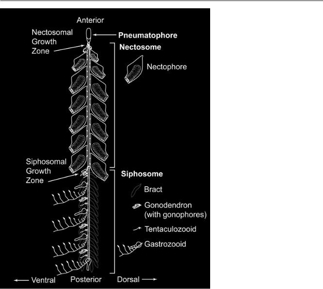
The siphonophores (Fig. 1), a group of about 160 described species of pelagic hydrozoans (Cnidaria), have the highest division of labor between zooids and the most precise organization of all colonial animals (Beklemishev, 1969, p 83). Siphonophores are among the most abundant carnivores of the oceans’ macroplankton (Pugh, 1984)
and include the longest animals in the world, with colonies of some species exceeding 40 m in length (Robison, 1995). The zooids and colonies of most siphonophores have an organization that is bilaterally symmetric at a first approximation (Totton, 1965; see Haddock et al., 2005, for a discussion of siphonophore organization and the terminology used to describe the major axes). This is not surprising, as it has long been known that many cnidarians show marked bilateral symmetry (e.g., Delage and Herouard, 1901; Hyman, 1940; Beklemishev, 1969; Martindale et al., 2002). Bilateral organization is not unique to the “Bilateria,” the monophyletic group of animals that includes almost all model systems, as is often misstated or implied (e.g., Meinhardt, 2001).
The zooids and colonies of many siphonophores are directionally asymmetric (e.g., Totton, 1932; Stepanjants, 1967; Pugh and Youngbluth, 1988; Pugh and Pages, 1997; Mapstone, 2003). Directional asymmetries are deviations from bilateral symme-
try that consistently occur in the same direction, and are found throughout the Bilateria (Neville, 1976). They include the displacement of the heart to the left in humans, the well-defined chirality of most spiraled gastropod shells, and the consistent asymmetry of the nervous system in Caenorhabiditis elegans (Hobert et al., 2002).
The directional asymmetries described in the siphonophore systematics literature have never been consolidated and have escaped wider notice. They do, however, indicate that the symmetry of at least some cnidarians can be of the same order as that of the most derived Bilateria. This raises questions as to how many times directional asymmetries have evolved in animals, and how old they are. It is still not clear if homologous developmental axes even exist in the Cnidaria and Bilateria, though expression data are largely consistent with the hypothesis that they do (Hayward et al., 2002; Finnerty et al., 2004). The axes of siphonophore colonies are labeled with the same names as the axes of
COLONY-LEVEL DEVELOPMENT OF A SIPHONOPHORE 837
bilaterian animals (reviewed by Haddock et al., 2005), but it should be noted that this is merely a semantic convenience and no homologies are implied by this nomenclature.
A recent molecular phylogeny (Dunn et al., 2005b) helps organize what is already known about the colony-level organization and development of siphonophores. Siphonophores are divided into two monophyletic groups, the Cystonectae and the Codonophora (Fig. 2). The Cystonectae is a small group of only five valid species, which include Physalia physalis, the familiar Portuguese Man
|
|
|
Haeckel |
(1869b), |
establishes |
two |
|
|
|
|
growth zones that are responsible for |
||||
|
|
|
further colony-level development (Fig. |
||||
|
|
|
1). These growth zones are the sites of |
||||
|
|
|
both stem elongation and the budding |
||||
|
|
|
process that gives rise to new zooids |
||||
|
|
|
throughout the life of the organism. |
||||
|
|
|
One growth zone gives rise to the |
||||
|
|
|
nectosome, a region that bears the |
||||
|
|
|
propulsive |
asexual |
medusae |
called |
|
|
|
|
nectophores. The other growth zone |
||||
|
|
|
gives rise to the more complex sipho- |
||||
|
|
|
some, a region that contains all other |
||||
|
|
|
types of zooids, including those for |
||||
|
|
|
feeding, reproduction, |
and defense. |
|||
|
|
|
The zooids of the siphosome are orga- |
||||
|
|
|
nized along the linear stem in a spe- |
||||
|
|
|
cies-specific repeating pattern, each |
||||
|
|
|
iteration of which is called a cor- |
||||
|
|
|
midium. |
|
|
|
|
|
|
|
The Codonophora contains two his- |
||||
|
|
|
torically recognized groups of siphono- |
||||
|
|
|
phores (Dunn et al., 2005b). These are |
||||
|
|
|
the Physonectae, a grade, and the Ca- |
||||
|
|
|
lycophorae, which |
is |
monophyletic |
||
|
|
|
and nested within the Physonectae |
||||
|
|
|
(Fig. 2). There is a large diversity of |
||||
|
|
|
colony-level organization in the sipho- |
||||
|
|
|
some of the Physonectae, while all of |
||||
|
|
|
the Calycophorae have a similar si- |
||||
|
|
|
phosomal |
structure |
(Bigelow, |
1911; |
|
|
|
|
Totton, 1954, 1965), which their phy- |
||||
|
|
|
logenetic position indicates is derived |
||||
|
|
|
and secondarily simplified. Siphoso- |
||||
|
|
|
mal budding has only been described |
||||
|
|
|
in detail for two Codonophora species, |
||||
|
|
|
both of which are calycophorans. |
||||
|
|
|
Chun (1885) found that each cor- |
||||
|
|
|
midium arises as a single bud in |
||||
|
|
|
Sphaeronectes gracilis, and Schneider |
||||
|
|
|
(1896) described some zooids as aris- |
||||
|
|
|
ing as independent buds in Abylopsis |
||||
|
|
|
tetragona, though his figures are not |
||||
|
|
|
completely clear on the matter. In a |
||||
|
|
|
later review of these studies, |
||||
|
|
|
Garstang (1946) raised several issues |
||||
o’ War. The embryology of the cys- |
with Schneider’s findings, and con- |
||||||
tonects is entirely unknown. Totton |
cluded that the subdivision of buds |
||||||
(1960) described several features of the |
was a general mechanism of colony- |
||||||
budding process of mature P. physalis |
level development in the Calycopho- |
||||||
colonies and showed that it is highly |
rae. He also coined the term “pro-bud” |
||||||
derived |
and |
fundamentally different |
for the bud that gives rise to the mul- |
||||
than any other siphonophore, including |
tiple zooids of a cormidium. |
|
|||||
the other cystonects. As such, it is diffi- |
Although it is critical to under- |
||||||
cult to apply the developmental find- |
standing the development of the an- |
||||||
ings from this species to other taxa. |
cestral Codonophora, the budding pro- |
||||||
The other monophyletic group, the |
cess in the physonects has proven |
||||||
Codonophora, contains the bulk of si- |
particularly problematic to study be- |
||||||
phonophore species. Their embryolog- |
cause “there is so much crowding to- |
||||||
ical development, which was first ob- |
gether of the siphosomal buds that it |
||||||
served |
by |
Gegenbaur (1853) and |
makes observation very difficult” (Tot- |
||||
838 DUNN
ton, 1954, p 22). The organization of all zooids within a mature cormidium has not even been described for any physonect. Totton (1965) noted that the siphosomal zooids of physonects arose on a protuberance in the growth zone rather than directly on the stem. Garstang (1946) suggested that each zooid of the physonects arises as an independent bud, but it is not clear how he arrived at this conclusion because he did not name any sources that describe the budding process in detail. The phylogenetic positions and derived colony organizations of the taxa that have been examined to date leave wide gaps in our knowledge of the colony-level development of siphonophores. Descriptions of colony structure and budding in physonects and other cystonects are essential if we are to understand the evolution, development, and origin of colonylevel complexity, as well as symmetry, of siphonophores as a whole.
The complex organization of siphonophores indicates the existence of a highly canalized colony-level developmental mechanism without parallel in other animals (Garstang, 1946), and provides an opportunity to explore the evolutionary origins of biological complexity in a novel context. Haeckel (1869a) recognized this, and made explicit comparisons between specialized cells in multicellular organisms and specialized zooids in siphonophore colonies. While complex multicellular organisms arose via the precise organization of functionally specialized cells in space and time, siphonophores arose by taking the process one step further and organized functionally specialized multicellular organisms into precise patterns. Interest has recently been rekindled in how new levels of biological organization arise (Buss, 1987; Michod, 2000), and this growing field now often goes under the name “the major transitions in evolution” (Maynard Smith and Szathma´ ry, 1995). Even so, there has only been occasional recent mention of siphonophores in this context (Mackie, 1963; Winsor, 1971; Gould, 1987; Wilson, 2000), and these animals have remained poorly known and largely forgotten in modern times. This is because siphonophores live in the open ocean, with many species being found only in the deep sea. They
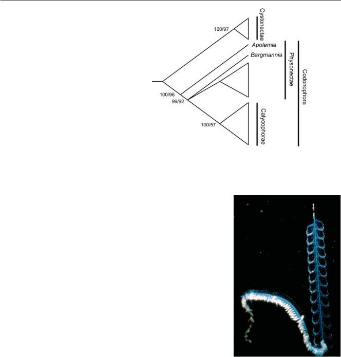
Fig. 2. A rooted siphonophore phylogeny, simplified to show the relationship of the taxa discussed here. Physalia physalis is in the Cystonectae, Abylopsis and Sphaeronectes are in the Calycophorae. This cladogram is based on an analysis by Dunn et al. (2005b) of sequence data from the 16S and 18S ribosomal RNA genes. Support is shown as (Bayesian posterior probability x 100)/(maximum likelihood bootstrap score).
are so fragile that their zooids often dissociate during collection and preservation (Pugh, 1989; Dunn et al., 2005a), and the resulting lack of intact material has largely precluded the study of the symmetry properties, colony-level organization, and developmental processes that make siphonophores interesting in a broader developmental and evolutionary context. Modern advances in oceanographic technology, however, alleviate the collecting problems that limited all previous work on siphonophores (Haddock, 2004).
The present study investigates the colony-level organization and development of a siphonophore, Bargmannia elongata (Fig. 3), using specimens collected with a remotely operated underwater vehicle (ROV) deployed from an ocean-going research ship. The general colony form of B. elongata (an elongate siphosomal stem, two growth zones, multiple identical nectophores, the possession of a gas-filled pneumatophore) is plesiomorphic for the Codonophora (Dunn et al., 2005b), unlike the colony form of the calycophorans that have previously been investigated. This makes B. elongata a good departure point for understanding the colony-level evolution and development, and symmetry properties, of other siphonophores.
Fig. 3. View of a living Bargmannia elongata colony. Photograph courtesy of Steve Haddock. The entire colony is about 40 cm long.
RESULTS
Collecting Bargmannia elongata
Nineteen specimens were collected by ROV Tiburon from depths of 350 –2,865 m. All collection sites were within 337 km of Moss Landing, California. Most specimens of Bargman-
COLONY-LEVEL DEVELOPMENT OF A SIPHONOPHORE 839
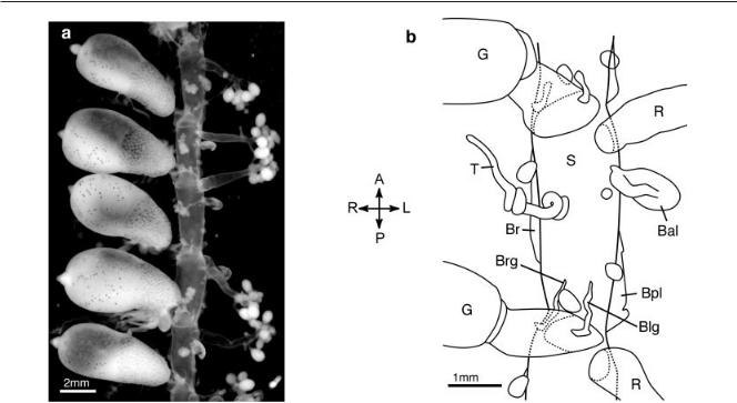
Fig. 4. The organization of the mature cormidia of Bargmannia elongata (ventral view, anterior up, the left side of the stem faces the right of the page). P, posterior; A, anterior; R, right; L, left. a: Photograph of a length of mature stem. The white mature gonophores can be seen on the gonozooids. The largest structures are the gastrozooids. b: Illustration of the organization of a cormidium. The gastrozooid shown at the top of the illustration belongs to the next cormidium to the anterior, and the gonozooid shown at the bottom of the illustration belongs to the next cormidium to the posterior. Bpl, Blg, Brg, and Br have been dissected away, leaving only their lamellae. S, stem; G, gastrozooid; T, tentaculozooid; R, gonozooid; Br, right lateral bract; Bpl, posterior left lateral bract; Bal, anterior left lateral bract; Brg, right gastrozooid-associated bract; Blg, left gastrozooid-associated bract. The unlabeled buds on the stem were inferred to be reserve bracts.
nia elongata were not visibly damaged during sampling, and survived the ascent to the surface intact. If not disturbed, they lasted for up to 2 days in a large volume of sea water ( 6 l at 4°C in the dark) before sinking to the bottom of the vessel and disintegrating. They would not feed in these vessels, so dynamic observations of growth were not possible. Instead, the developmental process was inferred from the morphology of the growth zones, each of which had a complete ontogenetic sequence of developing cormidia.
The Siphosome: Cormidial
Organization
Each mature cormidium consisted of a gastrozooid, a tentaculozooid, a gonozooid, five mature bracts, and several small buds. This is consistent with the zooid inventory described for this species by Pugh (1999). All of the gonophores of each colony were of the same sex, confirming that this species is dioecious. Aside from the gonophores, which were borne by the gonozooids,
all the zooids of mature cormidia were attached to the stem independently.
The organization of the zooids within the cormidia was entirely regular. The gastrozooid, tentaculozooid, and gonozooid were arranged from posterior to anterior within each cormidium (Fig. 4). While the gastrozooid and tentaculozooid were on the ventral midline, the gonozooid was displaced towards the left of the stem. The gonozooid was adjacent to the gastrozooid of the next younger cormidium to the anterior, so there was much more space within cormidia than between them (the stem is highly extensible, so it was not possible to make absolute measurements of these distances). There was an annular constriction in the stem at the point of attachment of each gastrozooid.
The bracts had to be plucked away in order to make observations of the stem, but the muscular lamella where each had been attached was clearly visible. There were two lateral bracts attached to the left side of the stem in each cormidium, one to the posterior of the
gonozooid (anterior left lateral bract, Bal) and one to the posterior of the tentaculozooid (posterior left lateral bract, Bpl). There was only one lateral bract on the right side of the stem (right lateral bract, Br), and its lamella was longer than those of the bracts on the left. These three lateral bracts were further from the ventral midline of the stem than any of the other zooids. Two other bracts were located on the anterior side of the gastrozooid, one slightly to the right (right gastrozooid-associ- ated bract, Brg) and the other slightly to the left (left gastrozooid-associated bract, Blg). The lamellae of the gastro- zooid-associated bracts ran mostly along the stem, but also ran a slight distance up the peduncle of the gastrozooid. There was a single small bud to the inside (i.e., towards the ventral midline) of the lamella of each lateral bract, and a single bud between the lamellae of the two gastrozooid-associated bracts. In some specimens, these buds were mature enough to see that they were reserve bracts.
All 19 of the Bargmannia elongata

840 DUNN
Fig. 5. Bargmannia elongata siphosomal growth zone. a: Scanning electron micrograph of the siphosomal growth zone (view from left, anterior up, ventral to the left of the page). b: Schematic of the siphosomal growth zone. The site of the youngest pro-buds is indicated with a gray circle. A gray line indicates the path of the developing zooids, which are organization in an ontogenetic sequence. c–i: Cormidia in various stages of development. The gray line indicates the order of the ontogenetic sequence (with the youngest cormidia shown being the closest to the circle). The view is indicated in parentheses for each pane. The orientation of the axes is shown to the right of each row and below each figure. P, posterior; A, anterior; V, dorsal; D, dorsal. j: The inferred lineage diagram of zooid origin. Gastrozooids have been broken away in g–i and most of a to allow for a clear view. The pictured specimens are: a, c: Tib675D5; d,e,f: Tib596SS12; g, h: Tib598SS6; i: Tib595D5. P, pro-bud; F, footbud; F1,F2, daughters of F; F21, F22, daughters of F2; G, gastrozooid; T, tentaculozooid; R, gonozooid; Br, right lateral bract; Bpl, posterior left lateral bract; Bal, anterior left lateral bract; Brg, right gastrozooid-associated bract; Blg, left gastrozooid-associated bract.
specimens examined were found to deviate from bilateral symmetry as described above (gonozooid displaced towards the left, two left lateral vs. one right lateral bract). The probability of these deviations occurring in the same direction due to chance alone is (0.5)n-1, which in a sample of 19 is less than 3.82 x 10⁻⁶. We can conclude, then, that B. elongata is directionally asymmetric.
The Siphosome: The Growth
Zone and Budding Sequence
The youngest cormidia were found on a protuberance, here called the horn. It was curled into a sinister coil of about one and a half turns, with a radius on the order of 0.5 mm, and bore the siphosomal elements along its outer side (Fig. 5a,b). The spiral arrangement of
the horn introduces some nomenclatural complexity for describing organizational axes. Throughout the present report, these axes are employed, in reference to the horn, as if the horn were straightened out into a linear anterior projection of the siphosomal stem. The outer zooid-bearing surface of the horn is continuous with the ventral zooid bearing side of the siphosomal stem,

COLONY-LEVEL DEVELOPMENT OF A SIPHONOPHORE 841
and is, therefore, referred to as ventral. The tip of the horn is taken to be anterior, and left and right are defined in relation to these two axes (as they are for the colony as a whole).
The siphosomal elements were arranged in a linear ontogenetic sequence, with structures to the posterior being more mature. The ventral tract of maturing zooids was continuous, and there was no boundary or discrete transition zone between the zooids on the horn and those on the rest of the siphosomal stem. Serially arranged pro-buds (P), all of the same type, were found at the anterior end of the horn (Fig. 5c). The least developed pro-buds were at the very tip. In some specimens, the youngest pro-buds were simple transverse ridges. In other specimens, no regular ridges were seen, and the smallest buds were closely packed at the tip of the horn.
Static observations of the ontogenetic sequence of siphosomal elements indicated that each pro-bud develops into a single cormidium. The complex organization of the cormidia of Bargmannia elongata results from a stereotypical series of divisions of the pro-bud into multiple zooid buds. The zooids of each cormidium were all attached to the stem by a common peduncle early in development, and only later in development (i.e., further to the posterior) do they come to be attached independently to the stem. It was not possible to consistently stage cormidia by counting posteriorly from the tip of the horn, as the difference in maturity between adjacent cormidia was not the same from specimen to specimen. Scanning electron microscopy (SEM) proved to be the most effective tool for examining the earlier stages of development. The youngest cormidia (i.e., those at the anterior end of the horn) were completely exposed and the developmental sequence could be observed by comparing each cormidium with its neighbors. Proceeding to the posterior, the pro-buds became more elongate and then formed a swelling on the left anterior side of their base. This swelling enlarged and became the footbud (Fig. 5d, F). The portion of the pro-bud distal to the footbud was seen, further to the posterior, to become the gastrozooid (G), including its single tentacle. The footbud then de-
veloped a bisecting furrow and split into two buds, F1 proximally and F2 distally (Fig. 5e). Each of these split again.
It was not possible to describe every developing cormidium in detail to the posterior of this point. While the distance between cormidia did not increase much, the bulk of the various products of the pro-buds did and they became crowded together. Even after physically breaking away the gastrozooids, which became larger than the other zooids, the developing zooids were too densely packed to see how they were attached relative to each other. Entire cormidia at various stages of development had to be removed so that the structure of adjacent cormidia still attached to the stem could be observed. This was most easily carried out on the dried SEM specimens, rather than in the hydrated material. The initial break along a given stretch of stem usually damaged several cormidia, and it was necessary to remove adjacent buds until an intact cormidium was fully exposed (this became easier as a wider gap was opened up). While this strategy did allow for the detailed description of cormidia in various stages of development, it left gaps in the ontogenetic series that had to be interpolated by keeping track of the relative positions of the various zooids and by identifying distinctive features of the various zooids as they matured.
The two products of the division of F1 were found to be the gonozooid (R) and the anterior left lateral bract (Bal). They were displaced further to the left than the products of F2. The gonozooid could be readily identified by its distinct shape. Not far to the posterior of the point where the gonozooid originated, it became elongate and pear-shaped, then elongated further and gave rise to the gonophore buds at its distal end. The anterior left lateral bract bud remained small, and did not begin to mature until much later. It was identified in developing cormidia by its position at the base of the gonozooid.
A bud at the right base of the developing cormidia was first observed in cormidia where the gonozooid was taking on its pear-shaped appearance. The disposition of this bud suggested that it arose from the peduncle of the
developing cormidium, but no cormidia were observed in the necessary stage of development to determine this with certainty. This bud could be traced from cormidium to cormidium by its location as the proximal-most zooid on the right side, and was found to mature into the right lateral bract (Br, Fig. 5g–i).
Both of the products of F2, here called F21 and F22, were found to be intermediate buds that split again into products that are here called F21 and F22. The products of F21, which were identified by their position to the anterior left of F22 and to the right of the anterior left lateral bract bud, were found to be the tentaculozooid
(T) and the posterior left lateral bract (Bpl). The tentaculozooid, like the gonozooid, had a very distinctive morphology that could be used to trace it from cormidium to cormidium. Soon after arising, its peduncle elongated slightly. It then formed a swelling and a posterior-facing hook arose at its distal end. This hook continued to elongate into the tentacle.
F22 was identified in cormidia at various stages of development by its position adjacent to the bud of the right lateral bract (Br). Shortly to the posterior of the point where it could be first identified, it took on a bi-lobed shape. Further to the posterior, where the zooids began to become attached independently to the stem, two buds were seen at its former position. These were the last two buds to form, and their position relative to the other zooids indicated that they were the gas- trozooid-associated bracts (Brg, Blg). A lineage diagram was constructed from these inferred aspects of the budding process (Fig. 5j).
Not far to the posterior of where the buds of the two gastrozooid-associated bracts formed, there was enough space between the spreading zooids that their organization could be readily observed without breaking away any cormidia (Fig. 5i). The cormidia of specimens that had not been prepared for SEM could also be examined in detail to the posterior of this point. The bracts did not mature at the same rate as each other. The maturing right lateral bract was the largest in each cormidium, followed in order of decreasing size by the posterior left lateral bract, the gastrozooid-as-
842 DUNN
sociated bracts, and finally the anterior left lateral bract. In the final stages of the development of each cormidium, the reserve bract buds formed on the stem just to the inside of the bracts, the annular constriction formed in the stem at the attachment point of each gastrozooid, and the gonophores matured at the distal end of the gonozooid. Every 7th to 10th gastrozooid grew larger and darker than the rest.
The Nectosome
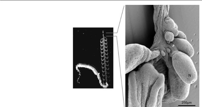
Bargmannia elongata has multiple identical nectophores (Pugh, 1999) that are all attached in a line on one side of the stem, though they come to be arranged biserially through the flexing of their peduncles (Figs. 1, 3). The nectophores were found to be attached to the opposite side of the stem as the siphosomal zooids (i.e., dorsally). The nectosomal growth zone was relatively simple (Fig. 6). The youngest nectophores (i.e., those furthest to the anterior) were only slight protrusions, which, further to the posterior, became elongate and then formed a peduncle. A second bud was found on the posterior side of each nectophore peduncle. This bud was not noted in the mature portion of the nectosome, which contained only nectophores and no other types of zooid.
DISCUSSION
The Structure of the Growth zones and Colony-Level Development in Bargmannia elongata
The present study has found that the siphosomal zooids of Bargmannia elongata arise on a protuberance within the siphosomal growth zone. Totton (1965, p 36) suggested that this was the general case for physonect siphonophores. Haeckel (1888, p. 279) originally described this protuberance in Athorybia, but called it the “nectostyle” because he mistook it as part of the nectosome rather than as an integral feature of the siphosomal growth zone. To avoid the mistaken connotations of this original name, the term “horn” has instead been used throughout the present report.
Each cormidium of Bargmannia
Fig. 6. Scanning electron micrograph of the Bargmannia elongata nectosomal growth zone (view from left, anterior up, dorsal to the right of the page). The arrow indicates one of the buds that form to the posterior of each nectophore. N, developing nectophore.
elongata arises as a single pro-bud at the tip of the horn, and the structure of the cormidium is established through the stereotypic subdivision of this bud into multiple zooids. The name “pro-bud subdivision” is suggested for this mechanism of cormidial development. The shape and orientation of some young pro-buds (those that arose as ridges) suggests that they may be generated by buckling caused by uneven growth on the curved surface of the horn. Recent studies indicate that a biophysical mechanism of this type may initiate the leaf primordia of angiosperms on the curved surface of the apical meristem (Green et al., 1996; Dumais and Steele, 2000). Morphogenesis resulting from the uneven physical stresses that can be associated with growth on a curved surface may prove to be important in very different biological systems.
The mechanism of zooid formation by pro-bud subdivision observed here in Bargmannia elongata is consistent with Chun’s (1885) observations of budding in the calycophoran Sphaeronectes gracilis, but contradicts the assertion made by Garstang (1946) that all the zooids of physonects arise as independent buds on the stem. The
origin of cormidia via pro-bud subdivision in taxa outside of the Calycophorae raises the possibility that it is a general mechanism of development in the Codonophora, rather than being restricted to the Calycophorae as Garstang believed. Only one species has been investigated here, so this hypothesis cannot be tested until colonylevel development is described in more taxa sampled across the Codonophora.
While most mature zooids of the physonects are attached independently to the stem, the zooids of each calycophoran cormidium are closely associated and usually attached by a common peduncle (Totton, 1965). This difference in mature organization may be what led Garstang to incorrectly infer that physonect buds arose independently on the stem. The calycophoran organization of zooids could be derived via paedomorphosis from the type of developmental found in B. elongata if zooids were to fail to spread out along the stem after differentiation.
The nectosomal growth zone of
Bargmannia elongata did not have a pronounced horn. There was a small bud immediately to the posterior of each developing nectophore, though it did not grow into a mature zooid. The

COLONY-LEVEL DEVELOPMENT OF A SIPHONOPHORE 843
Apolemiidae, which are sister to all other Codonophora (Fig. 2), have mature polyps adjacent to each nectophore (Totton, 1965). No structures other than nectophores have been found in the nectosome of other siphonophores until the present study. If the small nectosomal buds of B. elongata are vestigial polyps, then the most parsimonious reconstruction of the history of the nectosome would indicate that the common ancestor of all Codonophora possessed a nectosome with both polyps and nectophores. This would have important implications for the origin of the nectosome, indicating that it was originally more complex than previously believed. It may even have arisen as a tandem duplication of the siphosome.
There has been increasing interest in clonal budding, including molecular characterizations of clonal budding in cnidarians (e.g., Smith et al., 1999; Hobmayer et al., 2000; Spring et al., 2002; Reinhardt et al., 2004) and urochordates (e.g., Tiozzo et al., 2005). The budding process presented here differs from that of these other colonial animals in that one bud gives rise to multiple functionally specialized zooids. Previous studies of other hydrozoans have characterized differential gene expression between polyp and medusa buds in Podocoryna carnea (Spring et al., 2002; Bridge et al., 2004) and between functionally specialized polyps in Hydractinia symbiolongicarpus (Cartwright et al., 1999). This is directly relevant to understanding the instantiation and differentiation of functionally specialized zooids in siphonophores, but does not address how one bud can give rise to multiple zooids. It may be that probud subdivision arose through the evolutionary fusion of multiple sites of zooid budding, which would be similar to the fusion of zooid and shoot budding zones that has been proposed to have occurred in some benthic hydrozoans (Marfenin and Kosevich, 2004). Alternatively, the pro-bud (or perhaps the footbud) may have originally given rise to a single zooid, but now goes through a process of fission early in its development. A survey of colony-level development in other siphonophores may reveal variability in zooid budding that, with a phylogenetic per-
spective, could help differentiate between these two hypotheses.
Directional Asymmetry in
Siphonophores
Directional asymmetries are common within the Bilateria, and have long been of interest to comparative biologists (Palmer, 2004). They are also medically important because about 1 in 5,000 humans are born with variations from normal directional asymmetries that can seriously impact health (Casey and Hackett, 2000). There has been much recent progress in understanding the developmental mechanisms that initiate and control directional asymmetries in the Bilateria (reviewed by Hamada et al., 2002; Levin, 2005). Determining the evolutionary history of directional asymmetries, a prerequisite for learning how to relate findings from one model developmental system to another, would be greatly assisted by understanding the symmetry properties of the common ancestor of the Bilateria. This requires looking at the symmetry properties of outgroups to the Bilateria, such as the Cnidaria.
The present study has found that colonies of Bargmannia elongata are directionally asymmetric, and that this directional asymmetry arises through the consistent directionality in the subdivision of the pro-bud. Freeman (1983) demonstrated that the site of origin of the first cleavage furrow in the siphonophores Nanomia bijuga (which was misidentified as
Nanomia cara) and Muggiaea atlantica corresponds to the oral end of the planula. He also showed that the plane of bilateral symmetry in M. atlantica corresponds to the plane of the first cleavage, so that one blastomere forms the left side of the colony and the other the right side. His findings present an obvious starting point for the examination of the embryological mechanisms that establish directional asymmetry in siphonophores.
Gene expression data are consistent with the hypothesis that cnidarians and bilaterians share some anterior/ posterior and perhaps even dorsal/ ventral patterning mechanisms (e.g., Shenk et al., 1993; Hayward et al., 2002; Wikramanayake et al., 2003; Finnerty et al., 2004). If this is the
case, then it may also be that the left/ right patterning mechanisms responsible for directional asymmetries are also shared by the Bilateria and Cnidaria, and that these mechanisms are much older than currently believed. Alternatively, siphonophores and the Bilateria may have independently gained mechanisms for establishing directional asymmetries. The apparent lack of directional asymmetries in other cnidarians would seem to favor the latter hypothesis. But, as for siphonophores, directional asymmetries may already have been described in the systematics literature of other organisms but remained overlooked. Cnidarians that are radially or bilaterally symmetric at maturity may also have cryptic developmental directional asymmetries that are not evident at a morphological level. The directionally asymmetric development of Bargmannia elongata agrees with other recent findings (Spring et al., 2002; Kortschak et al., 2003; Martindale et al., 2004; Kusserow et al., 2005) that indicate that cnidarians have a greater degree of genetic, developmental, and morphological complexity than is usually acknowledged in the contemporary literature.
EXPERIMENTAL
PROCEDURES
All specimens were collected by the Monterey Bay Aquarium Research Institute’s remotely operated underwater vehicle Tiburon. These specimens, as well as space to work on them aboard the RV Western Flyer, were graciously provided by Dr. Steven Haddock. The material was collected in Monterey Bay, California, and adjacent waters, on cruises in March of 2002, July of 2003, May of 2004, and October of 2004.
Intact specimens were stored at 4°C until they were processed. A portion of the stem containing the siphosomal growth zone and the apical portion of the siphosome was excised from each of the specimens and transferred to a smaller vessel, where it was anaesthetized by adding 4°C isotonic magnesium chloride (7.5% MgCl2 6H20 in distilled water) to about 1/3 of the total volume. Once relaxed, the tentacles were cut away and all mature bracts and nectophores were plucked

844 DUNN
off with forceps. The remaining por- |
helpful critiques have been invalu- |
||||||||||
tion of the stem was pinned out in a |
able. Thanks to Erika Edwards, Terri |
||||||||||
dish lined with the clear silicone elas- |
Williams, and Mark Martindale for |
||||||||||
tomere Sylgard 184 (Dow Corning). |
reading earlier drafts of this article, |
||||||||||
Nectosomal growth zones were iso- |
and for the helpful comments of two |
||||||||||
lated in a similar way. |
|
|
|
anonymous |
reviewers. |
This |
work |
||||
Notes and photographs were made |
has been supported by the NSF Grad- |
||||||||||
from this anaesthetized material. It |
uate Research Fellowship, the NSF |
||||||||||
was then fixed by adding several |
Doctoral Dissertation |
Improvement |
|||||||||
drops of 50% glutaraldehyde while |
Grant DEB-0408014, and NSF Grant |
||||||||||
still pinned out. This proved critical to |
DEB-9978131 A000 (the Hydrozoan |
||||||||||
prevent the stem from contracting and |
PEET Grant, thanks to Cliff Cunning- |
||||||||||
twisting. After 0.5–1 h, the specimen |
ham). Thanks also to the crews of |
||||||||||
was transferred to a tube with 2% glu- |
ROV Tiburon and RV Western Flyer, |
||||||||||
taraldehyde in sea water and stored at |
and to Barry Piekos for SEM assis- |
||||||||||
4°C. |
|
|
|
|
|
|
tance. |
|
|
|
|
Back on land the specimens were |
|
|
|
|
|||||||
rinsed with 500 mM sodium chloride |
REFERENCES |
|
|
||||||||
and the regions of interest dissected |
|
|
|||||||||
out. Some were photographed under a |
Beklemishev WN. 1969. Principles of com- |
||||||||||
dissecting |
microscope, |
and |
others |
||||||||
parative anatomy, Vol. I. Promorphol- |
|||||||||||
were further prepared for scanning |
ogy. Edinburgh: Oliver and Boyd. 490 p. |
||||||||||
electron microscopy (SEM). SEM spec- |
Bigelow HB. 1911. The Siphonophorae. |
||||||||||
imens were fixed on ice for 0.5–1 h |
Mem Mus Comp Zool Harv 38:173–401. |
||||||||||
Blackstone NW, Buss LW. 1993. Experi- |
|||||||||||
with the |
following |
fixative: |
1% os- |
||||||||
mental heterochrony in hydractiniid hy- |
|||||||||||
mium tetroxide, 2.5 mM calcium chlo- |
|||||||||||
droids: why mechanisms matter. J Evol |
|||||||||||
ride, 500 mM sodium chloride, 50 mM |
Biol 6:307–327. |
|
|
||||||||
sodium cacodylate (pH 7.8). They were |
Boardman RS, Cheetham AH, Oliver WA, |
||||||||||
then rinsed 3 times (10 min each) with |
editors. 1973. Animal colonies. Strouds- |
||||||||||
burg: Dowden, Hutchinson, and Ross. |
|||||||||||
ice cold buffer containing 500 mM so- |
|||||||||||
Bridge DM, Ha CT, Nemir A, Renden A, |
|||||||||||
dium chloride and 50 mM sodium ca- |
|||||||||||
Rorick MM, Shaffer A, Underkoffler DM, |
|||||||||||
codylate (pH 7.8), and dehydrated as |
Wills AE, Martinez DE. 2004. Variations |
||||||||||
follows (15 min per step, ethanol di- |
on a theme? Polyp and medusa develop- |
||||||||||
luted with distilled water): 70% etha- |
ment in Podocoryna carnea. Hydrobiolo- |
||||||||||
gia 530-31:299 –307. |
|
|
|||||||||
nol, 80% ethanol, 90% ethanol, and 3 |
|
|
|||||||||
Buss L. 1987. The evolution of individual- |
|||||||||||
times with 100% ethanol. Specimens |
|||||||||||
ity. Princeton: Princeton University |
|||||||||||
were dried in a critical point drier (Po- |
Press. 201 p. |
|
|
||||||||
laron), sputter coated with gold (EMS |
|
´ |
|
|
|||||||
Carre´ D. 1967. Etude du de´veloppement |
|||||||||||
550x), and photographed using an ISI- |
larvaire de deux siphonophores: Lensia |
||||||||||
conoidea (Calycophore) et Forskalia ed- |
|||||||||||
SS40 scanning electron microscope. It |
|||||||||||
wardsi (Physonecte). Cah Biol Mar 8:233– |
|||||||||||
was sometimes necessary to dissect |
251. |
|
|
|
|||||||
these prepared specimens to deter- |
Cartwright P, Bowsher J, Buss LW. 1999. |
||||||||||
mine the later stages of zooid differen- |
Expression of a Hox gene, Cnox-2, and |
||||||||||
tiation. The gastrozooids were easily |
the division of labor in a colonial hydroid. |
||||||||||
Proc Natl Acad Sci 96:2183–2186. |
|||||||||||
broken away with a hypodermic nee- |
|||||||||||
Casey B, Hackett BP. 2000. Left-right axis |
|||||||||||
dle attached to a micromanipulator, |
malformations in man and mouse. Curr |
||||||||||
leaving their peduncles and all associ- |
Opin Genet Dev 10:257–261. |
|
|||||||||
ated buds. Entire cormidia were like- |
|
¨ |
|
|
|||||||
Chun C. 1885. Uber die cyklische Entwick- |
|||||||||||
wise removed at several locations to |
elung der |
Siphonophoren. SB |
Preuss |
||||||||
Akad Wiss 1885:511–529. |
|
||||||||||
get a complete view of neighboring |
|
||||||||||
Delage Y, Herouard E. 1901. Traite´ de Zo- |
|||||||||||
zooids. |
|
|
|
|
|
|
ologie Concre`te. Tome II-2me Partie. Les |
||||
|
|
|
|
|
|
|
Coelente´re´s. 848 p. |
|
|
||
|
|
|
|
|
|
|
Dudgeon SR, Buss LW. 1996. Growing |
||||
ACKNOWLEDGMENTS |
|
|
with the flow: on the maintenance and |
||||||||
I thank Gu¨ nter Wagner for his sup- |
malleability of colony form in the hy- |
||||||||||
droid Hydractinia. Am Nat 147:667–691. |
|||||||||||
port and advice throughout all stages |
Dumais J, Steele CS. 2000. New evidence |
||||||||||
of this |
project. None |
of this |
work |
for the role of mechanical forces in the |
|||||||
would have been possible without the |
shoot apical meristem. J Plant Growth |
||||||||||
Regul 19:7–18. |
|
|
|||||||||
specimens, ship space, and assistance |
|
|
|||||||||
Dunn CW, Pugh PR, Haddock SHD. 2005a. |
|||||||||||
provided by Steven Haddock. Philip |
|||||||||||
Marrus claudanielis, a new species of |
|||||||||||
Pugh’s |
insightful |
discussions |
and |
deep-sea physonect siphonophore (Si- |
|||||||
phonophora, Physonectae). Bull Mar Sci 76:699 –714.
Dunn CW, Pugh PR, Haddock SHD. 2005b. Molecular phylogenetics of the Siphonophora (Cnidaria), with implications for the evolution of functional specialization. Syst Biol (in press).
Finnerty JR, Pang K, Burton P, Paulson D, Martindale MQ. 2004. Origins of bilateral symmetry: Hox and dpp expression in a sea anemone. Science 304:1335– 1337.
Freedman G. 1983. Experimental studies on embryogenesis in hydrozoans (Trachylina and Siphonophora) with direct development. Biol Bull Mar Biol Lab Woods Hole 165:591– 618.
Garstang W. 1946. The morphology and relations of the Siphonophora. Q J Micr Sci 87:103–193.
Gegenbaur C. 1853. Beitra¨ ge zur na¨ heren Kenntniss der Schwimmpolypen (Siphonophoren). Z Wiss Zool 5:285–344.
Gould S. 1987. The flamingo’s smile: reflections in natural history. New York: W. W. Norton. 460 p.
Green PB, Steele CS, Rennich SC. 1996. Phyllotactic patterns: a biophysical mechanism for their origin. Ann Bot 77: 515–527.
Haddock SHD. 2004. A golden age of gelata: past and future research on planktonic ctenophores and cnidarians. Hydrobiologia 530/531:549 –556.
Haddock SHD, Dunn CW, Pugh PR. 2005. A reexamination of siphonophore terminology and morphology, applied to the description of two new prayine species with remarkable bio-optical properties. J Mar Biol Assoc UK 85:695–708.
Haeckel E. 1869a. Ueber Arbeitstheilung in Naturund Menschenleben. Berlin: Berliner Handwerker-Vereins. 40 p.
Haeckel E. 1869b. Zur Entwickelungsgeschichte der Siphonophoren. Natuurk Verh Prov Utrechtsh Genoots 6:1–120.
Haeckel E. 1888. Report on the Siphonophorae collected by H.M.S. Challenger during the years 1873–1876. Report of the Scientific Research Exploring Voyage of HMS “Challenger,” 1873–1876 Zoology 28:1–380.
Hamada H, Meno C, Watanabe D, Saijoh Y. 2002. Establishment of vertebrate left-right asymmetry. Nat Rev Genet 3: 103–113.
Harvell CD. 1994. The evolution of polymorphism in colonial invertebrates and social insects. Q Rev Biol 69:155–185.
Hayward DC, Samuel G, Pontynen PC, Catmull J, Saint R, Miller DJ, Ball EE. 2002. Localized expression of a dpp/ BMP2/4 ortholog in a coral embryo. Proc Natl Acad Sci USA 99:8106 –8111.
Hobert O, Johnston RJ Jr, Chang S. 2002. Left-right asymmetry in the nervous system: the Caenorhabditis elegans model. Nat Rev Neurosci 3:629 –640.
Hobmayer B, Rentzsch F, Kuhn K, Happel CM, von Laue CC, Snyder P, Rothbacher U, Holstein TW. 2000. WNT signalling molecules act in axis formation in the diploblastic metazoan Hydra. Nature 407: 186 –189.