

Gale Encyclopedia of Genetic Disorder / Gale Encyclopedia of Genetic Disorders, Two Volume Set - Volume 1 - A-L - I
.pdfAchondroplasia
K E Y T E R M S
Fibroblast growth factor receptor gene—A type of gene that codes for a cell membrane receptor involved in normal bone growth and development.
Rhizomelic—Disproportionate shortening of the upper part of a limb compared to the lower part of the limb.
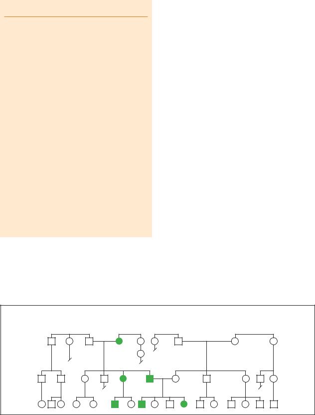
child with average stature, a 50% chance that they will have a child with one achondroplasia gene (a heterozygote), and a 25% chance that a child will get two copies of the achondroplasia gene (a homozygote). Babies with homozygous achondroplasia are much more severely affected than babies with a single achondroplasia gene. These infants generally die very shortly after birth because of breathing problems caused by an extremely small chest.
Demographics
Because individuals with other forms of dwarfism are often misdiagnosed with achondroplasia, the exact incidence of achondroplasia is unknown. Estimates of the incidence of achondroplasia vary between 1/10,000 to 1/40,000 births. It is estimated that there are approximately 15,000 individuals with achondroplasia in the United States and 65,000 worldwide. Achondroplasia affects males and females in equal numbers.
Signs and symptoms
Individuals with achondroplasia have disproportionate short stature, large heads with characteristic facial features, and rhizomelic shortening of their limbs. Rhizomelic means “root limb.” Rhizomelic shortening of the limbs means that those segments of a limb closest to the body (the root of the limb) are more severely affected. In individuals with achondroplasia, the upper arms are shorter than the forearms and the upper leg (thigh) is shorter than the lower leg.
In addition to shortened limbs, individuals with achondroplasia have other characteristic limb differences. People with achondroplasia have a limited ability to rotate and extend their elbows. They generally develop bowed legs and may have in-turned toes. Their hands and feet are short and broad, as are their fingers and toes. Their hands have been described as having a “trident” configuration. This term is based upon the trident fork used in Greek mythology and describes the unusual sep-
aration of their middle fingers. This unusual separation gives their hands a “three-pronged” appearance with the thumb and two small fingers on the side and the index and middle finger in the middle.
Individuals with achondroplasia have similar facial features and a large head (megalencephaly) due to the difference in the growth of the bones of the face and head. The exact reason for the increase in head size is not known, but it reflects increased brain size and can sometimes be due to hydrocephalus. People with achondroplasia have a protruding forehead (frontal bossing) and a relatively prominent chin. The prominent appearance of the chin is in part due to the relative flatness of their midface. While people with achondroplasia do resemble one another, they also resemble their family of origin.
Individuals with achondroplasia have shortening of their long bones. Women with achondroplasia have an average adult height of 48 in (122 cm). Men have an average adult height of 52 in (132 cm).
Diagnosis
Achondroplasia is generally diagnosed by physical examination at birth. The characteristic findings of short stature, rhizomelic shortening of the limbs, and specific facial features become more pronounced over time. In addition to being diagnosed by physical examination, individuals with achondroplasia have some specific bone changes that can be seen on an x ray. These include a smaller spinal canal and a small foramen magnum. The foramen magnum is the opening at the base of the skull. The spinal cord runs from the spinal canal through the foramen magnum and connects with the brain.
The diagnosis of achondroplasia can also be made prenatally either by ultrasound (sonogram) or by prenatal DNA testing. Sonograms use sound waves to provide an image of a fetus. The physical findings of achondroplasia (shortened long bones, trident hand) can be detected in the third trimester (last three months) of a pregnancy. Prior to the last three months of pregnancy, it is difficult to use a sonogram to diagnose achondroplasia because the physical features may not be obvious. Because of the large number of skeletal dysplasias, it can be very difficult to definitively diagnose achondroplasia by sonogram. Many other dwarfing syndromes can look very similar to achondroplasia on a sonogram.
Prenatal testing can also be done using DNA technology. A sample of tissue from a fetus is obtained by either chorionic villi sampling (CVS) or by amniocentesis. Chorionic villi sampling is generally done between 10-12 weeks of pregnancy and amniocentesis is done between 16-18 weeks of pregnancy. Chorionic villi sam-
18 |
G A L E E N C Y C L O P E D I A O F G E N E T I C D I S O R D E R S |

pling involves removing a small amount of tissue from the developing placenta. The tissue in the placenta contains the same DNA as the fetus. Amniocentesis involves removing a small amount of fluid from around the fetus. This fluid contains some fetal skin cells. DNA can be isolated from these skin cells. The fetal DNA is then tested to determine if it contains either of the two mutations responsible for achondroplasia.
Prenatal DNA testing for achondroplasia is not routinely performed in low-risk pregnancies. This type of testing is generally limited to high-risk pregnancies, such as those in which both parents have achondroplasia. It is particularly helpful in determining if a fetus has received two abnormal genes (homozygous achondroplasia). This occurs when both parents have achondroplasia and each of them passes on their affected gene. The baby gets two copies of the achondroplasia gene. Babies with homozygous achondroplasia are much more severely affected than babies with heterozygous achondroplasia. Infants with homozygous achondroplasia generally die shortly after birth due to breathing problems caused by an extremely small chest.
DNA testing can also be performed on blood samples from children or adults. This is usually done if there is some doubt about the diagnosis of achondroplasia or in atypical cases.
Treatment and management
There is no cure for achondroplasia. The recommendations for the medical management of individuals with achondroplasia have been outlined by the American Academy of Pediatrics’ Committee on Genetics. The potential medical complications of achondroplasia range from mild (ear infections) to severe (spinal cord compression). By being aware of the potential medical complications and catching problems early, it may be possible to avert some of the long-term consequences of these complications. An individual with achondroplasia may have some, all, or none of these complications.
All children with achondroplasia should have their height, weight, and head circumference measured and plotted on growth curves specifically developed for children with achondroplasia. Measurements of head circumference are important to monitor for the development of hydrocephalus—a known but rare ( 5%) complication of achondroplasia. Hydrocephalus (or water on the brain) is caused by an enlargement of the fluid-filled cavities of the brain (ventricles) due to a blockage that impedes the movement of the cerebrospinal fluid. Suspected hydrocephalus can be confirmed using imaging techniques such as a CT or MRI scan and can be treated with neurosurgery or shunting (draining) if it
This man has achondroplasia, a disorder characterized by short stature. (Photo Researchers, Inc.)
causes severe symptoms. Any child displaying neurologic problems such as lethargy, abnormal reflexes, or loss of muscle control should be seen by a neurologist to make sure they are not experiencing compression of their spinal cord. Compression of the spinal cord is common in individuals with achondroplasia because of the abnormal shape and small size of their foramen magnum (opening at the top of the spinal cord).
All children with achondroplasia should be monitored for sleep apnea, which occurs when an individual stops breathing during sleep. This can occur for several reasons, including obstruction of the throat by the tonsils and adenoids, spinal cord compression, and obesity. Individuals with achondroplasia are more prone to sleep apnea due to the changes in their spinal canal, foramen magnum, and because of their short necks. Treatment for sleep apnea depends on its cause. Obstructive sleep apnea is treated by surgically removing the tonsils and adenoids. Neurosurgery may be required to treat sleep apnea
Achondroplasia
G A L E E N C Y C L O P E D I A O F G E N E T I C D I S O R D E R S |
19 |

Achondroplasia
Achondroplasia
Autosomal Dominant
|
|
86y |
d.70y |
|
d.1y |
55y |
54y |
d.37y |
d.48y |
|
|
Emphysema |
Accident |
Hearing |
|
Leukemia |
|
Ovarian cancer |
|
|
|
|
loss |
|
|
|
|
|
|
|
|
|
|
|
2 |
56y |
52y |
49y |
40y 40y |
30y |
29y |
25y |
21y |
|
2 |
3 |
|
|
|
|
|
|
|
|
|
|
|
6y d.1day 6mos |
|
|
|
|
|
|
2 |
|
|
|
|
|
|
(Gale Group)
due to spinal cord compression. Weight management may also play a role in the treatment of sleep apnea.
Other potential problems in children with achondroplasia include overcrowding of the teeth (dental malocclusion), speech problems (articulation), and frequent ear infections (otitis media). Dental malocclusion (overcrowding of teeth) is treated with orthodontics. All children with achondroplasia should be evaluated by a speech therapist by two years of age because of possible problems with the development of clear speech (articulation). Articulation problems may be caused by orthodontic problems. Due to the abnormal shape of the eustachian tube in an individual with achondroplasia, they are very prone to ear infections (otitis media). Approximately 80% of infants with achondroplasia have an ear infection in the first year of life. About 78% of these infants require ventilation tubes to decrease the frequency of ear infections.
Weight management is extremely important for an individual with achondroplasia. Excess weight can exacerbate many of the potential orthopedic problems in an individual with achondroplasia such as bowed legs, curvature of the spine, and joint and lower back pain. Excess weight can also contribute to sleep apnea. Development of good eating habits and appropriate exercise programs should be encouraged in individuals with achondroplasia. These individuals should discuss their exercise programs with their health care provider. Because of the potential for spinal cord compression, care should be used in choosing appropriate forms of exercise.
The social adaptation of children with achondroplasia and their families should be closely monitored. Children with visible physical differences can have difficulties in school and socially. Support groups such as Little People of America can be a source of guidance on how to deal with these issues. It is important that children with achondroplasia not be limited in activities that pose no danger. In addition to monitoring their social adaptation, every effort should be made to physically adapt their surroundings for convenience and to improve independence. Physical adaptations can include stools to increase accessibility and lowering of switches and counters.
Two treatments have been used to try to increase the final adult height of individuals with achondroplasia –limb-lengthening and growth hormone therapy. There are risks and benefits to both treatments and as of 2001, they are still considered experimental.
Limb-lengthening involves surgically attaching external rods to the long bones in the arms and legs. These rods run parallel to the bone on the outside of the body. Over a period of 18-24 months, the tension on these rods is increased, which results in the lengthening of the underlying bone. This procedure is long, costly, and has potential complications such as pain, infections, and nerve problems. Limb-lengthening can increase overall height by 12-14 in (30.5-35.6 cm). It does not change the other physical manifestations of achondroplasia such as the appearance of the hands and face. This is an elective surgery and individuals must decide for them-
20 |
G A L E E N C Y C L O P E D I A O F G E N E T I C D I S O R D E R S |

selves if it would be of benefit to them. The optimal age to perform this surgery is not known.
Growth hormone therapy has been used to treat some children with achondroplasia. Originally there was doubt about the effectiveness of this treatment because children with achondroplasia are not growth hormone deficient. However, studies have shown that rate of growth in children with achondroplasia treated with growth hormone does increase during the first two years of treatment. It is too early to say how effective this treatment is because the children involved in this study are still growing and have not reached their final adult height.
Prognosis
The prognosis for most people with achondroplasia is very good. In general, they have minimal medical problems, normal IQ, and most achieve success and have a long life regardless of their stature. The most serious medical barriers to an excellent prognosis are the neurologic complications that can arise in achondroplasia. Spinal cord compression is thought to increase the risk for SIDS to 7.5% in infants with achondroplasia and can lead to life-long complications such as paralysis if untreated. Obesity can increase the risk for heart disease and some studies have revealed an increased risk of unexplained death in the fourth and fifth decade of life.
Successful social adaptation plays an important role in the ultimate success and happiness of an individual with achondroplasia. It is very important that the career and life choices of an individual with achondroplasia not be limited by preconceived ideas about their abilities.
Resources
BOOKS
Ablon, Joan. Living with Difference: Families with Dwarf Children. Westport, CT: Praeger Publishing, 1988.
PERIODICALS
American Academy of Pediatrics Committee on Genetics. “Health Supervision for Children With Achondroplasia.” Pediatrics 95, no 3 (March 1995): 443-51.
ORGANIZATIONS
Little People of America, Inc. National Headquarters, PO Box 745, Lubbock, TX 79408. (806) 737-8186 or (888) LPA2001. lpadatabase@juno.com. http://www.lpaonline
.org .
WEBSITES
The Human Growth Foundation. http://www.hgfound.org/ Little People of America: An Organization for People of Short
Stature. http://www.lpaonline.org/lpa.html
Kathleen Fergus, MS
I ACHOO syndrome
Definition
ACHOO syndrome is a generally benign condition characterized by sudden, uncontrollable sneezing after viewing a bright light.
Description
The ACHOO syndrome, standing for autosomal dominant compelling heliopthalmic outburst syndrome, is an inherited condition where a person will involuntarily sneeze after seeing a bright light. A person with this condition will sneeze multiple times, and in rare cases may sneeze 30-40 times. The syndrome is usually more intense if the person with the condition moves suddenly from darkness into an area with bright lights or sunlight.
Genetic profile
The ACHOO syndrome is thought to be inherited in an autosomal dominant pattern. This means that only one copy of the abnormal gene needs to be present for the syndrome to occur. If one parent has the condition, their children will have a 50% chance of also having the syndrome. One physician reported the condition in a family, where it was observed in the father and his brother, but not seen in the father’s mother or his wife. Both the father and brother would sneeze twice when going from an area of darkness to an area of light. At four weeks of age, the father’s daughter also started to sneeze whenever she was moved into bright sunlight.
Because of the relatively benign nature of the condition, there has been no reported scientific work trying to locate the gene responsible for the syndrome.
Demographics
Occurrence of the ACHOO syndrome is widespread in the general population. The few well-documented studies performed report the condition as being present in 23-33% of individuals. Men seem to be affected more than women. Studies on the occurrence of the syndrome in various ethnic groups are very limited. One study showed differences between whites and non-whites, while another study showed no difference.
Signs and symptoms
The prominent symptom of people with the ACHOO syndrome is sudden, involuntary sneezing when they see a bright light or sunlight. The way in which sneezing is
syndrome ACHOO
G A L E E N C Y C L O P E D I A O F G E N E T I C D I S O R D E R S |
21 |
ACHOO syndrome
K E Y T E R M S
Allergy—Condition in which immune system is hypersensitive to contact with allergens; an abnormal response by the immune system to contact with an allergen; condition in which contact with allergen produces symptoms such as inflammation of tissues and production of excess mucus in respiratory system.
Antibody—A protein produced by the mature B cells of the immune system that attach to invading microorganisms and target them for destruction by other immune system cells.
Antigen—A substance or organism that is foreign to the body and stimulates a response from the immune system.
Hypersensitivity—A process or reaction that occurs at above normal levels; overreaction to a stimulus.
Immune response—Defense mechanism of the body provided by its immune system in response to the presence of an antigen, such as the production of antibodies.
Immune system—A major system of the body that produces specialized cells and substances that interact with and destroy foreign antigens that invade the body.
triggered is not very well understood, but there are several theories that attempt to explain the syndrome.
One theory is that people who have the ACHOO syndrome have a hypersensitive reaction to light, just like some people have a sensitivity to cat hairs or pollen.
When a person with the syndrome is exposed to a bright light, the same mechanism in the body that triggers a sneeze due to an irritant such as pollen somehow confuses light with that irritant and causes a sneeze to occur. Another idea is that the sneeze reflex in people with the ACHOO syndrome is somehow linked to real nasal allergies, although this does not explain the syndrome in people without nasal allergies. A third theory is that people with the ACHOO syndrome are very sensitive to seeing bright light. The sneeze reflex of the syndrome can then be thought of as an involuntary defense reaction against bright light; when the person sneezes, they automatically close their eyes.
Diagnosis
The ACHOO syndrome is diagnosed simply by observing the sneezing pattern of a person, and by looking into the sneezing patterns of the person’s close relatives. If the person seems to sneeze every time they are exposed to a bright light, and if their parents and offspring do the same, then the diagnosis of the ACHOO syndrome can be made.
Currently, there are no known blood tests or other medical tests that can help diagnose the syndrome.
Treatment and management
There are no specific treatments for the ACHOO syndrome. Common measures, such as wearing sunglasses, can help people who are severely affected.
There have been reports that people who have nasal allergies have a higher incidence of the ACHOO syndrome. Therefore, it is sometimes assumed that medications that are used for allergies, such as antihistamines, could perhaps play a beneficial role in the ACHOO syn-
Achoo Syndrome |
(Gale Group)
22 |
G A L E E N C Y C L O P E D I A O F G E N E T I C D I S O R D E R S |

drome. However, no studies have successfully demonstrated that the syndrome is relieved by this type of medication. Alternative medicine, including homeopathy and herbal medicine, recommend a wide range of remedies for nasal allergies, these may accordingly also be helpful for the ACHOO syndrome.
Prognosis
People with the ACHOO syndrome generally have the condition for life. There is no evidence showing that the ACHOO syndrome in any way affects a person’s life span.
Resources
BOOKS
Knight, Jeffrey, and Robert McClenaghan. Encyclopedia of
Genetics. Pasadena: Salem Press, 1999.
PERIODICALS
Whitman, B. W., and R. J. Packer. “The Photic Sneeze Reflex.”
Neurology (May 1993): 868-871.
PERIODICALS
Askenasy, J. J. M. “The Photic Sneeze.” Postgraduate Medical
Journal (February 1990): 892-893.
Edward R. Rosick, DO, MPH, MS
I Acid maltase deficiency
Definition
Acid maltase deficiency, also called Pompe disease, is a non-sex linked recessive genetic disorder that is the most serious of the glycogen storage diseases affecting muscle tissue. It is one of several known congenital (present at birth) muscular diseases (myopathies), as distinct from a muscular dystrophy, which is a family of muscle disorders arising from faulty nutrition. The Dutch pathologist J. C. Pompe first described this genetic disorder in 1932.
Description
Acid maltase deficiency is also known as glycogen storage disease type II (GSD II) because it is characterized by a buildup of glycogen in the muscle cells. Glycogen is the chemical substance muscles use to store sugars and starches for later use. Some of the sugars and starches from the diet that are not immediately put to use are converted into glycogen and then stored in the mus-
cle cells. These stores of glycogen are then broken down into sugars, as the muscles require them. Acid maltase is the chemical substance that regulates the amount of glycogen stored in muscle cells. When too much glycogen begins to accumulate in a muscle cell, acid maltase is released to break down this excess glycogen into products that will be either reabsorbed for later use in other cells or passed out of the body via the digestive system. Individuals affected with acid maltase deficiency have either a complete inability or a severely limited ability to produce acid maltase. Since these individuals cannot produce the amounts of acid maltase required to process excess glycogen in the muscle cells, the muscle cells become overrun with glycogen. This excess glycogen in the muscle cells causes a progressive degeneration of the muscle tissues.
Acid maltase is an enzyme. An enzyme is a chemical that facilitates (catalyzes) the chemical reaction of another chemical or of other chemicals; it is neither a reactant nor a product in the chemical reaction that it catalyzes. As a result, enzymes are not used up in chemical reactions, but rather recycled. One molecule of an enzyme may be used to catalyze the same chemical reaction over and over again several hundreds of thousands of times. All the enzymes necessary for catalyzing the various reactions of human life are produced within the body by genes. Genetic enzyme deficiency disorders, such as acid maltase deficiency, result from only one cause: the affected individual cannot produce enough of the necessary enzyme because the gene designed to make the enzyme is faulty. Enzymes are not used up in chemical reactions, but they do eventually wear out, or accidentally get expelled. Also, as an individual grows, they may require greater quantities of an enzyme. Therefore, most enzyme deficiency disorders will have a time component to them. Individuals with no ability to produce a particular enzyme may show effects of this deficiency at birth or shortly thereafter. Individuals with only a partial ability to produce a particular enzyme may not show the effects of this deficiency until their need for the enzyme, because of growth or maturation, has outpaced their ability to produce it.
The level of ability of individuals with acid maltase deficiency to produce acid maltase, or their ability to sustain existing levels of acid maltase, are the sole determinants of the severity of the observed symptoms in individuals and the age of onset of these symptoms.
Acid maltase deficiency is categorized into three separate types based on the age of onset of symptoms in the affected individual. Type a, or infantile, acid maltase deficiency usually begins to produce observable symptoms in affected individuals between the ages of two and five months. Type b, or childhood, acid maltase defi-
deficiency maltase Acid
G A L E E N C Y C L O P E D I A O F G E N E T I C D I S O R D E R S |
23 |
Acid maltase deficiency
ciency usually begins to produce observable symptoms in affected individuals in early childhood. This type generally progresses much more slowly than infantile acid maltase deficiency. Type c, or adult, acid maltase deficiency generally begins to produce observable symptoms in affected individuals in the third or fourth decades of life. This type progresses even more slowly than childhood acid maltase deficiency.
Genetic profile
The locus of the gene responsible for acid maltase deficiency has been localized to 17q23. The severity of the associated symptoms and the age of onset in affected individuals have been closely tied to the particular mutation at this locus. Three specific mutations and one additional mutation type have been demonstrated to occur along the gene responsible for acid maltase deficiency. Each of these is associated with varying symptoms.
A gene is a particular segment of a particular chromosome. However, within the segment containing a particular gene there are two types of areas: introns and exons. Introns are sections of the segment that do not actively participate in the functioning of the gene. Exons are those sections that do actively participate in gene function. A typical gene consists of several areas that are exons divided by several areas of introns.
One mutation on the gene responsible for the production of acid maltase is a deletion of exon 18. A second mutation on the gene responsible for the production of acid maltase is the deletion of a single base pair of exon 2. Both these mutations are associated with a complete inability of the affected individual to produce acid maltase. Individuals with these mutations will invariably be affected with infantile (type a) acid maltase deficiency.
The third mutation on the gene responsible for the production of acid maltase is a complicated mutation within intron 1 that causes the cutting out of exon 2. This mutation is generally not complete in every copy of the gene within a given individual so it is associated with a partial ability of the affected individual to produce acid maltase. Individuals with this mutation will be affected with either childhood (type b), or, more commonly, adult (type c) acid maltase deficiency. In fact, greater than 70% of all individuals affected with adult acid maltase deficiency possess this particular mutation.
The final mutation class known to occur on the gene responsible for the production of acid maltase is missense at various locations along the various exons. Missense is the alteration of a single coding sequence (codon) that codes for a single amino acid that will be used to build the protein that is the precursor to the acid maltase molecule. These missense mutations generally
prevent the production of acid maltase and lead to infantile (type a) acid maltase deficiency.
The exact mutations responsible for the other 30% of the adult (type c) and the remainder of the childhood (type b) acid maltase deficiency cases have not yet been determined.
Demographics
Acid maltase deficiency is observed in approximately 1 in every 100,000 live births. In 2000, it was estimated that between 5,000 and 10,000 people were living somewhere in the developed world with a diagnosed case of acid maltase deficiency. It is observed in equal numbers of males and females and across all ethnic subpopulations.
Since acid maltase deficiency is a recessive disorder, both parents must be carriers of the disorder for it to be passed to their children. In the case of carrier parents with one child affected by acid maltase deficiency, there is a 25% likelihood that their next child will also be affected with the disorder. However, because type c (adult) acid maltase deficiency generally does not show symptoms in the affected individual until that individual is past 30, it is possible for an affected individual to parent children. In this case, the probability of a second child being affected with acid maltase deficiency is 50%. Should two affected individuals bear offspring; the probability of their child being affected with acid maltase deficiency is 100%.
In families with more than one affected child, the symptoms of the siblings will closely correspond. That is, if one child develops infantile acid maltase deficiency, a second child, if affected with the disorder, will also develop the infantile form.
Signs and symptoms
The symptoms of acid maltase deficiency vary depending on the severity of the deficiency of acid maltase in the affected individual. The most acid maltase deficient individuals will develop infantile acid maltase deficiency and will exhibit the most severe symptoms. Likewise, the least acid maltase deficient individuals will develop adult acid maltase deficiency and have less severe symptoms.
Infantile (type a) acid maltase deficiency is characterized by the so-called “floppy baby” syndrome. This condition is caused by extreme weakness and lack of tone of the skeletal muscles. This observed weakness in the skeletal muscles is accompanied by the much more serious problems of overall weakness of the heart muscle (cardiomyopathy) and the muscles of the respiratory sys-
24 |
G A L E E N C Y C L O P E D I A O F G E N E T I C D I S O R D E R S |

tem, primarily the diaphragm. Enlargement of the heart (cardiomegaly), tongue, and liver are also observed. Glycogen accumulation is observed in most tissues of the body.
Childhood (type b) acid maltase deficiency is characterized by weakness of the muscles of the trunk and large muscle mass with little muscle tone. This is due to a buildup of glycogen in the muscle cells. The heart and liver of those affected with childhood maltase deficiency are generally normal. However, there is a progressive weakening of the skeletal and respiratory muscles. The observed muscle weakness in childhood acid maltase deficiency affected individuals gradually progresses from the muscles of the trunk to the muscles of the arms and the legs. Glycogen accumulation is observed primarily in the muscle tissues.
Adult (type c) acid maltase deficiency is characterized by fatigue in younger affected individuals and by weakness of the muscles of the trunk in older affected individuals. The observed muscle weakness in adult acid maltase deficiency affected individuals gradually progresses from the muscles of the trunk to the muscles of the arms and the legs. High blood pressure in the artery that delivers blood to the lungs (pulmonary hypertension) is also generally observed in affected adults. Glycogen accumulation is observed primarily in the muscle tissues.
Diagnosis
Infantile acid maltase deficiency is generally diagnosed between the ages of two and five months when symptoms begin to appear. The first indicator of infantile acid maltase deficiency is general weakness and lack of tone (hypotonia) of the skeletal muscles, particularly those of the trunk.
A blood test called a serum CK test is the most commonly used test to determine whether muscular degeneration is causing an observed muscular weakness. It is used to rule out other possible causes of muscle weakness, such as nerve problems. To determine the CK serum level, blood is drawn and separated into the part containing the cells and the liquid remaining (the serum). The serum is then tested for the amount of creatine kinase (CK) present. Creatine kinase is an enzyme found almost exclusively in the muscle cells and not typically in high amounts in the bloodstream. Higher than normal amounts of CK in the blood serum indicate that muscular degeneration is occurring: that the muscle cells are breaking open and spilling their contents, including the enzyme creatine kinase (CK) into the bloodstream. Individuals affected with acid maltase deficiency have extremely high serum CK levels. Those affected with
K E Y T E R M S
Acid maltase—The enzyme that regulates the amount of glycogen stored in muscle cells. When too much glycogen is present, acid maltase is released to break it down into waste products.
Acidosis—A condition of decreased alkalinity resulting from abnormally high acid levels (low pH) in the blood and tissues. Usually indicated by sickly sweet breath, headaches, nausea, vomiting, and visual impairments.
Catalyze—Facilitate. A catalyst lowers the amount of energy required for a specific chemical reaction to occur. Catalysts are not used up in the chemical reactions they facilitate.
Enzyme—A protein that catalyzes a biochemical reaction or change without changing its own structure or function.
Exon—The expressed portion of a gene. The exons of genes are those portions that actually chemically code for the protein or polypeptide that the gene is responsible for producing.
Fibroblast—Cells that form connective tissue fibers like skin.
Glycogen—The chemical substance used by muscles to store sugars and starches for later use. It is composed of repeating units of glucose.
Hypoglycemia—An abnormally low glucose (blood sugar) concentration in the blood.
Intron—That portion of the DNA sequence of a gene that is not directly involved in the formation of the chemical that the gene codes for.
Myopathy—Any abnormal condition or disease of the muscle.
Serum CK test—A blood test that determines the amount of the enzyme creatine kinase (CK) in the blood serum. An elevated level of CK in the blood indicates that muscular degeneration has occurred and/or is occurring.
infantile acid maltase deficiency have much higher serum CK levels than those affected with the childhood or adult forms. The actual serum CK level, once observed to be higher than normal, can also be used to differentiate between various types of muscular degeneration.
Serum CK levels cannot be used to distinguish acid maltase deficiency from other glycogen storage diseases.
deficiency maltase Acid
G A L E E N C Y C L O P E D I A O F G E N E T I C D I S O R D E R S |
25 |

Acrocallosal syndrome
Acid maltase deficiency (type II glycogen storage disease) is differentially diagnosed from type I glycogen storage disease by blood tests for abnormally low levels of glucose (hypoglycemia) and a low pH, or high acidity, (acidosis). Hypoglycemia and acidosis are both characteristic of type I glycogen storage disease, but neither is characteristic of acid maltase deficiency.
It is sometimes possible to determine the abnormally low levels of the acid maltase enzyme in the white blood cells (leukocytes) removed during the above blood serum tests. If these levels can be determined and they are abnormally low, a definitive diagnosis of acid maltase deficiency can be made. When the results of this leukocyte test are not clear, acid maltase deficiency types a and b may be positively diagnosed by testing muscles cells removed from the affected individual (muscle biopsy) for the actual absence or lack of sufficient acid maltase. This test is 100% accurate for type a and type b acid maltase deficiency, but it may give improper results for type c acid maltase deficiency. In these hard-to-identify cases of type c acid maltase deficiency, an identical test to that performed on the leukocytes may be performed on cultured fibroblasts grown from a sample from the affected individual. This test is 100% accurate for type c acid maltase deficiency.
Treatment and management
As of early 2001, there is no treatment or cure for acid maltase deficiency. The only potential treatment for this deficiency is enzyme replacement therapy. This approach was initially undertaken in the 1970s for acid maltase deficiency with no success. A new enzyme replacement therapy is, however, currently in human clinical trials that began in 1999.
Prognosis
Acid maltase deficiency of all three types is 100% fatal. Individuals affected with infantile acid maltase deficiency generally die from heart or respiratory failure prior to age one. Individuals affected with childhood acid maltase deficiency generally die from respiratory failure between the ages of three and 24. Individuals affected with adult acid maltase deficiency generally die from respiratory failure within 10 to 20 years of the onset of symptoms.
Human clinical trials involving enzyme replacement therapy, in which a synthetic form of acid maltase is administered to affected individuals, were begun in 1999 at Duke University Medical Center in North Carolina and Erasmus University Rotterdam in the Netherlands. Genzyme Corporation and Pharming Group N. V. announced the first results of these trials in a joint press
release on October 5, 2000. These two companies currently own the worldwide patent rights to the synthetic enzyme being studied. As of early 2001, these clinical trials are still in phase I/II of the three-stage testing process for use in humans.
Resources
PERIODICALS
Chen, Y., and A. Amalfitano. “Towards a molecular therapy for glycogen storage disease type II (Pompe disease).”
Molecular Medicine Today (June 2000): 245-51. Poenaru, L. “Approach to gene therapy of glycogenosis type II
(Pompe disease).” Molecular Genetics and Metabolism
(July 2000): 162-9.
ORGANIZATIONS
Acid Maltase Deficiency Association (AMDA). PO Box 700248, San Antonio, TX 78270-0248. (210) 494-6144 or (210) 490-7161. Fax: (210) 490-7161 or 210-497-3810.http://www.amda-pompe.org .
Association for Glycogen Storage Disease (United Kingdom). 0131 554 2791. Fax: 0131 244 8926. http://www.agsd
.org.uk .
WEBSITES
Neuromuscular Disease Center. http://www.neuro.wustl.edu/ neuromuscular/msys/glycogen.html (February 12, 2001).
OMIM—Online Mendelian Inheritance in Man. http://www
.ncbi.nlm.nih.gov/htbin-post/Omim/dispmim?232300 (February 12, 2001).
The Pompe’s Disease Page. http://www.cix.co.uk/~embra/ pompe/Welcome.html (February 12, 2001).
OTHER
“Genzyme General and Pharming Group Reports Results From First Two Clinical Trials for Pompe Disease.” Genzyme Corporation Press Release (October 5, 2000).
“Pompe disease therapy to be tested.”Duke University Medical
Center Press Release (May 24, 1999).
Paul A. Johnson
I Acrocallosal syndrome
Definition
Acrocallosal syndrome is a rare congenital disorder in which the individual has absence or only partial formation of the corpus callosum. This is accompanied by skull and facial malformations, and some degree of finger or toe malformations. Individuals may display motor and mental retardation. The cause of this genetic disorder is unknown, and the severity of the symptoms vary by individual.
26 |
G A L E E N C Y C L O P E D I A O F G E N E T I C D I S O R D E R S |

Description
Acrocallosal syndrome was first described by Schinzel in 1979, and also may be referred to as Schinzel acrocallosal syndrome. The term acrocallosal refers to the involvement of the acra (fingers and toes) and the corpus callosum, the thick band of fibers joining the hemispheres of the brain. Reported in both males and females, the cause of the disorder is unknown. The major characteristic of the syndrome is the incomplete formation (hypoplasia) or absence (agenesis) of the corpus callosum. Facial appearance is typically similar among affected people. This includes a prominent forehead, an abnormal increase in the distance between the eyes (hypertelorism), and a large head (macrocephaly). Individuals have a degree of webbing or fusion (syndactyly), or duplication (polydactyly) of the fingers and toes. Occasionally, those affected may have a short upper lip, cleft palate, cysts that occur within the cranium (intracranial), hernias, or may develop seizure disorders. Less frequently, affected children have congenital heart defects, internal organ (visceral) or kidney (renal) abnormalities.
Moderate to severe mental retardation is reported with acrocallosal syndrome. Individuals usually display some form of poor muscle tone (hypotonia), and there may be a delay or absence of motor activities, walking, and talking. There is great variation of functioning and symptoms with this disorder, ranging from normal development to severe mental and motor retardation.
Genetic profile
The cause of acrocallosal syndrome is unknown. There are sporadic, or random, cases, and reports of multiple cases within families. Studies involving affected families have suggested an autosomal recessive pattern of inheritance. This means that both parents carry the altered form of the gene and the affected child inherited both copies. Following this pattern, each child born will have a 25% risk of being affected.
To help determine which chromosome or gene location causes the syndrome, acrocallosal syndrome has been compared with similar disorders. One condition that presents similar symptoms and has a known genetic cause is Greig cephalopolysyndactyly syndrome. However, there is no genetic similarity between the two conditions. To date, no specific genetic cause for acrocallosal syndrome is known, and the disorder can only be identified by clinical symptoms.
Demographics
Acrocallosal syndrome is extremely rare. Reports of this disorder may occur within family lines, or randomly.
K E Y T E R M S
Computed tomography (CT) scan—An imaging procedure that produces a three-dimensional picture of organs or structures inside the body, such as the brain.
Consanguinity—A mating between two people who are related to one another by blood.
Corpus callosum—A thick bundle of nerve fibers deep in the center of the forebrain that provides communications between the right and left cerebral hemispheres.
Hypertelorism—A wider-than-normal space between the eyes.
Hypotonia—Reduced or diminished muscle tone.
Polydactyly—The presence of extra fingers or toes.
Syndactyly—Webbing or fusion between the fingers or toes.
It affects both males and females. There are some reports of webbing of the fingers or toes (syndactyly) and relatedness (consanguinity) of the parents of affected children. However, affected children may also have unrelated, healthy parents and unaffected siblings.
Signs and symptoms
At birth, those with acrocallosal syndrome present the characteristic pattern of facial and limb malformations. Limb appearance ranges from minor webbing between the fingers or toes to near duplication of the hands or feet. Forehead prominence, increased distance between the eyes, and an enlarged head are the main features of facial appearance. X ray tests will reveal the absence or incomplete formation of the corpus callosum and the presence of any cysts within the cranium. The infant will usually display reduced muscle tone (hypotonia). This may lead to a drooling condition or feeding difficulties. Hypotonia can also contribute to a delay in growth and motor skills. Severe hypotonia is usually associated with a form of mental retardation.
Progress and functioning during the first year of life is dependent upon the severity of the symptoms. There has been a wide range of individual variation reported, and the degree to which symptoms affect each child may differ. Some children develop normally and will walk and talk within normal age limits, while others may experience a delay or absence of certain motor activities. Mental retardation may be moderate or severe. Some
syndrome Acrocallosal
G A L E E N C Y C L O P E D I A O F G E N E T I C D I S O R D E R S |
27 |