

Учебники / Rhinosinusitis - A Guide for Diagnosis and Management 2008
.pdf7 Endoscopic Sinus Surgery |
103 |
of subsequent symptoms possibly related to frontal sinus disease, such as frontal headaches, difficult. Additionally, the failure rate of frontal sinus fat obliteration increases over time, approaching at least 25% as the years go by.
Postoperative Management
Patients are usually instructed not to blow the nose for several days following the surgery and to avoid any bending or heavy lifting. Most patients do not have any facial swelling or ecchymosis following endoscopic sinus surgery. Pain is typically minimal or absent, and the classical uncomfortable postoperative packing is essentially never used today. However, patients will usually have a bloody nasal discharge for several days to weeks and intermittent nasal congestion, which may also continue for a number of weeks. Many physicians will either place a sponge into the middle meatus, which is removed after the first or second postoperative day, or use an absorbable spacer in the middle meatus, which slowly dissolves in the first 2 to 3 weeks after the surgery. The advantage of using a sponge that can be removed is that it allows the surgeon to suction out blood and mucus from the sinuses in the early postoperative period. As we believe that bacteria and fungi exacerbate the inflammatory response, removal of the sponge and cleaning of the area would appear to be preferable.
The natural sequela, arising from the combination of surgical intervention and the presence of significant inflammation, is scarring. Furthermore, the chronic inflammation that leads to chronic rhinosinusitis does not resolve immediately following surgical intervention. Since the primary goal of the surgery is to create widely patent sinuses that can drain easily, and because inflammation and surgery cause scarring, it is clear that significant postoperative care is required if the primary goal of the surgery is to be achieved. This care entails treatment of the underlying inflammation with antibiotics and antiinflammatory medications, repeated nasal endoscopy, and, not infrequently, some minor removal of scar tissue until the sinus cavities have healed and the residual inflammation has resolved [25]. Saline nasal sprays instituted in the early postoperative period will reduce nasal crusting, and we usually recommend that patients use these every hour or so while awake in the early postoperative period. Patients may also be asked to perform more significant nasal lavage with saline, antibiotic, or steroid solutions [26]. Whenever possible, antibiotic therapy is directed based upon endoscopic culture, and antibiotics are usually prescribed for at least 2 weeks following the surgical intervention. When significant bone inflammation is present, antibiotics may be required for a significantly longer period of time. Oral steroids, when required to control either asthma or nasal mucosal inflammation, are usually slowly tapered in the postoperative period. However, in the presence of severe polyposis or allergic fungal sinusitis, prolonged oral steroid therapy may be required if a recurrence of disease is to be avoided. Fortunately, patients with nasal and sinus diseases usually respond to low doses of oral steroids. Avoidance of allergens and environmental irritants, and, especially, avoidance of exposure to smoke is critically important in the postoperative period (Table 7.3).

104 |
D.W. Kennedy |
Table 7.3 Postoperative medical management
•Antibiotics (usually for 2 or more weeks)
•Nasal saline spray
•Long-term topical nasal steroids
•Possible slowly tapering oral steroids
•Possible antihistamines
•Possible antileukotrienes
Our postoperative regimen usually involves seeing the patient the day following the surgery and removing the middle meatal sponges and suctioning the nose under endoscopic visualization. The patient then returns weekly for 4 to 6 weeks, and less frequently thereafter until the cavity is fully healed and the residual inflammation has subsided. At each visit, a nasal endoscopy is performed under topical anesthesia and when the sinus cavities are raw. Particularly in the early postoperative period, an oral narcotic analgesic is beneficial before the visit because the cleanings are uncomfortable. When nasal endoscopy demonstrates a tendency to scarring, the immature scars are meticulously divided. Any residual areas of devitalized or inflamed bone are gently teased out, and careful attention is paid to ensure that the sinuses remain patent. Given the chronic and persistent nature of chronic rhinosinusitis and the tendency toward scarring in the postoperative period, it is particularly important that the patient returns for these postoperative visits until satisfactory healing is obtained. Convincing patients to return for these visits can be difficult because the patients are usually almost totally asymptomatic postoperatively, despite having ongoing persistent inflammation. Despite the relative lack of symptoms postoperatively, postoperative care, nasal endoscopy, and, when necessary, debridement are exceptionally important to the long-term outcome, and essential for most chronic disease if a late recurrence of chronic rhinosinusitis or nasal polyposis is to be avoided.
In the longer term, we regard these patients as “at-risk” patients for return of chronic sinusitis because the underlying factors associated with the onset of the disease probably have not been modified by the surgical intervention. Therefore, we recommend intermittent long-term nasal endoscopy and follow-up, until it is clear that the ethmoid cavity is stable and a recurrence of disease is unlikely. Early recurrent disease is usually asymptomatic, but it is important that it is identified at an early stage using nasal endoscopy if revision surgery is to be avoided [7]. Patients are usually recommended to use topical nasal steroids on a regular, ongoing, and long-term basis. The goal of the surgery should be to resolve the disease long term so as to avoid the necessity for subsequent surgical interventions, and not just reduce symptomatic relief, only to have the patient undergo revision surgery later.
Complications of Surgery
The most common complication of surgical intervention in the sinuses is persistence of inflammation, which may occur because of the underlying etiology of the disease, because of inadequate removal of the disease at the time of surgery, or
7 Endoscopic Sinus Surgery |
105 |
because of limited postoperative care and postoperative medical therapy. Although major bleeding is uncommon with this type of surgery, postoperative bleeding is also always a potential risk. Spinal fluid leak can occur as a result of inadvertent intracranial entry, but typically this is identified and closed at the time of the surgical procedure. If a spinal fluid leak is suspected postoperatively, the patient is frequently asked to collect some fluid for a beta-2 transferrin test. Because tears may also contain glucose, the use of Dextrostix to identify a CSF leak is considered misleading and to have low specificity. Trauma to the extraocular muscles, resulting in diplopia or intraorbital hemorrhage resulting in visual loss, has also been reported. Minor complications are much more common, but can be extremely bothersome to the patient and occasionally become almost crippling. These problems include persistent or worsening of inflammation or significant alterations in airflow resulting from extensive turbinate resection, whether inadvertent or deliberate (empty nose syndrome). A nose with abnormally large air passages is extremely bothersome to the patient, because of increased nasal resistance as a result of the loss of laminar airflow and because of an increased tendency toward mucosal inflammation and crusting. There is some evidence that inferior turbinate resection may also increase the incidence of maxillary sinus infections.
Summary
The introduction of endoscopic sinus surgery has dramatically improved the results of surgical management of rhinosinusitis in terms of decreasing the associated morbidity. Superior results have also been demonstrated when compared to open surgical intervention. It has been shown that nasal-specific symptoms improve. Using the SF-36 overall health survey form, overall quality of life is significantly adversely affected by chronic rhinosinusitis, but it returns to normal, and medication usage is decreased compared to the preoperative status, by 7.8 years postoperatively. However, the surgery is not a panacea. Sinus surgery is technically challenging and is best thought of as adjunctive to an overall long-term medical therapy plan. It takes a significant period of time for the underlying chronic inflammation to settle down, and fairly intensive medical therapy and endoscopic follow-up are often required during that period.
References
1.Muntz HR, Lusk RP. Nasal antral windows in children: a retrospective study. Laryngoscope 1990;100:643–646.
2.Stefansson P, Andreasson L, Jannert M. Caldwell-Luc operation: long-term results and sequelaes. Acta Otolaryngol Suppl 1988;449:97–100.
3.Kennedy DW, Zinreich SJ, Rosenbaum AE, et al. Functional endoscopic sinus surgery. Theory and diagnostic evaluation. Arch Otolaryngol 1985;111:576–582.
4.Zinreich SJ, Kennedy DW, Rosenbaum AE, et al. CT of the paranasal sinuses: imaging requirement for endoscopic surgery. Radiology 1987;163:769–775.
106 |
D.W. Kennedy |
5.Kennedy DW, Shaalan H. Reevaluation of maxillary sinus surgery: experimental study in rabbits. Ann Otol Rhinol Laryngol 1989;98(11):901–906.
6.Kennedy DW. Functional endoscopic sinus surgery. Technique. Arch Otolaryngol 1985;111:643–649.
7.Senior BA, Kennedy DW, Tanabodee J, et al. Long-term results of functional endoscopic sinus surgery. Laryngoscope 1998;108(2):151–157.
8.Stankiewicz JA. Complications of endoscopic nasal surgery: occurrence and treatment. Am J Rhinol 1987;1:45–49.
9.Conley DB, Tripathi A, Ditto AM, et al. Chronic sinusitis with nasal polyps: staphylococcal exotoxin immunoglobulin E and cellular inflammation. Am J Rhinol 2004;18(5):273–278.
10.Bernstein JM, Ballow M, Schlievert PM, et al. A superantigen hypothesis for the pathogenesis of chronic hyperplastic sinusitis with massive nasal polyposis. [Erratum appears in Am J Rhinol 2004;18(1):62.] Am J Rhinol 2003;17(6):321–326.
11.Slavin RG. Sinusitis in adults and its relation to allergic rhinitis, asthma, and nasal polyps. J Allergy Clin Immunol 1988;82:950–956.
12.Senior BA, Kennedy DW, Tanabodee J, et al. Long-term impact of functional endoscopic sinus surgery on asthma. Otolaryngol Head Neck Surg 1999;121:66–68.
13.Park AH, Lau J, Stankiewicz J, et al. The role of functional endoscopic sinus surgery in asthmatic patients. J Otolaryngol 1998;27(5):275–280.
14.Gwaltney JM Jr, Phillips CD, Miller RD, et al. Computed tomographic study of the common cold [see comments]. N Engl J Med 1994;330:25–30.
15.Havas TE, Motbey JA, Gullane PJ. Prevalence of incidental abnormalities on computed tomographic scans of the paranasal sinuses. Arch Otolaryngol 1988;114:856–859.
16.Zinreich SJ, Kennedy DW. Fungal sinusitis: diagnosis with CT and MR imaging. Radiology 1988;169:439-444.
17.Marple BF, Mabry RL. Allergic fungal sinusitis: learning from our failures. Am J Rhinol 2000;14(4):223–226.
18.Woodworth BA, Bhargave GA, Palmer JN, et al. Clinical outcomes of endoscopic and endoscopic-assisted resection of inverted papillomas: a 15-year experience. Am J Rhinol 2007;21(5):591–600.
19.Wolfe SG, Schlosser RJ, Bolger WE, et al. Endoscopic and endoscope-assisted resections of inverted sinonasal papillomas. Otolaryngol Head Neck Surg 2004;131(3):174–179.
20.Kennedy DW, Shaman P, Han W, et al. Complications of ethmoidectomy: a survey of fellows of the American Academy of Otolaryngology-Head and Neck Surgery. Otolaryngol Head Neck Surg 1994;111(5):589–599.
21.Wright ED, Agrawal S, Wright ED, et al. Impact of perioperative systemic steroids on surgical outcomes in patients with chronic rhinosinusitis with polyposis: evaluation with the novel Perioperative Sinus Endoscopy (POSE) scoring system. Laryngoscope 2007;117(11 pt 2 suppl 115):1–28.
22.Gross WE, Gross CW, Becker D, et al. Modified transnasal endoscopic Lothrop procedure as an alternative to frontal sinus obliteration. Otolaryngol Head Neck Surg 1995;113:427–434.
23.Brown CL, Bolger WE, Brown CL, et al. Safety and feasibility of balloon catheter dilation of paranasal sinus ostia: a preliminary investigation. [see comment]. Ann Otol Rhinol Laryngol 2006;115(4):293–299.
24.Bolger WE, Brown CL, Church CA, et al. Safety and outcomes of balloon catheter sinusotomy: a multicenter 24-week analysis in 115 patients. Otolaryngology - Head Neck Surg 2007;137(1):10–20.
25.Bugten V, Nordgard S, Steinsvag S, et al. The effects of debridement after endoscopic sinus surgery. Laryngoscope 2006;116(11):2037–2043.
26.Bachmann G, Hommel G, Michel O. Effect of irrigation of the nose with isotonic salt solution on adult patients with chronic paranasal sinus disease. Eur Arch Oto-Rhino-Laryngol 2000;257(10):537–541.

Chapter 8
Allergy and Rhinosinusitis
John H. Krouse
Clinicians have observed a relationship between allergy and acute and chronic rhinosinusitis (RS) for many years. This association appears to be common, and has been described by a number of authors over the past several decades [1]. As both RS and allergy reflect inflammatory disorders of the upper respiratory system, it is compelling to speculate that these two illnesses share common pathophysiological mechanisms and that treatment of nasal allergies would have an important benefit in the management of patients with RS. The science supporting this relationship, however, is largely anecdotal and epidemiologic, and mechanisms through which allergy predisposes a patient to RS are currently speculative.
This chapter discusses the relationship between nasal allergies and RS. The epidemiology and pathophysiology of nasal allergy are first reviewed, followed by a clinical overview of allergic rhinitis. Diagnostic methods used to evaluate the patient suspected of nasal allergy are outlined, and treatment methods to address allergy among patients with RS are discussed. It is important for physicians treating patients with RS to understand the role of allergy in the expression of symptoms among these individuals and to initiate relevant therapeutic options to decrease morbidity and improve quality of life.
Epidemiology and Burden of Nasal Allergy
Nasal allergies are common and are experienced by patients of all ages. The term that is used to describe a nasal disorder caused by allergic hypersensitivity is allergic rhinitis (AR). AR implies that nasal inflammation is triggered in an individual by hyperactive immune responses to known sensitizing agents. Epidemiologic studies have consistently positioned AR among the most common chronic illnesses in the United States (Fig. 8.1). In one recent study, it was estimated that more than 58 million Americans, or about 20% of the population, are bothered by symptoms
J.H. Krouse
Department of Otolaryngology, Wayne State University, Detroit, MI, USA e-mail: jKrouse@med.wayne.edu
E.R. Thaler, D.W. Kennedy (eds.), Rhinosinusitis, DOI: 10.1007/978-0-387-73062-2 8, |
107 |
C Springer Science+Business Media, LLC 2008 |
|

108 |
J.H. Krouse |
Fig. 8.1 Proportion of U.S. patients with allergic rhinitis by age. yo, years old
of AR [2]. It is also important to recognize that among patients with AR, many individuals also have symptoms that are worsened by nonallergic triggers, such as cigarette smoke, newsprint, and strong odors [3].
Although AR can affect individuals of any age, it is most commonly a disease of later childhood, adolescence, and young adulthood. Symptoms tend to be expressed most vigorously between the ages of 10 and 40 years [4], while the diagnosis of AR is usually made before the age of 10 [5]. In fact, there is strong evidence that the prevalence of AR among children has more than doubled since 1990 [6]. It is likely that the increasing prevalence of AR among children that has been noted will result in a greater number of adults with AR over the next several decades.
Because of the prevalence and symptomatic morbidity of AR, treatment of nasal allergies carries a significant economic cost. The financial burden of the direct treatment of AR has been estimated to range up to $9 billion (U.S.) [7], with up to $6 billion attributable to medication costs alone [8]. Since AR frequently coexists among patients with RS, treatment of AR among these individuals will add to the significant baseline costs associated with the diagnosis and treatment of RS.
Pathophysiology of Allergy
The allergic response involves a specific immune-mediated hypersensitivity to a generally innocuous substance in an individual who is genetically capable of generating such a response. This genetic predisposition to develop allergic diseases is
8 Allergy and Rhinosinusitis |
109 |
known as atopy. Not all individuals have inherited this predisposition to respond in this manner, and these individuals will not develop allergic hypersensitivities with exposure. A review of the immunology relevant to the allergic response will be useful. In addition, a list of terms commonly used in allergy diagnosis and treatment is presented in Table 8.1.
The term immunity is based on a Latin word meaning exemption. It refers to the processes through which the immune system is able to protect the individual from invasion by foreign substances that could be injurious. In carrying out this process, the immune system must be able to identify and neutralize substances that are potentially dangerous, yet exempt host tissues from injury. As such, the immune system is a primary line of defense through which the body is able to protect itself from pathogenic substances and organisms.
The immune system carries out its function through the coordinated response of a variety of humoral and cellular mediators. These components of the immune system must interact in a controlled and balanced manner to process foreign substances, identify host cells, and neutralize potentially harmful materials before they cause serious injury to the body. Cellular mediators involved in this system include director cells, such as macrophages and lymphocytes, and effector cells such as neutrophils, mast cells, and eosinophils. Lymphocytes are centrally involved in immune function and are divided into several subclasses, including B lymphocytes, T lymphocytes, and NK (natural killer) cells. B and T lymphocytes are both critical in the development and activation of the allergic response. In addition, eosinophils and mast cells are important leukocytes involved in the allergic response.
Humoral mediators include various cytokines, which are polypeptide messengers involved in coordination of the immune response, as well as immunoglobulins, which are antibodies involved in the binding of antigens and the stimulation of innate defense mechanisms. The primary immunoglobulin involved in the allergic response is immunoglobulin E (IgE), which is produced in very small amounts by B lymphocytes and plasma cells. IgE is developed in response to antigen exposure in an atopic individual and is bound to the surface of mast cells. These mast cells are present in both the nasal and sinus mucosa, as well as in other mucosal tissues and in the systemic circulation.
|
Table 8.1 Definitions of terms used in allergy |
Atopy |
The genetic predisposition to develop allergic sensitization |
Antigen |
Any substance capable of generating an immune response |
Allergen |
An antigen that is capable of generating an allergic response |
Allergy |
Type-1 hypersensitivity response, primarily mediated by IgE |
Histamine |
Primary mediator of the immediate hypersensitivity response |
Immunotherapy |
Desensitization therapy for allergic disease |
In vitro tests |
Allergy testing through serum sampling for immunoglobulins |
In vivo tests |
Allergy testing through patient exposure to suspected antigens |
Lymphocytes |
Director cells of the immune system |
RAST testing |
Type of in vitro test for allergic sensitivity |
Rhinitis |
Inflammatory condition affecting the nasal mucosa |
|
|
110 |
J.H. Krouse |
Although the normally functioning immune system protects host tissues from injury and avoids exaggerated responses to innocuous substances, in some circumstances the immune system reacts inappropriately, resulting in the identification of nonthreatening substances as dangerous or in the destruction of normal tissues. These processes are broadly referred to as hypersensitivity reactions and are classified into several types. The allergic response is classified as a type I hypersensitivity response and is mediated by sensitization to innocuous substances in atopic individuals, production of IgE antibodies, and histamine-mediated production of symptoms on exposure. The type I hypersensitivity response is seen in a variety of conditions, including allergic rhinitis, allergic conjunctivitis, urticaria, and asthma, and in its most pronounced form is expressed with anaphylaxis. In anaphylaxis, exposure to a sensitized antigen results in prompt and overwhelming release of histamine into the systemic circulation, resulting in wheezing, bronchoconstriction, hypotension, and potentially death. For purposes of this chapter, discussion of hypersensitivity responses is limited to those seen in allergic rhinitis.
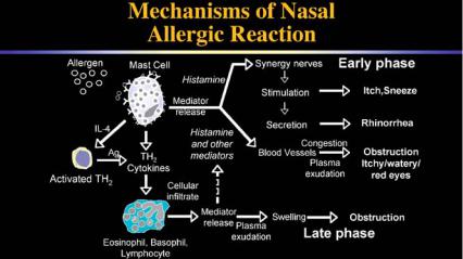
As noted earlier, AR is an immune-mediated nasal disorder that involves an inflammatory response of the nasal and sinus mucosa (Fig. 8.2). While it is primarily mediated by IgE, some evidence exists that may implicate other types of immune mechanisms [9]. When atopic individuals are exposed to antigens, they can become sensitized to those antigens. Antigen-presenting cells (APC), such as macrophages, capture these antigens and process them for presentation to T-helper lymphocytes. This APC–antigen complex binds to the T lymphocyte through a specific receptor and triggers activation of the T cell. Cytokines communicate with other cells of the immune system, including B lymphocytes, and stimulate amplification of the allergic response after sensitization. B lymphocytes are critical in the allergic response as they synthesize IgE antibodies specific for the sensitized antigen. These
Fig. 8.2 Earlyand late-phase allergic response: mediators and symptoms
8 Allergy and Rhinosinusitis |
111 |
IgE antibodies bind to the surface of mast cells and basophils and maintain memory for specific antigens that allows recognition of the antigen on future exposure.
On subsequent exposure, this antigen will cross-link adjacent IgE molecules bound to the surface of mast cells in the nasal mucosa, activating the mast cell through a calcium-dependent process, and allowing both release of preformed inflammatory mediators, such as histamine, and the synthesis of newly formed mediators, such as leukotrienes. Histamine provokes immediate nasal symptoms through prompt binding to histamine-1 (H1) receptors on cells of the nasal mucosa. Within 5 to 10 min after exposure, patients will complain of the four classic symptoms of AR: sneezing, itching, rhinorrhea (nasal discharge), and nasal congestion. While the effects of histamine decline within minutes after release, symptoms of AR are maintained and often worsened by prolonged mediators of inflammation such as the cysteinyl leukotrienes.
The nasal symptoms of AR are often accompanied by nonnasal symptoms as well. Ocular symptoms are common in patients with AR, as are symptoms such as palatal itching and ear fullness. In addition, patients with AR have a higher prevalence of lower respiratory inflammation and asthma than do nonallergic individuals. Nasal symptoms are also frequently exacerbated in patients with concurrent AR and RS.
Allergic Rhinitis: Overview
The term rhinitis refers to an inflammatory disorder of the nasal mucosa. The inflammation that is present in rhinitis can be caused by many different mechanisms, some of them allergic and many of them nonallergic. Nonallergic rhinitis can be stimulated by a variety of triggers, such as respiratory irritants, viral and bacterial infections, medications, and hormonal variations. The mucosal inflammation caused by rhinitis can be mild to severe; severe inflammation can be critical in contributing to the pathogenesis of RS by impairing ventilation and drainage through the sinus ostia.
AR is usually classified in the United States on the basis of its correspondence with well-defined geographic seasons. If AR shows a strong seasonal variability, it is referred to as seasonal allergic rhinitis (SAR), which is attributable to pollens and seasonal variations in mold spores. If AR is present throughout most or all of the year, it is referred to as perennial allergic rhinitis (PAR), which is generally attributable to animal dander, dust mites, other arthropods such as cockroaches, and indoor molds. Many patients have elements of both SAR and PAR, and complain of year-round symptoms with seasonal exacerbations. While this system can often be useful in classifying AR, especially in more temperate climates, it does not explain the nature of symptoms in climates with more frequent or prolonged periods of pollination, such as Southern California.
In patients with SAR, there is a clear association between the pollen counts in a specific region and the development and expression of symptoms. As pollen counts rise seasonally, inflammatory mediators in both the nasal mucosa and the serum
112 |
J.H. Krouse |
begin to rise. The presence of increased antigen loads causes mast cells to release histamine and other mediators, and proinflammatory cytokines increase cellular influx into the nasal tissues. Eosinophils are drawn into the nasal mucosa in AR as in chronic RS and cause tissue inflammation and local injury. As pollen counts decline at the end of the season, inflammation also begins to wane and symptoms become less prominent.
In contrast, among patients with PAR, while there can be fluctuations in symptoms with variations in antigen load throughout the year, symptoms are present at some level on an almost constant basis. Because the antigens are constantly present, wide fluctuations in symptoms are less common with PAR than with SAR. In addition, symptoms such as nasal congestion and postnasal drainage appear to be more common in PAR. Since PAR symptoms overlap with symptoms frequently expressed in chronic RS, diagnostic confusion can often be present. It is important to evaluate if PAR is a comorbidity of chronic RS in a particular individual, and to determine whether symptoms expressed by the patient are caused by allergy alone or by inflammatory changes related to RS.
A separate system that has come into common use outside the United States is a classification model for AR known as ARIA (allergic rhinitis and its impact on asthma). The ARIA guidelines present a system for categorizing AR that is similar conceptually to that used widely for the classification of asthma [10]. In the ARIA guidelines, AR is classified according to the severity of the patient’s symptoms and the time pattern over which they occur. Symptoms that occur throughout the majority of the year on essentially a daily basis are referred to as persistent, while symptoms that occur only a few days a week or only for a few weeks of the year are termed intermittent. Severity of symptoms is classified in the ARIA guidelines by the degree to which they effect the patient’s quality of life, sleep, and daytime function. If the patient’s symptoms are bothersome but do not interrupt sleep or interfere with daytime activities, the severity of AR is considered mild. On the other hand, if the patient’s symptoms cause significant difficulties with sleep, adversely affect daytime function, or bring about impairment in quality of life, the severity of AR is considered moderate-severe. ARIA allows for a consideration of the patient’s AR along four dimensions combining chronicity and severity, with the four classifications being (1) mild intermittent, (2) mild persistent, (3) moderate-severe intermittent, and (4) moderate-severe persistent. These categories are familiar to clinicians who are used to treating patients with asthma, and allow for treatment guidelines based upon an objective analysis of the patient’s symptoms.
Diagnosis of Allergic Rhinitis
History
The most important element of the diagnostic process among patients suspected of having AR is the history. The history will assist in the assessment of whether the patient’s symptoms are truly allergic in their pathophysiology or whether other