

Учебники / Pediatric Sinusitis and Sinus Surgery Younis 2006
.pdfPediatric Endoscopic Sinus Surgery |
185 |
64.Rosenfeld RM. Pilot study of outcomes in pediatric rhinosinusitis. Arch Otolaryngol Head Neck Surg 1995; 121:729–736.
65.Vandenberg SJ, Heatley DG. Efficacy of adenoidectomy in relieving symptoms of chronic sinusitis in children. Arch Otolaryngol Head Neck Surg 1997; 123:675–678.
66.Don DM, Yellon RF, Casselbrant ML, Bluestone CD. Efficacy of a step-wise protocol that includes intravenous antibiotic antibiotic therapy for the management of chronic sinusitis in children and adolescents. Arch Otolaryngol Head Neck Surg 2001; 127(9):1093–1098.
67.Buchman CA, Yellon RF, Bluestone CD. Alternative to endoscopic sinus surgery in the management of pediatric chronic rhinosinusitis refractory to oral antimicrobial therapy. Otolaryngol Head Neck Surg 1999; 120(2):219–224.
68.Tanner SB, Foeler KC. Intravenous antibiotics for chronic rhinosinusitis: are they effective? Curr Opin Otolaryngol Head Neck Surg 2004; 12(1):3–8.
69.Muntz HR, Lusk RP. Nasal antral windows in children: A retrospective study. Laryngoscope 1990; 100:643–646.
70.Stankiewicz JA, Newell DJ, Park AH. Complications of inflammatory diseases of the sinuses. Otolaryngol Clin North Am 1993; 26:629–655.
71.Pransky SW, Low WS. Pediatric ethmoidectomy. Otolaryngol Clin North Am 1996; 29:131–142.
72.Perko D. Endoscopic surgery of the formal sinus without external approach. Rhinology 1989; 27:119–123.
73.Stankiewicz JA. Complications of sinusitis and sinus surgery. In: Bailey B, ed. Otolaryngology-Head Neck Surgery. New York: Mosby CV, 1993.
74.Messerlinger W. Endoscopy of the Nose. Baltimore: Urban & Schwarszenburg, 1978.
75.Kennedy DW, Zinreich SJ, Rosenbum AE, Johns ME. Functional endoscopic sinus surgery: theory and diagnostic evaluation. Arch Otolaryngol 1985; 111(9): 576–582.
76.Draf W. Endoscopy of the paranasal and sinuses. Berlin: Springer-Verlag, 1983.
77.Mendelsohn MG, Gross CW. Soft-tissue shavers in pediatric sinus surgery. Otolaryngol Clin North Am 1997; 30(3):443–449.
78.Parsons DS. Rhinologic uses of powered instrumentation in children beyond sinus surgery. Otolaryngol Clin North Am 1996; 29:105–114.
79.Setliff RC, Parsons DS. The ‘‘Hummer’’: new instrumentation for functional endoscopic sinus surgery. Am J Rhinol 1994; 8:275–278.
80.Postec F, Bossard D, Disant F, Froehlich P. Computer-assisted navigation system in pediatric intranasal surgery. Arch Otoalryngol Head Neck Surg 2002; 128(7):797–800.
81.Hauser R, Westermann B, Probst R. Non-invasive tracking of patient’s head movements during computer-assisted intranasal microscopic surgery. Laryngoscope 1997; 107:491–499.
82.Gunkel AR, Freysinger W, Thumfart WF. Computer-assisted surgery in the frontal maxillary sinus. Laryngoscope 1997; 107:631–633.
83.Fried MP, Kleefield J, Gopal H, Reardon E, Ho BT, Kuhn FA. Image-guided endoscopic surgery. Laryngoscope 1997; 107:594–601.
186 |
Younis |
84.Anon J. Computer-aided endoscopic sinus surgery. Laryngoscope 1998; 108: 949–961.
85.Metson R, Gliklich RE, Cosenza M. A comparison of image guidance systems for sinus surgery. Laryngoscope 1998; 108:1164–1170.
86.Chang PH, Lee LA, Huang CC, Lai CH, Lee TJ. Functional endoscopic sinus surgery in children using a limited approach. Arch Otolaryngol Head Neck Surg 2004; 130(9):1033–1036.
87.Mitchell RB, Pereira KD, Younis RT, Lazar RH. Pediatric functional endoscopic sinus surgery: is a second look necessary? Laryngoscope 1997; 107: 1267–1269.
88.Walner DL, Falciglia M, Willging JP, Myer CM. The role of second-look endoscopy after pediatric functional endoscopic sinus surgery. Arch Otolaryngol Head Neck Surg 1998; 124:425–428.
89.Fakhri S, Manoukian JJ, Souaid JP. Functional endoscopic sinus surgery in the pediatric population: outcome of a conservative approach to postoperative care. J Otolaryngol 2001; 30:15–18.
90.Younis RT. The pros and cons of second-look Sinonasal Endoscopy after endoscopic sinus surgery in children. Arch Otolaryngol Head Neck Surg 2005; 131:1–3.
91.Herbert RL II, Bent JP III. Meta-analysis of outcomes of pediatric functional andoscopic sinus surgery. Laryngoscope 1998; 108(6):796–799.
92.Lusk RP, Muntz HR. Endoscopic sinus surgery in children with chronic sinusitis: a pilot study. Laryngoscope 1990; 100:654–658.
93.Clary RA. Is there a future for pediatric sinus surgery? An American perspective. Int J Pediatr Otorhinolaryngol 2003; 67(suppl 1):S213–S215.
94.Carpenter KM, Graham SM, Smith RJ. Facial skeletal growth after endoscopic sinus surgery in the piglet model. Am J Rhinol 1997; 11(3):211–217.
95.Mair EA, Bolger WE, Breisch EA. Sinus and facial growth after pediatric endoscopic sinus surgery. Arch Otolaryngol Head Neck Surg 1995; 121(5):547–552.
96.Bothwell MR, Piccirillo JF, Lusk RP, Ridenour BD. Long-term outcome of facial growth after functional endoscopic sinus surgery. Otolaryngol Head Neck Surg 2002; 126(6):628–634.
97.Senior B, Wirtschafter A, Mai C, Becker C, Belenky W. Quantative impact of pediatric sinus surgery on facial growth. Laryngoscope 2000; 110(11): 1866–1870.
98.Salvin RG, Cannon RE, Freidman WH, Patitang E, Sundaram M. Sinusitis and bronchial asthma. J All Clin Immunol 1980; 66(3):250–257.
99.Lieser JD, Derkay CS. Pediatric sinusitis: when do we operate? Curr Opin Otolaryngol Head Neck Surg 2005; 13(1):60–66.

12
Image-Guided Pediatric Sinus Surgery
Sam J. Daniel
Department of Otolaryngology, McGill University, Montreal,
Quebec, Canada
INTRODUCTION
Image-guided surgery (IGS) is a new technology that has become an important adjunctive tool in sinus and skull-base surgery. Image-guidance systems rely on preoperative CT and MRI data images to provide the surgeon with a real-time 3D visual localization of the surgical instruments relative to the patient’s anatomy during surgery. Therefore, movements of the surgical instruments are tracked by the system and projected onto the preoperative dataset displayed on the monitor, providing a precise surgical roadmap. The technology continues to progress at a fast pace with many systems currently available, each with its own benefits and drawbacks.
Image guidance can assist the endoscopic head and neck surgeon by confirming the position within distorted or pathological anatomic fields. It also helps identify anatomy landmarks in difficult cases, thus reducing the stress placed on the surgeon and augmenting patients’ safety (1). Undoubtedly, it does not bypass a thorough knowledge of the anatomy and should not confer a false sense of safety. However, it clearly improves surgical accuracy and greatly reduces the risk of major intracranial or intraorbital complications (2). The advantages of IGS are summarized in Table 1 (3).
187
188 |
Daniel |
Table 1 Advantages and Drawbacks of Image-Guided Surgery |
|
|
|
Advantages |
Drawbacks |
|
|
Accurate localization of pathology |
Longer set-up |
Three-dimensional visualization |
Danger of relying on equipment |
Increased patient safety |
Increased radiation exposure with 3D CT |
|
scan |
Simplified complex procedures |
Costly equipment |
Surgeon reassurance |
Danger of overconfidence |
Improved resident teaching |
|
|
|
TECHNOLOGY
Different IGS systems are currently available on the market, each with its own advantages, drawbacks, and limitations. Tracking systems can be classified into two types based on the type of signal they rely on for localization of instruments. These can be either electromagnetic, such as radiofrequency signals, or optical, such as infrared signals. Most currently available systems are optical-based and require that the patients wear special headsets during surgery in order to monitor head position (4). Electromagnetic systems also require headsets to be worn during the preoperative CT scan.
System Components
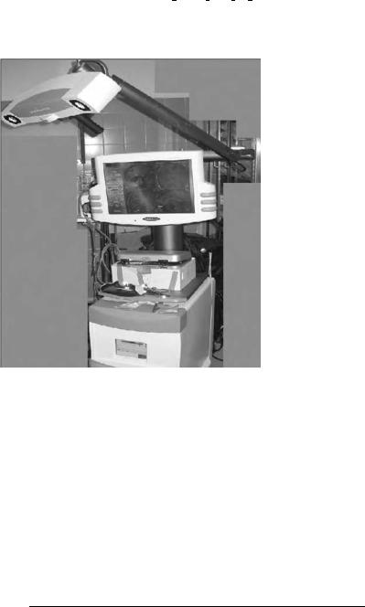
Standard IGS systems are made-up of a computer workstation, a display monitor, an image-processing software, a localization system, and specialized instrumentation that can be tracked digitally (Fig. 1).
Tracking Systems
Although both optical and electromagnetic image-guidance systems have proven to be valuable, individual preferences vary based on differences in design and operation. The system utilized at the Montreal Children’s Hospital is Vector Vision from Brainlab (Fig. 1). It consists of a powerful computer system, a touch screen monitor, and two cameras which emit infrared signals that determine the patient’s position in three dimensions as well as the position of the surgical instruments in relation to the patient’s preoperative CT or MR images. The image data is typically displayed in multiplanar format with options of videoscopic feeds.
The infrared cameras detect the reflective markers attached to the patient’s body and transmit the continuously collected data to the system. The system then calculates a 3D representation of the patient’s anatomy based on diagnostic data. During the operation, the surgeon can follow the movements of his or her instruments on the computer screen in real time. Other tracking systems available on the market are listed in Table 2.
0-8247-2881-5 Younis Ch12 R2 081705
Pediatric Sinus Surgery |
189 |
Figure 1 The standard image-guided surgical system includes a computer workstation, a display monitor, and a tracking system. This tracking system utilizes infrared cameras (Vector Vision, Brainlab).
Optical Systems
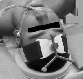
Optical-based systems rely on infrared light for the tracking of surgical instruments. Optical sensors are placed on a headframe worn by the patient (Fig. 2). There are two types of optical systems, referred to respectively as active or passive. Active optical systems use a camera to track the position of flashing light-emitting diodes attached to the instruments and headframe. Passive optical tracking systems detect the reflection of infrared
Table 2 Tracking Systems Most Commonly Utilized in Sinus Surgery
Digitizer |
Product’s name |
Company |
|
|
|
Electromagnetic |
Insta Trak |
GE Medical |
Optical |
Vector Vision |
Brainlab |
Optical |
LandmarX |
Medtronic |
Optical |
Stryker Navigation |
Stryker Corporation |
|
|
|
190 |
Daniel |
Figure 2 Headband worn by the patient during surgery. The reflective spheres have a known geometric configuration and their reflected infrared light is tracked by cameras, allowing the system to localize the patient’s head throughout the procedure.
light from reflective markers placed on cordless instruments (no need for cable attachment). In both types, line-of-sight must be maintained between the system components to allow tracking.
Electromagnetic Systems
Electromagnetic tracking systems use electromagnetic sensors to detect variations in the electromagnetic field caused by instrument or patient movement. They have a transmitter located near the surgical field and a receiver placed on the surgical instrument. A brief comparison between electromagnetic and optical systems is provided in Table 3.
Patient Registration
Registration is the process of matching preoperative images to the 3D space occupied by the patient during surgery. This allows accurate localization of the surgical tools within the surgical volume of interest (3,5). In fact, the registration process is a major factor in the accuracy of image-guided systems. There is a steep learning curve but set up and registration time do decrease over time (4,6). A synopsis of the main types of registration techniques currently in use is presented in Table 4.

Pediatric Sinus Surgery |
191 |
Table 3 Comparison of Electromagnetic and Optical Systems |
|
|
|
Electromagnetic |
Optical |
|
|
Relatively less expensive |
More expensive |
Does not require line-of-sight between |
Requires a clear line-of-sight |
transmitter and receiver |
between transmitter and receiver |
Can have signal interference from metallic |
Can have signal interference from the |
objects in the operating field |
operating room lights |
Requires specialized headsets to be |
Requires specialized headsets to be |
worn by patients during surgery |
worn by patients during surgery |
|
|
Table 4 Summary of Registration Techniques
Anatomic fiducial registration |
Anatomic landmarks such as tragus, |
|
nasion, menton, medical, and lateral |
|
canthus are selected on the patient. |
Extrinsic markers |
Skin-affixed fiducial markers are attached |
|
before CT scanning and are left until |
|
registration is complete. |
Surface mapping registration |
Multiple surface data points are gathered |
|
from the patient’s face using a laser |
|
device. The computer matches the |
|
patient’s facial contour to the |
|
preoperative image dataset. |
Autoregistration |
A special headframe provides a fixed |
|
arrangement of registration markers |
|
relative to the patient’s head. The |
|
patient must wear this frame at the |
|
time of the CT and in the OR during |
|
surgery. |
|
|
USE OF IGS IN PEDIATRIC OTOLARYNGOLOGY
Introduction
Intraoperative use of IGS is not considered standard care in all cases of sinus or skull-base surgery. However, it has undeniable value in many selected clinical settings as it assists the surgeon in confirming the position of normal or distorted anatomical structures as well as pathology. The American Academy of Otolaryngology-Head and Neck Surgery (AAO-HNS), in its policy statement on intraoperative use of computer-aided surgery, lists some examples of indications for image-guided sinus surgery; these are summarized in Table 5 (7). While this list is not exhaustive, usefulness of IGS in

192 |
Daniel |
Table 5 AAO-HNS Policy on Intraoperative Use of Computer-Aided Surgery
Revision sinus surgery
Distorted sinus anatomy of development, postoperative, or traumatic origin Extensive sinonasal polyposis
Pathologic conditions involving the frontal, posterior ethmoid, or sphenoid sinuses Disease abutting the skull base, orbit, optic nerve, or carotid artery
Cerebrospinal fluid rhinorrhea or conditions where there is a skull-base defect Benign and malignant sinonasal neoplasms
Source: From Ref. 7.
many of the indications listed in the table will be demonstrated in the next section using clinical case scenarios with radiological images.
Revision Sinus Cases
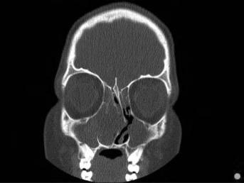
One of the principal uses of image guidance is in revision cases where the landmarks have been altered as in resected middle turbinates or distorted skull-base anatomy. This is particularly true in revision cases with extensive sinonasal polyposis and in children with cystic fibrosis. The following case of a 15-year-old girl with severe cystic fibrosis who has had two previous sinus operations is illustrative. She presented three years following her second operation with severe sinus symptoms and fever unresponsive to antibiotics. Flexible endoscopy and imaging revealed extensive polyposis and inflammation with shifting of the nasal septum and extension of the disease to the skull base. A representative CT scan section is shown below (Fig. 3). IGS was performed with complete eradication of the disease.
Disease Abutting Important Structures
Image guidance can enhance patient’s safety when used to confirm the anatomy or disease along risky areas. This includes the skull base, particularly along the superomedial portion of the ethmoid sinus near the cribriform plate attachment. Other problematic areas include the periorbital area and the frontal sinus, since the angles of approach and variability of this sinus can make the surgery very challenging. The sphenoethmoid recess can also be difficult to identify at times (8). Image guidance can be particularly useful in confirming the position of the anterior face of the sphenoid as well as vital structures in close proximity or within the sinus such as the carotid arteries, the cavernous venous sinuses, and the optic nerve. The latter can sometimes be found within an Onodi cell. The 3D orientation with coronal, axial, and sagittal sections simultaneously correlated in real time can be extremely helpful as illustrated in Figure 4.
Pediatric Sinus Surgery |
193 |
Figure 3 A 15-year-old girl suffering from fibrosis with severe disease recurrence following two previous surgeries. Note the extensive pathology deviating and fracturing the nasal septum.
Figure 5 is the MRI of a 15-year-old boy with pan-sinusitis and sphenoid sinus mucocele abutting the carotid arteries. Notice the compression of the right carotid artery (white arrow) by the mucocele, as well as the dural enhancement on that side. Image guidance was used to confirm the position of the carotid arteries during resection.
Image guidance can also be very helpful in cases of periorbital/orbital abscesses drained endoscopically, as well as in optic nerve decompression (Fig. 6).
Extensive Sinonasal or Skull-base Tumors
Image-guided surgery is extremely useful in extensive sinonasal and skullbase tumors. Again, IGS should be used as an adjunctive tool to confirm anatomy and/or disease, not to seek it. Benign or malignant tumors invading, distorting, or abutting important or vital structures can now be accessible endoscopically in a better and safer way. The following clinical cases are illustrative (Figs. 7–9).
Clinical case 1
An 18-year-old girl presented with acute visual loss and a one-month history of left eye disformat, nasal congestion, and frontal headache. She

194 |
Daniel |
Figure 4 A 16-year-old boy with neurofibroma of the sphenoid sinus resected under image guidance. The white wand represents the surgical instrument pointing to the anterior face of the sphenoid. The usefulness of the 3D orientation with the coronal, axial, and sagittal sections can be appreciated.
was diagnosed with bilateral retinoblastoma as an infant, and was treated with right eye enucleation and left eye radiotherapy. Figure 7 shows representative MRI sections demostrating a large tumor involving the left maxillary sinus and the orbit with skull-base and intracranial invasion. Pathology revealed an aggressive sarcoma.
Figure 5 A 15-year-old boy with right pan-sinusitis and sphenoid sinus mucocele.
White arrow is pointing to compressed right carotid artery.