

Human_Histology
.pdf218 Textbook of Human Histology
PLATE 18.1:
!" #
" $" % &
Key
'& "(& )*& + & +
B
General Structure of Seminiferous Tubules
Seminiferous tubules are highly convoluted structures present in each lobule of the testes. It has been estimated that each testis has about 200 lobules, and that each lobule has one to three seminiferous tubules. The total number of tubules is between 400 and 600.
When stretched out each tubule is 70–80 cm in length. It has a diameter of about 150 μm. The combined length of all seminiferous tubules in one testis is between 300
and 900 meters. These tubules are lined by cells that are concerned with the production of spermatozoa.
Within a lobule, the spaces between seminiferous tubules are filled by very loose connective tissue, containing blood vessels and lymphatics. Interstitial cells of Leydig are also present here. The wall of each tubule is made up of an outer layer of fibrous tissue that also contains musclelike (myoid) cells. Contractions of these cells probably help to move spermatozoa along the tubule.

Chapter 18 Male Reproductive System 219
PLATE 18.2:
B
, |
|
|
& - |
|
|
|
& |
|
$" % $
%
.
/
0# & 1 "
) 0&
) 0$ %&
Note: ) # & 23 0
3 &
Key
'& " $" % ( 4 *& "
5& "
6& "
7& " #
8& )
Between this connective tissue and the lumen of the tubule there are several layers of cells. The cells rest on a basal lamina. They are of various shapes and sizes. Most of the cells represent stages in the formation of spermatozoa: they are referred to as germ cells. Other cells that have a supporting function are called sustentacular cells or the cells of Sertoli (Plate 18.2).
Note: The appearance of the cellular lining of the seminiferous tubules is characteristic, and a student who has studied sections through them carefully (even at low magnification) is not likely to mistake the seminiferous tubules for anything else. The points to note are (a) the many layers of cells; (b) the great variety in size and shape of the cells and of their nuclei; (c) the lack of a well defined margin of the lumen; and (d) inconspicuous cell boundaries.

220Textbook of Human Histology
Spermatogenic Cells (Germ Cells)
Spermatogenic cells represent the various stages in the formation of spermatozoa. They are arranged in developmentally higher order from the basal lamina to the lumen, namely spermatogonia, spermatocytes, spermatids and spermatozoa (Plate 18.2).
However, all types of cells are not seen in any one part of the seminiferous tubule at a given time.
In a given segment of the tubule there is a gradual change in the type of cells encountered (with passage of time). Six phases have been recognized. At a given point of time different segments of a seminiferous tubule show cell patterns corresponding to the six phases.
It has, therefore, been suggested that over a period of time waves of maturation (of germ cells) pass along the length of a seminiferous tubule, this is referred to as spermatogenic cycle.
Sustentacular Cells or Cells of Sertoli (Fig. 18.3)
These are tall, slender cells having an irregularly pyramidal or columnar shape. The nucleus lies near the base of the cell. It is light staining and is of irregular shape. There is a prominent nucleolus. The base of each sustentacular cell rests on the basement membrane, spermatogonia being interposed amongst the sustentacular cells. The apex of the sustentacular cell reaches the lumen of the seminiferous tubule. Numerous spermatids, at various stages of differentiation into spermatozoa, appear to be embedded in the apical part of the cytoplasm (Fig. 18.3 and Plate 18.2). Near the basement membrane spermatocytes and spermatogonia indent the sustentacular cell cytoplasm.
With the EM it is seen that the sides and apices of these cells are marked by recesses that are occupied by spermatogonia, spermatocytes, and spermatids. However, there is no cytoplasmic continuity between these cells and the sustentacular cell. On the basis of light microscopic studies some workers were of the view that the sustentacular cells formed a syncytium. However, EM studies have shown that the cells are distinct.
The plasma membranes of adjoining sustentacular cells are connected by tight junctions that divide the wall of the seminiferous tubule into two compartments, superficial (or adluminal) and deep (abluminal). The deep compartment contains spermatogonia (and preleptotene spermatocytes) and the superficial compartment contains other stages of spermatogenesis.
The two compartments are believed to be separated by a blood-testis barrier. Sustentacular cells contain abundant mitochondria, endoplasmic reticulum, and other
Fig. 18.3: A sustentacular cell and some related germ cells
(Schematic representation)
organelles. Microfilaments and microtubules form a cytoskeleton that appears to be important in cohesive functions of these cells.
In the adult testis sustentacular cells are less prominent than germ cells. They are more prominent than germ cells before puberty, and in old age.
Functions
They provide physical support to germ cells, and provide them with nutrients. Waste products from germ cells are transferred to blood or lymph through them.
They phagocytose residual cytoplasm that remains after conversion of spermatids to spermatozoa.
They probably secrete fluid that helps to move spermatozoa along the seminiferous tubules. This fluid is rich in testosterone that may stimulate activity of cells lining the epididymis.
Sertoli cells produce a number of hormones. In the eighth month of fetal life they secrete a hormone (Mullerian inhibitory substance or MIS) that suppresses development of the paramesonephric (Mullerian) ducts (in male fetuses). They are also believed to produce a substance that inhibits spermatogenesis before puberty.
In the male adult, Sertoli cells produce androgen binding protein (ABP) which binds to testosterone (and hydroxytestosterone) making these available in high concentration to germ cells within seminiferous tubules. Secretion of ABP is influenced by FSH. A hormone inhibin produced by Sertoli cells inhibits production of FSH (Flowchart 18.1).
Interstitial Cells
The interstitial cells (of Leydig) are large, round or polyhedral cells lying in the connective tissue that intervenes between the coils of seminiferous tubules (Plate 18.2A). Their nuclei are eccentric. The cytoplasm stains lightly and

!" !# Scheme to show control of male genital system
often has a foamy appearance (because of the removal of lipids during processing of tissues). It contains yellow granules that are seen by EM to be vacuoles containing various enzymes. Rod-shaped crystalloids (Reinke’s crystalloids) are also present in the cytoplasm. Much agranular endoplasmic reticulum is present. Yellow-brown pigment (lipofuscin) is seen in some cells.
Interstitial cells secrete male sex hormone (testicular androgens). Secretion is stimulated by the interstitial cell stimulating hormone of the hypophysis cerebri. (This hormone is identical with the luteinising hormone present in the female).
Some interstitial cells may be present in the mediastinum testis, in the epididymis, or even in the spermatic cord.
Apart from interstitial cells, the interstitial tissue contains collagen fibers, fibroblasts, macrophages, mast cells, blood vessels, and lymphatics.
Structure of Rete Testis and Efferent Ductules
The rete testis consists of anastomosing tubules that are lined by flattened or cuboidal cells. They bear microvilli. The epithelium is surrounded by connective tissue of the mediastinum testis.
The efferent ductules are lined by ciliated columnar epithelium. Some non-ciliated cells bearing microvilli are also present. The tubules have some smooth muscle in their walls. Movement of spermatozoa through the tubules is facilitated by ciliary action, and by peristaltic contraction of smooth muscle.
Chapter 18 Male Reproductive System 221
!"$# Spermatogenesis
Pathological Correlation
Tumors of Testis
Testicular tumors can arise from germ cells (Germ cell tumors); stroma (sex cord stromal tumors) or both (combined germ cell-sex cord stromal tumors). Examples of germ cell tumors are seminoma, embryonal carcinoma, teratoma, and choriocarcinoma; sex cord stromal tumors are Leydig cell tumor, Sertoli cell tumor and granulosa cell tumor. Gonadoblastoma is an example of combined germ cell-sex cord-stromal tumors.
Germ cell tumors comprise approximately 95% of all testicular tumors and are more frequent before the age of 45 years. Testicular germ cell tumors are almost always malignant.
SPERMATOGENESIS
The process of the formation of spermatozoa is called spermatogenesis (Flowchart 17.2). This process occurs in waves along the length of seminiferous tubules taking about 64 days to complete. It consists of several stages as described below.

222 Textbook of Human Histology
Fig. 18.4: Some stages in spermatogenesis as seen in the walls of seminiferous tubules (Schematic representation)
Stages of Spermatogenesis
The stem cells from which all stages in the development of spermatozoa are derived are called spermatogonia. These cells lie near the basal lamina. Spermatogonia undergo several mitotic divisions and, because of this, spermatogonia of varied structure are seen in the wall of a seminiferous tubule. These mitoses give rise to more spermatogonia, and to primary spermatocytes.
Note: Three main types of spermatogonia are described: dark type-A (AD), light (or pale) type-A (AP), and type-B (Fig. 18.4). In type A spermatogonia (dark and light) the nuclei are oval and possess nucleoli that are eccentric, and are attached to the nuclear membrane. The terms dark and light, applied to these cells, refer to the intensity of staining of the nuclei. In type B spermatogonia th e nuclei are spherical. Each nucleus has a spherical nucleolus.
Dark type A spermatogonia (also called type A1) represent a reserve of resting stem cells. They divide to form more dark type A cells and also some light type A cells (or A2 cells). Light type A spermatogonia divide to form more light type A spermatogonia, and also some spermatogonia of type B. Each type B spermatogonium divides several times (probably four times in man). The resulting cells are designated B1, B2, B3, etc. Each of the resulting cells divides to form two primary spermatocytes.
Primary spermatocytes are formed by mitotic division of spermatogonia. These are largest cells with large spherical nuclei occupying the middle region of the seminiferous epithelium. Each primary spermatocyte
undergoes meiosis to give rise to two secondary spermatocytes (see below). This is the first meiotic division in which the number of chromosomes is reduced to half. (Each primary spermatocyte has 46 chromosomes, whereas each secondary spermatocyte has only 23).
The prophase of the first meiotic division is prolonged and passes through several stages (leptotene, zygotene, pachytene, diplotene) in which the appearance of the nucleus shows considerable alterations. As a result, primary spermatocytes at various stages of prophase (and subsequent stages of division) can be recognized in the wall of a seminiferous tubule (Fig. 18.4).
Secondary spermatocytes are smaller than primary spermatocytes, and so are their nuclei. Each secondary spermatocyte has the haploid number of chromosomes (i.e. 23). It divides to form two spermatids (see below). This is the second meiotic division and this time there is no further reduction in chromosome number. They are scarcely seen in sections as they undergo second meiotic division as soon as they are formed.
Each spermatid is a rounded cell with a spherical nucleus. Both cell and nucleus are much smaller than in the case of spermatogonia or spermatocytes. They lie in groups in association with Sertoli cells. The spermatid undergoes changes in shape, and in the orientation of its organelles, to form a spermatozoon. This process is called spermiogenesis.
% &
' ( )* + ') ( nt
We have seen that a typical cell contains 46 chromosomes, this being referred to as the diploid number. Spermatozoa (or ova) have only half this number, i.e. 23 which is the haploid number. At fertilization, the diploid number is restored, the zygote receiving 23 chromosomes from the ovum, and 23 from the sperm.
A primary spermatocyte contains the diploid number number is halved so that secondary spermatocytes have the haploid number (23).
Now let us look at these facts in relation to DNA content, rather than chromosome number. Let us designate the DNA content of a gamete (sperm or ovum) as n. The DNA content of a zygote formed as a result of fertilization is, therefore, n + n = 2n. Before the zygote can undergo division its DNA has to be replicated. In other words it has to become 4n, of which 2n goes to each daughter cell.
have2n DNA. After replication this becomes 4n meiotic division 2n goes to each secondary spermatocyte. The point to note is that although the chromosome number in a secondary spermatocyte is haploid, DNA is 2n. There is no replication of DNAin secondary spermatocytes.As a result the DNA content of spermatids formed during the second meiotic division is n. Therefore, note that although there is no reduction in chromosome number during the second meiotic division, DNA content is reduced from 2n to n.
Spermiogenesis
The process by which a spermatid becomes a spermatozoon is called spermiogenesis (or spermateleosis). The spermatid is a more or less circular cell containing a nucleus, Golgi complex, centriole and mitochondria. All these components take part in forming the spermatozoon.
The nucleus undergoes condensation and changes shape to form the head. The Golgi complex is transformed into the acrosomic cap that comes to lie over one side of the nucleus. The acrosome marks the future anterior pole of the spermatozoon. The centriole divides into two parts that are at first close together. They migrate to the pole of the cell that is away from the acrosome. The axial filament grows out of the distal centriole. The region occupied by the two centrioles later becomes the neck of the spermatozoon. The proximal centriole probably forms the basal body.
The part of the axial filament between the head and the annulus becomes surrounded by mitochondria, and together with them forms the middle piece. Most of the cytoplasm of the spermatid is shed, and is phagocytosed by Sertoli cells. The cell membrane persists as a covering for the spermatozoon.
Chapter 18 Male Reproductive System 223
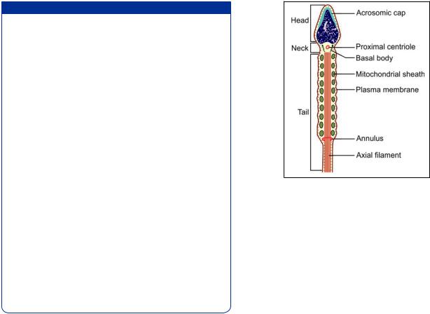
Fig. 18.5: Structure of spermatozoon as seen by electron microscope (Schematic representation)
Structure of a Mature Spermatozoon
The spermatozoa are motile male gametes. With their tails projecting into the lumen of seminiferous tubules, they are found in close association with the sertoli cells.
The spermatozoon has a head, a neck, and a principal piece or tail. The tail is made of three pieces i.e. middle piece, principal piece, and end piece.
The head is covered by a cap called the acrosomic cap, anterior nuclear cap, or galea capitis (Fig. 18.5). It is flattened from before backwards so that it is oval when seen from the front, but appears to be pointed (somewhat like a spear-head) when seen from one side, or in section. It consists of chromatin (mostly DNA) that is extremely condensed and, therefore, appears to have a homogeneous structure even when examined by EM. This condensation makes it highly resistant to various physical stresses.
Note: The acrosome is made up of glycoprotein. It can be regarded as a large lysosome containing numerous enzymes (proteases, acid phosphatase, neuraminidase, hyaluronidase).
The neck of the spermatozoon is narrow. It contains a funnel-shaped basal body and a spherical centriole.
The chief structure to be seen in the neck is the basal body. It is also called the connecting piece because it helps to establish an intimate union between the head and the remainder of the spermatozoon. The basal body is made up of nine segmented rod-like structures each of which is continuous distally with one coarse fiber of the axial filament. On its proximal side (i.e. towards the head of the spermatozoon) the basal body has a convex articular surface that fits into a depression (called the implantation fossa) present in the head.

224 Textbook of Human Histology
Fig. 18.6: Transverse section across the tail of a spermatozoon to
! "
An axial filament (or axoneme) begins just behind the centriole. It passes through the middle piece and extends into the tail. At the point where the middle piece joins the tail, this axial filament passes through a ring-like annulus. That part of the axial filament that lies in the middle piece is surrounded by a spiral sheath made up of mitochondria.
The axial filament is really composed of several fibrils arranged as illustrated in Figure 18.6. There is a pair of central fibrils, surrounded by nine pairs (or doublets) arranged in a circle around the central pair. (This arrangement of one central pair of fibrils surrounded by nine doublets is similar to that seen in cilia).
In addition to these doublets there are nine coarser petal-shaped fibrils of unequal size, one such fibril lying just outside each doublet. These coarse fibrils are present in the middle piece and most of the tail, but do not extend into the terminal part of the tail. The whole system of fibrils is kept in position by a series of coverings. Immediately outside the fibrils there is a fibrous sheath. In the region of the middle piece the fibrous sheath is surrounded by spirally arranged mitochondria. Finally, the entire sperm is enclosed in a plasma membrane.
From Figure 18.6 it will be seen that one of the coarse fibrils is larger than the others. This is called fibril 1, the others being numbered in a clockwise direction from it. The fibrous sheath is adherent to fibrils 3 and 8. The line joining fibrils 3 and 8 divides the tail into a major compartment containing 4 fibrils and a minor compartment containing 3 fibrils. This line also passes through both the central fibrils and provides an axis in reference to which sperm movements can be analyzed. The part of tail connected to neck is middle piece. The middle piece contains the mitochondrial sheath and provides energy for sperm maturation.
Principal piece contains the 9 + 2 pattern of microtubules in the central core surrounded by 9 coarse fibers enclosed in a fibrous sheath (Fig. 18.6). End piece contains 9 + 2 axonema enclosed by plasma membrane
Maturation and Capacitation of Spermatozoa
Maturation
As fully formed spermatozoa pass through the male genital passages they undergo a process of maturation. Spermatozoa acquire some motility only after passing through the epididymis. The secretions of the epididymis, seminal vesicles and the prostate have a stimulating effect on sperm motility, but spermatozoa become fully motile only after ejaculation.
When introduced into the vagina, spermatozoa reach the uterine tubes much sooner than their own motility would allow, suggesting that contractions of uterine and tubal musculature exert a sucking effect.
Capacitation
Spermatozoa acquire the ability to fertilize the ovum only after they have been in the female genital tract for some time. This final step in their maturation is called capacitation. During capacitation some proteins and glycoproteins are removed from the plasma membrane overlying the acrosome.
When the sperm reaches near the ovum, changes take place in membranes over the acrosome and enable release of lysosomal enzymes present within the acrosome. This is called the acrosome reaction. The substances released include hyaluronidase that helps in separating corona radiata cells present over the ovum. Trypsinlike substances and a substance called acrosin, help in digesting the zona pellucida and penetration of the sperm through it. Changes in the properties of the zona pellucida constitute the zona reaction.
ACCESSORY UROGENITAL ORGANS
EPIDIDYMIS
Epididymis is a comma shaped structure present on the postero-lateral aspect of testis. Structurally, the epididymis consists of a head and a duct. The body and tail of the epididymis are made up of the duct of the epididymis, that is greatly coiled on itself. The head is formed by highly convoluted continuations of the efferent ductules. These are lined by ciliated columnar epithelium (Plate 18.3). At the lower end of the head of the epididymis these tubules join to form a single tube called the duct of the epididymis.

Chapter 18 Male Reproductive System 225
PLATE 18.3: ,-
(
- (
B
&
"
! # &
Key
'& .(& " #*& "
The duct is lined by pseudostratified columnar epithelium which is made of 2 types of cells tall columnar cells, and shorter basal cells that do not reach the lumen.
The luminal surface of each columnar cell bears nonmotile projections that resemble cilia. These stereocilia are seen by EM to be thick microvilli. They do not have the structure of true cilia. The EM also shows the presence of agranular endoplasmic reticulum, lysosomes and a prominent Golgi complex in these cells. The basal cells are precursors of the tall cells. Beneath the epithelium there is a layer of circularly arranged smooth muscle fibers. This muscle layer increases in thickness gradually from head to tail and may be organized into inner circular and outer longitudinal layers in the tail region.
Functions
Phagocytosis of defective spermatozoa.
Absorption of excess fluid.
Secretion of substances (sialic acid, glyceryl-phosphoryl- choline) that play a role in maturation of spermatozoa.
DUCTUS DEFERENS
The ductus deferens (deferent duct or vas deferens) is a muscular tube extending from the lower end of epididymis to the prostatic urethra. The wall of the ductus deferens consists (from inside out) of:
Mucous membrane
Muscle
Connective tissue (Plate 18.4).

226 Textbook of Human Histology
PLATE 18.4: '* * ' &
9
/&
2 &
Key
'& "
|
|
|
|
(& |
|
|
|
*& 9 |
|
|
|
|
& ) |
|
|
|
+& 9 |
||
|
!& 2 |
|
|
5&
B
'* * ' &
Mucous Membrane
The mucous membrane shows a number of longitudinal folds so that the lumen appears to be stellate in section. The lining epithelium is simple columnar, but becomes pseudostratified columnar in the distal part of the duct. The cells are ciliated in the extra-abdominal part of the duct. The epithelium is supported by a lamina propria in which there are many elastic fibers.
Muscle
The muscle coat is very thick and consists of smooth muscle. It is arranged in the form of an inner circular layer and an outer longitudinal layer. An inner longitudinal layer is present in the proximal part of the duct.
Connective Tissue
The fibroelastic connective tissue forms the adventitial layer containing blood vessels and nerves.

Chapter 18 Male Reproductive System 227
PLATE 18.5: / ;
/
&
Key
'& :(& 9*&
B
/ <
The terminal dilated part of the ductus deferens is called the ampulla, which joints the duct of seminal vesicles to form ejaculatory duct. It has the same structure as that of the seminal vesicle.
THE SEMINAL VESICLE
The seminal vesicle is a sac-like mass that is really a convoluted tube. The tube is cut several times in any section (Plate 18.5). The wall of the seminal vesicle (from inside out) consists of:
Mucous membrane
Muscle
Connective tissue.
Mucous Membrane
The mucosal lining is thrown into numerous thin folds that branch and anastomose thus forming a network. The lining epithelium is simple columnar, or pseudostratified. Goblet cells are present in the epithelium.