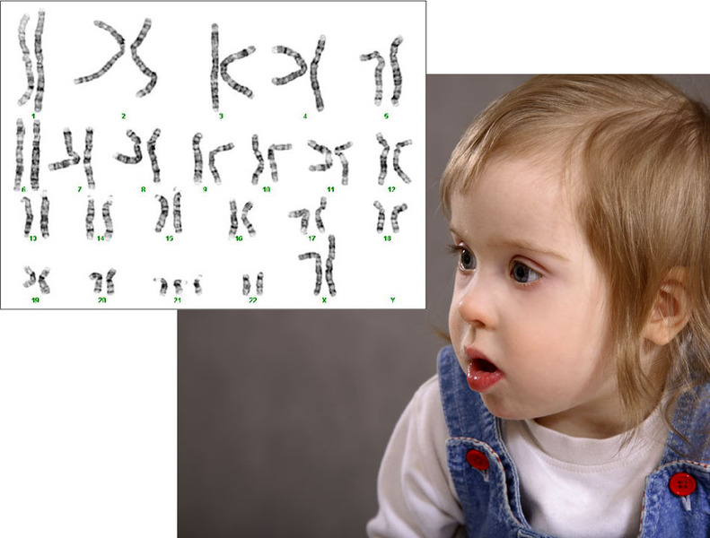
How do the extra genes lead to Down syndrome?
Exactly how the extra genes from chromosome 21 lead to Down syndrome is still not clear. Scientists believe that the increased presence of specific genes alters the interaction between these and other genes. Some genes will become more active and others less active than normal, leading to changes in the development and maintenance of the body. Why some individuals are more severely affected than others might have to do with how many and which specific extra genes were inherited.
Scientists are trying to find out which genes from chromosome 21, when present in three copies, are responsible for the different characteristics of Down syndrome. Currently, about 400 genes on chromosome 21 have been identified, but the functions of most are still unknown. Through human studies and animal models, scientists are making progress in understanding the functions of individual genes.
What are the risk factors for conceiving a child with Down syndrome?
The only well known risk factor for conceiving a child with Down syndrome is advanced maternal age. The older the woman is at conception, the greater the risk of having a child with Down syndrome.
Mother's age at conception Risk of Down syndrome
25 years 1 in 1,250
30 years 1 in 1,000
35 years 1 in 400
40 years 1 in 100
45 years 1 in 30
Parents who have conceived a child with Down syndrome have a 1% increased risk of conceiving another child with Down syndrome. If a parent is a carrier of a chromosome 21 translocation, the risk can be as high as 100%.
Women with Down syndrome have a 50% risk of conceiving a child with Down syndrome. If the father has Down syndrome, the risk of conceiving a child with Down syndrome is also increased.
What are the characteristic features and symptoms of Down syndrome?
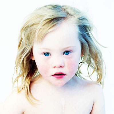
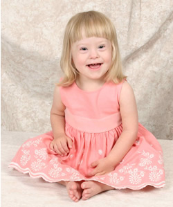
Although the severity of Down syndrome ranges from mild to severe, most individuals with Down syndrome have widely recognizable physical characteristics. These include:
a flattened face and nose, a short neck, a small mouth sometimes with a large, protruding tongue, small ears, upward slanting eyes that may have small skin folds at the inner corner (epicanthal fold);
white spots (also known as Brushfield spots) may be present on the colored part of the eye (iris);
the hands are short and broad with short fingers, and with a single crease in the palm;
poor muscle tone and loose ligaments are also common; and
development and growth is usually delayed and often average height and developmental milestones are not reached.
What type of prenatal screening is available for Down syndrome?
Several noninvasive screening options are offered to parents. If Down syndrome is suspected due to the screening outcome, a formal diagnosis can be made before the baby arrives. This gives parents time to gather information about Down syndrome before their baby is born and to make arrangements in case of medical complications.
Prenatal screening tests currently available include the expanded alpha-fetoprotein (AFP) screening test, the nuchal translucency test, and additional ultrasound screens which look for changes in certain anatomical features of the fetus. While these screening tests can assess the risk for Down syndrome, they cannot confirm Down syndrome with certainty.
The most widely used screening test is the AFP. Between weeks 15 and 20 of pregnancy, a small blood sample is taken from the mother and examined. The levels of AFP and three hormones called unconjugated estriol, human chorionic gonadotropin, and inhibin-A are measured in the blood sample. If the AFP and hormone levels are altered, Down syndrome can be suspected, but not confirmed. Likewise, a normal test result does not rule out Down syndrome.
The nuchal translucency test measures the thickness of the fold in the neck via ultrasound. This test can be done between 11 and 13 weeks of pregnancy. In combination with the mother's age, this test identifies about 80% of Down syndrome fetuses.
Women considered at high risk (advanced maternal age, positive AFP test, or a history of a previous child with Down syndrome) may benefit from additional ultrasound scans between 18 and 22 weeks of pregnancy. When certain anatomical features are altered, absent, or present in a fetus, it may indicate Down syndrome. Some of the markers that are examined include:
the length of the long arm (humerus) or leg bone (femur),
the length of the nasal bridge,
the size of the renal pelvis (hypoplasia, pyelectasis),
small bright spots in the heart (echogenic intracardiac foci),
small middle section of the little finger (hypoplastic fifth digit),
a large gap between the first and second toe,
increased brightness of the bowel (echogenic bowel), and
pelvic bone angle (widened iliac angle).