

Атлас по рентгенологии травмированных собак и кошек / an-atlas-of-radiology-of-the-traumatized-dog-and-cat
.pdf222 Radiology of Abdominal Trauma
Case 3.13
3
Signalment/History: “Tai Chi” was a 5-year-old, male Pekingese, who had been absent from home for several days. Upon return, the owners noted he was vomiting and then became anorectic. No stool had been passed.
Physical examination: Scrotal swelling was evident on physical examination with the exact nature of the scrotal contents not determined.
Radiographic procedure: Caudal radiographs were made in an effort to more fully evaluate the nature of a suspected hernia.
Radiographic diagnosis: Multiple, small, well-circum- scribed inguinal gas shadows were thought to be small bowel gas patterns, in which case an inguinal hernia was present (arrows). The pelvis was difficult to evaluate properly because of patient positioning.
Treatment/Management: Exploration of the suspected inguinal hernia revealed an incarcerated distal jejunum that required an intestinal anastomosis. An infarcted right testicle was removed surgically.
Comments: Herniated bowel loops cannot be considered a trivial lesion. Less likely etiologies for such an inguinal gas collection include the presence of a gas-producing organism causing an infectious lesion or a break in the skin permitting the entrance of subcutaneous air.

Inguinal hernias 223
3

224 Radiology of Abdominal Trauma
Case 3.14
3
Signalment/History: “Toot” was a 5-year-old, male DSH cat that was presented to the clinic following suspected trauma.
Physical examination: An inguinal hernia containing easily palpated bowel loops was found. Identification of the urinary bladder was questionable.
Radiographic procedure: Routine studies of the abdomen failed to identify the location or status of the urinary bladder, so a retrograde contrast study was performed.
Radiographic diagnosis (abdomen): The left-sided inguinal hernia contained multiple gas-filled, small bowel loops. The luminal diameter of the bowel loops was thought to be within normal limits (<11cm) and did not suggest bowel obstruction. The urinary bladder could not be identified on the noncontrast study. Left femoral head and neck fractures were seen.

Inguinal hernias 225
3
Retrograde cystogram
Radiographic diagnosis (retrograde cystogram): This showed the displaced and ruptured urinary bladder lying within the hernial sac. The bladder was partially filled and lay just ventral to the abdominal wall. The majority of the contrast agent spilled into the hernial sac.
Treatment/Management: Treatment was not permitted and the cat was euthanized.

226 Radiology of Abdominal Trauma
Case 3.15
3
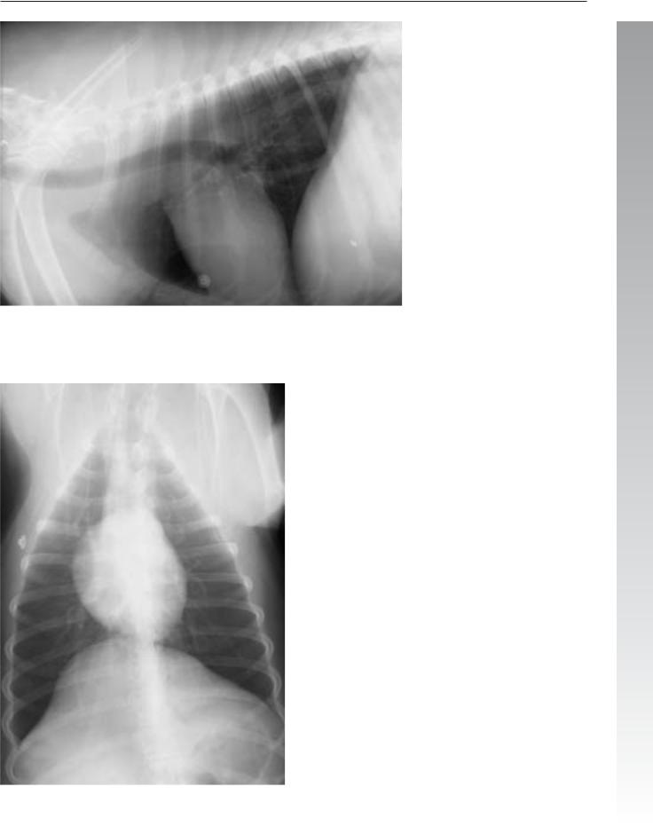
Signalment/History: “Canoe”, a 3-year-old, male mixedbreed dog, had received crushing injuries from an automobile accident and was presented in shock.
Physical examination: The examination was severely limited by the condition of the patient.
Radiographic procedure: Thoracic and abdominal radiographs were made.
Radiographic diagnosis (thorax): Generalized lung contusion was more severe on the right side. Fluid pooling adjacent to the sternum just cranial to the heart shadow suggested a minimal pleural effusion. Minimal pneumothorax was present on the left. Cranial mediastinal widening suggested the possibility of a hemomediastinum. The pulmonary vessels were small, indicative of shock. The diaphragm appeared to be intact. No thoracic wall injury was noted. The stomach was air-filled and distended, the result of panic breathing.

Inguinal hernias 227
3
Radiographic diagnosis (abdomen): Subcutaneous emphysema was associated with a right-sided inguinal hernia that contained intestinal loops. The urinary bladder could not be identified. Pneumoperitoneum was suggested by the indistinct linear gas patterns that were not compatible with the air within the bowel loops. The loss of contrast between the organs suggested peritoneal fluid. The diaphragm had remained intact.
Radiographic diagnosis (abdomen, horizontal beam):
Free peritoneal air was pocketed just beneath the diaphragm making it rather easy to identify (arrows).
Treatment/Management: The injuries were extensive and severe; however, “Canoe” was treated conservatively except for a surgical repair of the inguinal hernia. He was discharged a healthy dog.

228 Radiology of Abdominal Trauma
Case 3.16
3

Inguinal hernias 229
Signalment/History: “Rufus” was a 6-month-old, male mixed-breed cat admitted because he could not walk on the right pelvic limb. The lameness had had an acute onset.
Physical examination: Examination suggested a right femoral fracture. Additional crepitus was noted on palpation of the pelvis. The status of the hip joints was not determined. Soft tissue swelling was prominent especially around the right pelvic limb.
Radiographic diagnosis: A comminuted, midshaft, right femoral fracture was complicated by an apparent right inguinal hernia with bowel loops extending subcutaneously and distally into the pelvic limb. The bowel loops were thought to be
excessive in diameter and were considered obstructed. Pubic and ischial fractures had resulted in separation of the two halves of the pelvis. The right femoral neck was fractured; however, the exact nature of that fracture could not be determined because of the unique positioning of the pelvic limb. The right sacroiliac joint was separated. The urinary bladder could not be identified.
Treatment/Management: Surgery resulted in a reduction |
|
of the obstructed bowel loop and stabilization of the femoral |
|
fracture. A femoral head ostectomy was used to treat the |
3 |
femoral head/neck fracture. The urinary bladder was found to |
be uninjured.

230 Radiology of Abdominal Trauma
3.2.5Renal, ureteral, and urinary bladder injury
Case 3.17
3
Signalment/History: “Amber” was a 6-year-old, female mixed-breed dog with a clinical history of possible trauma. She was not able to walk normally.
Physical examination: On physical examination, a femoral fracture was detected. Abrasions of the skin suggested the possibility of more widespread injury.
Radiographic procedure: Both the thorax and abdomen were radiographed because the injury appeared to involve the whole body.
Radiographic diagnosis (abdomen): Marked subcutaneous emphysema was noted surrounding the injured pelvic limb. In addition, retroperitoneal air was evident ventral to the lumbar spine (arrows). The urinary bladder could not be identified and the presence of peritoneal fluid supported the diagnosis of a rupture of the bladder. Bilateral sacroiliac separation was present.
Renal, ureteral, and urinary bladder injury 231
3
Radiographic diagnosis (thorax): The thorax was examined for evidence of a pneumomediastinum, which could have resulted in air migrating from the thorax into the retroperitoneal space. The thorax appeared normal except for having a small cardiac silhouette and small pulmonary vessels both suggesting shock.
Treatment/Management: The femoral fracture was open and the retroperitoneal air may have gained entrance in that manner. A rupture at the bladder neck or proximal urethra could also have resulted in retroperitoneal air. Both of these causes for retroperitoneal air are considered uncommon, but as there was no evidence of pneumomediastinum in this dog, they were taken into consideration. No evidence of a puncture wound in the dorsal abdomen was found that could have resulted in abdominal air.
“Amber” recovered from surgery for the repair of the ruptured bladder and the fractured femur.