


472 11 The Reproductive Organs
Function and Structure of the Reproductive Organs
The reproductive organs have the function of forming germ cells, promoting their union, and giving the fertilized ovum an environment in which it can develop from an embryo to a fetus mature enough to be born. In addition, the reproductive organs take part in developing the body shape specific to each sex by the hormones they form.
The male and female reproductive organs include:
The gonads, which produce germ cells and sex hormones
The reproductive passages, which serve the transport of reproductive products
The reproductive glands, the secretions of which promote the union of ovum and sperm
The external sex organs, which allow sexual union to occur
Male Reproductive Organs
Overview
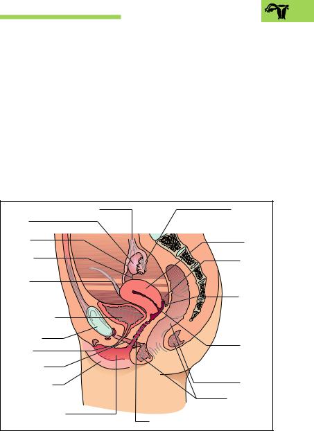
The male reproductive organs are divided according to their development into internal and external organs. The internal male reproductive organs include the testes, the epididymis, the vas deferens, the seminal vesicles, and the prostate. The external male reproductive organs include the penis and the scrotum (Fig. 11.1a, b).
From the testes, which produce the germ cells and the male reproductive hormones, the sperm cells (spermatozoa) pass through a system of small tubules into the epididymis, which stores them. Through the vas deferens, which runs through the inguinal canal, the spermatozoa reach the urethra at the level of the prostate. Shortly before they open into the urethra, they receive the ducts of the seminal vesicles. The ducts of the prostate and Cowper’s glands open directly into the urethra. The spermatozoa achieve mobility under the influence of secretions from these glands. The further transport of the spermatic fluid (semen) is taken over by the urethra, the corpora cavernosa of which generate the erection of the penis and so allow it to penetrate the vagina.

Male Reproductive Organs 473
Ureter |
|
|
|
Sacrum |
Urinary bladder |
|
|
|
Rectum |
|
|
|
|
|
(vesica urinaria) |
|
|
|
Seminal |
Vas deferens |
|
|
|
vesicle |
|
|
|
(glandula |
|
(ductus deferens) |
|
|
|
|
|
|
|
seminalis) |
|
|
|
|
|
|
Prostate |
|
|
|
Ampulla of |
Corpus |
|
|
|
vas deferens |
|
|
|
Excretory duct |
|
spongiosum |
|
|
|
|
|
|
|
|
of seminal vesicle |
|
|
|
|
Ejaculatory |
|
|
|
|
duct |
Corpus |
|
|
|
Prostatic part |
|
|
|
of urethra |
|
cavernosum |
|
|
|
|
|
|
|
|
|
a |
|
|
|
Cowper’s gland |
Glans |
Scrotum |
Testis |
Spongy |
Epididymis |
penis |
|
|
part of |
|
Prepuce |
|
|
urethra |
|
Efferent ductules |
|
|
|
|
Tunica albuginea |
|
|
|
|
|
|
|
|
Duct of epididymis |
Convoluted |
|
|
|
|
seminiferous |
|
|
|
|
ducts (tubuli |
|
|
|
Vas deferens |
seminiferi |
|
|
|
(ductus deferens) |
contorti) |
|
|
|
|
Rete testis |
|
|
|
|
Septa of testis |
|
|
|
|
|
b |
|
|
|
Fig. 11.1 a, b Internal and external male reproductive organs a Overview
b Ductules of the testis and epididymis

474 11 The Reproductive Organs
Testis (Orchis)
The two testes, each about the size of a plum, are the male gonads. They lie in a skin pocket, the scrotum (Fig. 11.1a). They develop on the posterior abdominal wall, and at the end of fetal development they leave a peritoneal pocket and descend along the posterior wall of the abdominal cavity through the inguinal canal into the scrotum (descensus testis). This mechanism withdraws them from experiencing the internal body temperature, which would impede maturation of the sperm. When the testes remain in the abdominal cavity or in the inguinal canal, the condition is called cryptorchidism.
Each testis is surrounded by a dense white connective tissue capsule
(tunica albuginea), from which connective tissue septa (septula testis) run inward (Fig. 11.1b). In this way the tissue of the testis is subdivided incompletely into more than 200 compartments (lobuli testis). Each lobule is made up of two to four extensively convoluted seminiferous tubules (tubuli seminiferi contorti) that measure 350 m in total and the epithelium of which forms the spermatozoa (spermatogenesis) (Fig. 11.1b).
Testosterone Production
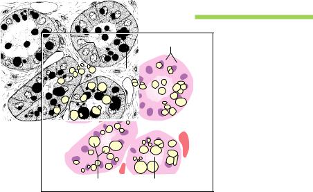
The connective tissue between the seminiferous tubules contains the endocrine interstitial cells of Leydig (Leydig cells) (Fig. 11.2) that produce the male sex hormones (chiefly testosterone) and secrete them into the bloodstream. Testosterone promotes the formation of spermatozoa, promotes the growth of the external reproductive organs, and determines sexspecific behavior. Additionally, the secondary sexual characteristics (e. g., hair distribution) develop under the influence of the male sex hormone. It also has an anabolic effect on metabolism. The production of testosterone in Leydig cells and spermatogenesis are controlled from the hypothalamus by releasing hormones that trigger the liberation of luteinizing hormone (lutropin, LH) and follicle-stimulating hormone (follitropin,
FSH). FSH directly promotes spermatogenesis in the seminiferous tubules and LH stimulates Leydig cells to secrete testosterone. The blood testosterone level in its turn influences the hypothalamopituitary system through a feedback mechanism (see Chapter 7: Hypothalamic−Hypophyseal Axis).

Male Reproductive Organs 475
Interstitial cells of Leydig |
Connective tissue sheath |
|
membrane |
|
Seminiferous |
|
epithelium |
|
Large |
|
spermatocytes |
|
(primary |
|
spermatocytes) |
|
Small |
|
spermatocytes |
|
(secondary |
|
spermatocytes) |
|
Spermatids |
|
Spermatozoa |
|
(sperm cells) |
|
Spermatogonia |
Fig. 11.2 Cross-section of a seminiferous tubule of the testis (tubulus seminiferus contortus)
Development of Sperm Cells (Spermatogenesis)
The wall of the seminiferous tubules is lined with seminiferous epithelium (Fig. 11.2) on a basement membrane. Within the seminiferous epithelium, the Sertoli cells form a support in which the germ cells are embedded. The formation of spermatozoa begins with puberty and continues into old age. It develops in several steps as the cells migrate from the periphery into the center of the seminiferous tubule:
1.Period of multiplication
2.Period of maturation
3.Period of differentiation
During the period of multiplication, the diploid primitive germ cells (spermatogonia) divide by mitosis to form more spermatogonia. During the maturation period, large spermatocytes (primary spermatocytes) are

476 11 The Reproductive Organs
formed, again by mitosis (Fig. 11.2). These then enter the first meiotic division (separation of the homologous chromosomes) (see Chapter 1: Reduction or Maturation Division [Meiosis] and Chapter 12: Germ Cells) and develop into the small spermatocytes (secondary spermatocytes) with only half a set of chromosomes. The secondary spermatocytes develop into smaller spermatids during the second meiotic maturation division (separation of the chromatids). In this way, at the end of the maturation period, which lasts about 72 hours, eight spermatids are generated from one spermatogonium. Of these, four contain an X and four a Y chromosome (see also Chapter 12: Germ Cells).
In the course of the period of differentiation, the spermatids are transformed into the motile form of germ cells, the spermatozoa, the tails of which protrude into the lumen of the seminiferous tubules. Every hour about 3−4 million spermatozoa pass from the testes into the epid- idymis—i.e., about 1000 spermatozoa are generated every second.
Spermatozoa
The spermatozoa are motile cells with tails, about 50−60 µm in length (1/20 mm). The head contains the haploid cell nucleus and possesses a caplike structure (acrosome) (Fig. 11.3) that allows the spermatozoon to penetrate the ovum (see Chapter 12: Fertilization). Below the head there follows a short neck, a relatively thick middle piece, a tail, and an end piece. The neck contains the centriole, which will form the cleavage spindle after the spermatozoon has united with the ovum. In the middle piece, the beginning of the flagellum (tail) is surrounded by numerous spirally arranged mitochondria that provide energy for the cell’s motion. The flagellum continues into the tail.
By wriggling their tail, spermatozoa can advance at a speed of about 3−4 mm a minute. In order to reach the ovum for fertilization, spermatozoa must migrate through the uterine cavity and along to the end of the fallopian tube, a passage that takes about 1−3 hours. Once they reach the ampulla of the fallopian tube, the spermatozoa remain fertile for up to 4 days.

Male Reproductive Organs 477
Fig. 11.3 Structure of a sperm cell
(magnified about 1650×)
Acrosome
Head
Neck
Middle piece
Tail
End piece
Epididymis
By way of the rete testis, a wide-meshed, dilated system of channels, the spermatozoa, swim in a fluid stream to reach the epididymis. This structure, which serves as a store for sperm (Fig. 11.4), sits on the testis like a horse’s tail. The epididymis is divided into a head, a body, and a tail. It includes the efferent ductules of the testis and the duct of the epididymis, which is about 5 m in length (Fig. 11.1b). The ducts are markedly convoluted and are packed tightly into a single mass by connective tissue. In the tail of the epididymis the duct of the epididymis continues into the vas deferens (ductus deferens) (Fig. 11.1b).

478 11 The Reproductive Organs
The epithelium of the duct of the epididymis reabsorbs much of the fluid and secretes substances that promote the definitive maturation of the spermatozoa and their protection against the acid medium of the duct. This acid medium depresses the spermatozoa’s motility, so that they do not use up their energy.
The transport of spermatozoa through the epididymis takes about 12 days. It takes several ejaculations in the course of 24 hours to empty the epididymis completely. In the absence of ejaculation for a prolonged period, the spermatozoa are broken down and the epithelium or macrophages take up the breakdown products.
Vas Deferens
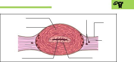
The vas deferens is 50−60 cm long. It transports the spermatozoa during ejaculation (Fig. 11.4). It is sheathed in connective tissue together with the testicular vessels and nerves, forming the spermatic cord (funiculus spermaticus), which runs through the inguinal canal and on into the pelvis. Toward its end, the vas deferens widens into a spindle-shaped ampulla (ampulla ductus deferens), is joined by the duct of the seminal vesicle to form theejaculatory duct, pierces the prostate, and opens into the prostatic part of the urethra (Fig. 11.1a).
The vas deferens has a smooth muscle coat, about 1.5 mm thick, with three layers. These are arranged in a spiral fashion, so that when they contract (during ejaculation) they widen and simultaneously shorten the vas. This mechanism actually sucks the spermatozoa out of the epididymis. Because of its firm consistency, the vas deferens can easily be palpated in the spermatic cord.
Seminal Vesicles (Vesiculae Seminales) or Seminal Glands (Glandulae Seminales)
The two seminal vesicles (seminal glands) are large thin-walled glands, about 10 cm long, that lie on the posterior wall of the urinary bladder, abutting on the rectum (Figs. 11.1a and 11.5). The excretory duct of the seminal vesicle runs at an acute angle into the vas deferens below the ampulla. The common ejaculatory duct so formed then pierces the pros-

Male Reproductive Organs 479
Ureter |
Vas deferens |
Bladder (vesica urinaria) |
(ductus deferens) |
|
|
Ampulla of vas |
Seminal vesicle |
|
|
deferens |
|
|
Ejaculatory duct |
Inguinal canal |
Inguinal ligament |
Prostate |
Urethra |
|
|
|
Epididymis |
|
Testis |
Fig. 11.4 Course of the spermatic pathways
tate from behind and laterally and drains into the prostatic part of the urethra.
Contrary to their name, the seminal vesicles do not contain spermatozoa, but produce a slightly alkaline, protein-rich secretion that mobilizes the sperm in the acid environment of the vagina. The secretion also contains fructose, a simple sugar, which provides energy for the motion of the spermatozoa.
Prostate Gland
The prostate has the shape and size of a large chestnut and lies between the pelvic floor (urogenital diaphragm) and the base of the bladder (Figs. 11.1a and 11.5). Its posterior aspect abuts directly on the rectum, through which it can be palpated with a finger (rectal examination of the prostate). It consists of 30−50 individual glands surrounded by a dense white connective tissue capsule. Their secretory ducts drain into the urethra, which runs vertically through the prostate. The prostatic stroma is largely composed of smooth muscle fibers.

480 11 The Reproductive Organs
|
Bladder (vesica urinaria) |
|
Ureter |
Inguinal |
|
|
||
|
ligament |
|
|
Vas deferens |
|
Ampulla of vas |
|
|
deferens |
|
|
|
Seminal vesicle |
|
|
vesiculosa) |
|
Excretory duct of |
|
|
seminal vesicle |
Prostate gland |
|
|
||
Urogenital diaphragm |
Cowper’s gland |
|
(m. transversus perinei |
||
Urethra |
||
profundus) |
Fig. 11.5 Urinary bladder and prostate seen from behind
The prostate forms a slightly acid, thinly fluid, opalescent secretion with an odor of chestnuts. It contains numerous enzymes (e. g., acid phosphatase), immunoglobulins, and prostaglandins to stimulate the uterus. Spermine, a protein found in prostatic secretion, promotes the motility and fertility of the spermatozoa. In dried semen it forms crystals, which, when demonstrated in the vagina, can document a rape in legal medicine.
Three poorly delineated zones can be made out in the glandular tissue of the prostate (Fig. 11.6): an outer zone, an inner zone, and a periurethral zone that lies in direct contact with the urethra. The outer zone lies under the connective tissue capsule and includes the major part of the glandular tissue. This zone is often a site for the development of malignant tumors (prostatic carcinoma). Prostatic cancer is one of the commonest tumors in elderly men. The periurethral zone is very often the site for the development of benign tumors (prostatic adenoma), which affect more than half of men over 60 years of age. Prostatic adenoma leads to a narrowing of the urethra, with consequent difficulty in emptying the bladder.
Male Reproductive Organs 481
Cowper’s Glands (Bulbourethral Glands)
The two pea-sized Cowper’s glands lie in the musculature of the pelvic floor (urogenital diaphragm) and their ducts drain into the first part of the spongy portion of the urethra (Figs. 11.1a and 11.5). Their weakly alkaline secretion precedes ejaculation and neutralizes the acid environment of the urethra.
Composition of the Ejaculate
The major portion of the fluid part of the ejaculate derives from the prostate (25 %) and the seminal vesicles (75 %). The semen as a whole is weakly alkaline and so protects against the acid environment of the vagina. After three days of sexual continence, one emission contains about 3−6 ml semen with at least 20 million spermatozoa per milliliter (normospermic). Among the spermatozoa of one ejaculate, as a rule 10− 20 % are not fully developed or are malformed. A sperm count of less than 20 million per milliliter is termed oligospermia. The condition in which there are no spermatozoa in the semen is azoospermia.
Castration and Sterilization
In castration, both testes are removed surgically, e. g., as treatment for a malignant testicular tumor. Castration leads not only to infertility but to profound hormonal disturbances. In sterilization, the vas deferens on each sides is merely divided; since the hormonal system remains intact, libido (sexual desire) and potency (ability to have an erection) remain intact.
External Male Sex Organs
The external male reproductive organs, the penis and scrotum, are developed from the abdominal wall.

482 11 The Reproductive Organs
Scrotal Sac (Scrotum)
The testes lie outside the abdominal cavity in the scrotal sac. Here the ambient temperature is about 3 °C lower than the body temperature in the abdominal cavity. This temperature difference is a prerequisite for optimal development of the sperm. The skin of the scrotum is provided with numerous smooth muscle cells (tunica dartos, dartos), that can wrinkle or smooth the skin surface and so play a role in temperature regulation (reduced surface).
Penis (Male Member)
The penis consists of a root, which is firmly anchored to the pelvic floor and the two pubic rami, and a freely movable body (shaft) ending in the glans penis. The skin is freely movable over the penis and is folded back over the glans as the prepuce (Fig. 11.1a). Constriction of the prepuce is called phimosis.
To facilitate copulation the penis has three cavernous bodies of erectile tissue that enable the penis to become erect (Figs. 11.1a and 11.7):
Two paired cavernous bodies (corpora cavernosa)
A single urethral cavernous body (corpus spongiosum)
The corpus spongiosum runs along the underside of the penis and surrounds the urethra. Posteriorly it widens into a bulb (bulb of the penis), while anteriorly it ends in the glans penis (Fig. 11.1a). The bulb of the penis is covered by the two bulbospongiosus muscles, which are joined in the midline. They help to express the content of the urethra. The dorsum of the penis is formed by the two corpora cavernosa, which are separated by a connective tissue septum. Two crura (legs) attach the corpora cavernosa to the pubic rami. All three cavernous bodies are surrounded by a dense white connective tissue sheath that is about 1−3 mm thick (tunica albuginea) (Fig. 11.7).
Structure of the Penis
The structure of the penis is determined principally by the blood-filled spaces of the cavernous bodies (Fig. 11.7). The paired cavernous body of the penis is a spongy network of collagen and elastic fibers lined with

|
Male Reproductive Organs 483 |
|
Periurethral |
Ventral |
Inner zone |
|
||
zone |
|
|
|
|
Seminal crest |
|
|
(colliculus |
|
|
seminalis) |
Orifices of |
|
|
the two |
|
Prostatic part |
ejaculatory |
|
of urethra |
ducts |
|
|
Connective |
|
Glandular tissue |
tissue capsule |
Dorsal |
of outer zone |
|
|
|
Fig. 11.6 Horizontal section through the prostate. (After Leonhardt)
Skin |
Dorsalis penis |
Dorsal artery |
vein |
|
|
of penis |
Corpus cavernosum |
|
|
Deep artery |
of penis |
of penis |
|
(a. profunda penis) |
Septum of |
|
|
Venous drainage |
penis |
from the corpus |
Spongy part of |
cavernosum |
|
|
urethra |
|
Corpus spongiosum |
Connective tissue |
of penis |
|
|
capsule (tunica albuginea) |
|
Fig. 11.7 Transverse section through the penis
endothelium. When empty, the spaces are slits; when they are filled with blood, they attain a diameter of several millimeters. The corpus spongiosum, on the other hand, is mainly filled with a dense network of veins.
In the middle of each cavernous body runs an artery (deep artery of the penis, arteria profunda penis), of which the branches run a helical

484 11 The Reproductive Organs
course (helicine arteries). They run into the spaces of the corpora cavernosa and their ends can be occluded. From the spaces, veins that can be occluded run through the tunica albuginea and drain into the dorsal vein of the penis.
Erection
During erection, the helicine arteries open and blood pours in, distending the tunica albuginea. At the same time, the veins running through the tunica albuginea become compressed, so that blood enters while drainage is occluded. The body of the penis therefore swells and becomes very hard. When the penis relaxes, the helicine arteries close, and as the tunica albuginea becomes less distended more blood can flow out through the vein.
During erection the venous network of the corpus spongiosum is repeatedly filled with blood, which, however, can flow out at any time. Hence the swelling remains relatively soft, allowing semen to flow through the urethra.
Ejaculation
Erection and ejaculation are complex processes that are regulated by the autonomic nervous system. While erection is a process influenced by the parasympathetic system, ejaculation is triggered by the sympathetic system. Ejaculation begins with contraction of the smooth musculature of the prostate, the seminal vesicles, and the vas deferens, as well as closure of the bladder neck. Once the semen has been positioned in the prostatic part of the posterior portion of the urethra, the pelvic floor contracts spasmodically. This movement drives the ejaculate in rhythmic thrusts out of the external urethral orifice.
 ligament of
 Vagina
486 11 The Reproductive Organs
migrates in a fluid stream into the uterus, where nidation (nesting) into a mucosa prepared by hormones follows. After just a few days the fertilized ovum signals the beginning pregnancy to the pituitary gland, which influences the ovary to secrete hormones that secure the maintenance of the pregnancy during the months that follow. The uterine muscle adapts to the size of the growing embryo by undergoing hypertrophy. As the pregnancy progresses a combination of embryonic and maternal tissue forms the placenta, by which the fertilized ovum receives nutrition and oxygen. During labor, repeated contractions of the uterine muculature push the infant out of the birth canal and the placenta becomes detached from the uterine mucosa as the afterbirth.
Ovaries
The ovaries, like the testes, originate on the posterior abdominal wall and in the course of development migrate downward (decensus) toward the true pelvis. They come to rest at the junction of the true and false pelves, about the level of the division of the common iliac artery. They are attached by ligaments to the pelvic wall (suspensory ligament of the ovary) and the uterus (ovarian ligament) (Figs. 11.8 and 11.13). Anteriorly they are loosely attached to the broad ligament of the uterus (ligamentum latum uteri) by a suspending ligament (mesovarium) (Fig. 11.13). In size and shape the ovaries resemble two almonds, each weighing about 14 g.
After maturation and preparation of the ovum, the ovary secretes hormones into the bloodstream that coordinate the processes in the uterus and vagina (estrogen, progesterone).
Structure of the Ovary
The ovary consists of a cortex and a medulla. The medulla contains blood vessels that enter the ovary through the mesovarium (Fig. 11.9). The cortex of an ovary in a woman during her reproductive years lies directly under the surface. It contains ovarian follicles in various stages of maturation (primary, secondary, and tertiary or graafian follicles), involuted follicles (atretic follicles), usually no more than one yellow body (corpus luteum), and the scarred remains of old corpora lutea (corpora albicantia) (Figs. 11.9 and 11.10).

|
Female Reproductive Organs 487 |
Peritoneum = perimetrium |
Primary follicle |
|
Scar of regressed corpus |
|
luteum (corpus albicans) |
Early graafian follicle |
|
|
Mature graafian |
Ovarian artery |
follicle |
and vein |
Ovum |
|
|
|
Follicular fluid |
|
(liquor folliculi) |
|
Primordial follicle |
|
Atretic follicle |
Suspensory |
|
ligament of ovary |
|
(mesovarium) |
Corpus luteum |
|
|
Secondary follicle |
Connective tissue capsule |
|
(tunica albuginea) |
Fig. 11.9 Prepared section of an ovary (longitudinal section, schematically represented)
Development of the Ovum (Oogenesis) and Follicle Maturation
Like spermatogenesis, the development of the ovum can be divided into a period of multiplication and a period of maturation. There is no period of differentiation. In contrast to the development of sperm in the male, which can continue into old age, the period of multiplication is already completed at birth. The germ cells (oogonia) develop into primary oocytes toward the end of the fetal period. These enter the prophase of the first maturation division (see Chapter 1: Reduction or Maturation Division [Meiosis]). They remain in this phase (dictyotene phase of the prophase) until the beginning of puberty or their atrophy. If an ovum matures after puberty (follicle maturation) (Fig. 11.10), the primary oocyte ends the first meiotic maturation division shortly before ovulation. It then forms two ova of different sizes, (a secondary oocyte and a first polar body, see also Chapter 12: Germ Cells), each with a haploid set of chromosomes. The second meiotic maturation division is initiated during ovulation, but is only completed if the egg is fertilized. During the


Female Reproductive Organs 489
nutrients. According to the type of follicular epithelium surrounding the oocyte, follicles are divided into primordial, primary, secondary, and graafian. At birth, both ovaries contain about one million primary follicles, of which a large number perish up to the time of puberty (follicle atresia). With puberty, some of the remaining follicles develop into secondary follicles, of which a small number continue to develop into graafian (tertiary) follicles during each menstrual cycle.
Ovulation
In the course of development from primary to secondary follicle and eventually to graafian follicle, the follicular epithelium divides and, under the influence of the follicle-stimulating hormone (FSH, follitropin) of the hypophysis, forms several layers (Figs. 11.10 and 11.11). The cells surrounding the follicle are hormonally active and secrete female sex hormones (e. g., estradiol), which reach the uterus by way of the bloodstream and cause the mucosal cells to proliferate (proliferative phase).
The cavity (antrum) of a tertiary follicle (Fig. 11.10) is filled with fluid rich in hyaluronic acid and proteoglycans (follicular fluid, liquor follicularis), and is enclosed by a multilayered follicular epithelium consisting of granulosa cells. The oocyte lies within a clump of granulosa cells called the cumulus oophorus. The granulosa cells surrounding the oocyte are called the corona radiata; they supply nutrients to the oocyte and are connected to it by gap junctions. The oocyte is closely enveloped by a layer of glycoproteins that it largely produces, the zona pellucida. This structure plays a major role in fertilization (Chapter 12: Fertilization) and in the early development of the embryo (segmentation; Chapter 12: Transport through the Uterine Tube and Segmentation).
In the middle of the menstrual cycle, one of the mature tertiary (graafian) follicles moves toward the surface of the ovary. The pressure of the fluid in the follicle, together with enzymes, initiates ovulation. The fluid pouring out of the follicle floats the ovum surrounded by a several surrounding follicular cells (corona radiata, Fig. 11.10) from the follicle into the distal end of the fallopian tube, which grasps the ovary with its fimbria at the time of ovulation. Suction in the fallopian tube brings the ovum into the ampulla (ampulla tubae), where fertilization takes place (Fig. 11.13). If the ovum is not fertilized within 12 hours, it perishes.

490 11 The Reproductive Organs
|
|
|
Hypothalamus |
|
|
|
|
Hypophysis |
|
|
|
|
|
|
Gonadotropins |
|
|
|
|
FSH |
|
LH |
|
Degeneration of |
|
|
Degeneration of false |
||
corpus luteum |
Maturation of |
|
|
corpus luteum |
|
|
|
Ovulation |
Corpus luteum |
||
|
|
follicle |
|||
|
|
|
|
Corpus luteum |
|
|
|
|
|
graviditatis |
HCG |
Structure of uterine mucosa |
|
Embryo |
|
||
(endometrium) |
|
|
|
||
Blood vessel |
Glands |
|
|
Functional |
|
|
|
|
|||
|
|
|
|
|
layer |
|
|
|
|
|
Basal layer |
0 |
4 |
|
14 |
|
28 |
Menstrua- |
Proliferative |
Secretory phase |
Pregnancy |
||
tion |
|
phase |
|
|
|
Fig. 11.11 Schematic representation of the ovarian cycle and the changes in the endometrium. The anterior pituitary hormones FSH (follicle-stimulating hormone) and LH (luteinizing hormone) induce growth of the follicle in the ovary, ovulation, and formation of the corpus luteum. Ovarian hormones (estradiol and progesterone) reach and act on the endometrium through the bloodstream. During pregnancy, the implanted fertilized ovum forms HCG (human chorionic gonadotropin), which stimulates the corpus luteum to secrete more progesterone (corpus luteum of pregnancy). (After Langman)
Corpus Luteum
Under the influence of pituitary luteinizing hormone (LH, lutropin) the follicular epithelium left in the ovary is transformed within a few days into a corpus luteum and begins to secrete luteal hormones (e. g., progesterone) (Figs. 11.10 and 11.11). This acts on the uterine mucosa to prepare it for nidation (nesting) of the fertilized ovum (secretory phase). If
Female Reproductive Organs 491
Events in the ovary
Ovulation |
Corpus luteum of |
||
pregnancy |
|||
Maturation |
Corpus |
||
|
|||
of the ovum |
luteum |
|
|
|
|
Scar |
|
|
|
Fertilization in the |
|
Events in the |
fallopian tube |
||
|
|||
uterine mucosa |
|
||
Proliferation |
Implantation |
||
Sloughing |
Recovery |
||
|
|||
|
|
|
|
|
|
|
|
|
|
|
|
|
|
|
|
|
|
|
|
|
|
|
|
|
|
|
|
|
|
|
|
|
|
|
|
|
|
|
|
|
|
|
|
|
|
|
|
|
|
|
|
|
|
|
|
|
|
|
|
|
|
|
|
|
|
|
20 |
25 |
5 |
10 |
15 |
20 |
25 |
5 |
10 |
15 |
20 |
25 |
5 |
10 |
15 |
20 |
25 |
5 |
10 |
15 |
20 |
25 30 |
35 |
40 |
||||||||
|
|
|
|
|
|
|
|
|
|
|
|
|
|
|
|
|
|
|
|
|
|
|
|
|
|
|
|
|
|
|
|
|
|
|
|
|
|
Ovulation |
|
|
|
|
|
Ovulation |
|
|
Ovulation |
|
|
|
|
Ovulation |
|
|
|
||||||||||
|
|
|
|
|
|
|
|
|
|
|
|
|
|
|
|
|
|
|
||||||||||||||
|
|
|
|
|
|
|
|
|
|
|
|
|
|
|
|
|
|
|
||||||||||||||
|
|
|
25-day cycle |
|
|
|
27-day cycle |
|
|
|
27-day cycle |
|
|
|
|
|
|
|
|
|
|
|||||||||||
|
|
|
|
|
|
|
|
|
|
|
|
|
|
|
Pregnancy |
|
|
|
||||||||||||||

 Days of menstruation and intensity of menstruation
Fertile days
Fig. 11.12 Simplified representation of the regular cyclic changes in the ovary and the endometrium
fertilization occurs, the germ cell takes over the secretion of chorionic gonadotropins (human chorionic gonadotropin, HCG), which in turn stimulates the corpus luteum to secrete more progesterone. The corpus luteum becomes a corpus luteum of pregnancy (corpus luteum graviditatis) and fulfills its task up to about the 4th month of pregnancy. After that it degenerates and the placenta takes over the functions of the corpus luteum.
If the ovum is not fertilized, the corpus luteum stops functioning after about 2 weeks (false corpus luteum, corpus luteum of menstruation = corpus luteum menstruationis). As the progesterone level declines, the uterine mucosa undergoes menstrual bleeding and the mucosa is sloughed off (Fig. 11.12).

492 11 The Reproductive Organs
The Menstrual Cycle
During the period of sexual maturity, the ovarian cycle causes cyclic changes in the uterine mucosa that lead to periodic sloughing of the mucosa (menstrual bleeding). The menstrual cycles begin (menarche) between the 10th and 15th years of life; they end (menopause) about the 50th year of life, beginning the “change” (climacteric). During this phase of life, the secretion of ovarian hormones gradually declines, follicular growth and ovulation no longer take place, and the uterine mucosa becomes increasingly thinner. The menstrual cycle is divided into three phases and lasts on an average 28 days (Fig. 11.12). Longer or shorter cycles are common. The day when menstrual bleeding begins is designated as the first day of the cycle. The phases are:
1.Phase of desquamation and regeneration (day 1−day 4)
2.Follicular (proliferative) phase (day 5−day 14)
3.Luteal (secretory) phase (days 15−28)
The proliferative phase shifts into the secretory phase on the day of ovulation (day 14 of a 28-day cycle). The secretory phase always lasts about 14 days, regardless of the duration of the cycle. Therefore, the day of ovulation shifts correspondingly.
If pregnancy does not occur, the corpus luteum involutes after 13−14 days. The interruption in progesterone secretion then causes menstrual sloughing of the uterine mucosa. At the same time, there is a transient diminution in the number of thrombocytes and blood clotting is reduced. Immediately afterward, the mucosa begins to regenerate from the basal layer (Fig. 11.11).
In the following proliferative phase, the endometrium grows and the mucosal glands enlarge. These processes are controlled by estrogens that are formed in the growing follicle and reach the uterine mucosa through the bloodstream. The proliferative phase ends on the day of ovulation (usually on or around the14th day).
The secretory phase that follows is controlled by the corpus luteum. It prepares the mucosa for the nidation (implantation) of the germ cell. In this phase the mucosal glands reach their greatest length and produce a mucous secretion. After ovulation, progesterone effects a rise in body temperature of 0.5−1 °C.

Female Reproductive Organs 493
Fallopian Tube (Uterine Tube, Salpinx)
The fallopian tubes are about 10−15 cm long and are incorporated into the broad ligament of the uterus by a ligament (mesosalpinx) (Figs. 11.13 and 11.14). It begins at the level of the ovary at the abdominal ostium (opening) as a funnel lined with fringes (infundibulum, fimbriated end) and runs into the uterus at an angle. It is usually divided into the narrow uterine end (isthmus, isthmus tubae uterinae) and the wider outer part (ampulla, ampulla tubae uterinae) in which fertilization takes place (Fig. 11.13).
The mucosal surface of the fallopian tube is greatly magnified by several longitudinal folds. It is lined with a simple columnar ciliated epithelium with numerous glandular cells that secrete mucus at certain stages of the cycle. The kinocilia of the epithelium direct the fluid stream toward the uterus, against which the spermatozoa must swim but which helps to transport the fertilized ovum toward the uterus. The migration of the germ cell through the tube, which lasts 4−6 days, is aided by peristalsis of the tubal musculature directed toward the uterus.
Supensory ligament of |
Ovarian ligament |
Isthmus of |
Ampulla of |
|
||
uterine tube (mesosalpinx) |
(lig. ovarium |
uterine tube |
uterine tube |
Suspen- |
||
|
|
proprium) |
|
|
||
|
|
|
|
|
sory liga- |
|
|
|
|
Fundus of uterus |
ment of ovary |
||
|
|
|
(mesovarium) |
|||
|
|
|
|
|
||
|
|
|
|
|
|
Ovary |
|
|
|
|
|
Fimbriated end |
|
|
|
|
|
|
of uterine tube |
|
|
|
|
|
|
Uterine tube |
|
Broad ligament of |
|
|
|
Mesovarium |
Meso- |
|
|
|
|
Ovary |
salpinx |
||
uterus (lig. latum uteri) |
|
|
|
|
||
Body of uterus |
|
|
Ureter |
Posterior |
Anterior |
|
|
|
|
Broad ligament |
|
||
(corpus uteri) |
|
|
|
|
||
|
Site of external os |
of uterus |
|
|||
|
|
|
|
|||
|
|
|
|
|
||
a |
Uterosacral ligament |
of uterus |
Cut line for b Parametrium |
|||
Fig. 11.13 a, b Uterus, fallopian tubes and ovaries a Dorsal view
b Section through a along the indicated cut line

494 11 The Reproductive Organs
Suspensory |
|
|
ligament of ovary |
Fundus of uterus |
Fimbriated end |
|
|
of tube |
Ovary |
|
Uterine tube |
|
|
|
Cavity of uterus |
|
Ovarian ligament |
|
Round ligament of uterus |
|
|
|
|
|
|
(lig. teres) |
Isthmus of uterus |
|
Body of uterus |
|
|
|
Cervix of uterus |
|
(corpus uteri) |
|
Uterine artery |
|
External os (ostium uteri) |
|
|
|
|
|
Vagina |
|
Vaginal part of cervix |
|
|
|
Clitoris |
|
|
Labium majus |
|
|
Labium minus |
|
External orifice of urethra |
|
|
|
|
|
Vestibule (vestibulum vaginae) |
|
|
Perineum |
|
|
Anus |
Fig. 11.14 Section through uterus and vagina, including view of the ovaries, fallopian tubes, and external sex organs (blue arrows indicate the path of the ovum from ovary to uterus)
Uterus
During pregnancy, the uterus supports the fertilized ovum. In shape and size it resembles a pear, and it lies between the urinary bladder and the rectum (Figs. 11.8, 11.13, and 11.14). It is divided into a body (corpus uteri), a fundus situated between the tubal ostia, and a narrow portion (isthmus uteri), situated at the transition from the uterine body to the uterine neck (cervix uteri). The cervix is conical and below is directed backward into the vaginal vault. There is an external opening in the part projecting into
|
Female Reproductive Organs 495 |
|
Muscle of uterus |
Branches of uterine artery |
|
(myometrium) |
Broad |
|
|
||
Peritoneal lining |
ligament of |
|
uterus (lig. |
||
(perimetrium = peritoneum) |
||
latum uteri) |
||
|
||
|
Connective |
|
|
tissue in |
|
|
broad liga- |
|
|
ment (par- |
|
|
ametrium) |
|
Uterine cavity |
Uterine mucosa |
|
(endometrium) |
Fig. 11.15 Cross-section through a human uterus
the vagina (portio vaginalis), the external os (ostium uteri externum) (Fig. 11.14).
The uterine cavity is a narrow slit lined with mucous membrane (endometrium). The rest of the wall consists of a muscular layer (myometrium) up to 2 cm thick and a peritoneal covering over the corpus and fundus (perimetrium). The connective tissue space on each side of the uterus is called the parametrium. It contains important structures including the ureter and vessels running toward the uterus (e. g., the uterine artery) (Figs.11.13 and 11.14).
Depending on the phase of the cycle, the uterine mucosa is 2−8 mm thick and is lined by a simple epithelium. The mucosal connective tissue contains numerous tubular glands with ducts opening into the uterine cavity. The mucosa is made up of two layers, one immediately adjacent to the muscular layer, the basal layer (stratum basale) and the functional layer (stratum functionale), which overlies it (Fig. 11.11). The cyclic changes of the uterine mucosa affect chiefly the functional layer.
Vagina
The vagina is a thin-walled tube, about 10 cm long, with a weakly developed muscle layer (Figs. 11.8 and 11.14). Its blind end surrounds the vaginal portion of the uterus, forming the vaginal vault (fornix of the vagina, fundus of the vagina). Its anterior end opens into the vaginal vesti-

496 11 The Reproductive Organs
bule. It is lined with a stratified squamous epithelium that shows changes with the menstrual cycle: during the second phase of the cycle, the superficial epithelial cells, which in this phase have an especially high glycogen content, are increasingly desquamated. The mucus of the cervical glands and the desquamated epithelial cells together form the acid vaginal secretion. The acidity (pH 4−4.5) of the vaginal milieu is due to a physiological lactobacillus (Lactobacillus acidophilus = Döderlein’s bacillus), that transforms the desquamated epithelial cells into lactic acid. This physiological vaginal flora is an effective barrier against invasion of the vagina by bacteria or other pathogens.
External Female Sex Organs (Vulva)
The external female sex organs as a whole are called the vulva.
Vestibule (Vestibulum Vaginae), Labia Majora and Minora,
and Clitoris
The urethra, the vagina, and the various vestibular glands all terminate in the vestibule (Fig. 11.14). It is bounded laterally and posteriorly by skin folds, the labia majora and minora, anteriorly by the clitoris, and posteriorly by a small skin fold, the frenulum (posterior commissure, fourchette, frenulum labiorum pudendi). The two bulbospongiosus muscles are not fused as in the male, but lie on either side of the labia minora and join at the level of the perineum. The two muscles cover the vestibular bulbs, two dense erectile venous networks that correspond to the corpus spongiosum in the male.
The labia majora contain adipose tissue and sebaceous, sweat, and scent glands. The clitoris arises by two crura from the inferior pubic rami and ends in the glans (glans clitoridis), an erectile body comparable to the male glans. The vaginal orifice can be partly occluded by a membrane (hymen) until the first sexual intercourse. The ducts of the large glands of the vestibule (Bartholin glands), about 1−2 cm long, end on each side of the vaginal opening and moisten it.

Female Reproductive Organs 497
The Female Breast (Mamma) and Mammary Gland
The female breast and mammary gland are structures derived from skin and are functionally related to the female sex organs. They develop during puberty under hormonal influence and are composed of glandular, adipose, and connective tissues (Figs. 11.16 and 11.17).
The sexually mature breast has the shape of a pliable hemisphere and is freely movable over the pectoralis major muscle at the level of the 3rd
Glandular lobe
Pectoralis major muscle
Lacteal sinus (sinus lactiferus)
Nipple |
|
Pectoralis fascia |
(mamilla) |
|
|
|
|
|
Mammary duct (ductus lactiferus)
Fatty body
Fig. 11.16 Longitudinal section through a female breast. (After Leonhardt)

Summary 499
Summary The Reproductive Organs
The role of the reproductive organs is to form haploid germ cells, to enable their union, and to ensure the development of the fertilized ovum through the embryonic and fetal stages until birth. The reproductive organs also secrete hormones to regulate the development of the germ cells and the cyclic changes in the endometrium. Finally, they take part in the expression of the specific sexual characteristics of the human body.
Structure and Function of the Reproductive Organs
Gonads: produce germ cells and sex hormones
Reproductive passages: transport the reproductive substances
Reproductive glands: produce secretions that promote the union of ovum and sperm
External sex organs: allow sexual union to occur
Male Reproductive Organs
Internal male reproductive organs: testes, epididymis, vas deferens, seminal vesicles, and prostate
External sex organs: penis and scrotum
Testes: paired organ, about the size of a plum with a dense connective tissue capsule (tunica albuginea); testicular tissue is divided into about 200 lobules each with 2−4 convoluted seminiferous tubules (tubuli seminiferi contorti), the walls of which form the germinal epithelium from Sertoli and germ cells.
Spermatogenesis is divided into periods of multiplication (mitotic division of spermatogonia throughout life), maturation (by mitosis the spermatogonia develop into primary spermatocytes that enter the first maturation division and form haploid secondary spermatocytes; the second maturation division creates spermatids), and differentiation (spermatids are transformed into the mobile form: sperm cells). It is regulated by the anterior pituitary (FSH); duration: about 72 days.
Testosterone production: interstitial cells of Leydig outside the seminiferous tubules; regulated by the anterior pituitary (LH).

500 11 The Reproductive Organs
Epididymis: through the rete testis, spermatozoa reach the epididymis, in which the spermatozoa mature and in which their motility is reduced; duration: 10−12 days.
Vas deferens: runs in the spermatic cord (funiculus spermaticus) is accompanied by vessels and nerves; about 50−60 cm in length; transports the spermatozoa from the epididymis to the urethra. It is wider at the end (ampulla of the vas deferens), is joined by the duct of the seminal vesicle, and opens as the ejaculatory duct into the prostatic urethra.
Seminal vesicles: also called seminal glands; secrete the major part of the ejaculate (about 75 %), of which most important component is the sugar fructose (delivers energy for spermatic motility).
Prostate: chestnut-sized gland between base of bladder and urogenital diaphragm; posteriorly abuts on rectum (rectal examination of the prostate). The glandular tissue is divided into an outer zone (most likely site for the development of malignant tumors), an inner zone, and a periurethral zone surrounding the urethra (commonest site for benign prostatic adenomata). The prostate secretes one-quar- ter of the spermatic fluid (major components: acid phosphatase, immunoglobulins, prostaglandins, spermine). The ducts of the prostatic glands open into the prostatic part of the urethra.
Semen (ejaculate): comprises 3−6 ml fluid (chiefly from the seminal vesicles and the prostate), with about 20 million spermatozoa/ml (normospermia) after three days of sexual continence (oligospermia corresponds to less than 20 million sperm cells; azoospermia is the absence of sperm cells in the ejaculate).
Cowper’s glands: pea-sized paired glands at the level of the urogenital diaphragm; open into the proximal portion of the spongy part of the urethra and with their alkaline secretion neutralize the urethral environment.
Scrotum: contains the two testes and makes possible an optimal ambient temperature (3 °C lower than body temperature) for spermatogenesis.
Penis: consists of a root, a body, and a glans, as well as the freely movable penile skin. Three erectile bodies (corpora cavernosa penis, paired erectile bodies; corpus spongiosum, unpaired urethral erectile body) permit the erection of the penis and are surrounded by a dense connective tissue sheath (tunica albuginea). The corpus spongiosum
Summary 501
protects the urethra and allows the sperm to be transported during an erection.
Erection (regulated by the parasympathetic system): blood flows from the deep arteries of the penis (aa. profundae penis) into the cavities of the erectile bodies and the tunica albuginea becomes tense. This compresses the penile veins and blood flows in while drainage is impeded.
Ejaculation (regulated by the sympathetic nervous system): contraction of the smooth muscle of the prostate, the seminal vesicles and the vas deferens, while semen is made available to the prostatic urethra (emission). Closure of the bladder neck and contraction of the urogenital diaphragm leads to rhythmic expulsion of the ejaculate from the urethral opening.
Female Reproductive Organs
Internal female reproductive organs: ovaries, fallopian tubes, uterus, and vagina
External female reproductive organs: labia majora and minora, vestibule, vestibular glands, and clitoris
Breast and mammary glands
Ovaries: paired almond-sized intraperitoneal organs, attached to the pelvic wall by the suspensory ligament of the ovary, to the uterus by the ovarian ligament, and to the broad ligament of the uterus by the mesovarium (entry of blood vessels). It is divided into a cortex (contains various stages of ovarian follicles) and a medulla (contains blood vessels).
Oogenesis, maturation of the follicle, and ovulation: oogenesis includes a multiplication period and a maturation period. The multiplication period is completed before birth, after which the primary oocytes are surrounded by a flattened simple follicular epithelium (about 1 million primordial or primary follicles at birth) and enter the first maturation division, which, however, is not completed until puberty (resting stage = dictyotene stage). After puberty, ovarian follicles mature in the cortex with the cycle (maturation sequence: primary, secondary, and graafian follicles). Shortly before ovulation in the middle of the cycle, the primary oocyte completes the first maturation division (separation of homologous

502 11 The Reproductive Organs
chromosomes) with two resulting haploid cells: a secondary oocyte and a first polar body. The second maturation division follows during ovulation (result: the mature ovum and another polar body), but it is not completed until successful fertilization has occurred.
Hormonal regulation of follicular maturation: The pituitary gonadotropic hormones (FSH = follicle-stimulating hormone and LH = luteinizing hormone) generate the cycle of follicle maturation (FSH) and trigger ovulation (LH). The follicular epithelium remaining after ovulation is transformed into a corpus luteum under the influence of LH). The female hormones secreted into the bloodstream by the follicular epithelium (estradiol) and the corpus luteum (progesterone) reach the uterine mucosa (endometrium), which undergoes cyclic changes under hormonal influence (menstrual cycle).
Fallopian tubes: paired tubes 10−15 cm in length with a funnelshaped opening near the ovary and an opening into the uterus at the fundus; by their ciliated epithelium they transport the ovum, after fertilization in the ampulla, into the uterine cavity over 4−6 days.
Uterus: The uterus is a pear-shaped organ lying between the bladder and the rectum. It consists of a uterine body, a fundus (ostia of the fallopian tubes), uterine cavity, isthmus, and neck, as well as a cervix, the vaginal part that projects into vagina. It has an opening, the external os. Layers of the uterine wall: endometrium, myometrium, and perimetrium. The endometrium is composed of a basal layer and a functional layer.
Menstrual cycle: menstruation begins (menarche) between the 10th and 15th years of life; it ends around the 50th year of life, followed by the menopause (“change”, climacteric). Average duration of one menstrual cycle is 28 ± 3 days. There are three phases, beginning with the first day of menstrual bleeding:
−Phase of desquamation and regeneration: days 1−4
−Follicular phase (proliferative phase): days 5−14
−Luteal phase (secretory phase): days 15−28
Mucosal (endometrial) changes during the cycle: if ovulation is not followed by fertilization, the corpus luteum stops secreting progesterone after 2 weeks (= corpus luteum of menstruation), lead-

Summary 503
ing to menstrual bleeding and sloughing of the endometrium (phase of desquamation). Regeneration begins at the basal layer. During the ensuing proliferative phase the endometrium is regenerated (triggered by estradiol), and the secretory phase (the mucosa is prepared for implantation) begins at ovulation (formation of the corpus luteum). Should pregnancy occur, the trophoblast, after nidation (days 20−23 of the cycle), secretes human chorionic gonadotropin (HCG), which stimulates the corpus luteum to continue secretion of progesterone (corpus luteum of pregnancy). Desquamation does not occur and pregnancy can begin.
Vagina: thin-walled tube with little muscle between the vestibule and the vaginal portion of the uterus. The stratified squamous epithelium shows changes with the menstrual cycle. During the second half of the cycle desquamation of superficial epithelial cells containing glycogen increases. These are transformed into lactic acid by a lactobacillus (Lactobacillus acidophilus = Döderlein’s bacillus). This permanent physiological vaginal flora (pH 4−4.5) protects against bacteria and pathogens, and is especially effective in the second half of the cycle (protecting nidation in case of a fertilized ovum).
External female reproductive organs (vulva): include the vestibule, the labia majora and minora, the vestibular glands, and the clitoris. The vestibule receives the openings of the urethra, the vagina, and the vestibular glands.
The female breast and mammary gland: Derivatives of skin that develop during puberty under hormonal influence and are made up of glandular, adipose, and connective tissue. The lacteal ducts (mammary ducts) open as 15−20 lacteal sinuses (sinus lactiferi) on the nipple (mamilla). During pregnancy, hormones (estradiol, progesterone) cause the glands and ducts to grow. The secretion of milk begins after birth, triggered by prolactin (anterior pituitary hormone). Lactation is facilitated by oxytocin (posterior pituitary hormone).

504