


259
6
Blood, the Immune System, and Lymphoid Organs
Contents
The Blood |
260 |
|
|
|
|
Functions of the Blood |
260 |
||||
The Cells of the Blood |
262 |
||||
− |
The Erythrocytes |
262 |
|||
− |
The Leukocytes |
263 |
|||
− |
The Platelets |
266 |
|
||
Blood Groups and Blood |
|||||
Transfusions |
266 |
|
|
|
|
− |
Blood Groups |
266 |
|
||
− |
Blood Transfusion |
|
267 |
||
−The Significance of the ABO System 267
−The Rh Factor 268
The Plasma 269
− Plasma Proteins 270
−Low-Molecular-Weight Plasma Constituents 273
−Plasma Electrolytes 273
The Erythrocyte Sedimentation Rate (ESR) 273
O2 and CO2 Transport in the Blood 274
− |
Hemoglobin Transport of O2 274 |
− |
CO2 Transport 274 |
−Hemoglobin and Carbon Monoxide 276
−Hemoglobin Concentration (Hb) 276 The Anemias 277
The Regulation of Erythrocyte Generation 278
Hemostasis and Coagulation of the
Blood 279 |
|
|
− |
Hemostasis |
280 |
− |
Coagulation of the Blood 280 |
|
− |
Fibrinolysis |
280 |
−The Regulation of Blood Clotting 281
The Immune System |
282 |
|
Nonspecific Immunity |
282 |
|
− |
Cellular Immunity |
282 |
− |
Humoral Immunity |
283 |
Specific Immunity 283
−Antigen Presentation by Macrophages 283
−T Lymphocytes (Cellular Immunity) 285
−B Lymphocytes (Humoral and Cellular Immunity) 286
−Memory Cells 287
The Lymphoid Organs (Immune
Organs) 287
The Thymus 290
The Lymph Nodes 291
The Spleen (Splen, Lien) |
293 |
Lymphoid Tissues of the |
Mucous |
Membranes |
295 |
Summary |
299 |

260 6 Blood, the Immune System, and Lymphoid Organs
The cells of the blood and the immune cells are free connective tissue cells and are in part of identical origin. Developmentally they both originate from the mesenchyme. For the most part they are formed in the same site, the bone marrow, but they differ considerably in their location and the site of their functioning (blood vs. connective tissue).
The Blood
Blood may be viewed as tissue—a sort of fluid transportation tissue, of which the intercellular substance is the blood plasma (plasma). The cellular components of this tissue are the red (erythrocytes) and white
(leukocytes) blood cells and the platelets (thrombocytes (Fig. 6.1a, b). The proportion of the total blood volume occupied by all blood cells in percent is called the hematocrit (see Fig. 6.3). It averages 45 %, and is usually a little higher in men (47 %) than in women (43 %).
Total Blood Volume. The total circulating blood volume in humans is about 8 % of body weight; i.e., the total blood volume of a person weighing 70 kg is about 5.6 liters.
Functions of the Blood
Blood has multiple functions closely connected with its components and with the vascular system. While a function of the blood vessels is to distribute the blood overall (heat regulation and distribution of substances), the formed and unformed blood components have some very specific functions.
The red blood cells, for instance, are responsible for the transport of blood gases from the lung to the tissues (oxygen) and from the tissues back to the lungs (carbon dioxide).
White blood cells serve to defend against pathogens and foreign bodies (immunity). They perform these tasks most of the time outside the blood vessels, in the connective tissues. In this case the blood serves solely as a means of transportation from the site of cell formation (bone marrow) to the site of action.
The fluid portion of the blood, the plasma, subserves several different transportation tasks. For instance, it undertakes the transport of
The Blood 261
Cells originating in the red bone marrow
Multilobar cell nucleus
Red blood cell |
|
|
(erythrocyte) |
Neutrophil granulocyte |
|
|
Specific |
Monocyte |
|
granules |
|
Platelets (thrombocytes)
a Basophil granulocyte |
Eosinophil granulocyte |
Cells originating in the lymphoid organs
Cell nucleus
Small lymphocyte
b |
Large lymphocyte |
Fig 6.1 a, b The cells of the blood. The cells of the blood are formed in the red bone marrow from a common ancestor, the blast cell, and released into the peripheral bloodstream after a certain maturation period. Throughout life all the cells of the blood are formed in the red bone marrow, with the exception of lymphocytes, which also multiply in the lymphoid organs

262 6 Blood, the Immune System, and Lymphoid Organs
nutrients from where they are absorbed (intestinal villi) to where they are utilized (organs), of metabolic products to the excretory organs (kidneys), and of substances acting inside the body to their sites of activity (hormones). Simultaneously, blood transports heat from the metabolically active organs to the surface.
Another task of blood is coagulation. When blood vessels are injured, the clotting factors carried in the blood, such as fibrinogen and platelets, are of vital importance. In addition to water, blood plasma contains a number of salts (electrolytes), proteins (albumins and globulins), lipids (fatty acids and cholesterol), and carbohydrates (blood glucose), and numerous vitamins, trace elements, and enzymes. Other noteworthy features of blood include its essentially constant composition, relatively constant osmotic pressure, and a pH value that varies only within narrow limits (7.2−7.4) (the so-called “constant internal milieu”).
The Cells of the Blood
The following are average values per microliter (1 µl = 1 mm3) for the formed elements of the blood:
Erythrocytes |
4.5−5.5 million |
|
|
Leukocytes |
4000−8000 |
|
Thrombocytes |
150 000−350 000 |
The white blood cells (leukocytes) are further subdivided into (differential white cell count):
Neutrophils (neutrophil granulocytes) |
60−70 % |
|
Eosinophils (eosinophil granulocytes) |
2−3 % |
|
Basophils (basophil granulocytes) |
0.5−1 % |
|
|
Lymphocytes |
20−30 % |
|
Monocytes |
4−5 % |
The Erythrocytes
The erythrocytes (red blood cells) are round, disk-shaped structures with an average diameter of 7.5 µm. They are concave on both sides and this gives them an optimal surface to volume ratio (Fig. 6.1a). The shape favors oxygen uptake and release (through short diffusion distances) and

The Blood 263
facilitates passive deformation during passage through narrow capillaries. The content of the cell consists almost entirely of the red iron-con- taining pigment hemoglobin, which binds oxygen reversibly. When hemoglobin is oxygen enriched (arterialized blood) it appears bright red, while it appears dark red when oxygen-poor (venous blood).
The normal red cell count in a man is about 5.3 × 10⁶/µl, while in a woman it is 4.6 × 10⁶/µl, the number depending on the oxygen needs of the body and the availability of oxygen in the lung. For instance, at high altitudes the number increases (polycythemia). If the formation or lifespan of the red cells is insufficient as a result of pathological processes, the result is anemia (p. 277). The commonest causes of anemia are iron deficiency, vitamin B12 deficiency, and folic acid deficiency.
Formation, Lifespan and Breakdown
The site of the formation and maturation of erythrocytes is the red bone marrow, where they develop from stem cells. In the course of their maturation they lose their nucleus and cell organelles and are extruded into the peripheral bloodstream. In humans, about 160 million red cells are formed every minute. In blood the least mature erythrocytes (reticulocytes; about 1 %) can be recognized by a granular structure visible with a special stain. The reticulocyte count in peripheral blood is increased, for example, after loss of blood.
The lifespan of red blood cells is on an average 120 days. They are mostly broken down in the spleen and in the liver. The part of the hemoglobin molecule that does not contain iron forms bile pigments (bilirubin). The liberated iron is stored and reused for the formation of hemoglobin.
In hypertonic solutions red cells lose water and shrink (crenated cells), while in hypotonic solutions they take up water and burst (hemolysis). When this happens, hemoglobin is liberated and the cells become transparent (ghost cells, achromocytes).
The Leukocytes
In addition to the red blood cells, the blood contains cells that are relatively colorless, the white blood cells (leukocytes). These include the granulocytes (polymorphonuclear leukocytes, polymorphs), the lym-

264 6 Blood, the Immune System, and Lymphoid Organs
phocytes, and the monocytes (Fig. 6.1a, b). Their lifespan, in contrast to that of the red cells, varies a great deal and can be from a few hours to years. Together with the lymphoid organs (the spleen, thymus, lymph nodes, tonsils, etc.), the white blood cells form the immune system, which is divided into a nonspecific and a specific immune system (p. 282).
The number of white blood cells varies between 4000 and 8000/µl. The number can increase considerably above 10 000/µl (leukocytosis). If the leukocytes are diminished to below 2000/µl, the condition is called leukopenia (e. g., after damage to the site of their formation). The leukocytes, like the erythrocytes, are generated in the red bone marrow and after maturation and proliferation they are released into the bloodstream. The lymphocytes form an exception, in that their stem cells are located in the bone marrow but they may multiply and differentiate in other lymphoid organs (e. g., in the thymus or the lymph nodes) (see Specific Immunity [T and B lymphocytes] below).
Most of the white blood cells use the blood only as a means of transportation from their generation in the bone marrow to the site of their activity. They fulfill their immune functions almost exclusively outside the vascular system, i.e., in the connective tissue and in the lymphoid organs. There they can move autonomously by their ameboid motion after passing through the walls of the capillaries and postcapillary venules (leukocyte diapedesis).
Granulocytes
Granulocytes are divided into neutrophils, eosinophils, and basophils according to the granules they contain (granular cell inclusions) (Fig. 6.1a). They all have a multilobar nucleus (polymorphonuclear leukocytes, polymorphs). In contrast, immature stages can be distinguished by their bandlike nuclei (band cells, stab cells).
Neutrophil granulocytes are also termed phagocytes because they phagocytose (Greek phagein = to ingest) foreign material. They are a part of the nonspecific immune system, and they are the first to reach the site of an inflammation. Their granules contain a number of lysosomal enzymes that destroy pathogens and cell debris, rendering them harmless. In so doing, the polymorphs mostly perish themselves (resulting in pus formation).

The Blood 265
Eosinophils are also capable of phagocytosis, especially of antigen− antibody complexes (see p. 286). They take part in allergic reactions by bonding to and inactivating excess histamine that has been released from mast cells or basophil granulocytes. Hence their chief task is to limit allergic reactions. Additionally, their granules contain a number of aggressive enzymes that are released when needed to damage their target cells.
Basophils form a very small part of human blood cells. Their granules contain mainly histamine and heparin. Histamine plays a part in immediate hypersensitivity (increase in vascular permeability, contraction of smooth muscle tissue), while heparin has anticoagulant properties.
The Lymphocytes
The lymphocytes present in the bloodstream (small lymphocytes) are about the size of erythrocytes, while the so-called large lymphocytes occur mainly in the lymphoid organs (Fig. 6.1b). Lymphocytes have a noticeably large nucleus and their cytoplasm is rich in cell organelles. They are the cells of specific immunity and they are also formed in the red bone marrow. However, they reach the various lymphoid organs by way of the bloodstream and there develop into specific immune cells (see The Immune System, p. 282).
Monocytes
The monocytes are the largest of the white blood cells (Fig. 6.1a). Their nucleus is oval or kidney-shaped and their cytoplasm contains numerous lysosomes. Like the other leukocytes they are generated in the red bone marrow, but after their release into the bloodstream they remain there only about 20−30 hours. After that time they leave the blood vascular system and are transformed into macrophages in the tissues. Monocytes and macrophages have multiple tasks in the immune system and take part especially in nonspecific immunity. Their function includes phagocytosis and intracellular destruction of bacteria, fungi, parasites, and autologous damaged cells. Beyond that they also take part in specific immunity, in that they pass information about foreign antigens to the lymphocytes (antigen presentation, see p. 283).

266 6 Blood, the Immune System, and Lymphoid Organs
The Platelets
Platelets or thrombocytes play a major role in coagulation of the blood and in hemostasis. They are formed in the bone marrow by cytoplasmic extrusions from giant cells (megakaryocytes), to be released into the bloodstream in the form of irregularly shaped platelets (Fig. 6.1a). Their cytoplasm has no nucleus and few cell organelles. Thrombocytes have a lifespan of about 5−10 days, after which they are destroyed in the spleen. They are deposited on the vessel wall when a blood vessel is injured and break down, liberating enzymes (e. g., thrombokinase) that combine with other factors (thrombin, fibrinogen) to coagulate the blood.
Blood Groups and Blood Transfusions
The surface membrane of erythrocytes contains a large number of various saccharides (glycolipids, glycoproteins), the so-called blood group antigens. They are called antigens because in foreign organisms they induce the formation of antibodies (see The Immune System, p. 282). Human blood contains more than 100 such antigens, of which especially the ABO and Rh systems are of clinical importance.
Blood Groups
The ABO (O = none, from German ohne) system consists of four blood groups: erythrocytes with the A antigen (blood group A), the B antigen (blood group B), antigens A and B (blood group AB), or neither antigen (blood group O). Additionally, plasma contains antibodies against whichever antigen is missing, i.e., persons with blood group A have antibodies against B (anti-B). Correspondingly, the plasma of blood of group B contains antigens against A (anti-A). In the case of blood group AB, the plasma contains neither antibody; and blood group O plasma contains both anti-A and anti-B. Unlike normal antibody formation, the formation of antibodies of the ABO system does not require contact with foreign antigens. They develop in the first months of life. Because of their agglutinating effect they are also called agglutinins, while the erythrocyte antigens are called agglutinogens (Table 6.1).

The Blood 267
Table 6.1 Blood group antigens (agglutinogens) and the corresponding antibodies
(agglutinins)
|
Erythrocyte antigen (blood group) |
Antibody in the serum* |
|
|
|
|
A |
Anti-B |
|
B |
Anti-A |
|
AB |
None |
0 |
Anti-A and Anti-B |
|
|
|
|
* Serum = plasma without fibrinogen |
|
|
Blood Transfusion
During the transfusion of incompatible blood, the red cells clump together (agglutinate) as a result of the interaction of the blood group antigens with their corresponding antibodies. This damages the red cells, which hemolyze. Such a transfusion reaction is especially severe when the plasma of the recipient contains antibodies against the red cells of the donor. In the reverse case, where the donor blood contains antibodies against the red cells of the recipient, the reaction is less marked, because the antibodies are diluted in the recipient’s bloodstream.
To avoid such reactions, the precise blood group of donor and recipient must be determined before any transfusion and matched serologically. For this, a few drops each of donor and recipient blood or banked blood is mixed with prepared test sera containing respectively antibodies against antigen A (anti-A) and antigen B (anti-B). This method is simply a test for blood groups of the ABO system (Fig. 6.2). To determine antigens and antibodies that are not part of the ABO system (e. g., the Rh system), blood is also subjected to Coombs’ tests. The direct Coombs’ test measures the presence of antibodies on the surface of erythrocytes, while the indirect Coombs’ test measures the presence of antibodies to erythrocytes in serum.
The Significance of the ABO System
In populations of middle European descent, blood group A occurs in about 44 % of people, group O in about 42 %, group B in about 10 %, and group AB about 4 %. In other ethnic populations, these percentages may

268 6 Blood, the Immune System, and Lymphoid Organs
Test serum
Anti-A
|
|
|
|
|
|
|
|
Fig. 6.2 Determining |
|
Blood group tested |
|
|
blood groups. Simplified |
||||
A |
|
|
B |
|
AB |
0 |
representation of the agglu- |
|
|
|
|
|
|
|
|
|
tination reaction after addi- |
|
|
|
|
|
|
|
|
|
|
|
|
|
|
|
|
|
tion of various test sera |
|
|
|
|
|
|
|
|
(anti-A and anti-B) |
Anti-B
|
Agglutination |
|
No agglutination |
vary considerably. The ABO blood group system is inherited according to Mendelian laws, and therefore the possible blood groups of children can be predicted if the blood groups of the parents are known. On the other hand, if the blood groups of the mother and child are known, it is possible to determine whether a man with a certain blood group is the father of that child (forensic paternity exclusion). The ABO antigens used to be important for forensic medical assessment because they occur in other bodily secretions, such as saliva, sperm, sweat, and gastric secretions, in about 85 % of subjects. Meanwhile this method has been superseded by DNA analysis, which is 100 % reliable.
The Rh Factor
Beside the ABO system the Rh (rhesus) blood group system, consisting of 40 different groups of antigens, has important clinical applications. These antigens were discovered by accident, when guinea-pigs were repeatedly injected with red cells of the rhesus monkey. It was found that they formed antibodies that agglutinated not only the erythrocytes of the rhesus monkey, but also human erythrocytes when these were injected in later studies. This reaction was caused by the most powerful and important Rh antigen (R1, D, Rh0 ). Hence people whose blood agglutinates in this way, that is, who possess the antigen, are labeled “Rh-posi- tive” (Rh+), while the rest, whose erythrocytes do not carry the antigen, are “Rh-negative” (Rh−). In most Rh+ people, agglutination is linked to antigen D on the erythrocyte.

The Blood 269
Unlike the ABO antibodies, Rh antibodies do not occur naturally, but are formed only when blood from Rh-positive donors is transfused into Rh-negative recipients. In such a case the recipients become sensitized to the Rh antigen, that is, they form antibodies against Rh-positive erythrocytes. Subsequently, during a second transfusion, large numbers of antibodies are formed rapidly, and these promptly agglutinate the erythrocytes of the Rh-positive donor.
Similarly, Rh antibodies may be formed automatically during pregnancy, for example, when the mother is Rh-negative and the father and child are Rh-positive. During labor, Rh-positive erythrocytes from the child can pass through leaks in the placenta into the maternal bloodstream, eliciting the formation of so-called anti-D antibodies (Rh antibodies) in the mother. These antibodies belong to the class G immunoglobulins (IgG) and are diffusible through the placenta; in case of another pregnancy, these antibodies can pass through the placental barrier from the maternal blood into the fetal circulation. If this child also possesses Rh-positive erythrocytes, the antibodies can cause the fetal cells to agglutinate and eventually destroy them (hemolysis). The unborn child develops a sometimes severe anemia, which can be fatal. This disease is known as hemolytic anemia of the newborn (erythroblastosis fetalis).
Often these children can only be saved by a timely blood transfusion. In order to prevent the formation of Rh antibodies in the mother after the first pregnancy, Rh-negative mothers are given anti-D prophylaxis. Immediately after delivery, previously prepared anti-D antibodies (immunoglobulins against Rh antigen developed in an animal) are injected into the mother. This renders harmless Rh-positive fetal erythrocytes that may have reached the maternal bloodstream, before the Rh-nega- tive mother can be sensitized.
The Plasma
If the cellular blood components (e. g., erythrocytes, leukocytes, thrombocytes) are separated by centrifugation from blood that has been made incoagulable, the resulting fluid is plasma (Fig. 6.3). In contrast, the liquid phase of blood that has already clotted is called serum (serum = plasma without the clotting factor fibrinogen, see

270 6 Blood, the Immune System, and Lymphoid Organs
Blood |
After centrifuging |
|
|
|
|
|
|
|
|
Albumin |
|
Globulins |
|
||
|
|
|
|
|
|
|
|
60 % |
α1 |
α2 |
β |
|
γ |
|
|
|
|
|
|
|
80g/l-65 (100%)Proteins |
4% |
8 % |
12 % |
|
16 % |
|
|
|
|
Plasma |
b |
Plasma |
||||||||
|
|
|
|
|
|
|
|
|
|||||
|
|
|
|
|
|
|
|
|
|
|
|
|
|
|
|
|
|
|
|
|
|
|
|
|
|
|
|
a |
|
|
|
|
|
|
|
|
|
|
|
|
|
|
|
|
|
|
|
|
|
|
|
|
|
||
|
|
|
Blood cells |
|
|
|
|
|
|
|
|
|
|
|
|
|
|
|
|
|
|
|
|
|
|
|
|
|
|
|
|
|
|
|
|
|
|
|
|
|
|
|
|
|
|
|
|
|
|
|
|
|
|
|
|
|
|
|
|
|
|
|
|
|
|
|
|
|
|
|
|
|
|
|
|
|
|
|
|
|
|
|
|
|
|
b |
= hematocrit |
|
|
|
Electrophoretic protein fractions |
||||||
|
|
a |
|
|
|
||||||||
Fig. 6.3 Composition of blood. (After Silbernagl and Despopoulos)
below). Plasma consists of 90 % water and 10 % dissolved substances. Of the dissolved substances, about 70 % are proteins (plasma proteins), 20 % are low-molecular-weight substances (e. g., nutrients, metabolic products, vitamins, trace elements, hormones), and 10 % are electrolytes.
Plasma Proteins
The 100 or so different proteins occurring in plasma (about 70 g/l) have a role in transportation (e. g., lipids, hormones, vitamins), form important constituents of the clotting system, and constitute the antibodies of the immune system. By electrophoresis, an analytical method, they can be roughly separated into five groups by their electric charge and their molecular size and shape (Fig. 6.3):
Albumins (3.5−5.5 g/dl plasma)
α1-Globulins (0.1−0.3 g/dl plasma
α2-Globulins (0.2−1.1 g/dl plasma)
-Globulins (0.5−1.2 g/dl plasma)
γ-Globulins (0.5−1.5 g/dl plasma)
Albumins
Albumins are proportionally the most important plasma proteins and their primary function is to maintain the osmotic pressure of the blood
The Blood 271
(p. 31). They also play a role in the transport of calcium ions, fatty acids, bilirubin (breakdown product of hemoglobin), bile acids and a few hormones and vitamins. They also serve as a protein reserve in albumin deficiency.
α1-, α2- and -Globulins
The proteins in these three groups serve chiefly for the transport of lipids (lipoproteins), free hemoglobin (haptoglobin), iron (transferrin), vitamin B12 (transcobalamin), and adrenal cortical hormones (e. g., transcortin). Some of them also form important constituents of the clotting system (e. g., fibrinogen, prothrombin).
Of major importance in this connection are the proteins that transport lipids that are insoluble in plasma (e. g., cholesterol). Lipoproteins are divided into several classes according to their proportion of fat and protein. They are classified in descending order of size and increasing density:
Chylomicrons
Chylomicron remnants
−very-low-density lipoproteins (VLDL)
−low-density lipoproteins (LDL)
−high-density-lipoproteins (HDL)
The various densities depend essentially on the proportion of fat (e. g., cholesterol, triglyceride, phospholipid) in each lipoprotein. This is very high in the VLDLs (about 90 %) and significantly lower in the HDLs (about 50 %). The LDL lipoproteins are rich in cholesterol and transport lipids from the liver, where they are formed, to the tissues. The HDL lipoprotein, on the other hand, transports excess cholesterol formed in the tissues back to the liver. There are a number of indications that a high plasma LDL level combined with a low HDL level is an important factor in the genesis of arteriosclerosis. In this disease, cholesterol is deposited in previously damaged cells in the walls of the blood vessel (e. g., when at the same time the blood pressure is high), and this markedly increases the risk of vascular occlusion by localized blood clots. Since the cholesterol content of blood is made up of VLDL, LDL, and HDL, the total cholesterol content has only conditional predictive value. A high HDL content is very favorable, a high LDL content, as previously noted, is very

272 6 Blood, the Immune System, and Lymphoid Organs
unfavorable. Hence, the HDL and LDL ratios in blood should always be determined.
γ-Globulins
The group of γ-globulins consists mainly of immune globulins, the immune substances in the blood plasma (antibodies). They are glycoproteins (proteins conjugated with carbohydrates) that are spilled into the plasma after being secreted by plasma cells, parts of the specific immune system (p. 286) derived from B lymphocytes. Human immunoglobulins can be divided according to their functions into five groups of immunoglobulins (prefixed “Ig”): IgA, IgD, IgE, IgG, and IgM.
Immunoglobulin A (IgA). These specialize in immune processes at mucosal surfaces and therefore occur chiefly in the gastrointestinal tract and in bodily secretions (saliva, sweat, tears, maternal milk, and intestinal secretions).
Immunoglobulins D (IgD). These are found only in small amounts in the plasma. Their function is still largely unknown. They may at times play a role as surface receptors during the differentiation and maturation of B lymphocytes.
Immunoglobulin E (IgE). Of all the immunoglobulins these occur in the plasma in the smallest concentration. They are increased especially in allergic reactions and parasite infections. IgE can bind e. g., to mast cells and induce the liberation of histamine from mast cells (anaphylaxis, anaphylactic shock).
Immunoglobulin G (IgG). Quantitatively these are the most important antibodies (75 % of all immunoglobulin). Apart from the plasma, they also occur in the interstitial fluid. IgG is the only human immunoglobulin able to pass through membranes and by crossing the placenta can reach the circulation of an unborn child. By this means maternal IgG provides immunological protection during the first six months.
Immunoglobulin M (IgM). These are the largest antibodies and are the first immunoglobulins to be formed after contact with an antigen (e. g., infection with a microorganism) (early antibodies). Their early forms are attached to B lymphocytes.

The Blood 273
Low-Molecular-Weight Plasma Constituents
The low-molecular-weight plasma constituents are mostly transported bound to proteins. They include the following substances:
Nutrients, vitamins, trace elements
Metabolic products (e. g., lactic acid, pyruvic acid)
Nitrogenous excretory products of protein and purine metabolism (e. g., urea, uric acid, creatinine)
Hormones and enzymes
Fatty substances (e. g., cholesterol, phospholipids, triglycerides, free fatty acids)
Plasma Electrolytes
The electrolyte composition of the plasma differs from the intracellular electrolyte concentration in specific ways. While sodium, calcium, and chloride ions have relatively high concentrations in plasma, potassium, magnesium, and phosphate ions have a higher concentration in the cells. The most important osmotic ion is common salt (NaCl) with about 0.6− 0.7 g/100 ml plasma. The total electrolyte osmotic pressure in plasma is about 290 mOsm/l (osmolarity = concentration of all osmotically active particles per unit volume). More accurate, however, is the determination of mOsm/kg H2O (osmolality), since the volume of a solution depends on the temperature and the volume of the dissolved substances.
When, for instance, saline solutions are needed for infusions, they must have the same osmotic pressure as the plasma, i.e., they must be isotonic. Solutions with a higher osmotic pressure are called hypertonic; solutions with lower osmotic pressure are hypotonic. Hypertonic solutions would lead to diffusion of water from the cells and so to shrinking of the cell. Hypotonic solutions, on the other hand, would allow water to flow into the cells and cause them to rupture.
The Erythrocyte Sedimentation Rate (ESR)
In incoagulable blood the red cells slowly sediment, because of their higher specific gravity. The erythrocyte sedimentation rate is usually determined by the Westergren method after 1 or 2 hours and is 6−10 mm in

274 6 Blood, the Immune System, and Lymphoid Organs
women and 3−6 mm in men in the first hour. The sedimentation rate depends on a number of factors (among others the quantity and composition of plasma proteins). An elevated ESR must be considered a nonspecific sign of illness. The sedimentation rate can reach 100 mm in the first hour, especially in cases of inflammation and of increased tissue destruction due to tumors. The cause is a strong tendency for the erythrocytes to form rouleaux (clumps). This reduces their surface-to-volume ratio, resulting in a reduced resistance to flow and more rapid sedimentation.
O2 and CO2 Transport in the Blood
Hemoglobin Transport of O2
The red pigment hemoglobin (Hb) consists of a protein moiety, globin, and the actual pigment, heme. The molecule is composed of four subunits, each with a heme group attached, with a bivalent iron atom arranged in its center. In the lung, each iron atom takes up one molecule of oxygen (O2). This is transported into the tissues where it is split off (Fig. 6.4). The taking up of O2 is called oxygenation, its splitting off deoxygenation.
CO2 Transport
About 10 % of carbon dioxide (CO2), the end product of oxidative metabolism in the tissue cells, is transported in the blood in physical solution and 90 % in chemically bound form. The greater portion of carbon dioxide first diffuses out of the tissue cells into the plasma, and from there into the red cells. There it is converted by enzymes into the much more soluble bicarbonate (HCO3−), bound chemically, and transported in the plasma. The creation of CO2 from HCO3− is greatly accelerated by an enzyme, carbonic anhydrase (new name: carbonate dehydratase) present in the erythrocytes (Fig. 6.4).
CO2 + H2O 
 HCO−3 + H+
The larger part (about 50−60 %) of the bicarbonate formed diffuses back into the plasma out of the erythrocytes in exchange for chloride ions. It is

The Blood 275
|
|
CO2 |
|
|
|
|
|
|
|
|
Tissue |
|
|
|
|
|
|
|
|
|
O2 |
|||||
|
|
|
|
|
|
|
|
|
|
|
|
|
|
|
||||||||||||
|
|
|
|
|
|
Carbonate dehydratase |
|
Hb – O2 |
|
|
Hb – H+ |
|||||||||||||||
|
|
|
|
|||||||||||||||||||||||
|
|
|
|
|
|
|
|
|
|
|
|
|
|
|
|
|
HCO–3 + H |
|
+ |
|
|
|
|
|
||
|
|
|
CO2 + H2O |
|
|
|
|
H2CO3 |
|
|
|
|
|
|
|
|
|
|||||||||
|
|
|
|
|
|
|
|
|
|
|
|
|
|
|
||||||||||||
|
|
|
|
|
|
|
|
|
|
|
|
|||||||||||||||
|
|
|
|
|
|
|
|
|
|
|
|
|
|
|
|
Cl– |
|
|
|
|
|
|
|
|
|
|
|
|
Erythrocyte |
|
|
|
|
|
|
|
|
|
|
|
|
|
|
|
|
|
|
|
|||||
|
|
|
|
|
|
|
|
|
|
|
|
|
|
|
|
|
|
|
|
|
|
|||||
|
|
|
|
|
Plasma |
|
|
|
|
|
|
|
|
|
Cl– |
HCO–3 |
|
|
|
|
||||||
|
|
|
|
|
|
|
|
|
|
|
HCO–3 |
Cl– |
|
|
|
|
|
|
|
|
||||||
|
|
|
|
|
|
|
|
|
|
|
|
|
|
|
|
|
|
|
|
|
|
|||||
|
|
|
|
|
|
|
|
|
|
|
|
|
|
|
|
|
|
|
|
|
|
|
|
|||
|
|
|
|
|
|
|
|
|
|
|
|
|
|
|
|
|
|
Cl– |
Erythrocyte |
|
||||||
|
|
|
|
|
|
|
|
|
|
|
|
|
|
|
|
|
– |
|
+ |
|
|
|
|
|
|
|
|
|
CO2 + H2O |
|
|
|
|
H2CO3 |
HCO3 |
+ H |
|
|
|
|
|
|
|
|
|||||||||
|
|
|
|
|
|
|
|
|
|
|
|
|
|
|
||||||||||||
|
|
|
|
|
Carbonate dehydratase |
|
H++ Hb – O2 |
|
|
Hb – H+ |
||||||||||||||||
|
|
|
|
|
|
|
|
|||||||||||||||||||
|
|
|
|
|
|
|
|
|
|
|
|
|
|
|
|
|
|
|
|
|
|
|
|
|
|
|
|
|
CO2 |
|
|
|
|
Lung |
|
|
|
|
|
|
|
|
O2 |
||||||||||
Fig. 6.4 Simplified representation of the reactions in erythrocytes during CO2 and O2 transport. Hb-O2 = oxygenated hemoglobin; Hb-H+ = deoxygenated hemoglobin
transported into the lungs (see respiratory organs) and exhaled after conversion into CO2. Both processes, formation of HCO3− and liberation of CO2 are linked respectively to the deoxygenation and oxygenation of hemoglobin. Deoxygenated hemoglobin is a markedly stronger base than oxygenated Hb and can take up more H+ ions (buffer function of hemoglobin), and so promotes the formation of HCO3− in the tissue capillaries. In the capillaries of the lungs, HCO3− again passes from the plasma into the erythrocytes, takes up H+ ions and is transformed back into CO2. This process is furthered by the fact that oxygenated blood liberates more H+ ions.
A much smaller portion of CO2 (about 5−10 %) is bound directly to hemoglobin and transported as carbaminohemoglobin.

276 6 Blood, the Immune System, and Lymphoid Organs
Hemoglobin and Carbon Monoxide
Carbon monoxide (CO) is a colorless, odorless gas that is formed during incomplete combustion and, like oxygen, can combine reversibly with hemoglobin. However, the affinity of carbon monoxide for hemoglobin is markedly greater than that of oxygen. Hence, with even a 0.3 % CO content in inspired air, 80 % of hemoglobin is bound to carbon monoxide (HbCO). Since carbon monoxide is released from its combination with hemoglobin 200−300 times more slowly than oxygen, the toxic effect of carbon dioxide is due to the fact that hemoglobin can no longer carry oxygen. In heavy smokers, for instance, 5−10 % of hemoglobin is present as HbCO, while at 20 % acute toxic symptoms appear (headache, dizziness, nausea), and 65 % can be fatal.
Hemoglobin Concentration (Hb)
The hemoglobin concentration in human blood is about 16 g/dl (160 g/l) in men and 14 g/dl (140 g/l) in women. Since every gram of hemoglobin can combine with 1.33 ml oxygen, about 200 ml O2/l can be carried in the blood. Because oxygen is poorly soluble in water, only about 3 ml O2/l of blood can be transported without hemoglobin; thus, hemoglobin allows an almost 70-fold increase in the amount moved.
Mean Corpuscular Hemoglobin (MCH)
Often, in order to assess hemopoiesis or to distinguish between different anemias, the mean corpuscular hemoglobin of a single erythrocyte is determined MCH). It is calculated by the formula:
MCH = Hemoglobin content (g/100 ml blood) × 10 Erythrocytes (106/µl)
The value of mean corpuscular hemoglobin lies between 38 and 36 picograms (pg) (1 pg = 10−12 g). Erythrocytes with a normal MCH are described as normochromic (orthochromic). If the MCH is low (e. g., due to chronic blood loss or iron deficiency), the erythrocytes are said to be hypochromic, if it is high (e. g., pernicious anemia due to vitamin B12 deficiency, see below), they are hyperchromic.

The Blood 277
The Anemias
Anemia is defined as an erythrocyte deficiency or a diminished hemoglobin content of the blood. The diagnosis of anemia is usually made by looking at the hemoglobin concentration, the lower limit of normal being about 14 g/dl in men and 12 g/dl in women. A noticeable symptom in almost all forms of anemia is pale color of the skin and mucous membranes. Often the heart rate increases markedly (accelerated circulation) during physical exertion and oxygen reduction in the tissues results in shortness of breath. Dizziness and easy fatigability can also occur.
Besides iron deficiency anemias and chronic blood loss due, e. g., to bleeding ulcers or tumors in the gastrointestinal tract (hypochromic anemias), anemia may also be due to deficiencies in vitamin B12, folic acid, or erythropoietin (see p. 278, The Regulation of Erythrocyte Generation). Vitamin B12 and folic acid are involved in the synthesis of DNA in immature bone marrow cells and hence especially influence the division and maturation of red cells (erythropoiesis). When they are deficient, fewer erythrocytes are formed, but these are markedly enlarged because of an increased hemoglobin content (macrocytes, precursors: megaloblasts), since the hemoglobin content of the blood is almost unchanged (hyperchromic, megalobastic, macrocytic anemia, see above).
Vitamin B12 deficiency is often due to a disturbance in absorption of the vitamin from the intestines, less often to insufficient nutritional intake. This so-called pernicious anemia is most often the result of chronic inflammation of the gastric mucosa, with reduction in the production of gastric secretions. Vitamin B12 can only be absorbed from the intestine when it combines with a factor found in gastric secretion (“intrinsic factor”) that prevents it from being destroyed by the digestive juices of the stomach. Since the liver can store large amounts of vitamin B12, it may take 2−5 years before an impairment of absorption in the intestine has an effect on erythrocyte formation. Like vitamin B12 deficiency, a deficiency of folic acid, another B vitamin, leads to disruption of erythropoiesis in the bone marrow.
There are two other causes of anemia. One is the destruction of the bone marrow (bone marrow aplasia) by radioactive radiation (e. g., after an accident in a nuclear power plant) or due to toxic reactions to medica-

278 6 Blood, the Immune System, and Lymphoid Organs
tion (e. g., by cytostatics) (aplastic anemias); the other is the reduction in erythrocyte lifespan due to destruction or increased breakdown (hemolytic anemias). With severe hemolytic anemias (e. g., following mismatched transfusions), a yellowish discoloration of the skin and mucous membranes may be observed besides anemic pallor. This jaundice (hemolytic icterus) is caused by the increased degradation of hemoglobin to bilirubin (yellow bile pigment) in the liver (see liver metabolism). This causes an increase in the plasma bilirubin level and bilirubin is deposited in the tissues.
An example of an anemia due to a genetic disorder of hemoglobin synthesis, clinically manifest as hemolytic anemia, is sickle cell anemia. In this disease, which occurs almost exclusively in black populations, there is a molecular defect leading to the substitution of sickle cell hemoglobin (Hb S) for normal hemoglobin. In Hb S the amino acid valine is substituted for glutamic acid. The erythrocytes containing this abnormal hemoglobin take on a sickle shape in the deoxygenated state. The sickle erythrocytes are inflexible and become wedged in the capillaries. Where the genetic defect occurs in homozygotes (proportion of Hb S in the total hemoglobin 70−99 %), it results in the occlusion of small vessels and hence permanent damage to organs. Those affected usually only reach adulthood with intensive treatment (e. g., partial exchange transfusions, analgesics, avoiding hypoxia, and at times bone marrow transplants). In some tropical African regions with a high incidence of malaria, 40 % of the population are heterozygous carriers of the gene (Hb S less than 50 %) and are asymptomatic. The defective gene gives them resistance to malarial infections (selective advantage).
The Regulation of Erythrocyte Generation
The generation of erythrocytes is regulated by the renal hormone erythropoietin. The body contains a simple but very effective regulatory system to keep oxygen content and with it the number of red cells relatively constant. If the oxygen content of the blood declines below a certain value, e. g., after large losses of blood or during a stay at high altitudes, formation of erythropoietin is repeatedly stimulated. The result is that the formation of erythrocytes in the bone marrow is stimulated, increasing oxygen carrying capacity. Once the oxygen deficiency has been re-

|
|
|
|
|
|
|
The Blood 279 |
|
|
|
||
|
|
|
|
|
|
|
|
|
|
|||
|
|
|
|
|
|
|
|
|
|
|
|
|
|
Hemostasis |
|
|
|
|
|
|
|
|
|
|
|
|
|
|
|
|
|
|
|
|
|
|
|
|
|
Vessel narrows |
|
Coagulation |
|
|
|
|
|
|
|
||
|
(vasoconstriction) |
|
Thrombokinase activation |
|
|
|
||||||
|
|
|
|
|
|
|||||||
|
|
|
Prothrombin |
|
|
|
Thrombin |
|
|
|
||
|
|
|
|
|
|
|
|
|
||||
|
Deposition of |
|
|
|
|
|
|
|
||||
|
Ca2+ |
|
|
|
||||||||
|
|
|
|
|
|
|
|
|
|
|||
|
thrombocytes |
|
|
|
|
|
|
|
|
|
|
|
|
(platelet plug) |
|
Fibrinogen |
|
|
|
|
Fibrin + red cells |
|
|
|
|
|
|
|
|
|
|
|
||||||
|
|
|
|
|
|
|
|
|
= thrombus |
|
|
|
|
Clot forms |
|
|
|
|
|
|
|
|
|
||
|
|
|
|
|
|
|
|
|
|
|
|
|
|
|
|
|
|
|
|
|
|
|
|
|
|
|
(thrombus |
|
Thrombus dissolution (fibrinolysis) |
|
|
|
||||||
|
formation) |
|
|
|
|
Blood and |
|
|
|
|
|
|
|
|
|
|
|
|
tissue |
|
|
|
|
|
|
|
|
|
Plasminogen activators |
|
Plasmin |
|
|
|
||||
|
Thrombus |
|
|
|
||||||||
|
|
|
|
|
|
|
|
|
|
|
|
|
|
consolidates |
|
Thrombus |
|
|
|
|
Resorption |
|
|
|
|
|
|
|
|
|
|
|
|
|
||||
|
(retraction) |
|
(Fibrin) |
|
|
|
|
|
|
|
||
|
|
|
|
|
|
|
|
|
|
|
|
|
|
|
|
|
|
|
|
|
|
|
|
|
|
Fig. 6.5 Schematic overview of the processes occurring during hemostasis, coagulation, and fibrinolysis
medied by the increased availability of erythrocytes, the formation of erythropoietin is again reduced. Patients requiring dialysis with disturbed renal functions (e. g., chronic renal failure) often suffer from marked deficiency in erythropoietin and therefore almost always suffer from concurrent anemia.
Hemostasis and Coagulation of the Blood
After injury to the vascular system, the body must be protected from lifethreatening blood loss. Immediately after an injury, physiological mechanisms of hemostasis and coagulation are set in motion to minimize blood loss as far as possible. Besides platelets (thrombocytes), various plasma components (coagulation factors) and the vessel walls play a role in this (Fig. 6.5). Bleeding time is defined as the time between injury and the arrest of bleeding (normally 2−4 minutes). Clotting time (coagulation time) is the time to definitive hemostasis by the formation of a solid thrombus. For a small stab wound it amounts to about 8−10 minutes.

280 6 Blood, the Immune System, and Lymphoid Organs
Hemostasis
Arrest of bleeding (hemostasis) can be divided into successive steps, ending in coagulation. Immediately after an injury, the vessels constrict (vasoconstriction) by contraction of the smooth muscle in the vessel wall. The second step is the formation of a platelet plug at the site of injury by deposition of thrombocytes on the injured vessel walls. At this stage the platelets are activated and change their shape, liberate substances from vesicles in which they were stored, and initiate the formation of the actual clot (thrombus) by aggregation (sticking together).
Coagulation of the Blood
At the same time, coagulation of the blood is set in motion. In the course of this process a cascade of reactions leads to enzymatic conversion of the soluble plasma protein fibrinogen into a fibrous meshwork of insoluble fibrin molecules. This process consists of numerous individual reactions, in which a total of 13 coagulation factors take part. According to the site of liberation of these factors, coagulation of the blood can be initiated in two different ways, namely, by tissue injury (extrinsic mechanism) or by processes that begin on the inside of a vessel (intrinsic mechanism). In both instances the enzyme thrombokinase is activated, either from thrombocytes or from the injured tissue (Fig. 6.5). In the presence of calcium ions this enzyme converts a protein precursor, prothrombin, that is carried in solution in the plasma, into the enzyme thrombin. Under the influence of thrombin, in turn, fibrinogen is converted to fibrin, which forms a feltlike meshwork of fibers. The deposition of more blood cells then leads to the formation of the final clot, which solidifies after coagulation and retracts.
Fibrinolysis
When the thrombus has fulfilled its task of wound closure, the fibrous clot must be dissolved in the course of wound healing (fibrinolysis). This is accomplished by the enzyme plasmin (Fig. 6.5), which is present in plasma as an inactive precursor (plasminogen) and is activated by a plasminogen activator present in blood or the surrounding tissues.
The Blood 281
The Regulation of Blood Clotting
In order to prevent uncontrolled blood clotting from occurring in the blood vessels, a number of mechanisms maintain a constant balance between activation and inhibition of blood clotting. The smooth walls of the vessels, with their negatively charged surfaces, prevent platelet adhesion and consequent activation of thrombokinase. Additionally, the thrombin created during coagulation is largely inactivated by the builtup fibrin. Finally, thrombin is inhibited in its activity by other substances in the plasma (e. g., antithrombin III, heparin). Some coagulation factors need calcium to effect coagulation. If, for instance, citrate or oxalate ions are added to blood that has been drawn, they bind calcium ions and inhibit coagulation of the blood.
In congenital classical hemophilia (hemophilia A), normal clotting does not occur, owing to a deficiency in a coagulation factor (factor VIII). Consequently, even minor injuries can be accompanied by considerable bleeding. Similarly, a lack of thrombocytes (thrombocytopenia) leads to impairment of clotting and hence to an increased bleeding tendency (hemorrhagic diathesis).
The coagulability of blood can be reduced by medications such as heparin or dicumarol (e. g., Dicumarin, warfarin). While heparin inhibits coagulation indirectly by inactivating thrombin (see above), dicumarol acts in its capacity as vitamin K antagonist (displaces vitamin K at its site of action) and so inhibits the vitamin K-dependent synthesis of various coagulation factors (e. g., prothrombin) in the liver. For this reason, bedridden patients for whom the danger of thrombosis (formation of thrombi because of the greatly slowed blood flow in some veins) is increased, e. g., by prolonged bed rest, are heparinized or, in the case of prolonged treatment, given dicumarol (anticoagulant therapy). There is always a danger that a thrombus, e. g., in a pelvic vein, can float free in the blood (embolus) and reach the lungs by way of the right heart, causing occlusion of a pulmonary vessel (pulmonary embolus). On the other hand, with an overdose of heparin or dicumarol, coagulation of the blood can be severely impaired, leading to hemorrhages that are difficult to control.

282 6 Blood, the Immune System, and Lymphoid Organs
The Immune System
The immune system, an effective network of immune cells (leukocytes), soluble proteins, and organs, is responsible for the ability of the human organism to defend itself against bacterial infections, viruses, fungi, and parasites. It can instantly react to and combat invading pathogens, foreign materials, or degenerated cells (e. g., tumor cells). The immune system is supported by external protective systems (skin and mucous membranes) that prevent the invasion of most infectious agents before they can penetrate. Should pathogens nevertheless invade the body, they must be rendered harmless by the immune system. The immune defenses are divided into a nonspecific and a specific immune system. Both systems include cells (cellular immunity) and soluble (humoral immunity) factors distributed throughout the body.
Nonspecific Immunity
Nonspecificimmunityisthefirstlineofdefense.Itsmechanismsareinnate.
Cellular Immunity
The most important cells included here are neutrophils, monocytes, and macrophages (see The Cells of the Blood above), the first to reach the site of attack, e. g., the infective focus, or to be attracted there by chemotaxis. They are designed to phagocytose foreign material and render it harmless. This process results in typical signs of infection, namely redness, swelling, heat, and pain. There follows the formation of pus, consisting of cellular debris, dead bacteria, and necrotic granulocytes.
Natural Killer Cells (NK Cells). Natural killer cells are immune cells specialized to defend against pathogens (e. g., viruses, bacteria) and tumor cells. They include special large lymphocytes (about 5 % of leukocytes) that are attracted by interferons liberated by virus-infected cells. They perforate cell membranes with the aid of special proteins (perforins). The infected cell dies and the virus cannot multiply without the enzyme apparatus of its host cell. T-killer cells (cytotoxic T cells, CTL) function more precisely, killing cells infected by viruses as part of specific immunity (see below).

The Immune System 283
Humoral Immunity
Monocytes and macrophages also produce a number of soluble factors (cytokines) that lead to the infiltration and activation of other cells of the nonspecific immune system. They are supported by the so-called complement system, a system of about 20 soluble plasma proteins, which are a part of an enzymatic cascade (complement cascade) that are activated either by the carbohydrates of bacterial cell walls (alternative pathway) or by definite antigen−antibody complexes (classical pathway). In the final stage, some of the plasma proteins (factors C6−C9) form a so-called membrane attack complex, which perforates the bacterial cell wall. At the same time, lysozyme (an enzyme that splits carbohydrates in the plasma, the lymphatic fluid, and the saliva among others) breaks down the bacterial cell walls enzymatically.
Specific Immunity
Specific immunity comprises mechanisms that are always directed against a specific agent. This type of immunity is not innate but is acquired or “learned”.
Antigen Presentation by Macrophages
Macrophages are a link between nonspecific and specific immunity. After phagocytosing and digesting a pathogen, they combine specific proteins of the pathogen (e. g., virus fragments) with autologous proteins (MHC,1 HLA I, HLA II) and incorporate them in their membranes. By this
1MHC (major histocompatibility complex), in the human also called HLA class I and class II proteins = human leukocyte antigen. These tissue-compatible proteins alow the immune system to recognize foreign cells. The formation of these proteins is regulated by a total of six genes. A consequence of their numerous combinatory possibilities is that even close relatives rarely possess the same set of MHC proteins. In tissue or organ transplantation, foreign MHC proteins act as tissue antigens and elicit the formation of antibodies. The tissue is rejected. The immune system learns the difference between autologous and foreign material at birth. The substances it touches at that time it recognizes normally throughout life as autologous (immune tolerance), while everything arriving later is foreign. When this distinction fails, the results are autoimmune diseases, in which antibodies are formed against autologous proteins.

284 6 Blood, the Immune System, and Lymphoid Organs
Virus |
MHC-protein |
|
(class I) |
B lymphocyte |
MHC-protein |
Macrophage |
(class II) |
Antibody |
|
(IgM) |
|
Macrophage |
|
T cell receptor  for MHC protein (class II)

 MHC protein
  (class II) with virus fragment
IL |
IL- 1 |
|
Plasma |
Activated |
cell |
helper cell |
MHC-protein (class I) with viruscoded peptide
T lymphocytes
Resting |
Resting |
helper cells |
cytotoxic |
|
T cells |
Activated |
IL- 1 |
|
helper cell |
||
|
||
IL- 2 |
IL- 2 |
|
|
Activated cytotoxic T cells
Perforin |
Any cell |
of the body |
Antibody: |
|
|
first IgM, |
|
|
then IgG |
Memory cells |
IL = interleukin |

The Immune System 285
means they present the antigen proteins (antigen presentation) to the T lymphocytes (T-helper and T-killer cells, see below) (Fig. 6.6).
T Lymphocytes (Cellular Immunity)
The soluble factors and the cells of nonspecific immunity do not always succeed in destroying the invading pathogens completely. Moreover, granulocytes, monocytes, and macrophages are not always in a position to adapt their reaction to the various pathogens or to recognize them after repeated invasion of the organism. This is the point where the specific immune system intervenes. It consists of the T and B lymphocytes (cellular immunity) (see Cells of the Blood, The Leukocytes above) and the soluble antibodies (humoral immunity) produced by the B lymphocytes.
In order to reach their immune competence, the lymphocytes must undergo a process of maturation. The T lymphocytes receive this stamp in the thymus (T = thymus). The lymphocytes are then extruded into the secondary lymphoid organs (see The Lymphoid Organs [Immune Organs] below). T lymphocytes create the specific cellular immunity; they include the following types.
T-helper cells (TH, stimulate the immune system). Certain T helper cells bind to the MHC−antigen fragment complex of the antigen-pre- senting macrophage (activated T-helper cell). The T cells carrying the corresponding receptor secrete a signaling substance (interleukin) to accelerate their clonal proliferation (clonal selection). At the same time, the activated T-helper cell binds to the B lymphocyte, which, after contact with the antigen, also presents the antigen fragment to the MHC protein on its surface. This leads to the selection of individual lymphocytes and their selective proliferation. Stimulated by interleukins (produced by T-helper cells), these B cells mature into antibody-producing plasma cells (see B lymphocytes below, and Fig. 6.6).
T-suppressor cells (TS, inhibit the immune system). These terminate the immune reaction. The T-helper and T-suppressor cells produce and secrete cytokinins that regulate their interaction. This interaction
Fig. 6.6 Simplified schema of the immune response. (Modified from Koolman and Röhm)

286 6 Blood, the Immune System, and Lymphoid Organs
is disturbed e. g., in the acquired immune deficiency syndrome (AIDS). The AIDS virus affects TH cells, but not TS cells, so that the im- mune-inhibiting TS cells predominate.
T-killer cells (TK, cytotoxic cells): these destroy autologous virus infected or degenerated (tumor) cells by direct contact. Their selective proliferation and activation is triggered in exactly the same way as the T-helper cells (Fig. 6.6). Like the natural killer cells, they destroy infected cells by perforin. However, their reaction is directed against a specific antigen, i.e., an antigen−antibody reaction. They are effective because the clonal proliferation of the T-killer cells makes antibodies available almost instantly.
B Lymphocytes (Humoral and Cellular Immunity)
B lymphocytes undergo maturation in the bone marrow (B = bone marrow). Antibodies of the IgM group (first antibodies) are attached to the surface of B lymphocytes and bind to the corresponding antigen. B lymphocytes take up an antigen−antibody complex (cellular immunity) and present an antigen fragment attached to a MHC protein at their surface. T-helper cells, activated by macrophages that also present antigen, bind to these B lymphocytes and, by releasing interleukins, stimulate their selective clonal proliferation and their transformation into antibody-producing plasma cells (Fig. 6.6). These processes take place predominantly in the secondary lymphoid organs (see below). For every antigen there are B lymphocytes that recognize it exclusively and form antibodies against it. The antibodies (immunoglobulins IgA, IgD, IgE, IgG, IgM; see Plasma Proteins) are released into the plasma and the surrounding body fluids (humoral immunity). Their function is to neutralize the antigens, to mark them as foreign materials for the nonspecific immune cells (“opsonization”), and to activate the complement system (see p. 283). Immunoglobulins cannot destroy pathogenic agents directly, only inactivate them. The elimination of the antigen−antibody complexes is again accomplished nonspecifically (by the complement system or by eosinophil granulocytes; see above The Leukocytes).
On contacting an antigen, the plasma cells first form IgM type antibodies (primary response). The early forms are attached to the surface of plasma cells, the later ones are secreted into the plasma. The

The Lymphoid Organs (Immune Organs) 287
primary response develops relatively slowly and does not last long. As the response proceeds, the plasma cells switch to IgG production (secondary response, faster and more lasting). The IgG are present in the plasma in the greatest amounts and are the only immunoglobulins that can cross the placenta, that is, that can be passed on to the fetus.
Memory Cells
Some of the lymphocytes (B lymphocytes, and T-helper and T-killer cells) when stimulated by antigens are transformed into so-called memory cells. These leave the blood and wander into the lymphoid tissues and organs. Here they circulate for long times, sometimes decades, until they meet the specific antigen again. Immunity rests on these memory cells. On repeated contact with the same antigen, the organism can react more rapidly and effectively than on first meeting, so that the disease does not recur or takes a more benign course (e. g., chickenpox, German measles). Active immunization occurs when the organism itself forms the antibodies during an interaction with a pathogenic agent. This can also be achieved if the organism is given attenuated, nonpathogenic agents (antigens) (active immunization). Such acquired active immunity can at times be maintained for years. Passive immunization is the administration not of antigens but of ready-made antibodies prepared by active immunization of another organism. Such passive immunization, however, usually only lasts a few weeks, since the antibodies are degraded by the organism.
The Lymphoid Organs (Immune Organs)
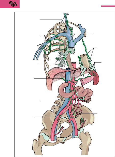
The surveillance of the body by immune cells and their rapid deployment presupposes not only a finely meshed transport system (blood and lymph vessels) but also the organization of cells in lymphoid organs. With the exception of the thymus (see below) the specific immune system in the form of lymphoid organs is localized at the danger sites, the entry portals of pathogens.
The lymphoid organs are divided into two types by their functions (Fig. 6.7):

288 6 Blood, the Immune System, and Lymphoid Organs
|
Adenoids |
|
|
Lingual tonsil |
|
|
Palatine tonsils |
|
Internal jugular vein |
Cervical lymph |
|
|
||
|
nodes |
|
|
Subclavian vein |
|
Thymus |
Left venous |
|
angle |
||
|
Axillary lymph |
|
|
nodes |
|
|
Thoracic duct |
|
|
Spleen |
|
Intestinal |
Cisterna chyli |
|
|
||
lymph node |
|
|
|
Lymphatic trunks |
|
Vermiform |
Lymphoid nodules (nodi |
|
lymphatici aggregati/ |
||
appendix |
||
Peyer’s patches) |
||
|
||
|
in the small intestine |
|
Bone |
Inguinal lymph nodes |
|
marrow |
||
|
||
|
Afferent |
|
|
lymph vessels |
Fig 6.7 Lymphoid organs, lymph vessels, and regional collecting lymph nodes

The Lymphoid Organs (Immune Organs) 289
Primary lymphoid organs, which generate, develop, and mature the immune cells. In the adult this includes especially the thymus (to develop and mature the T cells) and the bone marrow (generates all immune cells, develops and matures the B cells).
Secondary lymphoid organs, to which the immune cells migrate, including the spleen, the lymph nodes, and the lymphoid tissues of the mucous membranes (e. g., tonsils, Peyer’s patches of the small intestine, appendix).
The basic framework of all secondary lymphoid organs is a meshwork of reticular connective tissue, in which numerous lymphocytes are deposited. In places they form round collections of cells, the lymphatic follicles. These may be regarded as the functional units of the secondary lymphoid organs. About 98 % of all lymphocytes are found there and in the connective tissue, while only about 2 % are in the blood. A large proportion of the lymphocytes recirculate between the lymphoid organs and the blood (lymphocyte recirculation; see Gut-Associated Immune System below). The lymphocytes leave the bloodstream in postcapillary venules (transitional vessels between capillaries and veins) within the lymphoid organs, pass through the lymph vessels, and after a certain time return to the peripheral blood, e. g., by the thoracic duct (see Chapter 5: The Lymph Vessels). Under special circumstances, e. g., in inflammations, the lymphocytes can also leave the blood vessels outside the lymphoid organs.
The lymph vessels are a drainage system of the connective tissue. They return to the venous blood tissue fluid that has left the blood vessels and reached connective tissue while transporting substances (see Chapter 5: Substance Exchange between Blood and Tissues). They thus form a parallel pathway to the venous limb of the circulation. Into their course lymph nodes are inserted as “biological filters” where, for example, antigens meet with immune cells. Having proliferated, the lymphocytes leave the lymph nodes and return to the bloodstream and other parts of the body by way of the lymph vessels (see below).

The Lymphoid Organs (Immune Organs) 291
cytes return to the bloodstream and settle in the secondary lymphoid organs such as tonsils, lymph nodes, and spleen. The B lymphocytes that produce antibodies acquire their life-long immune competence in the bone marrow.
The Lymph Nodes
Lymph nodes are biological filters inserted like pearls on a string into the course of the lymph vessels (Fig. 6.7). By their situation they monitor lymph coming from the periphery. Lymph nodes near organs that are the first to receive lymph from an organ or a circumscribed region are designated regional lymph nodes. Lymph nodes that occur beyond these points and receive lymph from several regional lymph nodes are collecting nodes (Fig. 6.9), but they are distinguished from regional lymph nodes only by their location.
In the lymph node, lymphoid tissue is completely surrounded by a solid connective tissue capsule, forming a bean-shaped body several millimeters in size. Several connective tissue septa run from the capsule inward and, together with the basic framework of reticular connective tissue, divide the lymph node into a loose meshwork where numerous lymphatic follicles are deposited (Fig. 6.10). Several afferent lymph vessels pierce the capsule on one side, while on the opposite side usually only one or two vessels leave the lymph node. These are also the sites where the blood vessels enter and leave.
While passing through the lymph node, the lymphatic fluid has considerable contact with the surface of the lymphatic tissue. Cells of the macrophage system monitor and phagocytose foreign bodies, pathogens, and cell debris. When inflammations affect the area they drain, lymph nodes swell, become painful, and are easily palpable. At the same time, the macrophages stimulate (antigen presentation, see p. 283) the lymphocytes to proliferate (divide) and form specific antibodies. Cancer cells also reach the lymph nodes through the lymph, and in this way can develop lymph node metastases. From the lymph node, the antibodyforming plasma cells reach other lymph nodes and eventually the bloodstream by way of efferent lymph vessels.

The Lymphoid Organs (Immune Organs) 293
Fatty tissue |
|
|
|
Connective tissue |
|
Afferent |
septum |
|
lymph vessels |
|
|
|
Subcapsular sinus |
|
Intermediate |
Valve in |
|
sinus |
||
lymph vessel |
||
|
||
Connective |
Medullary cord |
|
tissue capsule |
Cortical |
|
|
||
|
lymphatic follicle |
|
Efferent |
|
|
lymph vessel |
|
Fig. 6.10 Structure of a lymph node
The Spleen (Splen, Lien)
The spleen is the only lymphoid organ in the bloodstream and can be regarded as an organ to monitor and filter the blood. It extracts aging erythrocytes, and provides immunological monitoring of the blood. It is soft, about the size of a fist (150−200 g), and is shaped like a coffee bean. The spleen lies in the left upper abdomen under the diaphragm (Fig. 6.11) and is normally well protected from the outside by the ribs.
If a fresh spleen is cut open, its gross structure can be seen by the naked eye to consist of red tissue (red pulp) interspersed with many small, white splenic nodules surrounded by a connective tissue capsule. The cut surface also reveals cut lymph vessel sheaths. Splenic nodules and lymphatic sheaths consist of lymphoid tissue (white pulp). Red and white pulp are embedded in a tough framework of septa of connective tissue that jut inward from the capsule (Fig. 6.12a, b). The red pulp (about 80 % of the splenic volume) consists of a framework of reticular connective tissue, traversed by a complex system of blood vessels. Each of the smallest branches of the blood vessels is a central vessel running through a splenic nodule. Inside the splenic nodules the lymphatic follicles are arranged in the form of lymphatic cords, where the B lympho-


The Lymphoid Organs (Immune Organs) 295
Numerous capillaries branch from the central artery, each surrounded by a spindle-shaped sheath (sheathed arteries) of densely packed macrophages. The capillaries then drain largely into the meshwork of reticular connective tissue (red pulp) surrounding every splenic sinus (open circulation). Aging erythrocytes are broken down as they pass through the reticular connective tissue. A few capillaries may drain directly into a sinus (closed circulation). The walls of the sinuses are lined with reticular cells, separated by more or less wide openings. At these points the erythrocytes must pass through narrow passages, which can only be surmounted by intact, pliable red cells. Unusable red cells are phagocytosed and broken down by the reticulum cells. In diseases that are accompanied by severe breakdown of blood (e. g., malaria), the spleen can enlarge markedly. Lastly, substances that can be used again can be stored here, e. g., iron from the breakdown of hemoglobin.
Lymphoid Tissues of the Mucous Membranes
The Tonsils
The faucial (palatine), adenoid (pharyngeal), and lingual tonsils together form Waldeyer’s tonsillar (lymphoid) ring (p. 400). To this must be added the lymphatic tissue on the pharyngeal wall, the eustachian tonsil (tubal tonsil, tonsilla tubaria), which lies close to the eustachian tube (connection between the middle ear and the pharynx). The tonsils lie under the epithelium of the oral cavity and their framework is also composed of reticular connective tissue containing lymphatic follicles. In many places the epithelium juts deeply into the lymphatic tissues, thus increasing their superficial contact. By this means, antigens invading through the nose and mouth can make timely contact with immune cells and activate the specific defenses. For instance, in the presence of a massive bacterial invasion, the lymphatic follicles enlarge as a result of the marked increase of antibody-producing lymphocytes. The tension in the connective tissue capsule can become very painful (tonsillitis). In early childhood there is often enlargement of the adenoids (pharyngeal polyps, commonly known as nasal polyps) at the transition from the nose to the pharynx (choanae). This can make it difficult to breathe through the nose.

296 6 Blood, the Immune System, and Lymphoid Organs
Gut-associated Lymphatic Tissue (e. g., Peyer’s Patches)
Because of their large surface, the intestines play a central role in immunity. After all, 70−80 % of all antibody-producing cells are situated in the intestinal wall, the rest being distributed among the other secondary lymphatic organs, the vascular system, and the connective tissue. Diffuse collections and loose associations of lymphocytes (lymphatic follicles) can be found throughout the gastrointestinal tract, which, because of its direct contact with ingested nutrients, is an ideal portal of entry for antigens.
Organized lymphatic tissue is present in the vermiform appendix and in the terminal portion of the small intestine (ileum), where it takes the form of Peyer’s patches in the submucosa and the connective tissue of the mucous membrane (mucosa) of the ileum. These are collections of lymphatic follicles, lying in platelike strands of five to one hundred. They are 1−12 cm in diameter and lie parallel to the axis of the intestine, usually on the side opposite the mesentery (Fig. 6.13a). The number of patches varies between 15 and 50 (up to 250) according to the individual. They are developed before birth and can be demonstrated in the small intestine even late in old age. The areas of the lymphatic follicles are devoid of villi and crypts. Over the lymphatic follicles the connective tissue of the intestinal mucosa, covered with mucosal cells, arches like a dome (Fig. 6.13a−c).
Into the epithelium of the intestinal mucosa are dispersed specific cells that apparently selectively recognize and take up antigenic substances. These M cells stand out as folded surface structures (microfolded cells = M cells) jutting into the intestinal lumen. They are horseshoeshaped, with their closed end toward the intestinal lumen, and appear to be the site where antigens are primarily recognized, as they take up microorganisms and other potentially pathogenic substances and initiate an immune reaction. In this way, the small intestine is protected against future absorption of these antigens.
The defense process may be described as follows. Under the M cell, flanked by its open limbs, embedded as it were in this horse-shaped membranous fold, are T and B lymphocytes and macrophages (Fig. 6.13c). The M cells take the antigenic substance from the intestine and pass it to the macrophages. The macrophages present it by way of the T lymphocytes (T-helper cells) to the B lymphocytes, thus activating them

The Lymphoid Organs (Immune Organs) 297
|
Mesentery |
|
(suspending membrane |
|
of the intestine) |
|
Ileum |
a |
|
|
Kerckring fold |
|
Lymphatic follicle under |
|
domed area without |
|
villi (Peyer’s patch) |
|
Villi of small |
|
intestine |
|
Submucosa |
b |
|
Muscle layer |
Lymphatic follicle under |
c |
mucosal epithelium |
Fig. 6.13 a−c Structure and function of the gut-associated lymphatic system a Site of Peyer’s patches in the ileum
b Lymphatic follicle under a domed area (magnification of a section from a)
(see above Specific Immunity). The B lymphocytes migrate through the lymph and blood vessel systems (lymphocyte circulation, which also takes place in the other lymphoid organs) and are transformed in part here already into antibody-producing plasma cells. The antibodies so
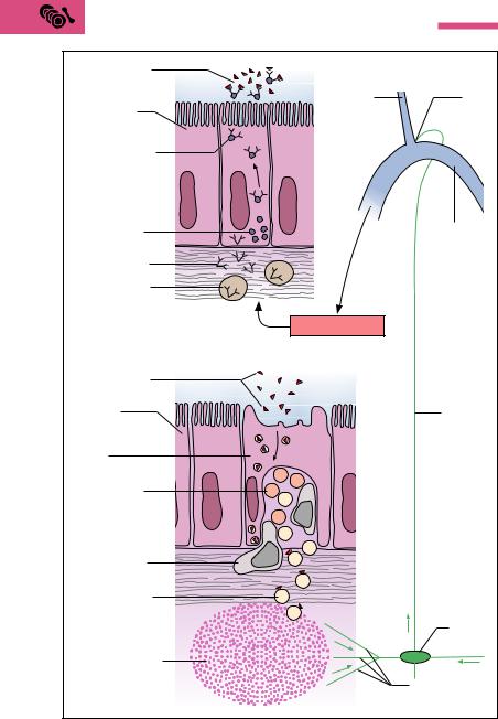
298 6 Blood, the Immune System, and Lymphoid Organs
IgA antibodies |
|
|
|
bind specific |
|
|
|
antigens |
|
Internal |
Left |
|
|
||
Enterocytes |
|
jugular vein |
venous |
of the small |
|
|
angle |
intestine |
|
|
|
Antibodies with |
|
|
|
secretory com- |
|
|
|
ponents are trans- |
|
|
|
ported through |
|
|
|
cells into the |
|
|
|
intestinal lumen |
|
|
|
Secretory |
|
|
Subclavian |
|
|
vein |
|
components |
|
|
|
|
|
|
|
IgA antibodies |
P |
|
|
|
|
|
|
Plasma cells in |
P |
|
|
|
|
|
|
the mucosa |
|
|
|
producing |
|
|
|
IgA antibodies |
|
Blood circulation |
|
|
|
|
|
Antigen in the |
|
|
|
intestinal lumen |
|
|
|
Mucosal |
|
|
Thoracic |
enterocytes |
|
|
duct |
with microvilli |
|
|
|
M cell |
|
|
|
T-helper cells |
T |
T |
|
T |
M |
|
|
|
B |
|
|
|
T |
|
|
|
B |
|
|
|
|
B |
|
Macrophages |
M |
B |
|
|
|
B |
|
B Lymphocytes |
B |
|
|
|
|
B |
|
|
|
|
Lymph |
|
|
|
node |
Lymphatic follicle |
|
|
|
|
|
|
Afferent lymph |
c |
|
|
vessels |

Summary 299
formed (IgA) are partly taken up by the liver cells and passed on to the bile, with which they reach the intestinal lumen. The antibodies so formed also reach other bodily secretions, including the mother’s milk. This benefits the nursing infant, who receives with the mother’s milk specific antigens against pathogens that the gut-associated immune system of the mother has already encountered.
The larger part of the B lymphocytes recirculate into the lymphatic follicles of the Peyer’s patches, where they are transformed into IgA-pro- ducing plasma cells. The antibodies are passed on to the cells of the intestinal mucosa, provided with a layer of glycoproteins (secretory components) to protect them from digestive enzymes and passed into the intestinal lumen. These antibodies bind to the corresponding antigens, which cannot now be absorbed and are therefore excreted.
Summary Blood, the Immune System and Lymphoid Organs
The Blood
The blood (about 8 % of body weight) consists of plasma and cells: erythrocytes (red cells, red corpuscles), leukocytes (white cells), and thrombocytes (platelets).
Erythrocytes
Erythrocytes number 4.5−5.5 million per microliter (mm3) of blood. Function: the transport of respiratory gases. The erythrocytes are formed (160 million per minute) in the bone marrow from nucleated stem cells, but later lose their nuclei and cell organelles. At this point they consist almost entirely of hemoglobin, which can bind oxygen reversibly. They are discoid and biconcave. Their lifespan is about 120 days and they are broken down in the spleen and liver.
In inflammations or increased tissue necrosis, erythrocytes tend to form clumps. In anticoagulated blood they therefore sediment more
Fig. 6.13 c Structure of the epithelium in the domed areas and the mechanism of intestinal mucosal immunity

300 6 Blood, the Immune System, and Lymphoid Organs
rapidly (100 mm/h, otherwise 3−10 mm/h). A raised erythrocyte sedimentation rate is a nonspecific sign of illness.
Leukocytes (Granulocytes, Lymphocytes, Monocytes)
Leukocytes number 4000−8000 per microliter of blood (in inflammation well over 10 000/µl). Function: Together with the lymphoid organs they form the immune system. They can move autonomously by ameboid movement and so can migrate through the capillary walls (connective tissue immunity). As a rule, generation, maturation, and proliferation take place in the bone marrow (the exception is lymphocytes); they are subsequently washed into the blood. The cells are nucleated.
Granulocytes: fine granules in the cytoplasm. (1) Neutrophils: 60− 70 % of leukocytes; phagocytosing cells of nonspecific immunity; decay in the process (forming pus). (2) Eosinophils: 2−3 % of leukocytes; cells of nonspecific immunity that phagocytose antigen−antibody complexes; limit allergic reactions. (3) Basophils: 0.5−1 % of leukocytes; elicit allergic reactions by histamine release; inhibit blood clotting by heparin.
Lymphocytes: 20−30 % of leukocytes; cells of specific immunity; they have a large nucleus and a cytoplasm rich in cell organelles. They are generated in the bone marrow, mature in the bone marrow (B lymphocytes) and thymus (T lymphocytes), and proliferate in the secondary lymphoid organs.
Monocytes: 4−5 % of leukocytes; the largest leukocytes; they leave the blood after 20−30 hours and outside the blood are transformed into macrophages. They are cells of nonspecific immunity (phagocytosis and digestion) but take part in specific immunity (antigen presentation).
Thrombocytes
Thrombocytes number 150 000−350 000 per microliter of blood. Function: hemostasis and triggering clotting of the blood. They are formed in the bone marrow by cytoplasmic extrusions from giant cells, and released into the blood as nonnucleated disks. Their lifespan is 5−10 days and they are broken down in the spleen.
Summary 301
Blood Groups
The heritable blood group antigens are glycolipids and glycoproteins in the cell wall of the erythrocytes that induce the formation of antibodies in foreign organisms:
The ABO system with four blood groups: (1) blood group A (erythrocytes with antigen A), 44 % of the population; (2) blood group B (erythrocytes with antigen B), 10 % of the population; (3) blood group AB (erythrocytes with antigens A and B), 4 % of the population; (4) blood group O (erythrocytes without antigens A or B), 42 % of the population.
The Rh blood group system (antigens = Rh factors C, D, E, mostly D): persons with the Rh factor are Rh-positive (Rh+, 85 % of the population); persons without the Rh factor are Rh-negative (Rh−, 15 % of the population).
From the first months of life, the plasma of each person contains the antibody against the missing antigen of the ABO system (in this case no contact with the foreign antigen is necessary): (1) for blood group A, anti-B; (2) for blood group B, anti-A; (3) for blood group AB, none;
(4) for blood group O, anti-A and anti-B.
Antibodies against Rh antigens are formed only after contact with the antigen.
Blood Transfusions
During a blood transfusion, if blood incompatible with the recipient is given, the reaction of the ABO antigens with their corresponding antibodies leads to erythrocyte agglutination. To prevent mismatched transfusions, the following measures are taken before every transfusion:
Test for ABO blood groups: serological compatibility testing (blood typing)
Test for other antigens and antibodies (e. g., Rh factor): direct and indirect Coombs’ test.
The Plasma
Plasma = blood without cells: 90 % water, 10 % dissolved substances (70 % plasma proteins, 20 % low-molecular-weight substances, 10 % electrolytes).

302 6 Blood, the Immune System, and Lymphoid Organs
Hematocrit = percent blood cells to volume of whole blood. Serum = plasma without fibrinogen.
Plasma proteins:
1.Albumins (35−40 g/l plasma): maintain the colloid osmotic pressure; transport calcium ions, fatty acids, bilirubin, bile acids, a few hormones and vitamins.
2.α1-, α2-, and -Globulins: transport lipids (lipoproteins), hemoglobin, iron, vitamin B12, adrenocortical hormones; constituent of fibrinogen and prothrombin. The lipoproteins are classified by decreasing size and increasing density. The proportion of fat declines with increasing density: very-low-den- sity lipoproteins (VLDL, proportion of fat 90 %), low-density lipoproteins (LDL), high-density lipoproteins (HDL, proportion of fat 50 %). LDL transport lipids (especially cholesterol) from the liver to the tissues (risk of arteriosclerosis); HDL returns excess cholesterol to the liver. Hence high HDL combined with low LDL reduces the risk of arteriosclerosis.
3.γ-Globulins (immunoglobulins, Ig): the antibodies of specific immunity produced by the B lymphocytes (plasma cells: mainly IgA (immune reaction of the mucous membranes, e. g., intestines); IgG (75 % of all immunoglobulins, can cross the placental barrier); IgM (early antibodies); also IgD and IgE.
Low-molecular-weight plasma constituents: nutrients, vitamins, trace elements, metabolic products, hormones, enzymes.
Plasma electrolytes: extracellular (i.e., in the plasma) preponderance of sodium, calcium, and chloride ions; intracellular preponderance of potassium, magnesium, and phosphate ions. NaCl is the most important for osmotic regulation.
Blood Gas Transport in the Blood
Oxygen is transported in the erythrocytes bound to hemoglobin (Hb). In the lung, four oxygen molecules are donated to the bivalent iron atoms of the four heme groups (oxygenation), transported by the blood to the tissues, and released there (deoxygenation). The Hb concentration is about 15 g/dl, so that about 200 ml O2/l can be transported (without Hb, only 3 ml/l).
90 % of carbon dioxide is transported in the plasma as soluble bicarbonate (generated from carbon dioxide with the aid of carbonic

Summary 303
anhydrase in the erythrocytes), 10 % as carbaminohemoglobin bound to hemoglobin and exhaled as carbon dioxide. These processes are linked to the oxygenation and deoxygenation of hemoglobin (buffer function of hemoglobin!).
Carbon monoxide has a higher affinity than oxygen for hemoglobin; 65 % HbCO is life-threatening.
The Anemias
Erythrocyte or hemoglobin deficiency in the blood. Consequence: reduced availability of oxygen in the tissues (symptoms of pale skin and mucous membranes, easy fatigability).
Hypochromic anemia: iron deficiency anemia, from chronic, mostly internal bleeding.
Hyperchromic anemia: deficiencies in vitamin B12 (pernicious anemia), folic acid, or erythropoietin (a hormone generated in the kidney that regulates erythrocyte formation). Consequence: formation of fewer, much larger erythrocytes with increased hemoglobin content (macrocytes).
Aplastic anemia: due to bone marrow aplasia (e. g., after treatment with cytostatics).
Hemolytic anemia: due to increased erythrocyte destruction (e. g., transfusion reactions, Rh incompatibility)
Hemostasis and Coagulation of the Blood
1.Arrest of bleeding (hemostasis): begins immediately after injury. The sequence in order is: vasoconstriction—platelet deposition on the injured vascular walls (platelet plug)—agglutination of thrombocytes and release of the enzyme thrombokinase (= initiation of blood clotting).
2.Coagulation: in the presence of calcium ions, thrombokinase transforms prothrombin present in the plasma into the enzyme thrombin. By the action of thrombin, the fibrous substance fibrin is generated from fibrinogen in a sequence of several reactions in which, apart from fibrinogen, 12 other coagulation factors take part. Together with blood cells deposited at the site, fibrin forms the thrombus, which solidifies after coagulation is complete (retraction).
3.Dissolution of the thrombus in the course of wound healing: catalyzed by the enzyme plasmin, which is formed from plasminogen

304 6 Blood, the Immune System, and Lymphoid Organs
dissolved in the plasma under the influence of activators in the blood and tissues.
A number of mechanisms and combinations provide a constant balance between activation and inhibition of coagulation of the blood (e. g., substances such as antithrombin and heparin in the plasma).
The Immune System
Divided into specific and nonspecific immunity. Nonspecific immunity is inborn; specific immunity is always directed against a definite pathogen and is acquired, i.e., learned. Both systems include cells (cellular immunity) and soluble factors (humoral immunity).
The Nonspecific Immune System
Cellular immunity: this first immune process includes inflammatory phagocytosing cells (neutrophil granulocytes, monocytes, and macrophages) and killer cells specialized against viruses. In addition, macrophages present antigen proteins (antigen fragment + MHC protein: antigen presentation) to the T lymphocytes of the specific immune system, initiating specific immunity.
Humoral immunity: this includes cytokines (generated by monocytes, macrophages), the complement system (enzyme cascade of ca. 20 plasma proteins, eliminating or perforating the walls of bacteria and antigen−antibody complexes) and lysosomes (break down bacterial cell walls enzymatically).
The Specific Immune System
Cellular immunity: includes the T and B lymphocytes.
1.T-helper cells—stimulate the immune system; bond first to the antigen-presenting macrophages, then to the antigen presenting B lymphocytes and stimulate their transformation into antibody-producing plasma cells by interleukins.
2.T-suppressor cells—inhibit the immune system by terminating the immune reaction. (The balance between T-helper and T- suppressor cells is upset in AIDS.)
3.T-killer cells (cytotoxic cells)—work similarly to natural killer cells, but each is directed against a particular virus.

Summary 305
4.B-lymphocytes—early antibodies (IgM) on B lymphocytes; bond to a specific antigen after ingesting it. Antigen fragment + MHC protein = antigen presentation.
5.Memory cells—B lymphocytes, T-helper cells and T-killer cells stimulated by antigens and so responsible for acquired immunity. With repeated contact with the antigen, the generation of antibodies is speeded up.
Humoral immunity: This consists of antibodies IgA, IgD, IgE, IgG, and IgM produced by plasma cells derived from B lymphocytes. The antibodies inactivate the antigens by forming antigen−antibody complexes, which are then eliminated by nonspecific mechanisms (complement system, eosinophil granulocytes).
The Lymphoid Organs
These are divided into primary lymphoid organs, in which generation, development, and maturation of immune cells takes place (thymus, bone marrow), and secondary lymphoid organs, to which immune cells migrate and in which in the course of an immune reaction lymphocytes proliferate and antibodies are formed (spleen, lymph nodes, lymphid tissues of the mucous membranes); 98 % of lymphocytes are found there. The lymphocytes recirculate between secondary lymphoid organs and the blood (lymphoid organ—blood—lymph vessels— thoracic duct—blood—lymphoid organ).
Structure of secondary lymphoid organs: collections of lymphocytes (lymphatic follicles) in reticular connective tissue.
Primary Lymphoid Organs
Thymus: the thymus is the overriding immune organ. This is where cellular immunity develops during embryonic development (e. g., generation of T lymphocytes; differentiation of T-helper cells, T- suppressor cells, and T-killer cells). The thymus gradually involutes throughout life.
Bone marrow: here all immune cells are constantly being generated. B lymphocytes attain their final form in the bone marrow.
Secondary Lymphoid Organs
Lymph nodes: lymph nodes are biological filters inserted into the course of the lymph vessels throughout the lymph vascular system

306 6 Blood, the Immune System, and Lymphoid Organs
and evenly distributed. Secondary lymphoid organs are therefore specific immune defenses localized at nearly every danger point in the body.
The spleen: the spleen is the regulatory and filtration organ of the blood, since it is the only secondary lymphoid organ inserted into the bloodstream.
Lymphoid organs of the mucous membranes: include the tonsils (Waldeyer’s tonsillar ring of pharyngeal, palatine, and lingual tonsils), the lymphatic tissue on the pharyngeal wall, and the gut-as- sociated lymphatic system. Because of its large surface area, the intestinal mucosa is of special significance for immunity; 70−80 % of all antibody-producing cells are located there. Lymphatic follicles are distributed throughout the gastrointestinal mucosa and form organized lymphoid tissue in the vermiform appendix and the terminal small intestine (Peyer’s patches)