

KAPLAN_USMLE_STEP_1_LECTURE_NOTES_2018_BIOCHEMISTRY_and_GENETICS
.pdfPart I ● Biochemistry
The most common types of blots are compared below. Most typically, DNA restriction fragments are analyzed on a Southern blot.
Table I-7-1. Types of Blots
|
|
|
|
|
|
|
Electro- |
|
|
|
|
|
|
|
|
Blot |
|
|
Material |
|
|
|
|
|
|
|
|||
|
|
|
|
|
phoresis |
|
|
|
|
|
|
|
||
|
|
|
|
|
|
|
|
|
|
|
|
|||
|
Type |
|
|
Analyzed |
|
|
Required |
|
Probe Used |
|
|
Purpose |
|
|
|
|
|
|
|
|
|
|
|
|
|
|
|
|
|
|
Southern |
|
DNA |
|
Yes |
|
32P-DNA |
|
To determine which |
|||||
|
|
|
|
|
|
|
|
|
|
|
|
|
restriction fragments |
|
|
|
|
|
|
|
|
|
|
|
|
|
|
of DNA are associ- |
|
|
|
|
|
|
|
|
|
|
|
|
|
|
ated with a particular |
|
|
|
|
|
|
|
|
|
|
|
|
|
|
gene |
|
|
|
|
|
|
|
|
|
|
|
|
|
|
|
|
|
Northern |
|
RNA |
|
Yes |
|
32P-DNA |
|
To measure sizes and |
|||||
|
|
|
|
|
|
|
|
|
|
|
|
|
amounts of specific |
|
|
|
|
|
|
|
|
|
|
|
|
|
|
mRNA molecules to |
|
|
|
|
|
|
|
|
|
|
|
|
|
|
answer questions |
|
|
|
|
|
|
|
|
|
|
|
|
|
|
about gene |
|
|
|
|
|
|
|
|
|
|
|
|
|
|
expression |
|
|
|
|
|
|
|
|
|
|
|
|
|
|
|
|
|
Western |
|
Protein |
|
Yes |
|
125I- or |
|
To measure amount |
|||||
|
|
|
|
|
|
|
|
|
|
enzyme- |
|
of antigen (proteins) |
||
|
|
|
|
|
|
|
|
|
|
linked |
|
or antibody |
||
|
|
|
|
|
|
|
|
|
|
antibody |
|
|
|
|
|
|
|
|
|
|
|
|
|
|
|
|
|
|
|
|
Dot (slot) |
|
RNA, DNA, |
|
No |
|
Same as for |
|
To detect specific |
|||||
|
(Figure |
|
or protein |
|
|
|
|
blots above |
|
DNA, RNA, protein, or |
||||
|
II-6-1) |
|
|
|
|
|
|
|
|
|
|
antibody |
||
|
|
|
|
|
|
|
|
|
|
|
|
|
|
|
Probes
DNA probes are radioactively labeled single-stranded DNA molecules that are able to specifically hybridize (anneal) to particular denatured DNA sequences. Examples include:
•Probes that bind to part of a specific gene region; often produced by cloning cDNA transcribed from the gene and labeling it with 32P,
a radioactive isotope of phosphorus
•Probes that bind to markers known to be in close proximity (closely linked) to a gene
•Probes that bind specifically to a single allele of a gene—allele-specific oligonucleotide (ASO) probes (Figure II-6-2)
When protein is separated and analyzed on a Western blot, 125I-labeled antibody specific for the protein of interest is used as a probe.
The probe is an important part of analyzing any blot because the only bands that will appear on the final autoradiogram are those to which the probe has hybridized (see figure below).
DNA probes are used to selectively detect DNA fragments. Staining with ethidium bromide can be used to visualize and detect all DNA fragments in a gel, provided the fragments are present in sufficient quantities. Ethidium bromide intercalates between stacked bases and fluoresces when exposed to UV light.
104
Southern Blots and Restriction Fragment
Length Polymorphisms (RFLPs)
Chapter 7 ● Techniques of Genetic Analysis
High-Yield
Although unrelated individuals share over 99.9% of their DNA sequences, the fact that the human genome contains over 3 billion base pairs means that 2 unrelated individuals’ DNA will differ at over a million base pairs (0.1% of a billion equals a million). These differences include mutations in restriction endonuclease sites that can be analyzed as RFLPs on Southern blots.
|
|
1.2 kB |
|
|
|
|
0.9 kB |
|
||
|
EcoR1 |
EcoR1 |
EcoR1 |
|||||||
A |
|
|
|
|
|
|
|
|
|
|
|
|
|
|
|
|
|
|
|
|
|
|
|
|
|
|
|
|
|
|
|
|
GAATTC |
GAATTC |
GAATTC |
||||||||
|
|
|
|
|
|
|
|
|
|
|
|
EcoR1 |
|
|
|
|
|
EcoR1 |
|||
B |
PROBE |
|
|
|
|
|||||
|
|
|
|
|
|
|
|
|
||
|
|
|
|
|
|
|
|
|
|
|
GAATTC |
GAGTTC |
GAATTC |
||||||||
|
|
|
|
|
|
|
|
|
|
|
QUESTION: Only one blot below correctly corresponds to the genotypes displayed. Which blot is correct?
2.1 kB |
|
|
|
2.1 kB |
|
|
|
|
|
|
|
|
|
||
1.2 kB |
|
|
|
1.2 kB |
|
|
|
0.9 kB |
|
|
|
0.9 kB |
|
|
|
0.6 kB |
|
|
|
0.6 kB |
|
|
|
|
AA |
AB |
BB |
|
AA |
AB |
BB |
Figure I-7-2. Restriction Fragment Length Polymorphism
Analysis on a Southern Blot
A pair of homologous chromosomes is shown above. They are designated chromosomes A and B. The figure shows the same region on both chromosomes and identifies sites (GAATTC) cut by the restriction endonuclease EcoR1. The probe used on the Southern blot binds to the area of the chromosomes indicated in the diagram. DNA samples from 3 individuals are tested: AA (homozygous for A at this region), AB (heterozygous at this region), and BB (homozygous for B at this region). At the bottom, the figure also presents 2 blots, only one of which correctly represents the results seen on the autoradiogram. Which blot is correct?
(Answer: The blot on the right is correct.)
105

Part I ● Biochemistry
Note
VNTR Sequences and RFLP
Variable number of tandem repeat (VNTR) sequences contribute to some restriction fragment length polymorphisms (RFLPs). A VNTR sequence designates a unit of nucleotides usually between 15 and 60 bp that is repeated in tandem multiple times at a particular location in the DNA. Although the repeated sequence is shared by all individuals, the number of repeated units is variable from person to person.
These VNTR sequences (boxes) can be flanked by restriction endonuclease sites (arrows). The probe used to detect the RFLP would bind to the repeated unit.
106
Repetitive sequences known as VNTRs (variable number of tandem repeats) make some contribution to RFLPs, predominantly in the centromeric and telomeric regions of chromosomes (see margin note: VNTR Sequences and RFLP).
RFLPs and genetic testing
RFLPs may be used in genetic testing to infer the presence of a disease-causing allele of a gene in a family with a genetic disease. If chromosome A in a family also carried a disease-producing allele of a gene in this region and chromosome B carried a normal allele, finding a 1.2 kilobase (kb) band would indicate that the dis- ease-producing allele was also present (both characteristics of chromosome A). Conversely, finding a 2.1-kb fragment would indicate that the normal allele of the gene was present (both characteristics of chromosome B). This type of genetic analysis is more fully discussed in the Medical Genetics section, Chapter 6.
A simple example is illustrated by the following case.
A phenotypically normal man and woman have an 8-year-old son with sickle cell anemia. They also have a 5-year-old daughter who does not have sickle cell anemia but has not been tested for carrier status. The mother is in her 16th week of pregnancy and wishes to know whether the fetus that she is carrying will develop sickle cell disease. The mutation causing sickle cell anemia (G6V) also destroys a restriction site for the restriction endonuclease MstII. DNA from each of the family members is cut with MstII, Southern blotted, and probed with a 32P-labeled cDNA probe for the 5′UTR of the β-globin gene. The results are shown below, along with the family pedigree. What is the best conclusion about the fetus?
|
Mst II Restriction Map of the β-Globin Gene |
||
Normal (A) |
5' |
0.2 |
3' |
|
1.15kb |
kb |
|
Sickle (S) |
5' |
|
3' |
|
1.35kb |
|
|
Pedigree Analysis |
Fetus |
1.35 kB |
1.15 kB |
Figure I-7-3. RFLP Diagnosis of Sickle Cell Disease
(Answer: Fetus is heterozygous, or a carrier, for the mutation.)

Chapter 7 ● Techniques of Genetic Analysis
Both the mother and the father have the same size restriction fragments marking the chromosome with the sickle allele. Because they are genetically unrelated, coming from different families this is not always the case. Additional examples will be discussed in Medical Genetics.
Northern Blots |
High-Yield |
|
Northern blots analyze RNA extracted from a tissue and are typically used to determine which genes are being expressed. One example is shown below. The goal is to determine which tissues express the FMR1 gene involved in fragile X syndrome. RNA samples from multiple tissues have been separated by electrophoresis, blotted, and probed with a 32P-cDNA probe from the FMR1 gene. The results are consistent with high-level expression (a 4.4-kb transcript) of this gene in brain and testis and lower-level expression in the lung. In the heart, the gene is also expressed, but the transcripts are only 1.4 kb long. Variability in the lengths of the mRNAs transcribed from a single gene may be the result of alternative splicing as discussed in Chapter 3, Transcription and RNA Processing.
Skeletal |
Liver |
Testes |
Lung |
Pancreas Heart |
muscle Brain |
4.4 kb
1.4 kb
Figure I-7-4. Northern Blot to Determine Pattern of FMR1 Expression
Gene expression profiling (microarrays)
It is now possible to embed probes for many different mRNA in a multi-well gel or even on a chip to simultaneously determine whether hundreds of genes are expressed in a particular tissue. This is referred to as gene expression profiling or microarray analysis. For example, previous research has suggested that cells from a breast cancer express a variety of genes that are either not expressed or expressed only at a low level in normal cells. Probes for the corresponding mRNAs can be embedded on a solid support and total mRNA from a particular woman’s breast tumor tested with each probe. The pattern of gene expression (gene expression profiling) may give information about the prognosis for that particular woman, aiding in making choices about the appropriate treatment protocol.
Clinical Correlate
Fragile X Syndrome
Fragile X syndrome is the leading known cause of inherited mental retardation. Other symptoms include large ears, elongated face, hypermobile joints, and macroorchidism in postpubertal males. The gene involved, FMR1, maps to the long arm of the X chromosome. See Section II, Chapter 1, for a further discussion of this single-gene disorder.
107

Part I ● Biochemistry
Western Blots
High-Yield
Western blots separate proteins by gel electrophoresis and use 125I-labeled probe antibodies to detect the proteins (antigens). For this reason, Western blots are also referred to as immunoblots.
One important application of Western blotting is to detect the presence of antibodies to the HIV virus in HIV testing (see Immunology Lecture Notes). Western blots may also be used to identify whether a particular protein is in a cell and therefore represent a way to test for gene expression at the level of translation.
Note
The PCR can be used for:
•Comparing DNA samples in forensic cases
•Paternity testing
•Direct mutation testing
•Diagnosing bacterial and viral infections
•HIV testing when antibody tests are uninformative (importantly, infants whose mothers are HIVpositive)
Note
Short tandem repeats (STRs, or microsatellites) are repeats of a dito tetranucleotide sequence. They are useful in genetic testing.
•Occur both in the spacer regions between genes and within gene regions
•In noncoding regions, show some variability in length as mutations have expanded or contracted the number of repeats throughout evolution
•Have known positions that are documented in chromosome maps
POLYMERASE CHAIN REACTION (PCR)
The polymerase chain reaction (PCR) is a technique in which a selected region of a chromosome can be amplified more than a million-fold within a few hours. The technique allows extremely small samples of DNA to be used for further testing. The PCR has many applications.
The region of a chromosome to be amplified by a PCR is referred to as the target sequence and may be an area containing a suspected mutation, a short tandem repeat (STR, or microsatellite sequence), or really any area of interest. The major constraint in performing a PCR is that one must know the nucleotide sequence bordering (flanking) the target region at each of its 3′ ends. This is no longer an obstacle because of the sequence data from the Human Genome Project.
The steps of the PCR include the following:
•Add the sample containing DNA to be amplified.
•Add excess amounts of primers complementary to both 3′ flanks of the target sequence. This selects the region to be amplified.
•Add a heat-stable DNA polymerase (Taq DNA polymerase) and deoxyribonucleotides (dNTPs) for DNA synthesis.
•Heat the sample to melt the DNA (convert dsDNA to ssDNA).
•Cool the sample to re-anneal the DNA. Because the ratio of primers to complementary strands is extremely high, primers bind at the 3′ flanking regions.
•Heat the sample to increase the activity of the Taq DNA polymerase. Primer elongation occurs, and new complementary strands are synthesized.
This process is repeated for approximately 20 cycles, producing over a million double-stranded copies of the target sequence.
108

Chapter 7 ● Techniques of Genetic Analysis
Region of DNA to be amplified
Strand 1 |
3' |
|
|
|
|
|
|
|
|
|
5' |
|
|
|
|
|
|
|||||
|
|
|
|
|
|
|
|
|
|
|
|
|
|
|||||||||
|
|
|
|
|
|
|
||||||||||||||||
|
|
|
|
|
|
|
|
|
|
|
||||||||||||
|
|
|
|
|
|
|
|
|
|
|
||||||||||||
Strand 2 |
5' |
|
|
|
|
|
|
|
|
3' |
|
|
|
|
|
|
||||||
|
|
|
|
|
|
|
|
|
|
|
||||||||||||
|
|
|
|
|
|
|
|
|
|
|
||||||||||||
|
|
|
|
|
|
|
|
|
|
|
|
|
|
|
|
|
|
|
|
|||
|
|
|
|
|
|
|
|
Cycle 1 |
• Add primers. |
|
|
|
||||||||||
|
|
|
|
|
|
|
|
• Heat to separate strands. |
|
|
|
|||||||||||
|
|
|
|
|
|
|
|
|
|
|
• Cool to allow primer-template hybridization. |
|||||||||||
Strand 1 |
3' |
|
|
|
|
|
|
|
|
|
Strand 2 |
5' |
|
|
|
|
|
|
|
|||
|
|
|
|
|
|
|
|
|
|
|
|
|
|
|
|
|||||||
|
|
|
|
|
|
|
|
|
|
|
|
|
|
|
||||||||
|
|
|
|
|
|
|
|
|
|
|
|
|
|
|
||||||||
|
|
|
|
|
|
|
|
|
|
|
|
|
|
|
||||||||
|
|
|
|
|
|
|
|
|
|
|
Add heat-stable DNA polymerase. |
|||||||||||
|
|
|
|
|
|
|
|
|
|
|
||||||||||||
|
|
|
|
|
|
|
|
|
|
|
||||||||||||
Strand 1 |
3' |
|
|
|
|
|
|
|
|
|
Strand 2 |
5' |
|
|
|
|
|
|
5' |
|
|
|
|
|
|
|
|
|
|
|
|
|
|
|
|
|
|
|
|
||||||
|
|
|
|
|
|
|
|
|
|
|
|
|
|
|
|
|||||||
|
|
|
|
|
|
|
|
|
|
|
|
|
|
|
|
|||||||
|
5' |
|
|
|
|
|
|
|
|
|
|
|
|
|
|
|
|
|||||
|
|
|
|
|
|
|
|
|
|
|
|
|
|
|
|
|
||||||
|
|
|
|
|
|
|
|
|
|
|
|
|
|
|
|
|
||||||
|
|
|
|
|
|
|
|
|
|
|
|
|
||||||||||
|
|
|
|
|
|
|
|
Cycle 2 |
Heat and cool (with primers and |
|
|
|
||||||||||
|
|
|
|
|
|
|
|
|
|
|
DNA polymerase present). |
|
|
|
||||||||
|
|
|
|
|
|
|
|
|
|
|
|
|
|
|
|
|
|
|
|
|
|
|
Strand 1 3'


Strand 2 5' 


Strand 1 3'


Strand 2 5' 


Cycle 3 Repeat heating and cooling cycle.
Strand 1 3'


Strand 2 5' 


Cycles 4 to 20 Multiple heating and cooling cycles.
Present in about 106 copies
Figure I-7-5. Polymerase Chain Reaction
109

Part I ● Biochemistry
Agarose gel electrophoresis of PCR products
If the known mutation changes the length of the gene (e.g., microsatellite repeats), this difference can be detected in the PCR-amplified DNA by electrophoresis on agarose gel.
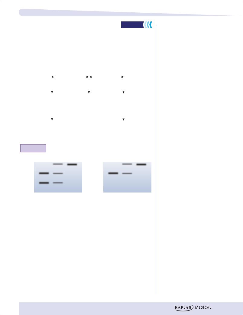
A 59-year-old man with increasing clumsiness, loss of balance, and irregular tremor and jerkiness in both arms seeks medical attention. The physician also notes impaired visual tracking. The patient has one sister, age 53. His father and mother died in a car accident at ages 45 and 43, respectively. There is no known history of neurologic dysfunction in his family. He takes a multiple vitamin tablet daily but no prescription drugs or supplements. MRI reveals visible atrophy of the caudate and total basal ganglia, and a tentative diagnosis of Huntington disease is made. To confirm the diagnosis, a sample of blood is sent for molecular genetic testing. PCR amplification is carried out on a region on 4p16.3 suspected to contain a mutation. The results are shown below, along with results from a normal, healthy, age-matched control. Is this result consistent with the diagnosis?
Patient Control
170 bp
104 bp
101 bp
95 bp
Figure I-7-6. Direct Genetic Diagnosis of Neurodegenerative
Disease Using PCR
(Answer: Yes, the results are consistent with Huntington disease. In comparison with the normal control PCR products, one of the patient’s PCR products (170 bp) is well out of the normal range. This is consistent with a triplet repeat expansion in that allele of the huntingtin gene. The 2 bands from the control are 95 base pairs and 101 base pairs, a difference of 2 triplet repeats. The patient sample shows a 104 base pair band, a difference of one triplet repeat from the larger control band. Significantly, there is a difference of 69 base pairs between the patient’s 2 bands: 104 base pairs versus 170 base pairs. This is equivalent to 23 triplet repeats.)
110


Chapter 7 ● Techniques of Genetic Analysis
•DNA sample to be sequenced
•Excess primers
•dNTPs
•DNA polymerase
ddATP ddCTP ddGTP ddTTP
Figure I-7-8. DNA Sequencing
The sequence of the newly synthesized DNA strand in this example is 5’CTTG-
GAACTGTA 3’.
If one wants the sequence of the original strand, serving as the template in the sequencing procedure, it would be complementary and anti-parallel to the sequence read from the gel. In this example the original strand sequence would be 5’TACAGTTCCAAG 3’.
PCR in HIV Testing
The enzyme-linked immunosorbent assay (ELISA) is used to screen individuals for antibodies to the HIV virus. The test has high sensitivity but somewhat lower specificity. A positive result in an ELISA must be confirmed by a Western blot for antibodies that are reactive with specific HIV protein antigens (see Immunology Lecture Notes).
In certain instances, ELISA/Western blot is not useful and PCR is the test of choice to detect HIV infection.
•PCR is designed to test for the integrated proviral genome, not for antibodies to HIV protein antigens.
•Primers that are specific for the HIV provirus are used for the PCR.
•If the person is infected, the proviral genome will be amplified and detected. If the person is not infected, there will be no PCR product detected.
113