

Опухоли легких, плевры, тимуса и сердца ВОЗ
.pdfChoriocarcinoma
M.R. Wick
A.Zettl J.K.C. Chan
C.Bokemeyer E.J. Perlman
A.Marx
Definition
Choriocarcinoma is a highly malignant neoplasm displaying trophoblastic differentiation. It is composed of syncytiotrophoblast, cytotrophoblast and variably intermediate trophoblast cells. Mediastinal choriocarcinomas are morphologically indistinguishable from their gonadal or uterine counterparts.
ICD-O code |
9100/3 |
Epidemiology
Pure mediastinal choriocarcinomas are exceedingly rare and virtually non-exis- tent in children. Only 2.5 to 5% of mediastinal germ cell tumours are pure choriocarcinomas {997,1033,1356}. Pure choriocarcinoma constitute 9% of malignant mediastinal nonseminomatous germ cell tumours, and 4.3% of nonteratomatous germ cell tumours {1033,1349, 1356}.
Clinical features
The patients` ages range from 17 to 63 (most commonly the 3rd decade of life), and almost all reported cases were male patients {520,1821,1955}. At diagnosis, mediastinal choriocarcinomas are mostly large anterior mediastinal masses (average size 10 cm) {1357}. Primary chorio-
carcinomas have also been observed in the posterior mediastinum {1357}.
The patients present with symptoms due to the mediastinal mass, such as chest pain, dyspnoea, cough, superior vena cava syndrome. Patients may show gynecomastia due to elevated beta-hCG levels {1356,1821}. Mediastinal choriocarcinomas are highly aggressive neoplasms with early haematogeneous dissemination. In a series of 8 cases, metastases were observed in the lungs (88%), liver (50%), kidney (38%) and spleen (25%). Metastatic disease to the brain, heart, adrenals and bone has also been observed. {1033,1357,1821}.
Morphology
Mediastinal choriocarcinomas are large tumours with soft consistency and extensive hemorrhage and necrosis. Microscopically, they are composed of syncytiotrophoblast, cytotrophoblast and intermediate trophoblastic cells. Syncytiotrophoblasts are large multinucleated cells with numerous, pleomorphic, dark-staining nuclei, distinct nucleoli, and abundant densly eosinophilic cytoplasm which may contain cytoplasmic lacunae. Cytotrophoblasts are uniform, polygonal cells with round nuclei, prominent nucleoli, and clear cytoplasm.
Syncytiotrophoblasts and cytotrophoblasts may grow intermingled in a bilaminar plexiform pattern or in disordered sheets. Occasionally, scattered clusters of syncytiotrophoblasts cap cytotrophoblast nodules. Atypical mitosis and cellular atypia are common. There can be sheets of nondescript mononuclear cells that resemble intermediate trophoblast. Choriocarcinomas are typically intimately associated with dilated vascular sinusoids. Partial or complete replacement of the walls of blood vessels are common. There are often vast areas of haemorrhage and necrosis.
Mediastinal choriocarcinoma cannot be distinguished morphologically from metastatic choriocarcinoma. Since gonadal choriocarcinoma often displays extensive regressive alterations but still may give rise to widespread metastasis, the exclusion of a primary gonadal choriocarcinoma is particularily difficult, although mediastinal metastasis of gonadal choriocarcinoma seem to be very rare {997,1356}.
Trophoblastic neoplasms other than choriocarcinoma, such as monophasic choriocarcinoma and placental site trophoblastic tumour have not been reported in the mediastinum.
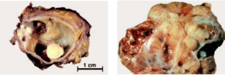
A B
Fig. 3.78 Choriocarcinoma. A High power showing multinucleated and eosinophilic syncytiotrophoblastic cells intertwined with mononuclear cytotrophoblastic cells. B beta-hCG staining of syncytiotrophoblastic cells.
Choriocarcinoma 209
Immunohistochemistry
Syncytiotrophoblasts and cytotrophoblasts react with pankeratin markers and CAM5.2, whereas they are negative for PLAP, AFP, CEA, CD30 and vimentin. The syncytiotrophoblasts additionally express beta-hCG, while the cytotrophoblasts are variably positive for human placental lactogen {1357,1920}.
Apart from metastasis of an extramediastinal choriocarcinoma, the differential diagnoses include mediastinal mixed germ cell tumour (in which a further germ
cell tumour component is found), sarcomatous component in teratoma, and mediastinal metastasis from a carcinoma with choriocarcinoma-like features/dedifferentiation.
Genetics
The genetic changes that have been described in mediastinal choriocarcinoma are the same as those reported in the testis and demonstrate the isochromosome i(12p) characteristic of postpubertal malignant germ cell tumours at all
Teratoma
Definitions
A germ cell tumour (GCT) that is composed of several types of organoid mature and/or immature somatic tissues derived from two or three germinal layers (ectoderm, endoderm and mesoderm).
Mature teratomas are tumours composed exclusively of mature, adult-type tissues. Dermoid cyst is a variant consisting of one or more cysts lined predominantly by keratinizing squamous epithelium with skin appendages. Monodermal teratomas analogous to struma ovarii have not been described in the mediastinum.
Immature teratomas contain immature, embryonic or fetal tissues exclusively or in addition to mature tissues. Mature and most immature mediastinal teratomas are benign tumours {1053,1244,1764, 1808}.
Teratomatous component is the term used to describe differentiated somatic tissues associated with a seminoma, embryonal carcinoma, yolk sac tumour or choriocarcinoma. The teratomatous component of mixed GCTs is very often immature {1808}.
Teratoma with somatic-type malignancy is a teratoma containing one or more
components of non-germ cell malignant tumour, which may be a sarcoma or a carcinoma (ICD-O code 9084/3).
ICD-O codes |
|
Mature teratoma |
9080/0 |
Immature teratoma |
9080/3 |
Epidemiology
Mediastinal teratomas account for 7- 9.3% of mediastinal tumours {708,1171} and 50–70% of all mediastinal germ cell tumours {466,520,759,1356,1458, 1888,2116} Among teratomas of all sites, up to 27% occur in the mediastinum in adults, and 4-13% in children {708,1053,1763}.
Overall, there is an equal sex distribution {1171} or a slight female preponderance (M:F =1:1.4) {1808}, but immature tera-
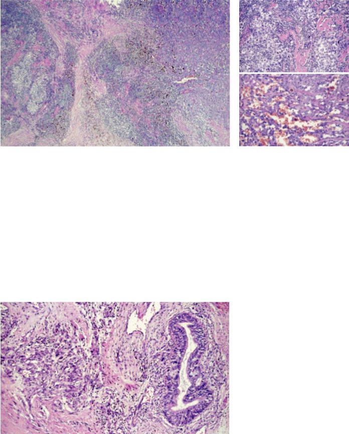
Fig. 3.79 Mature cystic mediastinal teratoma. The yellow areas represent mature fat.
sites {316}. Such tumours are most commonly aneuploid, with a minority having near-tetraploid DNA content.
Prognosis
In most of the reported cases, patients died of disseminated disease shortly after diagnosis (average survival time 1 to 2 months) {520,1357,1821}. However, treament with cisplatin-based chemotherapy may improve the prognosis {1955}.
M.R. Wick |
P. Ströbel |
E.J. Perlman |
J.C.K. Chan |
U. Göbel |
A. Marx |
D. T. Schneider |
|
tomas occur almost exclusively in males {1808}.
The mean age of adults is 28 years (range 18–60) {1171}. In children, teratoma is the predominant mediastinal tumour during the first year and has been detected in fetuses as young as 28 weeks of gestation {708}. The proportion of immature teratomas (up to 40%) is much higher in the first year of life than at older age (~4-6%) {1053,1356,2116}. Mature teratoma can be associated with classical (47, XXY) and very rarely, mosaic Klinefelter syndrome {480}.
Clinical signs and symptoms
30-59% of mediastinal mature teratomas, particularly those in adults {1955}, are asymptomatic {708,1171, 1337}. Other cases can be associated
Fig. 3.80 Immature mediastinal teratoma with a variegated cut surface. Cystic areas are less prominent than in mature teratoma.
210 Tumours of the thymus - Germ cell tumours

A B
Fig. 3.81 Mature teratoma. A Dermoid cyst-like area (left), mature cartilage (top, right), mature intestinal type glands and villi (bottom, right). B High power of pancreatic tissue, including islets.
with chest, back or shoulder pain, dyspnoea, cough, and fever due to chronic pneumonia {708,1764}. Rare symptoms include superior vena cava syndrome, erosion of bronchi or vessels, Horner syndrome, or pneumothorax {696,708, 1764,1808}. Due to the occurrence of exocrine pancreatic tissue, rupture is more common in mediastinal teratomas than teratomas of other sites {1808,2038} and can result in pleural effusions or cardiac tamponade. Endocrine pancreatic component can cause hyperinsulinism and hypoglycaemia {1808}. Hydrops fetalis is a complication of congenital intraand extrapericardial mediastinal teratoma {708}.
Imaging
Mature teratomas show multilocular cystic structures in almost 90% of cases {1888}. Attenuation is heterogeneous with varying combinations of soft tissue,
fluid, fat and calcium {1337}. Calcifications occur in in 26% {1171} to 53% {1337}. A shell-like tumour wall calcification or identifiable bone and teeth occur in up to 8% each {1171, 1337}. Immature teratomas appear more often solid {1888}. With rare exceptions {1244}, the usual serum tumour markers (AFP; beta-hCG) are not elevated.
Site of involvement
More than 80% of mature teratomas occur in the anterior mediastinum, 3-8% in the posterior mediastinum and 2% in the middle mediastinum, while 13-15% involve multiple mediastinal compartments {963,1337,1829}. Teratomas can extend deeply into one or both thoracic cavities and elicit atelectasis.
Macroscopy
Mature mediastinal teratomas are usually encapsulated masses with a mean dia-
meter of 10 cm (range 3-25 cm) {1655, 2008,2116}. There can be adhesions to the surrounding lung or great vessels. The cut surface is variegated, showing cystic spaces with fluid or grumous materials, hair, fat, flecks of cartilage, and rarely teeth or bone {1808,1888}.
Immature teratomas are often very large (up to 40 cm) {1975} and solid. They exhibit a soft to fleshy consistency or are extensively fibrous or cartilaginous {1808}. Haemorrhage and necrosis can be present.
Histology
Mature teratomas
These are characterized by a haphazard admixture of organoid mature tissues derived from 2 or 3 germinal layers. Skin and cutaneous appendages are consistent constituents and form cyst linings. Bronchial, neural, gastrointestinal, smooth muscle and adipose tissue com-
A B
Fig. 3.82 Immature teratoma. A Immature neural tissue forming tubes. B Immature cartilage.
Teratoma 211
ponents are very frequent (>80%), while skeletal muscle, bone and cartilage are less common {708,1808}. Salivary gland, prostate, liver and melanocytes are even less frequent; thyroid tissue has not been reported {1808}. Pancreatic tissue is typical of mediastinal teratomas and found in up to 60% of cases, but is rare or absent in teratomas of other sites {521, 1808}.
Regressive changes, such as rupture of cystic structures, can be accompanied by a granulomatous inflammation {1808, 2116}. Remnant thymic tissue is found outside the capsule in 75% of mature teratomas {708}.
Immature teratoma
These lesions are characterized by embryonic or fetal tissues derived from the various germinal layers, such as immature glands lined by tall columnar epithelial cells, fetal lung, immature cartilage and bone, rhabdomyoblasts, blastema-like stromal cells. The most common immature components are neuroectodermal tissues, with neuroepithelial cells forming tubules, rosettes or retinal anlage {708,1808,2116}. By definition, pure immature teratoma should not harbour a morphologically malignant component.
Immunohistochemistry
The main role of immunohistochemistry in teratomas is: (i) to define the nature of immature components, such as rhab
domyoblasts (desmin, myogenin), neural components (S100; NSE) or immature cartilage (S100; GFAP) {1490}, and (ii) to exclude other germ cell or somatic malignancies. Pure teratomas are negative for PLAP, beta-hCG and CD30. AFP is usually negative, although liver cells and immature neuroepithelium in teratomas may express AFP.
Grading of immature teratoma
There are insufficient data to support a particular grading system for immature teratomas of the mediastinum. Grading according to Gonzalez-Crussi {708} was of no prognostic significance in children {696,1244,1764,1808}. However, it is important to realize the following: 1) the more immaturity is present in a teratoma, the higher the risk to find a yolk sac tumour component; 2) immaturity in a teratoma in an adolescent male is highly suspicious of a malignant i(12p)-contain- ing germ cell tumour. Therefore, the pathologist should communicate clearly in the report the quantity (rough percentage) of immaturity.
Genetics
The pure mature and immature teratomas analyzed and reported to date do not show recurrent genetic gains and losses. This is in contrast to malignant germ cell tumours {1765}. Mature teratoma can be associated with classical and very rarely mosaic Klinefelter syndrome {480}.
Differential diagnosis
The main differential diagnosis is mixed germ cell tumour with a teratomatous component. Immature teratoma may be difficult to distinguish from teratoma with somatic type malignancy; the latter usually shows frank cytologic atypia and invasiveness that are absent in pure immature teratomas.
Prognostic factors
Mature teratoma is a benign tumour irrespective of the patient´s age. The prognosis of pure immature teratoma is agedependent. In children, pure immature teratoma has an excellent prognosis with no risk of recurrence and metastasis {1244,1764}. The presence of an admixed malignant germ cell tumour component (detected in up 30% of immature teratomas after extensive sampling {1244}, and most commonly yolk sac tumour) is associated with a recurrence rate of 25%. In children, such mixed GCTs have a good prognosis after cisplatin-based chemotherapy (>80% 3- years-survival) {695,1244}.
In adults, the prognosis of pure immature teratoma is more guarded but experience is limited {2116}. Apparently pure immature teratomas with pulmonary metasases have been reported in adults, with only the metastasis showing a germ cell and/or somatic type malignancy {1808}.
212 Tumours of the thymus - Germ cell tumours
Mixed germ cell tumours
M.R. Wick |
F. Mayer |
E.J. Perlman |
Ph. Ströbel |
U. Göbel |
J.K.C. Chan |
D.T. Schneider |
A. Marx |
Definition
A neoplasm composed of two or more types of germ cell tumours (GCTs). The diagnosis should be complemented by listing each component and its approximate proportion.
Polyembryoma represents a variant with a unique growth pattern that is characterized by the predominance of embryoid body-like structures. Embryonal carcinoma, yolk sac tumour, syncytiotrophoblastic cells and teratomatous components can usually be recognized in polyembryoma.
Embryonal carcinomas or seminomas containing scattered syncytiotrophoblastic cells do not qualify as mixed GCTs, but are classified as the respective “pure” GCTs.
ICD-O code
Polyembryoma 9072/3
Synonyms
Malignant teratoma intermediate, teratocarcinoma. The use of terms that do not precisely qualify the type and quantity of tumour components is discouraged.
Epidemiology
In adults, mixed GCTs account for 1325% of all mediastinal GCTs {466,520, 1005,1033,2116}, second only to teratomas (40-60%) and as common as seminomas (15-20%) {520,1005,1356, 1808,2116}. Virtually all patients are male {1005,1356}.
In children, mixed GCTs account for about 20% of cases, and yolk sac tumour with mature or immature teratoma is the characteristic constellation. Other types of mixed GCTs are virtually nonexistent during the first four years of life {1764}. Among children <8 years of age, some authors {1005,1765} but not others {167} see a preponderance of females, while almost all adolescent patients > 8 years are males {1765}.
After the onset of puberty, mixed germ cell tumours can be associated with Klinefelter syndrome {150,795,1106, 1290,1765}.
Clinical features
Only ~10% of mixed GCTs are asymptomatic at diagnosis {1955}. Most patients present with general and local symptoms identical to those in other mediastinal GCT: chest pain, cough, dyspnoea, hoarseness, superior vena cava syndrome and cardiac tamponade {757, 1955}. Precocious puberty and gynecomastia are rare in polyembryoma {150} and other mixed GCTs {1106,1808}. In some cases, endocrinologic symptoms induced by beta-hCG production may precede tumour diagnosis by years {1769}.
A minority of patients present with symptoms attributable to metastases {709, 757,1955}. Clonally related leukaemias are rare (~2%) {40,729,789,1518}.
Imaging studies typically show a large inhomogeneous mass with necrosis, hemorrhage and infiltration of adjacent structures. Cystic spaces or adipose tissue hint to the presence of a teratomatous component {757,1888}.
Most cases (~90%) show elevated serum tumour marker levels {1005}. Raised AFP (~80%) is strongly correlated with a yolk sac tumour component, although teratomatous hepatoid cells and teratomatous neuroepithelium can also produce small amounts of AFP. Increased beta-hCG (~30%) levels occur in mixed GCTs with a choriocarcinoma component or with syncytiotrophoblast cells {2169}.
Post-chemotherapy findings, including the Growing Teratoma Syndrome
During or following chemotherapy, patients with GCT can alternatively show {2169}: (1) Normalization of tumour markers and resolution of the tumour mass (10%), (2) persistence of elevated tumour markers and the tumour mass due to resistance to chemotherapy (10%), or (3) normalization of tumour markers with residual tumour mass (80%).
In the latter group, 10-20% of patients exhibit tumour enlargement. This phenomenon can be due to a) chemo- therapy-resistant GCT components that
do not secrete AFP or beta-hCG; b) development of somatic-type malignancies; c) the “growing teratoma syndrome” (GTS). GTS is a rare complication of mixed GCTs {24} and defined by 1) an increase in tumour size during or after chemotherapy; 2) normalization of serum tumour markers; 3) identification exclusively of mature teratoma on histological analysis of the resected tumour specimen {1199}. The growing mediastinal mass is usually asymptomatic but can be accompanied by fever and dyspnoea {24,1256}. Lymphatic spread can involve mediastinal and supraclavicular lymph nodes {56}. Late GTS complications are local or metastatic development of malignant non-seminomatous GCTs and development of GCT-related sarcomas, carcinomas or leukaemias {56,1256}. The pathogenesis of GTS is largely unknown.
Tumour spread
Most mixed GCT exhibit extensive infiltration into mediastinal structures and adjacent organs. Rates of metastasis at time of diagnosis vary widely in different reports, from 20-36% {520,1356,1764, 1955} up to >80% {997,2169}. Metastasis to lung, pleura, lymph node, liver, bone and brain have been reported {156,1005,1765,1955}. Metastases to supraclavicular lymph nodes and lung due to occult mediastinal mixed GCTs are rare {708}.
Macroscopy
The tumours are often poorly circumscribed or frankly infiltrative, and show a heterogeneous cut surface with solid areas, haemorrhage and necrosis. Cystic spaces usually indicate presence of a teratomatous component. The size ranges between 3 and 20 cm (mean 10 cm) {908}. Tumours in the context of the “growing teratoma syndrome” measure up to 28 cm {24}.
Histopathology
Various types of GCTs can occur in any combination in mediastinal mixed GCTs. Their morphologies are identical to those
Mixed germ cell tumours 213
of pure GCTs. The reported frequencies of the various GCT subtypes vary widely in the literature, but the following conclusions can be drawn:
In adults, the two most frequent components are teratoma (50-73%; mean 65%) and embryonal carcinoma (22–100%; mean 66%) {237,520,1005,1356,1955}. Less common are yolk sac tumour (0-83%; mean 48%), seminoma (22-50%, mean 38%), and choriocarcinoma (10–67%, mean 28%) {520,1005,1033, 1356,1955}. The teratoma components are more often immature than mature {17, 1033,1808}. The most common combination is teratoma and embryonal carcinoma (previously called teratocarcinoma), encounterd in 15-56% of cases (mean 40%) {237,1005,1808,1955}.
In children, a yolk sac tumour component occurs in most (>90%) mixed GCTs, followed by teratoma (~30%), and, in adolescents, seminoma, choriocarcinoma and embryonal carcinoma (~20% each) {709,1764}. In contrast to adults, the teratoma components in paediatric mixed GCTs are more often mature than immature {167,1725,1765}.
Polyembryomas {150} show a unique growth pattern mimicking embryoid bodies. These GCTs are composed of EC, YST, syncytiotrophoblast cells and teratoma. Adult, but not childhood, mediastinal mixed GCTs are frequently associated with non-germ cell malignancies (sarcomas, carcinomas, and/or leukaemias).
Histology of metastasis
The histology of metastases usually reflects the histology of the primary GCT or one of its components {17} but other GCT histologies and somatic type malignancies may occur, particularly after chemotherapy {56,406,1230,1256,2046}.
Postchemotherapy histology
After chemotherapy, viable non-tera- tomatous tumour occurs in up to 50% of cases even after normalization of serum tumour markers {1808,2049}. In the remaining cases, areas of necrosis, teratoma structures, inflammatory infiltrates including xanthogranulomatous reactions, and fibrosis can be encountered. Chemotherapy may unmask a previously overlooked somatic-type tumour or a teratomatous component. Metastases do not necessarily reflect the histology of remnant viable tumour cells in the primary location {56,1256}.
Immunohistochemistry
The immunohistochemical profiles reflect those of the various germ cell tumour components contributing to a given mixed GCT. AFP is expressed in virtually all mixed GCTs, at least focally, due to the frequent occurrence of yolk sac tumour components.
Genetics
In adults and children > 8 years old, gain of 12p and sex chromosomal abnormalities (often associated with Klinefelter syndrome) are the most common recurrent abnormalities of mediastinal mixed GCTs {258,1765}, including polyembryoma {150}. Additional recurrent changes include gain of chromosome 21 and loss of chromosome 13. These abnormalities are also encountered in the mature teratoma component and/or somatic-type malignant components of mixed GCTs, while pure teratomas are typically devoid of genetic imbalances {1394,1765}.
In children < 8 years old, i(12p) does not occur {1765}, and gain of the X chromosome and trisomy 21 {258,1765} are rare findings. Instead, gain of 1q, 3, and 20q and loss of 1p, 4q, and 6q are common {258,1765} in yolk sac tumour; teratomatous elements show no chromosomal abnormalities.
Postulated cell of origin
Totior pluripotent primordial germ cell.
Differential diagnosis
Embryonal carcinoma components may be difficult to recognize against a background of yolk sac tumour due to the heterogeneity of yolk sac tumour growth patterns. CD30 staining is a helpful diagnostic adjunct to resolve this differential. Due to the lack of cytotrophoblastic cells, scattered syncytiotrophoblasts in “pure” seminomas and embryonal carcinomas can be distinguished from the choriocarcinoma components of mixed GCTs.
Prognostic factors
In adults, mixed GCTs exhibit a long-term survival rate of 40-45% {1359} and there appears to be no significant difference between mixed and pure NSGCTs {1955}. Therefore, tumour stage, particularly metastasis to brain, liver, lung, and bone, and elevated beta-hCG levels might be major risk factors for mixed GCTs as for NSGCTs {199,790}. Modern
cisplatin-based chemotherapies and resection are the treatment of choice {199,236,1462,2172}.
In children, mixed GCTs usually harbour only yolk sac tumour and teratomatous components and their prognosis is not different from the prognosis of pure yolk sac tumour {1764}, suggesting that 5-year overall survival rates of >80% can be achieved with modern therapies {1764}. Local stage, distant metastasis and AFP levels have not been shown to be of prognostic significance in a recent paediatric series of NSGCTs that includes 24% mixed GCTs {1764}. In young children, mixed GCTs exhibiting microscopically small foci of NSGCTs in teratomas have a good prognosis after complete resection and chemotherapy {1244}.
Small series suggest that histology, specifically an extensive seminoma component of mixed GCTs, has a beneficial impact on survival {1349,1359}, while a choriocarcinoma component might indicate a more aggressive clinical course {466,2116}.
Postchemotherapy prognostic factors
Postchemotherapy findings are the most important prognostic factors in the era of multimodality treatments. Primary complete response, i.e. normalization of tumour marker levels and disappearance of the mediastinal mass after chemotherapy occurs in 10% of NSGCT patients and is associated with 80% long-term survival {587}. 20% of such patients relapse usually within 2 years after chemotherapy and may be amenable to salvage therapy after early detection of the relapse {2169}.
Among patients that show normalization of tumour markers and a residual tumour mass (80% of cases) {587,588,2169, 2172}, completeness of resection is the most important prognostic factor in adults {199} and children {11,1764}: salvage rates after incomplete resection are <10% in adults and <50% in children.
In addition, postchemotherapy histology has a bearing on prognosis {660,1655}: complete lack of viable tumour cells is associated with a 90% disease-free survival rate, while the rate drops to 60% if viable teratoma, including the growing teratoma syndrome, is encountered. Viable non-teratomatous GCT tumour or somatic-type malignant cells are associated with a 30% and <10% survival rate, respectively.
214 Tumours of the thymus - Germ cell tumours
Patients with persistently elevated tumour markers have a worse prognosis than patients with normalization of tumour markers, although viable tumour cells are detectable in only half of the respective resection specimens {997}.
Relapses after chemotherapy and surgery and primary resistance to chemotherapy are poor prognostic factors due to low salvage rates {199}.
Mixed germ cell tumours 215

Germ cell tumours with somatic-type malignancy
M.R. Wick
E.J. Perlman
Ph. Ströbel
J.K.C. Chan
A. Marx
Definition
A germ cell tumour (GCT) accompanied by a somatic-type malignant component of sarcoma, carcinoma or both. Leukaemias or lymphomas are also somatic-type neoplasms that can accompany mediastinal GCTs
Synonyms
Teratoma with malignant transformation {1394}; Malignant teratoma with nongerminal malignant tumour {1808}; Teratoma with non-germ cell malignancy.
Comments
Tumours included in this category have been collectively called “Teratomas with malignant components” etc. in the literature. However, since somatic-type malignancies are more common in mixed germ cell tumours than in teratomas {709,1230,1515,1808,2046} and can also occur in pure yolk sac tumours {2048} and seminomas {879,2047}, the germ cell tumour component that accompanies the somatic malignancy should be specified accordingly.
A minimum size of one low-power field has been suggested as the threshold for the diagnosis of somatic-type malignancy in GCTs {1655,2047}. However, this size criterion is arbitary. More important is the independent growth pattern demonstrated by the somatic-type malignancy. It would be helpful to estimate the size and percentage areas occupied by the somatic malignancy and give this information in the patholgy report.
Epidemiology
GCTs with somatic-type malignancies are rare (~ 2% of all male GCTs) {30}. About 25-30% of cases occur in the mediastinum {2047}. They account for up to 29% of all mediastinal GCTs of adults {40,199,1230,1394,1450,1808,1955}, but are almost non-existent in children {277,406,428,1005,1232,1764}. With few exceptions {277,428,1033}, the tumours occur in males. The age range is from 4- 66 years {277,406,1005,1033,1230,
1356,1385}, with most cases occurring between 20-40 years.
The somatic-type malignancies may arise in the mediastinum or only in the metastases {277,1394,1808}. They are more common after chemotherapy and in tumours of late recurences {1665}. After removal of apparently mature teratomas, metastases with pure sarcomatous features have been rarely reported {277,1808}.
according to the malignant components that are present.
Imaging studies typically reveal a solid mass (representing the sacoma or carcinoma component) associated either with a cystic teratomatous structure or with a lesion showing heterogeneous attenuation, predominant areas of enhancing soft tisuue elements, calcifications and massive necrosis {1888}.
Tumour spread
Clinical features
Signs and symptoms
The tumours show the same local symptoms as other mediastinal GCTs, but they are more frequently symptomatic (~90%) than pure teratomas (~50%) {1808}. Symptoms due to metastatic disease may accompany or follow local symptoms {1394}.
Most but not all cases show elevated AFP and/or beta-hCG levels in the serum. Other tumour markers (e.g. carcinoembyonic antigen [CEA] or neuronspecific enolase [NSE]) may be elevated
Table 3.11
Sarcoma and carcinoma components can infiltrate into the mediastinal structures and the lung {1230}. Metastases have been reported in the majority of cases {1230,1505} and can be composed either of the somatic-type tumour {406,1230}, the GCT or one of its components {1230}, or of both somatic-type and germ cell tumour {1230,2046}. Metastatic spread may involve lung {406,1394}, regional lymph nodes {40,997}, bone {1515,2186}, brain {2046,2186}, liver {40,2046} and spleen {1394,2046}.
Mediastinal germ cell tumours and associated somatic-type malignancies.
Germ cell tumour component |
Frequency1 |
Somatic-type malignancies2 |
Teratoma (mature; immature) |
~ 10-20% |
Sarcomas/Neurogenic Tumours |
|
|
Rhabdomyosarcoma |
|
|
Angiosarcoma |
|
|
Neuroblastoma |
|
|
Liposarcoma |
|
|
Leiomyosarcoma |
Non-teratomatous GCT of one |
< 5% |
Osteo-, Chondrosarcoma, Ewing |
histological type (most commonly |
|
sarcoma/PNET3 |
seminoma or yolk sac tumour) |
|
Malignant fibrous histiocytoma |
|
|
MPNST4 |
|
|
Glioblastoma |
|
|
|
Mixed germ cell tumours |
> 75% |
Epithelial Malignancies |
(almost all cases contain |
|
Adenocarcinoma |
teratoma components) |
|
Adenosquamous carcinoma |
|
|
Squamous cell carcinoma |
|
|
Undifferentiated carcinoma |
|
|
Haematological malignancies |
|
|
|
1Percentage of all mediastinal GCTs with somatic-type malignancies
2More than one type of sarcoma and/or carcinoma can occur in a single GCT, and haematologic neoplasias can accompany sarcomas {1394}
3PNET, primitive neuroectodermal tumour; 4 MPNST, malignant peripheral nerve sheath tumour
216 Tumours of the thymus - Germ cell tumours
A
B
Fig. 3.83 Germ cell tumour (GCT) with somatic-type malignancy: Seminoma (left) and angiosarcoma (right).
Fig. 3.85 GCT with somatic-type malignancy (STM): A Seminoma and B epithelioid angiosarcoma.
Macroscopy
The tumours range in size from 6 to 30 cm {1230,1356}. They usually exhibit a partially cystic and often variegated cut surface with focally necrotic areas. The carcinoma or sarcoma areas are firm and gray or haemorrhagic (e.g. angiosarcoma) and often adherent to adjacent mediastinal structures {2046}.
Histopathology
Mature {277,428,1033,1385} and immature {520,1195,1505,2186} teratomas, in addition to seminomas, yolk sac tumours or mixed germ cell tumours can be asso-
ciated with various sarcomas (63% of cases) {428,2046,2047}, carcinomas (37%) {1385,1808}, combinations of both {1033,1808,2047} or carcinosarcoma {1808}. The somatic malignancy can be intimately intermingled with the GCT component, or forms an expansile nodular proliferation of atypical cells, often with increased mitotic rate and necrosis. Embryonal rhabdomyosarcoma {428,1230,1450} is the single most frequent somatic-type malignancy. Angiosarcoma {1230,2046}, leiomyosarcoma {1450} and neuroblastoma {397,1505} are also common. Any other
Fig. 3.84 Germ cell tumour with somatic-type malignancy. Immature teratoma and angiosarcoma.
type of sarcoma or combinations {1230} may occur, including chondrosarcoma, osteosarcoma, malignant fibrous histiocytoma, malignant peripheral nerve sheath tumour, glioblastoma {1808}, and liposarcoma {1359}.
The non-mesenchymal component can be adenocarcinoma (usually of colonic type) {1385,1394,1808,2047}, adenosquamous carcinoma {2047}, squamous cell carcinoma {1655} or primitive neuroectodermal tumours (PNET) {1655}. Melanocytic neuroectodermal tumours {49} and carcinoids {1707} are rare.
Immunohistochemistry
Somatic-type malignancies stain like their counterparts occurring elsewhere in the body. PLAP, AFP, beta-hCG, and CD30 are generally not expressed, while they can be detected in “pure” GCTs and the respective components of mixed GCTs. One should keep in mind that rhabdomyoblasts, embryonal rhabdomyosarcomas and leiomyosarcomas can express PLAP {700} and that hepatoid carcinomas can be AFP-positive.
Genetics
An isochromosome i(12p) genotype shared by the somatic-type neoplasia and the associated germ cell tumour component is typical {789,1113,1394}. In a case of teratoma-associated rhabdomyosarcoma, an add(2)q35-q37 genetic abnormality that is characteristic
Germ cell tumours with somatic-type malignancy 217

for rhabdomyosarcoma was detected in the sarcoma but not the germ cell component {1394}. Thus, tissue-specific secondary chromosomal aberrations may be necessary for the development of somat- ic-type tumour components in GCTs. Klinefelter syndrome has been reported in association with GCT with somatictype malignancy {2186}.
Postulated cell of origin
Malignant transformation of mature teratoma cells or divergent differentiation of a plurior totipotent primordial germ cell towards a germ cell tumour and the somatic-type malignancy have been suggested {314}. The latter hypothesis is favoured by the finding that “pure” mature mediastinal teratomas show no chromosome 12 abnormalities {1765} while a shared i(12p) abnormality is characteristic of teratomas that are clonally related with somatic type malignancies, including leukaemias {1394}.
Differential diagnosis
Immature teratoma may be difficult to distinguish from teratoma with somatictype malignancy. Frank atypia and infiltrative growth favour the latter interpretation. Likewise, chemotherapy-induced atypia is usually diffusely distributed throughout the tumour, while somatictype malignancy is a focal process often forming a recognizable mass and invading adjacent structures {1888}.
Scattered rhabdomyoblasts are a frequent feature of mature and immature teratomas and do not justify a diagnosis of rhabdomyosarcoma unless they show nodular tumour formation and/or infiltration of adjacent structures.
Rhabdomyoblasts can rarely occur in thymic carcinomas. The thymic carcino-
A B
C D
Fig. 3.86 Germ cell tumour with somatic-type malignancy. A Angiosarcoma component of the case shown in Fig. 3.85. B CD31 expression of the same case. C Immature teratoma and rhabdomyosarcoma. D Adenocarcinoma component in immature teratoma.
ma is morphologically different form GCT and commonly expresses CD5, while the rhabdomyoblasts are devoid of atypia and proliferative activity.
Prognostic factors
Presence of somatic-type malignancy in a GCT confers a dismal prognosis {406,520,709,997,1005,1230,1394,1515, 1655,2047}. There is no response to chemotherapy used for treatment of germ cell tumours. Only a minority of patients will survive after chemotherapy and complete surgical removal of mediastinal tumour remnants {879,1394, 2047}. Advanced local infiltration, metastatic disease, and incomplete resection are bad prognostic factors {997,1230,
2047}, while the type of somatic malignancy in the primary biopsy has no major impact on survival {1394}. Persistence of viable tumour after chemotherapy heralds an unfavourable outcome {660, 997,2169}. The median survival is only approximately 9 months {406,520, 709, 997,1005,1230,1394,1515,1655, 2047}.
218 Tumours of the thymus - Germ cell tumours