World Health Organization Classification of Tumours
WHO |
OMS |
International Agency for Research on Cancer (IARC)
Pathology and Genetics of
Tumours of the Digestive System
Edited by
Stanley R. Hamilton
Lauri A. Aaltonen
IARCPress
Lyon, 2000

World Health Organization Classification of Tumours
Series Editors Paul Kleihues, M.D.
Leslie H. Sobin, M.D.
Pathology and Genetics of Tumours of the Digestive System
Editors |
Stanley R. Hamilton, M.D. |
|
Lauri A. Aaltonen, M.D., Ph.D. |
Clinical Editor |
René Lambert, M.D |
Editorial Assistance |
Wojciech Biernat, M.D. |
|
Norman J. Carr, M.D. |
|
Anna Sankila, M.D. |
Layout |
Sibylle Söring |
|
Felix Krönert |
Illustrations |
Georges Mollon |
|
Sibylle Söring |
Printed by |
Team Rush |
|
69603 Villeurbanne, France |
Publisher |
IARCPress |
|
International Agency for |
|
Research on Cancer (IARC) |
|
69372 Lyon, France |

This volume was produced in collaboration with the
International Academy of Pathology (IAP)
and
with support from the
Swiss Federal Office of Public Health, Bern
The WHO Classification of Tumours of the Digestive System presented in this book reflects the views of a
Working Group that convened for an Editorial and Consensus Conference in Lyon, France, November 6-9, 1999.
Members of the Working Group are indicated in the List of Contributors on page 253.

Published by IARC Press, International Agency for Research on Cancer, 150 cours Albert Thomas, F-69372 Lyon, France
© International Agency for Research on Cancer, 2000 reprinted 2006
Publications of the World Health Organization enjoy copyright protection in accordance with the provisions of Protocol 2 of the Universal Copyright Convention. All rights reserved.
The International Agency for Research on Cancer welcomes
requests for permission to reproduce or translate its publications, in part or in full. Requests for permission to reproduce figures or charts from this publication should be directed to
the respective contributor (see section Source of Charts and Photographs).
The designations used and the presentation of the material in this publication do not imply the expression of any opinion whatsoever on the part of the Secretariat of the
World Health Organization concerning the legal status of any country, territory, city, or area or of its authorities, or concerning the delimitation of its frontiers or boundaries.
The mention of specific companies or of certain manufacturers' products does not imply
that they are endorsed or recommended by the World Health Organization in preference to others of a similar nature that are not mentioned. Errors and omissions excepted,
the names of proprietary products are distinguished by initial capital letters.
The authors alone are responsible for the views expressed in this publication.
Enquiries should be addressed to the
Editorial & Publications Service, International Agency for Research on Cancer, 69372 Lyon, France, which will provide the latest information on any changes made to the text and plans for new editions.
Format for bibliographic citations:
Hamilton S.R., Aaltonen L.A. (Eds.): World Health Organization Classification of Tumours. Pathology and Genetics of Tumours of the Digestive System. IARC Press: Lyon 2000
IARC Library Cataloguing in Publication Data
Pathology and genetics of tumours of the digestive system / editors, S.R. Hamilton and L.A. Aaltonen
(World Health Classification of tumours ; 2)
1. Digestive System Neoplasms I. Aaltonen, L.A. II. Hamilton, S.R.
III. Series
ISBN 92 832 2410 8 |
(NLM Classification: W1) |

Contents
Diagnostic terms and definitions |
8 |
|
Endocrine tumours |
137 |
|
|
|
|
B-cell lymphoma |
139 |
|
1 Tumours of the oesophagus |
9 |
|
Mesenchymal tumours |
142 |
|
|
|
|
|||
WHO and TNM classifications |
10 |
7 |
Tumours of the anal canal |
145 |
|
Squamous cell carcinoma |
11 |
||||
|
WHO and TNM classifications |
146 |
|||
Adenocarcinoma |
20 |
|
|||
Endocrine tumours |
26 |
|
Tumours of the anal canal |
147 |
|
Lymphoma |
27 |
|
|
|
|
Mesenchymal tumours |
28 |
8 |
Tumours of the liver and |
|
|
Secondary tumours and melanoma |
30 |
157 |
|||
|
intrahepatic bile ducts |
||||
|
|
|
|||
2 Tumours of the oesophagogastric junction |
31 |
|
WHO and TNM classifications |
158 |
|
|
Hepatocellular carcinoma |
159 |
|||
Adenocarcinoma |
32 |
|
Intrahepatic cholangiocarcinoma |
173 |
|
|
|
|
Combined hepatocellular and cholangiocarcinoma 181 |
||
3 Tumours of the stomach |
37 |
|
Bile duct cystadenoma and cystadenocarcinoma |
182 |
|
|
Hepatoblastoma |
184 |
|||
|
|
|
|||
WHO and TNM classifications |
38 |
|
Lymphoma |
190 |
|
Carcinoma |
39 |
|
Mesenchymal tumours |
191 |
|
Endocrine tumours |
53 |
|
Secondary tumours |
199 |
|
Lymphoma |
57 |
|
|
|
|
Mesenchymal tumours |
62 |
9 |
Tumours of the gallbladder and |
|
|
Secondary tumours |
66 |
|
|||
|
extrahepatic bile ducts |
203 |
|||
|
|
|
|||
4 Tumours of the small intestine |
69 |
|
WHO and TNM classifications |
204 |
|
|
Carcinoma |
206 |
|||
|
|
|
|||
WHO and TNM classifications |
70 |
|
Endocrine tumours |
214 |
|
Carcinoma |
71 |
|
Neural and mesenchymal tumours |
216 |
|
Peutz-Jeghers syndrome |
74 |
|
Lymphoma |
217 |
|
Endocrine tumours |
77 |
|
Secondary tumours and melanoma |
217 |
|
B-cell lymphoma |
83 |
|
|
|
|
T-cell lymphoma |
87 |
10 |
Tumours of the exocrine pancreas |
219 |
|
Mesenchymal tumours |
90 |
||||
|
WHO and TNM classifications |
220 |
|||
Secondary tumours |
91 |
|
|||
|
|
|
Ductal adenocarcinoma |
221 |
|
5 Tumours of the appendix |
93 |
|
Serous cystic neoplasms |
231 |
|
|
Mucinous cystic neoplasms |
234 |
|||
|
|
|
|||
WHO and TNM classifications |
94 |
|
Intraductal papillary-mucinous neoplasm |
237 |
|
Adenocarcinoma |
95 |
|
Acinar cell carcinoma |
241 |
|
Endocrine tumours |
99 |
|
Pancreatoblastoma |
244 |
|
Miscellaneous tumours |
102 |
|
Solid-pseudopapillary neoplasm |
246 |
|
|
|
|
Miscellaneous carcinomas |
249 |
|
6 Tumours of the colon and rectum |
103 |
|
Mesenchymal tumours |
249 |
|
|
Lymphoma |
250 |
|||
WHO and TNM classifications |
104 |
|
|||
|
Secondary tumours |
250 |
|||
Carcinoma |
105 |
|
|
|
|
Familial adenomatous polyposis |
120 |
Contributors |
253 |
||
Hereditary nonpolyposis colorectal cancer |
126 |
||||
Source of charts and photographs |
261 |
||||
Juvenile polyposis |
130 |
||||
References |
265 |
||||
Cowden syndrome |
132 |
||||
Hyperplastic polyposis |
135 |
Subject index |
307 |
||

Diagnostic terms and definitions1
Intraepithelial neoplasia2. A lesion characterized by morphological changes that include altered architecture and abnormalities in cytology and differentiation. It results from clonal alterations in genes and carries a predisposition for progression to invasion and metastasis.
High-grade intraepithelial neoplasia.
A mucosal change with cytologic and architectural features of malignancy but without evidence of invasion into the stroma. It includes lesions termed severe dysplasia and carcinoma in situ.
Polyp. A generic term for any excrescence or growth protruding above a mucous membrane. Polyps can be pedunculated or sessile, and are readily seen by macroscopic examination or conventional endoscopy.
Adenoma. A circumscribed benign lesion composed of tubular and/or villous structures showing intraepithelial neoplasia. The neoplastic epithelial cells are immature and typically have enlarged, hyperbasophilic and stratified nuclei.
Tubular adenoma. An adenoma in which branching tubules surrounded by lamina propria comprise at least 80% of the tumour.
Villous adenoma. An adenoma in which leaf-like or finger-like processes of lamina propria covered by dysplastic epithelium comprise at least 80% of the tumour.
Tubulovillous adenoma. An adenoma composed of both tubular and villous structures, each comprising more than 20% of the tumour.
Serrated adenoma. An adenoma composed of saw-toothed glands.
Intraepithelial neoplasia (dysplasia) associated with chronic inflammatory diseases. A neoplastic glandular epithelial proliferation occurring in a patient with a chronic inflammatory bowel disease, but with macroscopic and microscopic features that distinguish it from an adenoma, e.g. patchy distribution of dysplasia and poor circumscription.
Peutz-Jeghers polyp. A hamartomatous polyp composed of branching bands of smooth muscle covered by nor- mal-appearing or hyperplastic glandular mucosa indigenous to the site.
Juvenile polyp. A hamartomatous polyp with a spherical head composed of tubules and cysts, lined by normal epithelium, embedded in an excess of lamina propria. In juvenile polyposis, the polyps are often multilobated with a papillary configuration and a higher ratio of glands to lamina propria.
Adenocarcinoma. A malignant epithelial tumour with glandular differentiation.
Mucinous adenocarcinoma. An adenocarcinoma containing extracellular mucin comprising more than 50% of the tumour. Note that ‘mucin producing’ is not synonymous with mucinous in this context.
Signet-ring cell carcinoma. An adenocarcinoma in which the predominant component (more than 50%) is composed of isolated malignant cells containing intracytoplasmic mucin.
Squamous cell (epidermoid) carcinoma. A malignant epithelial tumour with squamous cell differentiation.
Adenosquamous carcinoma. A malignant epithelial tumour with significant components of both glandular and squamous differentiation.
Small cell carcinoma. A malignant epithelial tumour similar in morphology, immunophenotype and behaviour to small cell carcinoma of the lung.
Medullary carcinoma. A malignant epithelial tumour in which the cells form solid sheets and have abundant eosinophilic cytoplasm and large, vesicular nuclei with prominent nucleoli. An intraepithelial infiltrate of lymphocytes is characteristic.
Undifferentiated carcinoma. A malignant epithelial tumour with no glandular structures or other features to indicate definite differentiation.
Carcinoid. A well differentiated neoplasm of the diffuse endocrine system.
______________
1 This list of terms is proposed to be used for the entire digestive system and reflects the view of the Working Group convened in Lyon, 6 – 9 November, 1999. Terminology evolves with scientific progress; the terms listed here reflect current understanding of the process of malignant transformation in the digestive tract. The Working Group anticipates a further convergence of diagnostic terms throughout the digestive system.
2 In an attempt to resolve confusion surrounding the terms ‘dysplasia’, ‘carcinoma in situ,’ and ‘atypia’, the Working Group adopted the term ‘intraepithelial neoplasia’ to indicate preinvasive neoplastic change of the epithelium. The diagnosis does not exclude the possibility of coexisting carcinoma. Intraepithelial neoplasia should not be used as a generic description of epithelial abnormalities due to reactive or regenerative changes.

CHAPTER 1
Tumours of the Oesophagus
Carcinomas of the oesophagus pose a considerable medical and public health challenge in many parts of the world. Morphologically and aetiologically, two major types are distinguished:
Squamous cell carcinoma
In Western countries, oesophageal carcinomas with squamous cell differentiation typically arise after many years of tobacco and alcohol abuse. They frequently carry G:C >T:A mutations of the TP53 gene. Other causes include chronic mucosal injury through hot beverages and malnutrition, but the very high incidence rates observed in Iran and some African and Asian regions remain inexplicable.
Adenocarcinoma
Oesophageal carcinomas with glandular differentiation are typically located in the distal oesophagus and occur predominantly in white males of industrialized countries, with a marked tendency for increasing incidence rates. The most important aetiological factor is chronic gastro-oesophageal reflux leading to Barrett type mucosal metaplasia, the most common precursor lesion of adenocarcinoma.

WHO histological classification of oesophageal tumours
Epithelial tumours |
|
Non-epithelial tumours |
|
|
Squamous cell papilloma |
8052/01 |
Leiomyoma |
8890/0 |
|
Intraepithelial neoplasia2 |
|
Lipoma |
8850/0 |
|
Squamous |
|
Granular cell tumour |
9580/0 |
|
|
Gastrointestinal stromal tumour |
8936/1 |
||
Glandular (adenoma) |
|
|||
|
benign |
8936/0 |
||
Carcinoma |
|
|||
|
uncertain malignant potential |
8936/1 |
||
Squamous cell carcinoma |
8070/3 |
|||
malignant |
8936/3 |
|||
Verrucous (squamous) carcinoma |
8051/3 |
|||
Leiomyosarcoma |
8890/3 |
|||
Basaloid squamous cell carcinoma |
8083/3 |
|||
Rhabdomyosarcoma |
8900/3 |
|||
Spindle cell (squamous) carcinoma |
8074/3 |
|||
Kaposi sarcoma |
9140/3 |
|||
Adenocarcinoma |
8140/3 |
|||
Malignant melanoma |
8720/3 |
|||
Adenosquamous carcinoma |
8560/3 |
|||
Others |
|
|||
Mucoepidermoid carcinoma |
8430/3 |
|
||
|
|
|||
Adenoid cystic carcinoma |
8200/3 |
Secondary tumours |
|
|
Small cell carcinoma |
8041/3 |
|
||
|
|
|||
Undifferentiated carcinoma |
8020/3 |
|
|
|
Others |
|
|
|
|
Carcinoid tumour |
8240/3 |
|
|
|
|
|
|
|
_____________
1 Morphology code of the International Classification of Diseases for Oncology (ICD-O) {542} and the Systematized Nomenclature of Medicine (http://snomed.org). Behaviour is coded /0 for benign tumours, /1 for unspecified, borderline or uncertain behaviour, /2 for in situ carcinomas and grade III intraepithelial neoplasia, and /3 for malignant tumours.
2Intraepithelial neoplasia does not have a generic code in ICD-O. ICD-O codes are available only for lesions categorized as glandular intraepithelial neoplasia grade III (8148/2), squamous intraepithelial neoplasia, grade III (8077/2), and squamous cell carcinoma in situ (8070/2).
TNM classification of oesophageal tumours
TNM classification1 |
|
|
|
|
|
|
T – Primary Tumour |
|
|
|
|
|
|
TX |
Primary tumour cannot be assessed |
For tumours of upper thoracic oesophagus |
|
|||
T0 |
No evidence of primary tumour |
M1a |
Metastasis in cervical lymph nodes |
|||
Tis |
Carcinoma in situ |
|
M1b |
Other distant metastasis |
||
T1 |
Tumour invades lamina propria or submucosa |
For tumours of mid-thoracic oesophagus |
|
|||
T2 |
Tumour invades muscularis propria |
M1a |
Not applicable |
|
||
T3 |
Tumour invades adventitia |
M1b |
Non-regional lymph node |
|||
T4 |
Tumour invades adjacent structures |
|
or other distant metastasis |
|||
N – Regional Lymph Nodes |
|
Stage Grouping |
|
|
|
|
NX |
Regional lymph nodes cannot be assessed |
|
|
|
|
|
N0 |
No regional lymph node metastasis |
Stage 0 |
Tis |
N0 |
M0 |
|
N1 |
Regional lymph node metastasis |
Stage I |
T1 |
N0 |
M0 |
|
|
|
|
Stage IIA |
T2 |
N0 |
M0 |
M – Distant Metastasis |
|
|
T3 |
N0 |
M0 |
|
MX |
Distant metastasis cannot be assessed |
Stage IIB |
T1 |
N1 |
M0 |
|
M0 |
No distant metastasis |
|
|
T2 |
N1 |
M0 |
M1 |
Distant metastasis |
|
Stage III |
T3 |
N1 |
M0 |
|
For tumours of lower thoracic oesophagus |
|
T4 |
Any N |
M0 |
|
|
M1a |
Metastasis in coeliac lymph nodes |
Stage IVA |
Any T |
Any N |
M1a |
|
M1b |
Other distant metastasis |
Stage IVB |
Any T |
Any N |
M1b |
_____________
1 {1, 66}. This classification applies only to carcinomas.
2 A help desk for specific questions about the TNM classification is available at http://tnm.uicc.org.
10 Tumours of the oesophagus

Squamous cell carcinoma of the oesophagus
H.E. Gabbert |
Y. Nakamura |
T. Shimoda |
J.K. Field |
P. Hainaut |
H. Inoue |
Definition
Squamous cell carcinoma (SCC) of the oesophagus is a malignant epithelial tumour with squamous cell differentiation, microscopically characterised by keratinocyte-like cells with intercellular bridges and/or keratinization.
ICD-O Code |
8070/3 |
Epidemiology
Squamous cell carcinoma of the oesophagus shows great geographical diversity in incidence, mortality and sex ratio. In Western countries, the age-standar- dized annual incidence in most areas does not exceed 5 per 100,000 population in males and 1 in females. There are, however, several well-defined high-risk areas, e.g. Normandy and Calvados in North-West France, and Northern Italy, where incidence may be as high as 30 per 100,000 population in males and 2 in females {1020, 1331}. This type of cancer is much more frequent in Eastern countries and in many developing countries. Regions with very high incidence rates have been identified in Iran, Central China, South Africa and Southern Brazil. In the city of Zhengzhou, capital of Henan province in China, the mortality rate exceeds 100 per 100,000 population in males and 50 in females {1116, 2191}.
In both high-risk and low-risk regions, this cancer is exceedingly rare before the age of 30 and the median age is around 65 in both males and females.
Recent changes in the distribution pattern in France indicate that the rate of SCC has increased steadily in low-risk areas, particularly among females, whereas there may be a slight decrease in high-risk areas. In the United States, a search in hospitalisation records of military veterans indicates that SCC is 2-3 times more frequent among blacks than among Asians, Whites or Native Americans {453}.
Aetiology
Tobacco and alcohol. In Western countries, nearly 90% of the risk of SCC can be attributed to tobacco and alcohol. Each of these factors influences the risk of oesophageal cancer in a different way. With regard to the consumption of tobacco, a moderate intake during a long period carries a higher risk than a high intake during a shorter period, whereas the reverse is true for alcohol. Both factors combined show a multiplicative effect, even at low alcohol intake. In high-risk areas of North-West France and Northern Italy, local drinking customs may partially explain the excess incidence of SCC {523, 1020}. In Japanese alcoholics, a
polymorphism in ALDH2, the gene encoding aldehyde dehydrogenase 2, has been shown to be significantly associated with several cancers of the upper digestive tract, including squamous cell cancer. This observation suggests a role for acetaldehyde, one of the main carcinogenic metabolites of alcohol in the development of oesophageal carcinoma {2177}.
Nutrition. Risk factors other than tobacco and alcohol play significant roles in other regions of the world. In high-risk areas of China, a deficiency in certain trace elements and the consumption of pickled or mouldy foods (which are potential sources of nitrosamines) have been suggested.
Hot beverages. Worldwide, one of the most common risk factors appears to be the consumption of burning-hot beverages (such as Mate tea in South America) which cause thermal injury leading to chronic oesophagitis and then to precancerous lesions {1116, 2191, 387}.
HPV. Conflicting reports have proposed a role for infectious agents, including human papillomavirus (HPV) infection. Although HPV DNA is consistently detected in 20 to 40% of SCC in high-risk areas of China, it is generally absent in the cancers arising in Western countries {954, 679}.
|
|
|
7.8 |
|
China, Henan |
|
|
|
|
|
|
|
|
|
|
|
|
1% |
|
|
|
|
|
|
|
|
|
|
|
|
|
|
|
|
|
|
|
||||
5.2 |
|
|
|
Iran, North East |
|
|
|
|
|
|
|
|
|
|
|
|
|
20% |
|
|
|
|
|
|
|
|
|
|
|
|
|
|
|
|
|
|
|
||||
|
|
|
|
|
|
|
|
|
|
|
|
|
|
|
|
|
|
|||
|
|
|
|
|
|
|
|
|
|
|
|
|
|
|
|
|
|
|
||
|
|
|
|
19.3 |
South Africa |
|
|
|
50% |
|
|
|
|
|
|
|
|
|
|
|
|
|
|
51.6 |
India, Bombay |
|
50% |
|
|
|
|
|
|
|
|
|
|
|
|
||
|
|
|
|
|
|
|
|
|
|
|
|
|
|
|
|
|
||||
|
|
|
|
|
Urugay |
|
70% |
|
|
|
|
|
|
|
|
|
|
|
|
|
|
|
|
|
|
|
|
|
|
|
|
|
|
|
|
|
|
|
|||
|
|
|
|
|
China, Hong Kong |
|
|
|
|
|
|
|
|
|
|
|
|
|
|
|
|
|
|
|
|
|
70% |
|
|
|
|
|
|
|
|
|
|
|
|
||
11.0 |
|
11.9 |
|
4.6 |
Italy, North East |
|
90% |
|
|
|
|
|
|
|
|
|
|
|
|
|
|
|
|
|
|
|
|
|
|
|
|
|
|
|
|
|
|||||
|
|
|
|
USA, New York |
|
90% |
|
|
|
|
|
|
|
|
|
|
|
|
|
|
|
|
|
|
|
France, Calvados |
|
|
90% |
|
|
|
|
|
|
|
|
|
|
|
|
|
|
|
|
|
|
|
|
|
|
|
|
|
|
|
|
|
|
|||
|
|
|
|
|
|
|
|
|
|
|
|
|
|
|
|
|
|
|
|
|
|
< 3.8 |
< 5.8 |
< 9.5 |
|
0 |
50 |
100 |
150 |
200 |
|||||||||||
< 2.2 |
< 51.7 |
|
|
|
|
|
|
|
|
|
|
|
|
|
|
|
|
|||
|
|
|
|
|
|
|
|
|
|
|
|
|
|
|
||||||
Fig. 1.01 Worldwide annual incidence (per 100,000) of oesophageal cancer in |
Fig. 1.02 Squamous cell carcinoma of the oesophagus. Age-standardized incidence |
|||||||||||||||||||
males. Numbers on the map indicate regional average values. |
rates per 100,000 and proportions (%) due to alcohol and tobacco (dark-blue). |
|||||||||||||||||||
Squamous cell carcinoma 11

A |
B |
C |
D |
E |
F |
Fig. 1.03 Macroscopic images of squamous cell carcinoma (SCC) of the oesophagus. A Flat superficial type. B Lugol iodine staining of the specimen illustrated in A. C Polypoid SCC. D Longitudinal sections of carcinoma illustrated in C. E Deeply invasive polypoid SCC. F Longitudinal sections of carcinoma illustrated in E.
Associations between achalasia, Plum- mer-Vinson syndrome, coeliac disease and tylosis (focal nonepidermolytic palmoplantar keratoderma) with oesophageal cancer have also been described.
Localization
Oesophageal SCC is located predominantly in the middle and the lower third of the oesophagus, only 10-15% being situated in the upper third {1055}.
Clinical features
Symptoms and signs
The most common symptoms of advanced oesophageal cancer are dysphagia, weight loss, retrosternal or epigastric pain, and regurgitation caused by narrowing of the oesophageal lumen by tumour growth {606}. Superficial SCC usually has no specific symptoms but sometimes causes a tingling sensation, and is, therefore, often detected incidentally during upper gastrointestinal endoscopy {464, 1874}.
Endoscopy and vital staining
Superficial oesophageal cancer is commonly observed as a slight elevation or shallow depression on the mucosal surface, which is a minor morphological change compared to that of advanced cancer. Macroscopically, three types can be distinguished: flat, polypoid and ulcerated. Chromoendoscopy utilizing toluidine blue or Lugol iodine spray may be of value {465, 481}. Toluidine blue, a metachromatic stain from the thiazine group, has a particular affinity for RNA and DNA, and stains areas that are richer in nuclei than the normal mucosa. Lugol solution reacts specifically with glycogen in the normal squamous epithelium, whereas precancerous and cancerous lesions, but also inflamed areas and gastric heterotopia, are not stained. However, the superficial extension of carcinomas confined to the mucosa can not be clearly recognized by simple endoscopy.
Endoscopic ultrasonography
Endoscopic ultrasonography is used to evaluate both depth of tumour infiltration and para-oesophageal lymph node involvement in early and advanced stages of the disease {1509, 1935}. For the evaluation of the depth of infiltration, high frequency endoscopic ultrasonography may be used {1302}. In general,
12 Tumours of the oesophagus
Fig. 1.04 Catheter probe ultrasonograph of a squamous cell carcinoma, presenting as hypoechoic lesion (arrow).
oesophageal carcinoma presents on endosonography as a circumscribed or diffuse wall thickening with a predominantly echo-poor or echo-inhomoge- neous pattern. As a result of tumour penetration through the wall and into surrounding structures, the endosonographic wall layers are destroyed.
Computed tomography (CT) and magnetic resonance imaging (MRI)
In advanced carcinomas, CT and MRI give information on local and systemic spread of SCC. Tumour growth is characterized as swelling of the oesophageal wall, with or without direct invasion to surrounding organs {1518}. Cervical, abdominal and mediastinal node enlargement is recorded. Three-dimensional CT or MRI images may be presented as virtual endoscopy, effectively demonstrating T2-T4 lesions, but not T1 lesions.
Macroscopy
The gross appearance varies according to whether it is detected in an early or an advanced stage of the disease. Among early SCC, polypoid, plaque-like, depressed and occult lesions have been described {161, 2183}. For the macroscopic classification of advanced oesophageal SCC, Ming {1236} has proposed three major patterns: fungating, ulcerative, and infiltrating. The fungating pattern is characterized by a predominantly exophytic growth, whereas in the ulcerative pattern, the tumour growth is predominantly intramural, with a central ulceration and elevated ulcer edges. The infiltrative pattern, which is the least common one, also shows a predominantly intramural growth, but causes only a small mucosal defect. Similar types of macroscopic
growth patterns have been defined in the classification of the Japanese Society for Esophageal Diseases {58}.
Tumour spread and staging
For the staging of SCC, the TNM system (tumour, node, metastasis) established by the International Union Against Cancer (UICC) is the most widely used system. Its usefulness in the planning of treatment and in the prediction of prognosis has been validated {1104, 895, 66, 1, 772}.
Superficial oesophageal carcinoma.
When the tumour is confined to the mucosa or the submucosa, the term superficial oesophageal carcinoma is used irrespective of the presence of regional lymph node metastases {58, 161}. In China and in Japan, the term early oesophageal carcinoma is often used defining a carcinoma that invades no deeper than the submucosa but has not metastasised {609}. In several studies from Japan, superficial carcinomas accounted for 10-20% of all resected carcinomas, whereas in Western countries
CA
A
CA
B
Fig. 1.05 A Endoscopic view of a superficial squamous cell carcinoma presenting as a large nodule (CA) in a zone of erosion. B After spraying of 2% iodine solution, the superficial extent of the tumour becomes visible as unstained light yellow area (CA, arrows).
CA
M
Fig. 1.06 Primary squamous cell carcinoma (CA) of oesophagus with an intramural metastasis (M) near the oesophagogastric junction.
superficial carcinomas are much less frequently reported {543}. About 5% of superficial carcinomas that have invaded the lamina propria display lymph node metastases, whereas in carcinomas that invade the submucosa the risk of nodal metastasis is about 35% {1055}. For tumours that have infiltrated beyond the submucosa, the term advanced oesophageal carcinoma is applied.
Intramural metastases. A special feature of oesophageal SCC is the occurrence of intramural metastases, which have been found in resected oesophageal specimens in 11-16% of cases {896, 987}. These metastases are thought to result from intramural lymphatic spread with the establishment of secondary intramural tumour deposits. Intramural metastases are associated with an advanced stage of disease and with shorter survival.
Second primary SCC. Additionally, the occurrence of multiple independent SCC has been described in between 14 and 31% of cases, the second cancers being mainly carcinomas in situ and superficial SCC {1154, 989, 1507}.
Treatment groups. Following the clinical staging, patients are usually divided into two treatment groups: those with locoregional disease in whom the tumour is potentially curable (e.g. by surgery, radiotherapy, multimodal therapy), and those with advanced disease (metastases outside the regional area or invasion of the airway) in whom only palliative treatment is indicated {606}. Oesophageal SCC limited to the mucosa may be treated by endoscopic mucosal resection due to its low risk of nodal metastasis. Endoscopic mucosal resection is also indicated for high-grade intraepithelial neoplasia. Tumours that have invaded the submucosa or those in more advanced tumour stages have
Squamous cell carcinoma 13

more than 30% risk of lymph node metastasis, and endoscopic therapy is not indicated {465}. Additionally, clinical staging is performed in order to determine the success of treatment, e.g. following radioand/or chemotherapy.
Tumour spread
The most common sites of metastasis of oesophageal SCC are the regional lymph nodes. The risk of lymph node metastasis is about 5% in carcinomas confined to the mucosa but over 30% in carcinomas invading the submucosa and over 80% in carcinomas invading adjacent organs or tissues {772}. Lesions of the upper third of the oesophagus most frequently involve cervical and mediastinal lymph nodes, whereas those of the middle third metastasise to the mediastinal, cervical and upper gastric lymph nodes. Carcinomas of the lower third preferentially spread to the lower mediastinal and the abdominal lymph nodes {28}. The most common sites of haematogenous metastases are the lung and the liver {1153, 1789}. Less frequently affected sites are the bones, adrenal glands, and brain {1551}. Recently, disseminated tumour cells were identified by means of immunostaining in the bone marrow of about 40% of patients with oesophageal SCC {1933}. Recurrence of cancer following oesophageal resection can be locoregional or distant, both with approximately equal frequency {1185, 1027}.
Histopathology
Oesophageal SCC is defined as the penetration of neoplastic squamous epithelium through the epithelial basement membrane and extension into the lamina propria or deeper tissue layers. Invasion commonly starts from a carcinoma in situ with the proliferation of rete-like projections of neoplastic epithelium that push into the lamina propria with subsequent dissociation into small carcinomatous cell clusters. Along with vertical tumour cell infiltration, usually a horizontal growth undermines the adjacent normal mucosa at the tumour periphery. The carcinoma may already invade intramural lymphatic vessels and veins at an early stage of disease. The frequency of lymphatic and blood vessel invasion increases with increasing depth of invasion {1662}. Tumour cells in lymphatic vessels and in blood vessels may be found progressively several centimetres beyond the gross
M
Fig. 1.07 Squamous cell carcinoma with transmural invasion. M, remaining intact mucosa.
tumour. The carcinoma invades the muscular layers, enters the loose fibrous adventitia and may extend beyond the adventitia, with invasion of adjacent organs or tissues, especially the trachea and bronchi, eventually with the formation of oesophagotracheal or oesophagobronchial fistulae {1789}.
Oesophageal SCC displays different microscopic patterns of invasion, which are categorised as ‘expansive growth’ or ‘infiltrative growth’. The former pattern is characterized by a broad and smooth invasion front with little or no tumour cell dissociation, whereas the infiltrative pattern shows an irregular invasion front and a marked tumour cell dissociation.
The degree of desmoplastic or inflammatory stromal reaction, nuclear polymorphism and keratinization is extremely variable. Additionally, otherwise typical oesophageal SCC may contain small foci of glandular differentiation, indicated by the formation of tubular glands or mucinproducing tumour cells {987}.
Verrucous carcinoma (ICD-O 8051/3)
This rare variant of squamous cell carcinoma {19} is histologically comparable to verrucous carcinomas arising at other sites {969}. On gross examination, its appearance is exophytic, warty, cauli- flower-like or papillary. It can be found in any part of the oesophagus. Histologically, it is defined as a malignant papillary tumour composed of well differentiated and keratinized squamous epithelium with minimal cytological atypia, and pushing rather than infiltrating margins {2066}. Oesophageal verrucous carcinoma grows slowly and invades locally, with a very low metastasising potential.
Spindle cell carcinoma (ICD-O 8094/3)
This unusual malignancy is defined as a squamous cell carcinoma with a variable
Fig. 1.08 Squamous cell carcinoma invading thinwalled lymphatic vessels.
sarcomatoid spindle cell component. It is also known by a variety of other terms, including carcinosarcoma, pseudosarcomatous squamous cell carcinoma, polypoid carcinoma, and squamous cell carcinoma with a spindle cell component {1055}. Macroscopically, the tumour is characterized by a polypoid growth pattern. The spindle cells may be capable of maturation, forming bone, cartilage and skeletal muscle cells {662}. Alternatively, they may be more pleomorphic, resembling malignant fibrous histiocytoma. In the majority of cases a gradual transition between carcinomatous and sarcomatous components has been observed on the light microscopic level. Immunohistochemical and electron microscopic studies indicate that the sarcomatous spindle cells show various degrees of epithelial differentiation. Therefore, the sarcoma-
A
B
Fig. 1.09 Verrucous carcinoma. A Typical exophytic papillary growth. B High degree of differentiation.
14 Tumours of the oesophagus

A B C
Fig. 1.10 Spindle cell carcinoma. A Typical polypoid appearance. B Transition between conventional and spindle cells areas. C Malignant fibrous histiocytoma-like area in a spindle cell carcinoma.
tous component may be metaplastic. However, a recent molecular analysis of a single case of a spindle cell carcinoma showed divergent genetic alterations in the carcinomatous and in the sarcomatous tumour component suggesting two independent malignant cell clones {823}.
Basaloid squamous cell carcinoma (ICD-O 8083/3)
This rare but distinct variant of oesophageal SCC {1961} appears to be identical to the basaloid squamous cell carcinomas of the upper aerodigestive tract {109}. Histologically, it is composed of closely packed cells with hyperchromatic nuclei and scant basophilic cytoplasm, which show a solid growth pattern, small gland-like spaces and foci of comedo-type necrosis. Basaloid squamous cell carcinomas are associated
A
B
Fig. 1.11 Basaloid squamous cell carcinoma. A Typical comedo-type necrosis. B Small gland-like structures.
with intraepithelial neoplasia, invasive SCC, or islands of squamous differentiation among the basaloid cells {2036}. The proliferative activity is higher than in typical SCC. However, basaloid squamous cell carcinoma is also characterized by a high rate of apoptosis and its prognosis does not differ significantly from that of the ordinary oesophageal SCC {1663}.
Precursor lesions
Most studies on precursor lesions of oesophageal SCC have been carried out in high-risk populations, especially in Iran and Northern China, but there is no evidence that precursor lesions in low-risk regions are substantially different. The development of oesophageal SCC is thought to be a multistage process which progresses from the conversion of normal squamous epithelium to that with basal cell hyperplasia, intraepithelial neoplasia (dysplasia and carcinoma in situ), and, finally, invasive SCC {354, 1547, 377}.
Intraepithelial neoplasia. This lesion is about eight times more common in high cancer-risk areas than in low-risk areas {1547}, and is frequently found adjacent to invasive SCC in oesophagectomy specimens {1154, 988}. Morphological features of intraepithelial neoplasia include both architectural and cytological abnormalities. The architectural abnormality is characterized by a disorganisation of the epithelium and loss of normal cell polarity. Cytologically, the cells exhibit irregular and hyperchromatic nuclei, an increase in nuclear/cytoplasmic ratio and increased mitotic activity. Dysplasia is usually graded as low or high-grade. In low-grade dysplasia, the abnormalities are often confined to the lower half of the epithelium, whereas in high-grade dysplasia the abnormal cells also occur in
the upper half and exhibit a greater degree of atypia. In carcinoma in situ, the atypical cells are present throughout the epithelium without evidence of maturation at the surface of the epithelium {1154}. In a two-tier system, severe dysplasia and carcinoma-in-situ are included under the rubric of high-grade intraepithelial neoplasia, and may have the same clinical implications {1055}.
Epidemiological follow-up studies suggest an increased risk for the subsequent development of invasive SCC for patients with basal cell hyperplasia (relative risk: 2.1), low-grade dysplasia (RR: 2.2), moderate-grade dysplasia (RR: 15.8), high-grade dysplasia (RR: 72.6) and carcinoma in situ (RR: 62.5) {377}.
Fig. 1.12 Low-grade intraepithelial neoplasia with an increase in basal cells, loss of polarity in the deep epithelium and slight cytological atypia.
Squamous cell carcinoma 15

A B
C D
Fig. 1.13 High grade intraepithelial neoplasia of oesophageal squamous epithelium. Architectural disarray, loss of polarity and cellular atypia are much greater than shown in Fig. 1.12. Changes in D extend to the parakeratotic layer of the luminal surface.
Fig. 1.14 Squamous cell papilloma of distal oesophagus. This lesion was negative for human papillomavirus by in situ hybridisation.
Basal cell hyperplasia
This lesion is histologically defined as an otherwise normal squamous epithelium with a basal zone thickness greater than 15% of total epithelial thickness, without elongation of lamina propria papillae {377}. In most cases, basal cell hyperplasia is an epithelial proliferative lesion in response to oesophagitis, which is frequently observed in high-risk populations for oesophageal cancer {1547}.
Squamous cell papilloma (ICD-O 8052/0)
Squamous cell papilloma is rare and usually causes no specific symptoms. It is a benign tumour composed of hyperplastic squamous epithelium covering finger-like processes with cores derived from the lamina propria. The polypoid lesions are smooth, sharply demarcated, and usually 5 mm or less in maximum diameter {249, 1428}. Rarely, giant papillomas have been reported, with sizes up to 5 cm {2037}. Most squamous cell papillomas represent single isolated lesions, typically located in the distal to
middle third of the oesophagus, but multiple lesions occur.
Histologically, cores of fibrovascular tissue are covered by mature stratified squamous epithelium. The aetiological role of human papillomavirus (HPV) infection has been investigated in several studies, but the results were inconclusive {248}. Malignant progression to SCC is extremely rare.
In Japan, oesophageal squamous cell carcinoma is diagnosed mainly based on nuclear criteria, even in cases judged to be non-invasive intraepithelial neoplasia (dysplasia) in the West. This difference in diagnostic practice may contribute to the relatively high rate of incidence and good prognosis of superficial squamous cell carcinoma reported in Japan {1682}.
Grading
Grading of oesophageal SCC is traditionally based on the parameters of mitotic activity, anisonucleosis and degree of differentiation.
Well differentiated tumours have cytological and histological features similar to those of the normal oesophageal squamous epithelium. In well differentiated oesophageal SCC there is a high proportion of large, differentiated, keratinocytelike squamous cells and a low proportion of small basal-type cells, which are located in the periphery of the cancer cell nests {1055}. The occurrence of keratinization has been interpreted as a sign of differentiation, although the normal oesophageal squamous epithelium does not keratinize.
Poorly differentiated tumours predominantly consist of basal-type cells, which usually exhibit a high mitotic rate.
Moderately differentiated carcinomas, between the well and poorly differentiated types, are the most common type, accounting for about two-thirds of all oesophageal SCC. However, since no generally accepted criteria have been identified to score the relative contribution of the different grading parameters, grading of SCC suffers from a great interobserver variation.
Undifferentiated carcinomas are defined by a lack of definite light microscopic features of differentiation. However, ultrastructural or immunohistochemical investigations may disclose features of squamous differentiation in a subset of lightmicroscopically undifferentiated carcinomas {1881}.
16 Tumours of the oesophagus

A B C
Fig. 1.15 Squamous cell carcinoma. A Moderately differentiated. B Well differentiated with prominent lymphoid infiltrate. C Well differentiated areas (left) contrast with immature basal-type cells of a poorly differentiated carcinoma (right).
Genetic susceptibility
Familial predisposition of oesophageal cancer has been only poorly studied except in its association with focal nonepidermolytic palmoplantar keratoderma (NEPPK or tylosis) {1279, 1278, 752}. This autosomal, dominantly inherited disorder of the palmar and plantar surfaces of the skin segregates together with oesophageal cancer in three pedigrees, two of which are extensive {456, 1834, 693}. The causative locus has been designated the tylosis oesophageal cancer (TOC) gene and maps to 17q25 between the anonymous microsatellite markers D17S1839 and D17S785 {1594, 899}. The genetic defect is thought to be in a molecule involved in the physical structure of stratified squamous epithelia whereby loss of function of the gene may alter oesophageal integrity thereby making it more susceptible to environmental mutagens.
Several structural candidate genes such as envoplakin (EVPL), integrin β4 (ITGB4) and plakoglobin have been excluded as the TOC gene following integration of the genetic and physical maps of this region {1595}. The importance of this gene in a larger population than those afflicted with the familial disease is indicated by the association of the genomic region containing the TOC gene with sporadic squamous cell oesophageal carcinomas {2020, 823}, Barrett adenocarcinoma of the oesophagus {439}, and primary breast cancers {549} using loss of heterozygosity studies.
Genetics
Alterations in genes that encode regulators of the G1 to S transition of cell cycle are common in SCC. Mutation in the TP53 gene (17p13) is thought to be an early event, sometimes already
detectable in intraepithelial neoplasia. The frequency and type of mutation varies from one geographic area to the other, suggesting that some TP53 mutations may occur as the result of exposure to region-specific, exogenous risk factors. However, even in SCC from Western Europe, the TP53 mutation spectrum does not show the same tobacco-associ- ated mutations as in lung cancers {1266}. Amplification of cyclin D1 (11q13) occurs in 20-40% of SCC and is frequently detected in cancers that retain expression of the Rb protein, in agreement with the notion that these two factors cooperate within the same signalling cascade {859}. Inactivation of CDKN2A occurs essentially by homozygous deletion or de novo methylation and appears to be associated with advanced cancer. Other potentially important genetic alterations include transcriptional inactivation of the FHIT gene (fragile histidine triad, a
presumptive tumour suppressor on 3p14) by methylation of 5’ CpG islands, and deletion of the tylosis oesophageal cancer gene on 17q25 {2020, 1264}. Furthermore, analysis of clones on 3p21.3, where frequent LOH occurs in oesophageal cancer {1274}, recently led to identification of a novel gene termed DLC1 (deleted in lung and oesophageal cancer-1) {365}. Although the function of the DLC1 gene remains to be clarified, RT-PCR experiments indicated that 33% of primary cancers of lung and oesophagus lacked DLC1 transcripts entirely or contained increased levels of nonfunctional DLC1 mRNA. Recent evidence suggests that LOH at a new, putative tumour suppressor locus on 5p15 may occur in a majority of SCC {1497}. Amplification of several proto-oncogenes has also been reported (HST-1, HST-2, EGFR, MYC) {1266}. How these various genetic events correlate with phenotypic
Location of the tylosis oesophageal cancer gene by haplotype analysis
1cM/ 500 Kb
Fig. 1.16 Location of the tylosis oesophageal cancer gene on chromosome 17q.
Squamous cell carcinoma 17

changes and co operate in the sequence of events leading to SCC is still speculative.
Prognosis and prognostic factors
Overall, the prognosis of oesophageal SCC is poor and the 5-year survival rates in registries are around 10%. Cure is foreseen only for superficial cancer. The survival varies, depending upon tumour stage at diagnosis, treatment received, patient’s general health status, morphological features and molecular features of the tumour. In the past, studies on prognostic factors were largely focused on patients who were treated by surgery, whereas factors influencing survival of patients treated by radiotherapy or by multimodal therapy have been investigated only rarely.
Morphological factors
The extent of spread of the oesophageal SCC is the most important factor for prognosis, the TNM classification being the most widely used staging system.
Staging. All studies indicate that the depth of invasion and the presence of nodal or distant metastases are independent predictors of survival {1104, 895, 772}. In particular, lymph node involvement, regardless of the extent of the primary tumour, indicates a poor prognosis {1862, 912, 1873}. More recently, the prognostic significance of more sophisticated methods for the determination of tumour spread have been evaluated, including the ratio of involved to resected lymph nodes {1603}, immunohistochemically determined lymph node micrometastases {824, 1327} and micrometastases in the bone marrow {1933}. However, current data are still too limited to draw final conclusions on the prognostic value.
Differentiation. The prognostic impact of tumour differentiation is equivocal, possibly due to the poor standardisation of the grading system and to the high prognostic power of tumour stage. Although some studies have shown a significant influence of tumour grade on survival {709, 772}, the majority of studies have not {443, 1858, 1601, 1660}. Other histopathological features associated with a poor prognosis include the presence of vascular and/or lymphatic invasion {772, 1662} and an infiltrative growth pattern of the primary tumour {1660}.
Lymphocytic infiltration. Intense lympho-
Table 1.01
Genetic alterations in squamous cell carcinoma of the oesophagus.
Gene |
Location |
Tumor abnormality |
Function |
|
|
|
|
TP53 |
17p13 |
Point mutation, LOH |
G1 arrest, apoptosis, |
|
|
|
genetic stability |
p16, p15, |
9p22 |
Homozygous loss |
CDK inhibitor |
ARF/CDKN2 |
|
Promoter methylation |
(cell cycle control) |
Cyclin D1 |
11q13 |
Amplification |
Cell cycle control |
EGFR |
17p13 |
Amplification, overexpression |
Signal transduction |
|
|
|
(membrane Tyr kinase) |
c-myc |
8q24.1 |
Amplification |
Transcription factor |
Rb |
13q14 |
LOH |
Cell cycle control |
|
|
Absence of expression |
|
TOC |
17q25 |
LOH |
Tumour suppressor |
FEZ1 |
8p22 |
Transcription shutdown |
Transcription factor |
DLC1 |
3p21.3 |
Transcription shutdown |
Growth inhibition |
|
|
|
|
cytic response to the tumour has been associated with a better prognosis {1660, 443}.
Proliferation. The cancer cell proliferation index, determined immunohistochemically by antibodies such as PCNA or Ki67 / MIB-1, have been studied extensively. However, the proliferation index does not appear to be an independent prognostic factor {2189, 1005, 1659, 779}.
DNA ploidy. Aneuploidy of cancer cells, as determined by flow cytometry or by image analysis, has been identified in 55% to 95% of oesophageal SCC {935}. Regarding the prognostic impact, patients with diploid tumours usually survive longer than those with aneuploid tumours. However, a prognostic impact independent of tumour stage has been shown only in two studies {422, 1195}, whereas the majority of studies have not verified this
|
|
|
|
|
|
|
|
|
|
|
G:C>A:T |
|
|
|
|
|
|
||
|
|
|
|
|
|
|
|
|
|
|
|
|
|
|
|
|
|||
|
|
|
|
|
|
|
|
|
|
|
|
|
|
|
|
|
|||
|
|
|
|
|
|
|
|
|
|
|
G:C>A:T (CpG) |
|
|
|
|||||
SCC |
|
|
|
|
|
|
|
|
|
G:C>C:G |
|
|
|
|
|
|
|||
|
|
|
|
|
|
|
|
|
|
|
G:C>T:A |
|
|
|
|
|
|
||
|
|
|
|
|
|
|
|
|
|
|
Deletions, insertions, complex mutations |
||||||||
|
|
|
|
|
|
|
|
|
|
|
G:C>A:T |
|
|
|
|
|
|
||
|
|
|
|
|
|
|
|
|
|
|
|
|
|
|
|
|
|||
|
|
|
|
|
|
|
|
|
|
|
|
|
|
|
|
|
G:C>A:T (CpG) |
||
ADC |
|
|
|
|
|
|
|
|
|
G:C>C:G |
|
|
|
|
|
|
|||
|
|
|
|
|
|
|
|
|
|
|
G:C>T:A |
|
|
|
|
|
|
||
|
|
|
|
|
|
|
|
|
|
|
Deletions, insertions, complex mutations |
||||||||
|
|
|
|
|
|
|
|
|
|
|
|
|
|
|
|
|
|
|
|
|
|
|
|
|
|
|
|
|
|
|
|
|
|
|
|
|
|
|
|
0 |
10 |
|
20 |
30 |
40 |
50 |
60 |
70 |
|
||||||||||
|
|
|
|
|
|
|
|
|
|
|
|
|
|
|
|
|
|
|
|
Fig. 1.17 Spectrum of TP53 mutations in squamous cell carcinoma (SCC) and adenocarcinoma (ADC) of the oesophagus.
18 Tumours of the oesophagus

Fig. 1.18 TP53 immunoreactivity in squamous cell carcinoma of the oesophagus.
finding {935}. Therefore, the determination of DNA ploidy is currently not considered to improve the prognostic information provided by the TNM system {1055}.
Extent of resection. The frequency of locoregional recurrence is negatively correlated with the distance of the primary tumour to the proximal resection margin and possibly to preoperative chemotherapy {1890, 1027}.
Molecular factors
The TP53 gene is mutated in 35% to 80%
Fig. 1.19 Immunoreactivity for epidermal growth factor receptor (EGFR) in oesophageal squamous cell carcinoma.
of oesophageal SCC {1266}. Whereas some studies indicated a negative prognostic influence of p53 protein accumulation in cancer cell nuclei {1743, 277}, others did not observe any prognostic value of either immunoexpression or TP53 mutation {2014, 1661, 1008, 779, 319}.
Other potential prognostic factors include growth factors and their receptors {927}, oncogenes, including c-erbB-2 and int-2 {778}, cell cycle regulators {1748, 1297}, tumour suppressor genes {1886}, redox
Fig. 1.20 Fluorescence in situ hybridisation demonstrating cyclin D1 in squamous carcinoma cells.
defence system components, e.g., metallothionein and heat shock proteins {897}, and matrix proteinases {1303, 1947, 2155}. Alterations of these factors in oesophageal SCC may enhance tumour cell proliferation, invasiveness, and metastatic potential, and thus may be associated with survival. However, none of the factors tested so far has entered clinical practice.
Multiple LOH
Amplification of CMYC, EGFR, CYCLIND1, HST1...
Overexpression of CYCLIN D1
LOH at 3p21; LOH at 9p31
LOH 3p14 (FHIT); LOH 17q25 (TOC)
TP53 mutations
Normal oesophagus |
Oesophagitis |
Low-grade |
High-grade |
Invasive SCC |
|
|
intraepithelial neoplasia |
intraepithelial neoplasia |
|
Fig. 1.21 Putative sequence of genetic alterations in the development of squamous cell carcinoma of the oesophagus.
Squamous cell carcinoma 19

Adenocarcinoma of the oesophagus
M. Werner |
R. Lambert |
J.F. Flejou |
G. Keller |
P. Hainaut |
H.J. Stein |
H. Höfler |
|
Definition
A malignant epithelial tumour of the oesophagus with glandular differentiation arising predominantly from Barrett mucosa in the lower third of the oesophagus. Infrequently, adenocarcinoma originates from heterotopic gastric mucosa in the upper oesophagus, or from mucosal and submucosal glands.
ICD-O Code |
8140/3 |
Epidemiology
In industrialized countries, the incidence and prevalence of adenocarcinoma of the oesophagus has risen dramatically {1827}. Population based studies in the U.S.A. and several European countries indicate that the incidence of oesophageal adenocarcinoma has doubled between the early 1970s to the late 1980s and continues to increase at a rate of about 5% to 10% per year {152, 153, 370, 405, 1496}. This is paralleled by rising rates of adenocarcinoma of the gastric cardia and of subcardial gastric carcinoma. It has been estimated that the rate of increase of oesophageal and oesophagogastric junction adenocarcinoma in the U.S.A. during the past decade surpassed that of any other type of cancer {152}. In the mid 1990s the incidence of oesophageal adenocarcinoma has been estimated between 1 and 4 per 100,000 per year in the U.S.A. and several European countries and thus approaches or exceeds that of squamous cell oesophageal cancer in these regions. In Asia and Africa, adenocarcinoma of the oesophagus is an uncommon finding, but increasing rates are also reported from these areas.
In addition to the rise in incidence, adenocarcinoma of the oesophagus and of the oesophagogastric junction share some epidemiological characteristics that clearly distinguish them from squamous cell oesophageal carcinoma and adenocarcinoma of the distal stomach. These include a high preponderance for the male sex (male:female ratio 7:1), a
higher incidence among whites and an |
sia at or immediately below the gastric |
|
average age at the time of diagnosis of |
cardia {715, 1797, 1722} is discussed in |
|
around 65 years {1756}. |
|
the chapter on adenocarcinoma of the |
|
|
oesophagogstric junction. Despite the |
Aetiology |
|
broad advocation of endoscopic surveil- |
Barrett oesophagus |
|
lance in patients with known Barrett |
The epidemiological features of adeno- |
oesophagus, more than 50% of patients |
|
carcinoma of the distal oesophagus and |
with oesophageal adenocarcinoma still |
|
oesophagogastric junction match those |
have locally advanced or metastatic dis- |
|
of patients with known intestinal metapla- |
ease at the time of presentation {1826}. |
|
sia in the distal oesophagus, i.e. Barrett |
Chronic gastro-oesophageal reflux is the |
|
oesophagus {1605, 1827}, which has |
usual underlying cause of the repetitive |
|
been identified as the single most impor- |
mucosal injury and also provides an |
|
tant precursor lesion and risk factor for |
abnormal environment during the healing |
|
adenocarcinoma of the distal oesopha- |
process that predisposes to intestinal |
|
gus, irrespective of the length of the seg- |
metaplasia {1799}. Data from Sweden |
|
ment with intestinal metaplasia. |
|
have shown an odds ratio of 7.7 for oeso- |
Intestinal metaplasia of the oesophagus |
phageal adenocarcinoma in persons |
|
develops when the normal squamous |
with recurrent reflux symptoms, as com- |
|
oesophageal epithelium is replaced by |
pared with persons without such symp- |
|
columnar epithelium during the process |
toms {1002, 1001}. |
|
of healing after repetitive injury to the |
The more frequent, more severe, and |
|
oesophageal mucosa, typically associat- |
longer-lasting the symptoms of reflux, the |
|
ed with gastro-oesophageal reflux dis- |
greater the risk. Among persons with |
|
ease {1798, 1799}. |
|
long-standing and severe symptoms of |
Intestinal metaplasia can be detected in |
reflux, the odds ratio for oesophageal |
|
more than 80% of patients with adenocar- |
adenocarcinoma was 43.5. Based on |
|
cinoma of the distal oesophagus. {1756, |
these data a strong and probably causal |
|
1824}. A series of prospective endoscop- |
relation between gastro-oesophageal |
|
ic surveillance studies in patients with |
reflux, one of the most common benign |
|
known intestinal metaplasia of the distal |
disorders of the digestive tract, and |
|
oesophagus has shown an incidence of |
oesophageal adenocarcinoma has been |
|
oesophageal adenocarcinoma in |
the |
postulated. |
order of 1/100 years of follow up {1799}. |
Factors predisposing for the development |
|
This translates into a life-time risk for |
of Barrett oesophagus and subsequent |
|
oesophageal adenocarcinoma of about |
adenocarcinoma in patients with gastro- |
|
10% in these patients. The length of the |
oesophageal reflux disease include a |
|
oesophageal segment with intestinal |
markedly increased oesophageal expo- |
|
metaplasia, and the presence of ulcera- |
sure time to refluxed gastric and duode- |
|
tions and strictures have been implicated |
nal contents due to a defective barrier |
|
as further risk factors for the development |
function of the lower oesophageal sphinc- |
|
of oesophageal adenocarcinoma |
by |
ter and ineffective clearance function of |
some authors, but this has not been con- |
the tubular oesophagus {1823, 1827}. |
|
firmed by others {1799, 1797, 1827}. |
|
Experimental and clinical data indicate |
The biological significance of so-called |
that combined oesophageal exposure to |
|
ultrashort Barrett oesophagus or intestin- |
gastric acid and duodenal contents (bile |
|
al metaplasia just beneath a normal Z |
acids and pancreatic enzymes) appears |
|
line has yet to be fully clarified {1325}. |
to be more detrimental than isolated |
|
Whether adenocarcinoma of the gastric |
exposure to gastric juice or duodenal |
|
cardia or subcardial gastric cancer is |
contents alone {1241, 1825}. Combined |
|
also related to foci of intestinal metapla- |
reflux is thought to increase cancer risk |
|
20 Tumours of the oesophagus

by promoting cellular proliferation, and by exposing the oesophageal epithelium to potentially genotoxic gastric and intestinal contents, e.g. nitrosamines {1825}.
Tobacco
Smoking has been identified as another major risk factor for oesophageal adenocarcinoma and may account for as much as 40% of cases through an early stage carcinogenic effect {562, 2204}.
Obesity
In a Swedish population-based case control study, obesity was also associated with an increased risk for oesophageal adenocarcinoma. In this study the adjusted odds ratio was 7.6 among persons in the highest body mass index (BMI) quartile compared with persons in the lowest. Obese persons (BMI > 30 kg/m2) had an odds ratio of 16.2 as compared with the leanest persons (persons with a BMI < 22 kg/m2) {1002}. The pathogenetic basis of the association with obesity remains to be elucidated {310}.
Alcohol
In contrast to squamous cell oesophageal carcinoma, there is no strong relation between alcohol consumption and adenocarcinoma of the oesophagus.
Helicobacter pylori
This infection does not appear to be a predisposing factor for the development of intestinal metaplasia and adenocarcinoma in the distal oesophagus. According to recent studies, gastric H. pylori infection may even exert a protective effect {309}.
Localization
Adenocarcinoma may occur anywhere in a segment lined with columnar metaplastic mucosa (Barrett oesophagus) but develops mostly in its proximal verge. Adenocarcinoma in a short segment of Barrett oesophagus is easily mistaken for adenocarcinoma of the cardia. Since adenocarcinoma originating from the distal oesophagus may infiltrate the gastric cardia and carcinoma of the gastric cardia or subcardial region may grow into the distal oesophagus these entities are frequently difficult to discriminate (see chapter on tumours of the oesophagogastric junction). As an exception, adenocarcinoma occurs also in the middle or proximal third of the oesophagus, in the
Fig. 1.22 Endoscopic ultrasonograph of Barrett T1 adenocarcinoma. The hypoechoic tumour lies between the first and second hyperechoic layers (markers). The continuity of the second layer (submucosa) is respected.
latter usually from a congenital islet of heterotopic columnar mucosa (that is present in up to 10% of the population).
Barrett oesophagus
Symptoms and signs
Barrett oesophagus as the precursor of most adenocarcinomas is clinically silent in up to 90% of cases. The symptomatology of Barrett oesophagus, when present, is that of gastro-oesophageal reflux {1011}. This is the condition where the early stages of neoplasia (intraepithelial and intramucosal neoplasia) should be sought.
Endoscopy
The endoscopic analysis of the squamocolumnar junction aims at the detection of columnar metaplasia in the distal oesophagus. At endoscopy, the squamocolumnar junction (Z-line) is in the thorax, just above the narrowed passage across the diaphragm. The anatomical landmarks in this area are treated in the chapter on tumours of the oesophagogastric junction.
If the length of the columnar lining in this distal oesophageal segment is 3 cm, it is termed a long type of Barrett metaplasia. When the length is < 3 cm, it is a short type. Single or multiple finger-like (1-3 cm) protrusions of columnar mucosa are classified as short type. In patients with short segment (< 3 cm) Barrett oesophagus the risk for developing adenocarcinoma is reported to be lower compared to those with long segment Barrett oesophagus {1720}.
As Barrett oesophagus is restricted to cases with histologically confirmed intestinal metaplasia, adequate tissue sampling is required.
Histopathology
Barrett epithelium is characterized by two different types of cells, i.e. goblet cells and columnar cells, and has also been termed ‘specialized’, ‘distinctive’ or Barrett metaplasia. The goblet cells stain positively with Alcian blue at low pH (2.5). The metaplastic epithelium has a flat or villiform surface, and is identical to gastric intestinal metaplasia of the incomplete type (type II or III). Rarely, foci of complete intestinal metaplasia (type I) with absorptive cells and Paneth cells may be found. The mucous glands beneath the surface epithelium and pits may also contain metaplastic epithelium. Recent studies suggest that the columnar metaplasia originates from multipotential cells located in intrinsic oesophageal glands {1429}.
Intraepithelial neoplasia in Barrett oesophagus
Macroscopy
Intraepithelial neoplasia generally has no distinctive gross features, and is detected by systematic sampling of a flat Barrett mucosa {634, 1573}. The area involved is variable, and the presence of multiple dysplastic foci is common {226, 1197}.
In some cases, intraepithelial neoplasia presents as one or several nodular masses resembling sessile adenomas. Rare dysplastic lesions have been considered true adenomas, with an expanding but localised growth resulting in a well demarcated interface with the surrounding tissue {1459}.
Microscopy
Epithelial atypia in Barrett mucosa is usually assessed according to the system
Table 1.02
Pattern of endoscopic ultrasound in oesophageal cancer. There are three hyperand two hypoechoic layers; the tumour mass is hypoechoic.
T1 |
The 2nd hyperechoic layer |
|
(submucosa) is continuous |
|
|
T2 |
The 2nd hyperechoic layer |
|
(submucosa) is interrupted |
|
The 3rd hyperechoic layer |
|
(adventitia) is continuous |
|
|
T3 |
The 3rd hyperechoic layer |
|
(aventitia) is interrupted |
|
|
T4 |
The hypoechoic tumour is |
|
continuous with adjacent structures |
|
|
Adenocarcinoma 21

A B
Fig. 1.23 Barrett oesophagus. A Haphazardly arranged glands (right) adjacent to hyperplastic squamous epithelium (left). B Goblet cells and columnar cells form vil- lus-like structures over chronically inflamed stroma. There is no intraepithelial neoplasia.
devised for atypia in ulcerative colitis, |
changes with enlarged, hyperchromatic |
|||
namely: negative, positive or indefinite |
nuclei, prominence of nucleoli, and |
|||
for intraepithelial neoplasia. If intra- |
occasional mild stratification in the lower |
|||
epithelial neoplasia is present, it should |
portion of the glands. However, towards |
|||
be classified as low-grade (synonymous |
the surface there is maturation of the |
|||
with mild or moderate dysplasia) or high- |
epithelium with few or no abnormalities. |
|||
grade (synonymous with severe dyspla- |
These changes meet the criteria of atypia |
|||
sia and carcinoma in situ) {1582, 1685}. |
negative for intraepithelial neoplasia, and |
|||
The criteria used to grade intraepithelial |
can usually be separated from low-grade |
|||
neoplasia comprise cytological and archi- |
intraepithelial neoplasia. |
|||
tectural features {75}. |
|
|
Atypia indefinite for intraepithelial neo- |
|
|
|
|
|
plasia. One of the major challenges for |
Negative for |
intraepithelial |
neoplasia |
the pathologist in Barrett oesophagus is |
|
Usually, the lamina propria of Barrett |
the differentiation of intraepithelial neo- |
|||
mucosa contains a mild accompanying |
plasia from reactive or regenerative |
|||
inflammatory |
infiltrate of |
mononuclear |
epithelial changes. This is particularly |
|
cells. There |
may be |
mild |
reactive |
difficult, sometimes even impossible, if |
Fig. 1.24 Barrett oesophagus with low-grade intraepithelial neoplasia on the left and high-grade on the right. Note the numerous goblet cells showing a clear cytoplasmic mucous vacuole indenting the adjacent nucleus.
erosions or ulcerations are present {1055}. In areas adjacent to erosions and ulcerations, the metaplastic epithelium may display villiform hyperplasia of the surface foveolae with cytological atypia and architectural disturbances. These abnormalities are usually milder than those observed in intraepithelial neoplasia. There is a normal expansion of the basal replication zone in regenerative epithelium versus intraepithelial neoplasia, where the proliferation shifts to more superficial portions of the gland {738}. If there is doubt as to whether reactive and regenerative changes or intraepithelial neoplasia is present in a biopsy, the category atypia indefinite for intraepithelial neoplasia is appropriate and a repeat biopsy after reflux control by medical acid suppression or anti-reflux therapy is indicated.
Low-grade and high-grade intraepithelial neoplasia. Intraepithelial neoplasia in Barrett metaplastic mucosa is defined as a neoplastic process limited to the epithelium {1582}. Its prevalence in Barrett mucosa is approximately 10%, and it develops only in the intestinal type metaplastic epithelium.
Cytological abnormalities typically extend to the surface of the mucosa. In lowgrade intraepithelial neoplasia, there is decreased mucus secretion, nuclear pseudostratification confined to the lower half of the glandular epithelium, occasional mitosis, mild pleomorphism, and minimal architectural changes.
High-grade intraepithelial neoplasia shows marked pleomorphism and decrease of mucus secretion, frequent mitosis, nuclear stratification extending
22 Tumours of the oesophagus
A B
Fig. 1.25 High-grade intraepithelial neoplasia in Barrett oesophagus. A Marked degree of stratification with nuclei being present throughout the thickness of the epithelium. Foci of cribriform, back-to-back glands. B Highly atypical cells lining tubular structures.
to the upper part of the cells and glands, and marked architectural aberrations. The most severe architectural changes consist of a cribriform pattern that is a feature of high-grade intraepithelial neoplasia as long as the basement membrane of the neoplastic glands has not been disrupted. The diagnostic reproducibility of intraepithelial neoplasia is far from perfect; significant interobserver variation exists {1572}.
Adenocarcinoma
Symptoms and signs
Dysphagia is often the first symptom of advanced adenocarcinoma in the oesophagus. This may be associated with retrosternal or epigastric pain or cachexia.
Endoscopy
The endoscopic pattern of the early tumour stages may be that of a small polypoid adenomatous-like lesion, but more often it is flat, depressed, elevated or occult {1011, 1009}. Areas with high
Fig. 1.26 Mucinous adenocarcinoma arising in Barrett oesophagus. Large mucinous lakes extend throughout the oesophageal wall.
grade intraepithelial neoplasia are often multicentric and occult. Therefore a systematic tissue sampling has been recommended when no abnormality is evident macroscopically {483}. The usual pattern of advanced adenocarcinoma at endoscopy is that of an axial, and often tight, stenosis in the distal third of the oesophagus; with a polypoid tumour, bleeding occurs at contact.
Radiology
This approach is still proposed in the primary diagnosis of oesophageal cancer when endoscopic access is not easily available {1058}. Today, barium studies are helpful mostly for the analysis of stenotic segments; they are less efficient than endoscopy for the detection of flat abnormalities. Computerised tomography will detect distant thoracic and abdominal metastases.
Endoscopic ultrasonography
At high frequency, some specificities in the echoic pattern of the mucosa and submucosa of the columnar lined oesophagus are displayed. However, the procedure is only suitable for the staging of tumours previously detected at endoscopy; the tumour is hypoechoic. Lymph nodes adjacent to the oesophageal wall can also be visualised by this technique {1614}.
Macroscopy
The majority of primary adenocarcinomas of the oesophagus arise in the lower third of the oesophagus within a segment of Barrett mucosa {1055}. Adjacent to the tumour, the typical salmon-pink mucosa
of Barrett oesophagus may be evident, especially in early carcinomas. In the early stages, the gross findings of Barrett adenocarcinoma may be subtle with irregular mucosal bumps or small plaques. At the time of diagnosis, most tumours are advanced with deep infiltration of the oesophageal wall. The advanced carcinomas are predominantly flat and ulcerated with only one third having a polypoid or fungating appearance. Occasionally, multifocal tumours
Fig. 1.27 Highly infiltrative adenocarcinoma in Barrett oesophagus (pT3), with extension into the cardia.
Adenocarcinoma 23

A B
C
Fig. 1.28 Adenocarcinoma, tubular type. A Well differentiated, B moderately differentiated and C poorly differentiated.
may be present {1055, 1770}. The rare adenocarcinomas arising independently of Barrett oesophagus from ectopic gastric glands and oesophageal glands display predominantly ulceration and polypoid gross features, respectively. These tumours are also found in the upper and middle third of the oesophagus {265, 1204}, but are rare.
Histopathology
Adenocarcinomas arising in the setting of Barrett oesophagus are typically papillary and/or tubular. A few tumours are of
the diffuse type and show rare glandular formations, and sometimes signet ring cells {1458, 1770}. Differentiation may produce endocrine cells, Paneth cells and squamous epithelium. Mucinous adenocarcinomas, i.e. tumours with more than 50% of the lesion consisting of mucin, also occur.
Grading
Most adenocarcinomas arising from Barrett mucosa are well or moderately differentiated {1458}, and display well formed tubular or papillary structures.
The well differentiated tumours may pose a diagnostic problem in biopsy specimens because the infiltrating component may be difficult to recognize as invasive {1055} since Barrett mucosa often has irregular dispersed glands. Glandular structures are only slightly formed in poorly differentiated adenocarcinomas and absent in undifferentiated tumours.
Small cell carcinoma may show foci of glandular differentiation. It is discussed in the chapter on endocrine neoplasms of the oesophagus.
Tumour spread and staging
Adenocarcinomas spread first locally and infiltrate the oesophageal wall. Distal spread to the stomach may occur. Extension through the oesophageal wall into adventitial tissue, and then into adjacent organs or tissues is similar to squamous cell carcinoma. Common sites of local spread comprise the mediastinum, tracheobronchial tree, lung, aorta, pericardium, heart and spine {1055, 1789}. Barrett associated adenocarcinoma metastasizes to para-oesophageal and paracardial lymph nodes, those of the lesser curvature of the stomach and the celiac nodes. Distant metastases occur late. The TNM classification used for SCC is applicable to Barrett adenocarcinoma and provides prognostically significant data {1945}.
Other carcinomas
Adenosquamous carcinoma (ICD-O code: 8560/3)
This carcinoma has a significant squamous carcinomatous component that is intermingled with a tubular adenocarcinoma.
Mucoepidermoid carcinoma (ICD-O code: 8430/3)
This rare carcinoma shows an intimate mixture of squamous cells, mucus secreting cells and cells of an intermediate type.
Adenoid cystic carcinoma (ICD-O code: 8200/3)
This neoplasm is also infrequent and believed to arise, like the mucoepidermoid variant, from oesophageal glands {265, 2066}. Both lesions tend to be of salivary gland type, and small tumours may be confined to the submucosa. However, the ordinary oesophageal adenocarcinoma can also arise from ectopic gastric glands, or oesophageal glands {1204, 1055}.
24 Tumours of the oesophagus

Genetic susceptibility
Several lines of evidence suggest that there is a genetic susceptibility to oesophageal adenocarcinoma arising from Barrett oesophagus. The almost exclusive occurrence of Barrett oesophagus in whites and its strong male predominance hint at the involvement of genetic factors {1605}. Several reports describe familial clustering of Barrett oesophagus, adenocarcinoma and reflux symptoms in up to three generations, with some families showing an autosomal dominant pattern of inheritance with nearly complete penetrance {470, 480, 482, 569, 861, 1537, 1610, 1959}. Although shared dietary or environmental factors in these families could play a role, the earlier age of onset of Barrett in some families suggests the influence of genetic factors {861}. The molecular factors that determine this genetic susceptibility are largely unknown and linkage analysis in families has not been reported. Recently, an association between a variant of the GSTP1 (glutathione S-transferase P1) gene and Barrett oesophagus and adenocarcinoma has been demonstrated {1994}. GSTs are responsible for the detoxification of various carcinogens, and inherited differences in carcinogen detoxification capacity may contribute to the development of Barrett epithelium and adenocarcinoma.
Genetics
In Barrett oesophagus a variety of molecular genetic changes has been correlated with the metaplasia-dysplasia-carci- noma sequence (Fig. 1.21) {2091}. Prospective follow-up of lesions biopsied at endoscopy show that alterations in TP53 and CDKN2A occur at early stages {112, 1337}.
TP53. In high-grade intraepithelial neoplasia a prevalence of TP53 mutations of approximately 60% is found, similar to adenocarcinoma {789}. Mutation in one allele is often accompanied by loss of the other (17p13.1). Mutations occur in diploid cells and precede aneuploidy. The pattern of mutations differs significantly from that in squamous cell carcinomas. This is particularly evident for the high frequency of G:C>A:T transition mutations, which prevail in adenocarcinomas but are infrequent in SCC (Fig. 1.17). CDKN2A. Alterations of CDKN2A, a locus on 9p21 encoding two distinct tumour suppressors, p16 and p19arf
Fig. 1.29 Adenoid cystic carcinoma showing typical cribriform pattern resembling its salivary gland counterpart.
include hypermethylation of the p16 promotor and, more rarely, mutations and LOH {948}.
FHIT. Among other early changes in the premalignant stages of metaplasia are alterations of the transcripts of FHIT, a presumptive tumour suppressor gene spanning the common fragile site FRA3B {1222}.
LOH and gene amplification. A number of other loci are altered relatively late during the development of adenocarcinoma, with no obligate sequence of events. Prevalent changes (> 50%) include LOH on chromosomes 4 (long arm) and 5 (several loci including APC) and amplification of ERBB2 {1266, 1264}.
Phenotypic changes in Barrett oesopha-
Table 1.03
Genes and proteins involved in carcinogenesis in Barrett oesophagus.
Factor |
Comment |
|
|
Tumour suppressor genes |
|
TP53 |
60% Mutation – high-grade intraepithelial neoplasia and carcinoma |
APC |
Late in intraepithelial neoplasia-carcinoma sequence |
FHIT |
Common, early abnormalities |
CDKN2A (p16) |
Hypermethylation common in intraepithelial neoplasia |
|
|
Growth factor receptors |
|
CD95/APO/Fas |
Shift to cytoplasm in carcinoma |
EGFR |
Expressed in 60% carcinomas, gene amplification |
c-erbB2 |
Late in dysplasia-carcinoma sequence, gene amplification |
|
|
Cell adhesion |
|
E-cadherin |
Loss of expression in intraepithelial and invasive carcinoma |
Catenins |
Similar loss of expression to E-cadherin |
|
|
Proteases |
|
UPA |
Prognostic factor in carcinoma |
|
|
Proliferation |
|
Ki-67 |
Abnormal distribution in high-grade intraepithelial neoplasia |
|
|
Membrane trafficking |
|
rab11 |
High expression in low-grade intraepithelial neoplasia |
|
|
Adenocarcinoma 25

gus include expansion of the Ki-67 proliferation compartment correlating with the degree of intraepithelial neoplasia {738}. Molecules involved in membrane trafficking such as rab11 have been reported to be specific for the loss of polarity seen in low-grade intraepithelial neoplasia {1566}. In invasive carcinoma, reduced expression of cadherin/catenin complex and increased expression of various proteases are detectable. Non-neoplastic Barrett oesophagus expresses the MUC2 but not the MUC1 mucin gene product, whereas neither is expressed in intraepithelial neoplasia in Barrett
oesophagus {298}. Invasive lesions exhibit variable expression of MUC1 and MUC2.
Prognostic factors
The major prognostic factors in adenocarcinoma of the oesophagus are the depth of mural invasion and the presence or absence of lymph node or distant metastasis {734, 1049, 1458, 1945}. Gross features and histological differentiation do not influence prognosis. The overall 5-year survival rate after surgery is less than 20% in most series including a majority of advanced carcinomas. The
survival rates are better in superficial (pT1) adenocarcinoma, ranging from 65% to 80% in different series {735, 1219}.
Since the stage at the time of diagnosis is the most important factor affecting outcome, endoscopic surveillance of Barrett patients with early detection of their adenocarcinomas, results in better prognosis in most cases {1995}.
Endocrine tumours of the oesophagus |
C. Capella |
|
|
|
E. Solcia |
|
L.H. Sobin |
|
R. Arnold |
Definition
Endocrine tumours of the oesophagus are rare and include carcinoid (well differentiated endocrine neoplasm), small cell carcinoma (poorly differentiated endocrine carcinoma), and mixed endocrine-exocrine carcinoma.
ICD-O codes |
|
Carcinoid |
8240/3 |
Small cell carcinoma |
8041/3 |
Mixed endocrine-exocrine |
|
carcinoma |
8244/3 |
Epidemiology
In an analysis of 8305 carcinoid tumours of different sites, only 3 (0.04%) carcinoids of the oesophagus were reported {1251}. They represented 0.05% of all gastrointestinal carcinoids reported in this analysis and 0.02% of all oesophageal cancers. All cases were in males and presented at a mean age of 56 years {1251}. Small cell carcinoma occurs mainly in the sixth to seventh decade and is twice as common in males as females {190, 421, 765, 1026}. The reported frequencies among all oesophageal cancers were between 0.05% to 7.6 % {190, 421, 765, 1026}.
The few mixed endocrine-exocrine carcinomas were in males at the sixth decade {256, 301}.
Aetiological factors
Patients with small cell carcinomas often have a history of heavy smoking and one reported case was associated with long standing achalasia {93, 1539}. A case of combined adenocarcinoma and carcinoid occurred in a patient with a Barrett oesophagus {256}. Small cell carcinoma has also been associated with Barrett oesophagus {1678, 1813}.
Localization
Carcinoid tumours are typically located in the lower third of the oesophagus {1329, 1567, 1754}. Almost all small cell carcinomas occur in the distal half of the oesophagus {190, 421}.
Clinical features
Dysphagia, severe weight loss and sometimes chest pain are the main symptoms of endocrine tumours of the oesophagus. Patients with small cell carcinomas often present at an advanced stage {765, 1026}. Inappropriate antidiuretic hormone syndrome and hypercalcemia have been reported {421}. In addition, a case
of watery diarrhoea, hypokalaemia- achlor-hydria (WDHA) syndrome, due to ectopic production of VIP by a mixed-cell (squamous-small cell) carcinoma of the oesophagus has been described {2070}.
Fig. 1.30 Small cell carcinoma of the oesophagus.
26 Tumours of the oesophagus

Macroscopy
All reported oesophageal carcinoids were of large size (from 4 to 7 cm in diameter) and infiltrated deeply the oesophageal wall {1329, 1567, 1754}. Small cell carcinomas usually appear as fungating or ulcerated masses of large size, measuring from 4 to 14 cm in greatest diameter.
Histopathology
Carcinoid (well differentiated endocrine neoplasm)
All carcinoids so far reported in the literature have been described as deeply infiltrative tumours, with high mitotic rate and metastases {1329, 1567, 1754}. Microscopically, they are composed of solid nests of tumour cells that show positive stain for Grimelius and neuron-spe- cific enolase {1567}, and characteristic membrane-bound neurosecretory granules at ultrastructural examination {1754}.
Small cell carcinoma (poorly differentiated endocrine carcinoma)
Small cell carcinoma of the oesophagus is indistinguishable from its counterpart
in the lung according to histological and immunohistochemical features as well as clinical behaviour. The cells may be small with dark nuclei of round or oval shape and scanty cytoplasm, or be larger with more cytoplasm (intermediate cells) forming solid sheets and nests. There may be foci of squamous carcinoma, adenocarcinoma, and/or mucoepidermoid carcinoma, a finding that raises the possibility of an origin of tumour cells from pluripotent cells present in the squamous epithelium or ducts of the submucosal glands {190, 1887}. Argyrophylic granules can be demonstrated by Grimelius stain, and small dense-core granules are always detected by electron microscopy {781}.
Immunohistochemical reactions for neu- ron-specific enolase, synaptophysin, chromogranin and leu7 usually are positive and represent useful diagnostic markers {723}. Some cases have been associated with calcitonin and ACTH production {1272}.
Mixed endocrine-exocrine carcinoma
In the few reported cases {256, 301}, the
tumours combined a gastrointestinaltype adenocarcinoma with the trabecu- lar-acinar component of a carcinoid. In one case the carcinoid component was positive for Grimelius stain, Fontana argentaffin reaction and formaldehyde induced fluorescence for amines {301}.
Prognostic factors
Two of three oesophageal carcinoids from the analysis of 8305 cases of carcinoid tumours {1251} were associated with distant metastases and one {1567} of the three reported cases {1329, 1567, 1754} died 29 months after surgery.
The prognosis of small cell carcinoma of the oesophagus is poor, even when the primary growth is limited {190, 421}. The survival period is usually less than 6 months {816 and thus similar to that of patients with small cell carcinoma of the colon {765, 1026}. Multidrug chemotherapy may offer temporary remission {765, 816, 1026, 1678}.
Lymphoma of the oesophagus
A.Wotherspoon
A.Chott
R.D. Gascoyne
H.K. Müller-Hermelink
Definition
Primary lymphoma of the oesophagus is defined as an extranodal lymphoma arising in the oesophagus with the bulk of the disease localized to this site {796}. Contiguous lymph node involvement and distant spread may be seen but the primary clinical presentation is in the oesophagus with therapy directed at this site.
Clinical features
The oesophagus is the least common site of involvement with lymphoma in the digestive tract, accounting for less than 1% of lymphoma patients {1399}. Oesophageal involvement is usually second-
ary either from the mediastinum, from nodal disease or from a primary gastric location. Patients are frequently male and usually over 50 years old. Tumours involving the distal portion of the oesophagus may cause dysphagia {644}.
Histopathology
Primary oesophageal lymphomas may be of the large B-cell type or may be lowgrade B-cell MALT lymphomas {1794}. MALT lymphomas show morphological and cytological features common to MALT lymphomas found elsewhere in the digestive tract. Lymphoid follicles are surrounded by a diffuse infiltrate of cen- trocyte-like (CCL) cells showing a vari-
able degree of plasma cell differentiation. Infiltration of these cells into the overlying epithelium is usually seen. Characteristically the CCL cells express pan-B-cell markers CD20 and CD79a and they are negative for CD5 and CD10. They express bcl-2 protein and may be positive with antibodies to CD43. Due to the rarity of these lesions, molecular genetics data are not available.
In common with other sites in the digestive tract, secondary involvement of the oesophagus may occur in dissemination of any type of lymphoma.
Primary oesophageal T-cell lymphoma has been described but is exceedingly rare {547}.
Lymphoma 27

Mesenchymal tumours of the oesophagus
Definition
A variety of rare benign and malignant mesenchymal tumours that arise in the oesophagus. Among these, tumours of smooth muscle or ‘stromal’ type are most common.
ICD-O codes |
|
Leiomyoma |
8890/0 |
Leiomyosarcoma |
8890/3 |
Gastrointestinal |
|
stromal tumour (GIST) |
8936/3 |
Granular cell tumour |
9580/0 |
Rhabdomyosarcoma |
8900/3 |
Kaposi sarcoma |
9190/3 |
Classification
The morphological definitions of these lesions follow the WHO histological classification of soft tissue tumours {2086}. Stromal tumours are described in detail in the chapter on gastric mesenchymal tumours.
Epidemiology
Leiomyoma is the most common mesenchymal tumour of the oesophagus. It occurs in males at twice the frequency
A
B
Fig. 1.31 Leiomyoma of oesophagus. A Haematoxylin and eosin stain. B Immunoreactivity for desmin.
as females and has a median age distribution between 30 and 35 years {1712, 1228}. Sarcomas of the oesophagus accounted for 0.2% of malignant oesophageal tumours in SEER data from the United States from 1973 to 1987. Males were more frequently affected than females by nearly 2:1 {1928}. Adults between the 6th and 8th decades are primarily affected. Oesophageal stromal tumours show demographics similar to those of sarcomas {1228}.
Localization
Leiomyomas and stromal tumours are most frequent in the lower oesophagus and begin as intramural lesions. The larger tumours can extend to mediastinum and form a predominantly mediastinal mass. Leiomyomatosis forms worm-like intramural structures that may extend into the upper portion of the stomach.
Clinical features
Dysphagia is the usual complaint, but many leiomyomas and a small proportion of stromal tumours are asymptomatic and are incidentally detected by X-ray as mediastinal masses. Since most sarcomas project into the lumen, they are relatively easy to diagnose by endoscopy or imaging studies. The endoscopic pattern is that of a submucosal tumour with a swelling of a normal mucosa. Endoscopic ultrasound helps in determining the actual size of the tumour, its position in the oesophageal wall and its eventual position in the mediastinum. A CT scan of the mediastinum is then a useful compliment. Most tumours less than 3 cm in diameter are benign. Endoscopic tissue sampling (large biopsy or fine needle aspiration) is difficult and not very reliable for the assessment of malignancy.
Macroscopy
Leiomyomas vary in size from a few millimeters up to 10 cm in diameter (average 2-3 cm). They may be spherical, or when larger they can form sausage-like masses with a large longitudinal dimen-
M. Miettinen
J.Y. Blay
L.H. Sobin
sion or dumb-bell shaped masses with circular involvement {1712, 1228}. Large leiomyomas (over 0.5 kg) have been described {968}. Sarcomas, most of them representing malignant gastrointestinal stromal tumours (GISTs), are typically multinodular or less commonly plaque-like masses resembling sarcomas of the soft tissues. Many oesophageal sarcomas protrude into the mediastinum.
Histopathology
Leiomyoma is composed of bland spindle cells and shows low or moderate cellularity and slight if any mitotic activity. There may be focal nuclear atypia. The cells have eosinophilic, fibrillary, often clumped cytoplasm. Eosinophilic granulocytes and spherical calcifications are sometimes present. Leiomyomas are typically globally positive for desmin and smooth muscle actin, and are negative for CD34 and CD117 (KIT) {1228}.
Fig. 1.32 Stromal tumour of the oesophagus, involving the oesophageal muscle layer beneath a normal mucosa.
28 Tumours of the oesophagus

Fig. 1.33 Granular cell tumour of oesophagus.
Leiomyosarcoma, a malignant tumour featuring differentiated smooth muscle cells, is rare in the oesophagus. In a recent series, such tumours comprised 4% of all combined smooth muscle and stromal tumours. They were large tumours that presented in older adults, and all patients died of disease. Diagnosis is based on demonstration of smooth muscle differentiation by α- smooth muscle actin, desmin or both, and lack of KIT expression {1228}.
Stromal tumours (GISTs) are rare in the oesophagus, and comprise 20-30% of the combined cases of smooth muscle and stromal tumours. Like elsewhere in the digestive system, they predominantly occur in older adults between the 6th and 8th decades; oesophageal stromal tumours may have a male predominance. Most oesophageal examples are spindle cell tumours, and a minority are epithelioid. Oesophageal GISTs are identical with their gastric counterparts by their positivity for KIT and CD34, variable reactivity for smooth muscle actin and general negativity for desmin. Most are clinically malignant, and commonly develop liver metastases. The oesophageal tumours analyzed to date have shown similar c-kit mutations (exon 11) as observed in gastric and intestinal GISTs {1228}. The pathological features are described with gastric GISTs.
Granular cell tumours are usually detected endoscopically as nodules or small sessile polyps predominantly in the distal oesophagus {1216, 7}. Benign behaviour is the rule, but a case of malignant oesophageal granular cell tumour has been reported. The tumours are usually small, up to 1-2 cm in diameter, and are grossly yellow, firm nodules. Histologically they are composed of sheets of oval to polygonal cells with a small central nucleus and abundant granular slightly basophilic cytoplasm. This is due to extensive accumulation of lysosomes filled with lamellar material. Granular cell tumours are typically PASand S100-pro- tein positive and negative for desmin, actin, CD34 and KIT. Tumours that encroach upon the mucosa may elicit a pseudocarcinomatous squamous hyperplasia {862, 1710}.
Rhabdomyosarcoma has been reported in older adult patients in distal oesophagus. A few well-documented cases have shown features similar to embryonal rhabdomyosarcoma {2002}. Demonstration of skeletal muscle differentiation by the presence of cross-striations, electron microscopy, or immunohistochemistry is required for the diagnosis.
Synovial sarcoma has been reported in children and in older adults. These tumours usually present as polypoid masses in the proximal oesophagus {168, 149}.
Kaposi sarcoma may appear as a mucosal or less commonly more extensive mural lesion, usually in HIV-positive patients. Histologically typical are spindle cells with vascular slit formations and scattered PAS-positive globules. The tumour cells are positive for CD31 and CD34.
Grading
Histological grading follows the systems commonly used for soft tissue tumours. Mitotic activity is the main criterion for grading stromal sarcomas and leiomyosarcomas, namely those tumours with over 10 mitoses per 10 HPF are considered high-grade.
Genetics
Somatic deletions and gene rearrangements involving the genes encoding alpha5 and 6 chains of collagen type IV have been described in oesophageal leiomyomatosis associated with Alport syndrome {1704, 1982} and in sporadic
1 cm
Fig. 1.34 Kaposi sarcoma in a patient with acquired immunodeficiency syndrome.
leiomyoma {683}, whereas these tumours do not have c-kit gene mutations commonly found in GISTs {1018}. Comparative genomic hybridization studies have shown that oesophageal leiomyomas do not have losses of chromosome 14, as often seen in GIST, but instead have gains in chromosome 5 {450, 1664}. Oesophageal stromal tumours show similar c-kit mutations as observed in gastric and intestinal GISTs (see stomach mesenchymal tumours) {1228}.
Kaposi sarcoma is positive for human herpesvirus 8 by PCR.
Prognosis
The prognosis of oesophageal sarcomas, like carcinomas, is largely dependent on the size, depth of invasion, and presence or absence of metastasis.
Mesenchymal tumours 29

Secondary tumours and melanoma of the oesophagus
Secondary tumours
Definition
Tumours of the oesophagus that originate from but are discontinuous with a primary tumour elsewhere in the oesophagus or an extra-oesophageal neoplasm.
Incidence
Metastatic spread to the oesophagus is uncommon. An unusually high frequency (6.1% of autopsy cases) was reported from Japan {1249}.
Origin of metastases
The concept of intramural metastasis in oesophageal squamous cell carcinoma is discussed in the chapter on squamous cell carcinoma of the oesophagus. Neoplasms of neighbouring organs such as pharynx or gastric cardia {714} can spread to the oesophagus via lymphatics. Haematogenous metastases from any primary localization may occur. Reported primary sites include thyroid {335}, lung {1416, 1249}, breast {2143,
1249, 545}, skin {1569, 1203}, kidney {1956}, prostate {1318} and ovary {1249}.
Localization
The most common site of involvement is the middle third of the oesophagus.
Clinical features
The leading symptom is dysphagia, whereas achalasia and upper gastrointestinal bleeding with anemia are unusual {545}. Barium swallow examination, endoscopy, computed tomography and magnetic resonance imaging demonstrate in most cases a submucosal tumour, but any aspect resembling a primary oesophageal carcinoma may be observed {545, 1318, 714}.
Histopathology and predictive factors
Submucosal localization without invasion of the mucosa is characteristic for a metastasis. Early metastases of gastric and oesophageal tumours into the oesophagus may be local indicators of systemic spread {896, 714}. The pres-
ME
G. Ilyés
A. Kádár
N.J. Carr
ence of metastasis in the oesophagus is a sign of poor prognosis, but the outcome is much better when the primary tumour growth rate is slow, and when other metastases are excluded {1416, 1249}.
Melanoma
ICD-O Code |
8720/3 |
Malignant melanoma in the oesophagus is much more commonly metastatic than primary. Primary oesophageal melanomas are usually polypoid and are clinically aggressive lesions {400, 353}. They are believed to arise from a zone of atypical junctional proliferation of melanocytes and such a proliferation is often present adjacent to the invasive tumour, although it may not be observed in advanced disease. The histology of the invasive component is indistinguishable from cutaneous melanoma {409}. Growth is typically expansile rather than infiltrative.
Fig. 1.35 Primary melanoma of the oesophagus (ME). The gastro-oesophageal |
Fig. 1.36 Primary malignant melanoma of the distal oesophagus. Zone of atypi- |
junction is on the left (arrows). |
cal junctional proliferation of melanocytes located adjacent to the invasive |
|
tumour. This supports the diagnosis of a primary melanoma. |
30 Tumours of the oesophagus


Adenocarcinoma of the oesophagogastric junction
S.J. Spechler |
P. Hainaut |
M.F. Dixon |
R. Lambert |
R. Genta |
R. Siewert |
Definition
Adenocarcinomas that straddle the junction of the oesophagus and stomach are called tumours of the oesophagogastric (OG) junction. This definition includes many tumours formerly called cancers of the gastric cardia.
Squamous cell carcinomas that occur at the OG junction are considered carcinomas of the distal oesophagus, even if they cross the OG junction.
ICD-O code |
8140/3 |
Definition of the oesophagogastric junction
The OG junction is the anatomical region where the tubular oesophagus joins the stomach. The squamo-columnar (SC) epithelial junction may occur at or above the OG junction. The gastric cardia has been defined conceptually as the region of the stomach that adjoins the oesophagus {1568}. The gastric cardia begins at the OG junction, but its distal extent is poorly defined.
Figure 2.01 shows endoscopically recognizable landmarks that can be used to identify structures at the OG junction. The squamocolumnar junction (SCJ or Z-line) is the visible line formed by the juxtaposition of squamous and columnar epithelia. The OG junction is the imaginary line at which the oesophagus ends and the
Fig. 2.01 Topography of the oesophagogastric junction and cardia {1797}.
stomach begins anatomically. The OG junction is defined endoscopically as the level of the most proximal extent of the gastric folds {1200}. In normal individuals, the proximal extent of the gastric folds generally corresponds to the point at which the tubular oesophagus flares to become the sack-shaped stomach at the distal border of the lower oesophageal sphincter. In patients with hiatus hernias, in whom there may be no clear-cut flare at the OG junction, the proximal margin of the gastric folds is determined when the distal oesophagus is minimally inflated with air because over-inflation obscures this landmark {1271}. Whenever the squamocolumnar junction is located above the OG junction, there is a colum- nar-lined segment of oesophagus. When the squamocolumnar junction and the OG junction coincide, the entire oesophagus is lined by squamous epithelium (i.e. there is no columnar-lined oesophagus). By definition, the gastric cardia starts at the OG junction, but there are no endoscopic landmarks that define the distal extent of the gastric cardia.
A potential source of confusion is the histological terminology used to describe the most proximal part of the stomach. Cardiac mucosa is characterized by tortuous, tubular glands that are comprised almost exclusively of mucus-secreting cells with few or no parietal (oxyntic) cells. The histological finding of cardiac mucosa does not establish that the specimen has been obtained from the cardia of the stomach, for the following reasons:
(1)Cardiac mucosa can be found in the distal oesophagus {1479, 678}.
(2)Cardiac mucosa rarely extends more than 2 to 3 mm below the SC epithelial junction in the distal oesophagus {1430, 911}. Therefore it will not line the larger anatomical area often called cardia.
(3)Recent studies have shown that the proximal stomach is lined predominantly, if not exclusively, by oxyntic epithelium {272, 1388}. Therefore, even a tumour that is unquestionably located at the cardia may not have arisen from cardiac epithe-
lium. Conversely, a tumour that clearly is located in the distal oesophagus could have arisen from oesophageal cardiac epithelium.
Some investigators actually contend that cardiac mucosa is not a normal mucosa at all, but one that is acquired as a consequence of chronic inflammation in the distal oesophagus {272, 1388}.
Diagnostic criteria
Various criteria have been used to categorize tumours in the region of the OG junction as cancers of the gastric cardia {1240, 314, 877, 1271, 638, 767, 684}. In most of these classification systems, the anatomic location of the epicenter or predominant mass of the tumour is used to determine whether the neoplasm is oesophageal or gastric in origin. Due to the use of divergent classification systems, the patient populations in studies on cancers of the gastric cardia are heterogeneous, and often include patients with gastric tumours and others with tumours of oesophageal origin. The following guidelines are based on the definition of the OG junction described above:
(1)Adenocarcinomas that cross the oesophagogastric junction are called adenocarcinomas of the OG junction, regardless of where the bulk of the tumour lies.
(2)Adenocarcinomas located entirely above the oesophagogastric junction as defined above are considered oesophageal carcinomas.
(3)Adenocarcinomas located entirely below the oesophagogastric junction are considered gastric in origin. The use of the ambiguous and often misleading term ‘carcinoma of the gastric cardia’ is discouraged; depending on their size , these should be called carcinoma of the proximal stomach or carcinoma of the body of the stomach.
Epidemiology
Reliable data on the incidence of tumours of the OG junction are not avali-
32 Adenocarcinoma of the oesophagogastric junction
Fig. 2.02 Incidence of adenocarcinoma of the stomach (left) compared to adenocarcinoma of the distal oesophagus and oesophagogastric junction (right). Rate per 10,000 hospitalisations from North America.
able at this time. Tumour registries typically distinguish only the adenocarcinoma in Barrett oesophagus and the carcinoma of the cardia.
Adenocarcinomas of the OG junction and ‘cardia’ share similar epidemiologic characteristics. At both sites, there is a strong predilection for middle-aged and older white males {1133, 2205, 1473}, with a marked increase in incidence in recent years. This is in contrast to the worldwide decline of adenocarcinoma of the gastric body and antrum (Fig. 2.02). Despite the increasing incidence, the cumulative rates at the OG junction and the cardia are still much lower than those observed in the ‘non cardia’ stomach. In the Norwegian cancer registry data for the period 1991/92 {664}, the age adjusted incidence rate for the combined adenocarcinoma of the distal third of the oesophagus and proximal stomach was 3.0 for males and 0.8 for females, while the incidence for all subsites of the stomach was 13.8 in males and 6.5 in females.
However, there appear to be significant differences in the pathogenetic, morphological and histochemical characteristics, as well as in the clinical importance of intestinal metaplasia in the two organs (Fig. 2.03).
In the oesophagus, gastro-oesophageal reflux disease (GERD) is accepted as the cause of intestinal metaplasia (Barrett oesophagus); chronic reflux oesophagitis is a strong risk factor for adenocarcinoma of the oesophagus {1001}. The cancer risk for patients with intestinal metaplasia in the oesophagus appears to be substantially higher than for patients with intestinal metaplasia in the stomach {1797} In contrast to the stomach, infection with H. pylori does not appear to play a direct role in the pathogenesis of oesophageal inflammation and metapla-
Oesophagus
GERD
sia {1381, 1076, 1617, 1889, 501, 1087, 6, 1579}. Indeed, recent reports suggest that gastric infection with H. pylori may actually protect the oesophagus from cancer by preventing the development of reflux oesophagitis and Barrett oesophagus {2090, 998, 2094, 309, 2012, 615, 350, 504, 1948, 2213, 837, 1957}. In biopsies from the SC epithelial junction of patients with Barrett oesophagus, a peculiar hybrid cell type has been observed that has both microvilli (a feature of columnar cells) and intercellular bridges (a feature of squamous cells) on its surface {1740, 1651, 155}.
The relationship between intestinal metaplasia in the proximal stomach and in the oesophagus is disputed {1797}. Intestinal metaplasia has been found in the proximal stomach (gastric cardia) of only a minority of patients with Barrett oesophagus {1498}.
Recent studies indicate that specialized intestinal metaplasia at a normal-looking OG junction carries a much lower rate of malignancy than in Barrett oesophagus {715}. Indeed, intestinal metaplasia at the oesophagogastric junction has been found with similar frequencies in Caucasians with GERD (a high risk group for adenocarcinoma at the junction) and in African Americans without GERD (a low risk group) {269}.
Cancers of the gastric cardia resemble oesophageal adenocarcinomas in terms of their association with GERD {1133, 2205, 1473}.
Stomach
H. Pylori
Aetiology
The most consistent association described for carcinoma at the OG junction is with gastro-oesophageal reflux. In contrast with the aetiological factors involved in ‘non cardia’ gastric cancer, there is no consistent association with diet (salty food in excess and lack of fruits and vitamins) nor Helicobacter pylori infection, while in the body and antrum of the stomach, intestinal metaplasia occurs in relation to chronic gastritis due to H. pylori infection {1829, 88, 343}.
Intestinal metaplasia is judged to be the precursor of adenocarcinoma both in the oesophagus and in the stomach {1797}.
Reflux Oesophagitis |
Gastritis |
Intestinal Metaplasia
Dysplasia
Adenocarcinoma
Fig. 2.03 Pathogenetic pathways operative in the evolution of oesophageal and gastric carcinoma. Intestinal metaplasia is a common precursor lesion that may result from gastro-oesophageal reflux disease (GERD) or chronic H. pylori infection.
Adenocarcinoma of the oesophagogastric junction 33

Table 2.01
Features of intestinal metaplasia in the oesophagus and stomach.
|
Stomach |
Oesophagus |
|
|
|
H. pylori association |
Yes |
No |
|
|
|
GERD association |
No |
Yes |
|
|
|
Usual type of metaplasia |
Complete |
Incomplete |
|
|
|
Barrett cytokeratin pattern |
No |
Yes |
|
|
|
Cancer risk |
Lower |
Higher |
|
|
|
Clinical features
Common presenting symptoms for patients with adenocarcinomas of the oesophagogastric junction include dysphagia, weight loss, and abdominal pain. Early cancers, and the metaplastic and dysplastic lesions that spawn them, usually cause no symptoms. Consequently, symptomatic patients usually have advanced, incurable disease. Oesophagogastric junction tumours are discovered at an early stage during endoscopic surveillance in patients known to have Barrett oesophagus.
Endoscopy and imaging
The diagnosis of cancer at the oesophagogastric junction is typically established by endoscopic examination with biopsy. Endoscopy. The distal oesophagus should be examined carefully for evidence of intestinal metaplasia (Barrett oesophagus), and biopsy specimens of the metaplastic epithelium should be taken to determine whether the tumour is oesophageal in origin. The finding of intestinal metaplasia with dysplastic features above an OG junction tumour is strong evidence that the cancer began in the oesophagus. The location of the tumour in reference to the landmarks shown in Figure 2.01 should be noted. The proximal stomach is examined carefully, preferably by retroversion of the endoscope, to determine the gastric extent of the tumour. Early tumours may be polypoid, but flat lesions are more frequent. These flat lesions may appear depressed, elevated, or completely flush with the surrounding mucosa {1010}. Mucosal hyperplasia immediately distal to the squamo-columnar junction, occurs in carditis and can, without biopsy sampling, be mistaken for an elevated neoplastic lesion. In advanced adenocarci-
noma, the tumour is often polypoid and circumferential. Tight stenoses can be difficult to explore endoscopically and dangerous to dilate, especially when there is tortuosity.
Endoscopic ultrasonography is the modality of choice for tumour staging, and accuracy can be improved even further by using high frequency (20 or 30 MHz) miniprobes {669}. Endosonography accurately identifies the depth of tumour invasion and regional lymph node involvement in approximately 77% and 78% of cases, respectively {1301}. Endosonography is also useful in assessing the proximal extent of submucosal tumour invasion in the oesophagus. Endosonographic study of the wall of the oesophagus reveals 3 hyperechoic layers that are separated by 2 hypoechoic layers. The inner (1st) and external (3rd) hyperechoic layers correspond to the interfaces of the wall with the gut lumen and surrounding tissues, respectively. The intermediate (2nd) hyperechoic layer corresponds to the submucosa. The inner (1st) and outer (2nd)
Fig. 2.04 Endoscopic ultrasonograph demonstrating adenocarcinoma at the oesophagogastric junction (CA) with deep infiltration and several lymph node metastases (arrows).
hypoechoic layers represent part of the muscularis mucosae and the muscularis propria, respectively.
Computed tomography is necessary to detect distant thoracic and abdominal metastases.
Barium swallow has a limited role as a diagnostic test for cancer at the oesophagogastric junction {1058, 1180} but may be helpful in the analysis of malignant stenoses that are too narrow to be traversed by the endoscope.
Tumour spread and staging
According to TNM, in this junction area, carcinomas that are mainly on the gastric side should be classified according to the TNM for gastric tumours, while those predominantly on the oesophageal side should be staged according to the TNM for oesophageal carcinomas {698}.
Adenocarcinomas at the oesophagogastric junction exhibit a great propensity for upward lymphatic spread mainly in the submucosa of the oesophagus. For this reason, intraoperative frozen-section examination of the proximal oesophageal resection margin is recommended. Upward spread can also involve lower mediastinal nodes. Lymphatic spread from the cardia frequently extends downwards to nodes in the oesophagogastric angles and around the left gastric artery, and may involve para-coeliac and paraaortic lymph nodes {26, 949}.
There are differences in the criteria for stage grouping oesophageal and gastric malignancies, and the pathological staging recommended by the AJCC {1} for lymph node involvement by gastric cancers is not easily adapted for use by endosonographers. Involvement of the coeliac lymph nodes is usually deemed regional disease for gastric cancers, whereas coeliac node involvement is considered distant metastatic disease (M1) for cancers of the thoracic oesophagus. The regional nodes of the OG junction are not well enough defined to stage OG junction cancers properly.
Histopathology
Adenocarcinoma
The vast majority of cancers arising at the cardia are adenocarcinomas {1790}. Histologically, four types are usually distinguished in the WHO classification: papillary, tubular, mucinous, and signetring cell adenocarcinoma. The latter two types are uncommon. The signet-ring
34 Adenocarcinoma of the oesophagogastric junction

Fig. 2.05 Adenocarcinoma of the proximal stomach (‘pylorocardiac type’). A Macroscopic appearance resembles other adenocarcinmomas. B Glands with tall cells, pale cytoplasm, and basal or central nuclei.
type is much less common in the proximal than in the distal stomach, and usually not accompanied by atrophic gastritis {2045}. Well differentiated tubular adenocarcinomas can present considerable diagnostic difficulty as the neoplastic tubules may have a deceptively regular appearance and can be readily mistaken for low-grade dysplasia or even hyperplastic glands.
Pylorocardiac carcinoma. Mulligan and Rember {1847} termed lesions resembling normal pyloric glands as ‘pylorocardiac carcinomas’. They predominate in the cardiac region and typically have tall epithelial cells with clear or pale cytoplasm and nuclei in a basal or central position. However, this pattern is difficult to distinguish reliably from other glandforming adenocarcinomas {1847}.
Adenosquamous carcinoma
Of the less common forms of cancers in the oesophagogastric junction region, adenosquamous carcinoma is the one most likely to be encountered. The diagnosis rests on the finding of a mixture of glandular and squamous elements and not merely on the presence of small squamoid foci in an otherwise typical adenocarcinoma. The latter is a frequent finding in tumours at this site. Such composite tumours should also be distinguished from the rare mucoepidermoid carcinoma of the oesophagus, which arises from mucous glands and is similar to the salivary gland tumour of that name. Although the term mucoepidermoid has been used synonymously for adenosquamous carci-
nomas {1476}, the latter are distinguished by increased nuclear pleomorphism, occasional keratin pearls, and the separation of the two components with some areas of purely glandular epithelium and mucin. While in the past there were claims that adenosquamous carcinoma represented a ‘collision tumour’, it is now generally accepted that this malignancy results from dual differentiation and that it is analogous to other cancers arising at junctional sites in the body (e.g. uterine cervix and anal canal).
Fig. 2.06 Adenocarcinoma of the oesophagogastric junction. pT2 lesion.
Small cell carcinoma can occur at this site.
Grading
Adenocarcinomas in the oesophagogastric junction region can be graded as well, moderately, or poorly differentiated. However, agreement on tumour grading is notoriously poor. Blomjous et al. {151} reported that 3.6% of gastric cardiac cancers were well differentiated, 31% moderately differentiated, and 43% poorly differentiated, but others consider a greater proportion well differentiated, particularly when early carcinomas are included {1271, 1903, 1363}.
Precursor lesions
Intraepithelial neoplasia
Interobserver agreement on the grading of intraepithelial neoplasia in the absence of invasion of the lamina propria is poor, particularly in the identification of low-grade changes, and different terms have been applied to identical appearances {1683}. Such differences in nomenclature have been reduced by the widespread acceptance of a new classification that embraces the previously discordant terminology in a unified scheme {1637}.
Intramucosal non-invasive neoplasia can be classified as flat (synonymous with dysplasia) or elevated (synonymous with adenoma); lesions can be low grade or high grade, the latter including lesions previously designated as intraglandular carcinoma.
Intestinal metaplasia
Putative precancerous lesions other than intraepithelial neoplasia are controversial.
Adenocarcinoma of the oesophagogastric junction 35

Intestinal metaplasia is widely regarded as carrying an increased risk of malignant change, but the frequency at which it is found in the OG junction region (5.3% to 23% of dyspeptic patients) limits its value as a criterion for surveillance {716, 1960, 1800, 2028, 1269}. Some of the variability in the reported prevalence of intestinal metaplasia can be attributed to differences in diagnostic criteria. Some authors accept the finding of columnar cells containing acidic glycoproteins (‘columnar blues’ in Alcian blue / PAS stained sections) as evidence for intestinal metaplasia {1398}. This staining pattern reflects immature, regenerative cells and is a common finding in biopsy specimens of the cardia in children with GERD. This finding alone is not sufficient to identify intestinal metaplasia; intestinal metaplasia should only be diagnosed if goblet cells are present.
Genetic changes
The best characterized somatic alteration found in tumours of this region are mutations of TP53 which are present in up to 60% of carcinomas of the oesophagogastric junction. In 5 patients who had adenocarcinomas at the junction associated with Barrett oesophagus, the same mutation was detected in the tumour and in the surrounding oesophageal intestinal metaplasia, indicating an oesophageal origin. No association has been found between p53 status and tumour stage or subtype. The TP53 alterations noted in tumours at the oesophagogastric junction show a
predominance of transition mutations at CpG sites, similar to the pattern seen in adenocarcinomas in Barrett oesophagus {585}. Transitions at CpG dinucleotides in TP53 are generally assumed to result from endogenous mutational mechanism (deamination of 5-methylcytosine) which may be enhanced by oxidative or nitrosative stress. In colon cancers that frequently exhibit CpG mutations, excess nitric oxide production resulting from nitric oxide synthase-2 expression may contribute to the transition from adenoma to carcinoma {51}.
In a study of cancers at the oesophagogastric junction that did not show evidence of associated Barrett oesophagus, the prevalence of TP53 mutations was only 30% {1641}. Overexpression of the MDM2 gene was found frequently in these tumours, suggesting that TP53 may be inactivated either by mutation or by overexpression of the MDM2 gene.
Comparative genomic hybridization has been used to compare tumours of the ‘gastric cardia’ and tumours in Barrett oesophagus. Gains and losses of genetic material were identified at a number of common regions in cancers from both sites {1718}. Common altered regions included chromosome 4q (loci not yet identified), 3p14 (FHIT, RCA1), 5q 14-21
(APC, MCC), 9p21 (MTS1/CDKN2),
14q31-32.1 (TSHR), 16q23, 18q21 (DCC, p15), and 21q21. Minimal overlapping amplified sites were seen at 5p14
(MLV12), 6p12-21.1 (NRASL3), 7p12
(EGFR), 8123-24.1 (MYC), 15q25
(IGF1R), 17q12-21 (ERBB2/HER2-neu), 19q13.1 (TGFB1, BCL3, AKT2), 20p12 (PCNA), and 20q12-13 (MYBL2, PTPN1).
The distribution of these imbalances was similar in both groups. However, loss of 14q31-32.1 (TSHR) was significantly more frequent in Barrett-related adenocarcinomas than in cardiac cancers.
Overall, the available genetic data suggests that within cancers of the oesophagogastric junction, a subset of tumours is genetically similar to adenocarcinomas in Barrett oesophagus, whereas another subset is genetically distinct from adenocarcinomas of both the oesophagus and distal stomach {314, 1133}.
Prognosis and predictive factors
There is a significant relationship between grade and prognosis by univariate analysis. For example, Blomjous et al. found that 31% of patients with well or moderately differentiated cardia tumours survived 5 years, whereas the survival for patients with poorly or undifferentiated tumours was only 17% {151}. When T, N, and M status were included in the analysis, however, grade was significantly related to survival only in those patients with negative lymph nodes (53% 5-year survival for well and moderate compared to 21% for poor and undifferentiated tumours).
36 Adenocarcinoma of the oesophagogastric junction

CHAPTER 3
Tumours of the Stomach
The incidence of adenocarcinoma of the stomach is declining worldwide. In some Western countries, rates have been reduced to less than one third within just one generation. In countries with a traditionally high incidence, e.g. Japan and Korea, the reduction is also significant but it will take more time to diminish the still significant disease burden. The main reasons for these good news is a change in nutrition, in particular the avoidance of salt for meat and fish preservation, the lowering of salt intake from other sources, and the availability in many countries of fresh fruits and vegetables throughout the year. Mortality has been further dercreased by significant advances in the early detection of stomach cancer.
Infection with Helicobacter pylori appears to play an important additional aetiological role since it leads to chronic atrophic gastritis with intestinal metaplasia as an important precursor lesion.
The stomach is the main gastrointestinal site for lymphomas and most of these are also pathogenetically linked to H. pylori infection. Regression of such tumours often follows H. pylori eradication.

WHO histological classification of gastric tumours1
Epithelial tumours |
|
Intraepithelial neoplasia – Adenoma |
8140/02 |
Carcinoma |
|
Adenocarcinoma |
8140/3 |
intestinal type |
8144/3 |
diffuse type |
8145/3 |
Papillary adenocarcinoma |
8260/3 |
Tubular adenocarcinoma |
8211/3 |
Mucinous adenocarcinoma |
8480/3 |
Signet-ring cell carcinoma |
8490/3 |
Adenosquamous carcinoma |
8560/3 |
Squamous cell carcinoma |
8070/3 |
Small cell carcinoma |
8041/3 |
Undifferentiated carcinoma |
8020/3 |
Others |
|
Carcinoid (well differentiated endocrine neoplasm) |
8240/3 |
_____________
Non-epithelial tumours |
|
Leiomyoma |
8890/0 |
Schwannoma |
9560/0 |
Granular cell tumour |
9580/0 |
Glomus tumour |
8711/0 |
Leiomyosarcoma |
8890/3 |
GI stromal tumour |
8936/1 |
benign |
8936/0 |
uncertain malignant potential |
8936/1 |
malignant |
8936/3 |
Kaposi sarcoma |
9140/3 |
Others |
|
Malignant lymphomas |
|
Marginal zone B-cell lymphoma of MALT-type |
9699/3 |
Mantle cell lymphoma |
9673/3 |
Diffuse large B-cell lymphoma |
9680/3 |
Others |
|
Secondary tumours
1The classification is modified from the previous WHO histological classification of tumours {2066} taking into account changes in our understanding of these lesions. In the case of endocrine neoplasms, the classification is based on the recent WHO clinicopathological classification {1784}, but has been simplified to be of more practical utility in morphological
classification.
2Morphology code of the International Classification of Diseases for Oncology (ICD-O) {542} and the Systematized Nomenclature of Medicine (http://snomed.org). Behaviour is coded /0 for benign tumours, /3 for malignant tumours, and /1 for unspecified, borderline or uncertain behaviour. Intraepithelial neoplasia does not have a generic code in ICD-O. ICD-O codes are available only for lesions categorized as glandular intraepithelial neoplaia grade III (8148/2), and adenocarcinoma in situ (8140/2).
TNM classification of gastric tumours
TNM classification1
T – Primary Tumour
TX |
Primary tumour cannot be assessed |
T0 |
No evidence of primary tumour |
Tis |
Carcinoma in situ: intraepithelial tumour |
|
without invasion of the lamina propria |
T1 |
Tumour invades lamina propria or submucosa |
T2 |
Tumour invades muscularis propria or subserosa2 |
T3 |
Tumour penetrates serosa (visceral peritoneum) |
|
without invasion of adjacent structures2,3,4,5 |
T4 |
Tumour invades adjacent structures2,3,4,5 |
N – Regional Lymph Nodes |
|
NX |
Regional lymph nodes cannot be assessed |
N0 |
No regional lymph node metastasis |
N1 |
Metastasis in 1 to 6 regional lymph nodes |
N2 |
Metastasis in 7 to 15 regional lymph nodes |
N3 |
Metastasis in more than 15 regional lymph nodes |
M – Distant Metastasis
MX Distant metastasis cannot be assessed M0 No distant metastasis
M1 Distant metastasis
Stage Grouping |
|
|
|
Stage 0 |
Tis |
N0 |
M0 |
Stage IA |
T1 |
N0 |
M0 |
Stage IB |
T1 |
N1 |
M0 |
|
T2 |
N0 |
M0 |
Stage II |
T1 |
N2 |
M0 |
|
T2 |
N1 |
M0 |
|
T3 |
N0 |
M0 |
Stage IIIA |
T2 |
N2 |
M0 |
|
T3 |
N1 |
M0 |
|
T4 |
N0 |
M0 |
Stage IIIB |
T3 |
N2 |
M0 |
Stage IV |
T4 |
N1, N2, N3 M0 |
|
|
T1, T2, T3 |
N3 |
M0 |
|
Any T |
Any N |
M1 |
____________
1 {1, 66}. This classification applies only to carcinomas.
2 A help desk for specific questions about the TNM classification is available at http://tnm.uicc.org.
3A tumour may penetrate muscularis propria with extension into the gastrocolic or gastrohepatic ligaments or the greater and lesser omentum without perforation of the visceral peritoneum covering these structures. In this case, the tumour is classified as T2. If there is perforation of the visceral peritoneum covering the gastric ligaments or omenta, the tumour
is classified as T3.
4 The adjacent structures of the stomach are the spleen, transverse colon, liver, diaphragm, pancreas, abdominal wall, adrenal gland, kidney, small intestine, and retroperitoneum. 5 Intramural extension to the duodenum or oesophagus is classified by the depth of greatest invasion in any of these sites including stomach.
38 Tumours of the stomach

Gastric carcinoma
C. Fenoglio-Preiser |
N. Muñoz |
F. Carneiro |
S.M. Powell |
P. Correa |
M. Rugge |
P. Guilford |
M. Sasako |
R. Lambert |
M. Stolte |
F. Megraud |
H. Watanabe |
Definition
A malignant epithelial tumour of the stomach mucosa with glandular differentiation. Its aetiology is multifactorial; most commonly it develops after a long period of atrophic gastritis.
Tumours of the oesophagogastric junction are dealt with in the preceding chapter.
ICD-O codes |
|
Adenocarcinoma |
8140/3 |
Intestinal type |
8144/3 |
Diffuse type |
8145/3 |
Papillary adenocarcinoma |
8260/3 |
Tubular adenocarcinoma |
8211/3 |
Mucinous adenocarcinoma |
8480/3 |
Signet-ring cell carcinoma |
8490/3 |
Epidemiology
Geographical distribution
Gastric cancer was the second commonest cancer in the world in 1990, with an estimated 800,000 new cases and 650,000 deaths per year; 60% of them occurred in developing countries {1469}. The areas with the highest incidence rates (> 40/100,000 in males) are in Eastern Asia, the Andean regions of South America and Eastern Europe. Low rates (< 15/100,000) are found in North America, Northern Europe, and most countries in Africa and in Southeastern
Asia {1471}. There is about a 20-fold difference in the incidence rates when comparing the rates in Japan with those of some white populations from the US and those of some African countries. A predominance of the intestinal type of adenocarcinoma occurs in high-risk areas, while the diffuse type is relatively more common in low-risk areas {1296}.
Time trends
A steady decline in the incidence and mortality rates of gastric carcinoma has been observed worldwide over the past several decades, but the absolute number of new cases per year is increasing mainly because of the aging of the population {1296}. Analysis of time trends by histological types indicates that the incidence decline results from a decline in the intestinal type of carcinoma {1296}.
Age and sex distribution
Gastric carcinoma is extremely rare below the age of 30; thereafter it increases rapidly and steadily to reach the highest rates in the oldest age groups, both in males and females. The intestinal type rises faster with age than the diffuse type; it is more frequent in males than in females.
Diffuse carcinoma tends to affect younger individuals, mainly females; it
frequently has hereditary characteristics, perhaps modulated by environmental influences {1738, 1633}.
Aetiology
Diet
Epidemiological studies in different populations show that the most consistent association is diet. This is especially true of intestinal type carcinomas. An adequate intake of fresh fruits and vegetables lowers the risk {1450}, due to their antioxidant effects. Ascorbic acid, carotenoids, folates and tocopherols are considered active ingredients. Salt intake strongly associates with the risk of gastric carcinoma and its precursor lesions {869}.
Other foods associated with high risk in some populations include smoked or cured meats or fish, pickled vegetables and chili peppers.
Alcohol, tobacco and occupational exposures to nitrosamines and inorganic dusts have been studied in several populations, but the results have been inconsistent.
Bile reflux
The risk of gastric carcinoma increases 5-10 years after gastric surgery, especially when the Bilroth II operation, which increases bile reflux, was performed.
7.4 |
45.5 |
|
|
18.0 |
77.9 |
|
49.1 |
7.4
7.4
25.9
10.8
< 6.7 < 11.6 < 17.1 < 25.0 < 77.9
Fig. 3.01 Worldwide annual incidence (per 100,000) of stomach cancer in males. |
Fig. 3.02 The mortality of stomach cancer is decreasing worldwide, including |
Numbers on the map indicate regional average values. |
countries with a high disease burden. |
Gastric carcinoma 39

|
|
|
|
|
|
|
|
|
|
|
|
|
|
|
|
|
|
|
H. Pylori Infection |
|
|
|
|
|
|
|
|
|
Gastritis |
|
|
|
Nitrate Reductase |
||||||||
|
|
|
|
|
|
|
|
|
|
|
Diet. Saliva |
||
iNOS Gene Expression |
|
|
|
|
|||||||||
|
|
|
|
|
|
|
|||||||
|
|
|
|
|
|
|
|
|
|
|
Acid (HCI) |
||
|
|
|
|
|
|
|
|
|
|
|
|||
|
NO |
|
|
|
|
|
|
|
|
||||
|
|
|
|
|
|
|
Nitrite |
||||||
|
|
|
|
|
|
|
|||||||
|
|
|
|
|
|
|
|
|
|
|
|
|
|
|
ONOOH |
|
|
|
|
|
N2O3 |
||||||
Ascorbic Acid |
|
|
|
|
|
|
|
|
|
|
|||
|
|
|
|
|
|
|
|
|
|
|
|
|
|
|
|
|
|
|
Cell Damage |
|
|
|
|
Antimicrobial |
|||
β-Carotene |
|
(DNA, lipids, mitochondria...) |
|
|
|
|
|
||||||
|
|
|
|
|
|
|
|
Nitrosamines |
|||||
|
|
|
|
|
|
|
|
|
|
|
|||
|
|
|
|
|
|
|
|
|
|
|
|
|
|
Apoptosis |
|
Repair |
|
|
|
Mutation |
|
||||||
|
|
|
|
|
|
|
|
|
|
|
|
||
|
|
|
|
|
|
|
|
|
|
|
|
|
|
Atrophic gastritis |
|
|
|
|
|
|
|
|
|
|
|||
|
|
|
|
|
|
|
CANCER |
|
|||||
|
|
|
|
|
|
|
|
|
|
|
|
|
|
Fig. 3.03 Pathogenetic scheme of carcinogenesis in the stomach.
Helicobacter pylori infection
The most important development in the epidemiology of adenocarcinoma is the recognition of its association with Helicobacter pylori infection. Strong epidemiological evidence came from three independent prospective cohort studies reporting a significantly increased risk in subjects who 10 or more years before the cancer diagnosis had anti-H. pylori antibodies, demonstrable in stored serum samples {1371, 1473, 519}. At the pathological level, H. pylori has been shown to induce the phenotypic changes leading up to the development of adenocarcinoma (i.e. mucosal atrophy, intestinal metaplasia and dysplasia) in both humans and in experimental animals {1635, 350, 2069}.
A prolonged precancerous process, lasting decades, precedes most gastric cancers. It includes the following sequential steps: chronic gastritis, multifocal atrophy, intestinal metaplasia, and intraepithelial neoplasia {342}. Gastritis and atrophy alter gastric acid secretion, elevating gastric pH, changing the flora and allowing anaerobic bacteria to colonize the stomach. These bacteria produce active reductases that transform food nitrate into nitrite, an active molecule capable of reacting with amines, amides and ureas to produce carcinogenic N-nitroso compounds {2167}.
H. pylori acts as a gastric pathogen and it is important in several steps in the car-
cinogenic cascade. H. pylori is the most frequent cause of chronic gastritis. It decreases acid-pepsin secretion and interferes with anti-oxidant functions by decreasing intragastric ascorbic acid (AA) concentrations. The organisms predominantly occur in the mucus layer overlying normal gastric epithelium. They are absent in areas overlying intestinal metaplasia where neoplasia originates. Thus, H. pylori’s carcinogenic influences are exerted from a distance, via soluble bacterial products or the inflammatory response generated by the infection.
H. pylori genome. H. pylori is genetically heterogeneous, and all strains may not play the same role in the development of malignancy. Strains containing a group of genes named cag pathogenicity island {264} induce a greater degree of inflammation than strains lacking these genes. The mechanism involves epithelial production of interleukin 8 via a nuclear factor KappaB pathway. There is an association between an infection with a cag positive H. pylori strain and the development of gastric carcinoma {1549}.
The determination of the complete DNA sequence of two H. pylori strains has shown other similar 'islands’ are also present in the H. pylori genome. Research is ongoing to determine whether strain-specific genes located in one of these islands named the plasticity zone, or outside on the rest of the chromo-
some, could be associated with gastric carcinogenesis. H. pylori can also produce a vacuolating cytotoxin named VacA. This cytotoxin, responsible for epithelial cell damage, also associates with gastric carcinogenesis {1771}. The aetiological role of H. pylori in gastric carcinogenesis was confirmed when inoculation of a cag and VacA positive strain was able to induce intestinal metaplasia and gastric carcinoma in Mongolian gerbils {2069}.
Excessive cell proliferation. Cell replication, a requisite of carcinogenesis, potentiates action of carcinogens targeting DNA. The higher the replication rate, the greater the chance that replication errors become fixed and expressed in subsequent cell generations. Spontaneous mutations lead to subsequent neoplastic transformation, but whether or not they cause epidemic increases in cancer rates is debatable. The latter is better explained by the presence of external or endogenous carcinogens. Proliferation is higher in H. pylori infected than in noninfected stomachs; it declines significantly after infection eradication {187} supporting the mitogenic influence of H. pylori on gastric epithelium. Ammonia, a substance stimulating cell replication, is abundantly liberated by the potent urease activity of H. pylori in the immediate vicinity of gastric epithelium.
Oxidative stress. Gastritis is associated with increased production of oxidants and reactive nitrogen intermediates, including nitric oxide (NO). There is an increased expression of the inducible isoform of nitric oxide synthase in gastritis {1157}. This isoform causes continuous production of large amounts of NO.
NO can also be generated in the gastric lumen from non-enzymatic sources. Acidification of nitrite to NO produces the reactive nitrogen species dinitrogen trioxide (N2O3), a potent nitrosating agent that forms nitrosothiols and nitrosamines {628}. Nitrosated compounds are recognized gastric carcinogens in the experimental setting.
Interference with antioxidant functions.
Ascorbic acid (AA), an antioxidant, is actively transported from blood to the gastric lumen by unknown mechanisms. Its putative anti-carcinogenic role is by preventing oxidative DNA damage. H. pylori infected individuals have lower AA intragastric concentrations than noninfected subjects. Following H. pylori
40 Tumours of the stomach

treatment, intragastric AA concentrations increase to levels resembling those of non-infected individuals {1613}.
DNA damage. Free radicals, oxidants and reactive nitrogen species all cause DNA damage {344}. These usually generate point mutations, the commonest being G:C→A:T, the commonest type of transformation in cancer with a strong link to chemical carcinogenesis. Peroxynitrite forms nitro-guanine adducts that induce DNA damage, generating either DNA repair or apoptosis. The latter process removes cells containing damaged DNA from the pool of replicating cells in order to avoid introduction of mutations into the genome and an associated heightened cancer risk. NO impairs DNA repair by compromising the activity of Fpg, a DNA repair protein. Thus, NO not only causes DNA damage but it also impairs repair mechanisms designed to prevent the formation of genetic mutations.
As noted, cell proliferation increases in H. pylori infection. This increased replication is balanced by increased cell death. It is likely that the increased mitoses are a response to increased epithelial loss. However, the replicative rate exceeds apoptotic rates in patients infected with the virulent cagA vacA s1a H. pylori {1481} suggesting that cell loss also occurs via desquamation in patients infected by toxigenic H. pylori strains. Antitoxin derived from H. pylori also induces apoptosis. In patients with H. pylori gastritis, treatment with anti-oxi- dants attenuates the degree of apoptosis and peroxynitrite formation {1481}.
It seems more than coincidental that dietary nitrite, nitrosamines and H. pylori- induced gastritis share so much chemistry and their association with cancer. As this process is chronic, the opportunity for random hits to the genome to occur at critical sites increases dramatically.
Localization
The most frequent site of sub-cardial stomach cancer is the distal stomach, i.e. the antro-pyloric region. Carcinomas in the body or the corpus of the stomach are typically located along the greater or lesser curvature.
Clinical features
Symptoms and signs
Early gastric cancer often causes no symptoms, although up to 50% of patients may have nonspecific gastroin-
testinal complaints such as dyspepsia. Among patients in Western countries who have endoscopic evaluations for dyspepsia, however, gastric carcinoma is found in only 1-2% of cases (mostly in men over the age of 50). Symptoms of advanced carcinoma include abdominal pain that is often persistent and unrelieved by eating. Ulcerated tumours may cause bleeding and haematemesis, and tumours that obstruct the gastric outlet may cause vomiting. Systemic symptoms such as anorexia and weight loss suggest disseminated disease.
The lack of early symptoms often delays the diagnosis of gastric cancer. Consequently, 8090% of Western patients with gastric cancers present to the physician with advanced tumours that have poor rates of curability. In Japan, where gastric cancer is common, the government has encouraged mass screening of the adult population for this tumour. Approximately 80% of gastric malignancies detected by such screening programs are early gastric cancers. However, many individuals do not choose to participate in these screening programs, and consequently only approximately 50% of all gastric cancers in Japan are diagnosed in an early stage.
Imaging and endoscopy
Endoscopy is widely regarded as the most sensitive and specific diagnostic test for gastric cancer. With high resolution endoscopy, it is possible to detect slight changes in colour, relief, and architecture of the mucosal surface that suggest early gastric cancer. Endoscopic detection of these early lesions can be improved with chromoendoscopy (e.g. using indigo carmine solution at 0.4 %). Even with these procedures, a substantial number of early gastric cancers can be missed {745A}.
Type I
Protruded
Type IIa
Elevated
Type IIb
Flat
Type IIc
Depressed
Type III
Excavated
Fig. 3.04 Growth features of early gastric carcinoma.
Gastric cancers can be classified endoscopically according to the growth pattern {1298, 63} The patterns I. II and III of superficial cancer (Fig. 3.03) reflect the gross morphology of the operative specimen. The risk of deep and multifocal penetration into the submucosa and the risk of lymphatic invasion is higher in type IIc, the depressed variant of type II. Infiltration of the gastric wall (linitis plastica) may not be apparent endoscopically. This lesion may be suspected if there is limited flexibility of the gastric wall. Diagnosis may require multiple, jumbo biopsies. The depth of invasion of the tumour is staged with endoscopic ultrasound. A 5-layer image is obtained at 7.5/12 MHz: in superficial (T1) cancer the second hyperechoic layer is not interrupted.
Radiology with barium meal is still used in mass screening protocols in Japan, followed by endoscopy if an abnormality has been detected. For established gas-
A B
Fig. 3.05 Endoscopic views of early, well differentiated adenocarcinoma. A Polypoid type. B Elevated type.
Gastric carcinoma 41

A B A
C D
Fig. 3.06 Endoscopic views of gastric cancer (A, C) and corresponding images with dye enhancement (B, D). A, B Depressed early gastric cancer. C, D Deep ulcer scar surrounded by superficial early gastric cancer infiltrating the mucosa and submucosa.
tric cancers, radiology usually is not necessary, but may complement endoscopic findings in some cases. Tumour staging prior to treatment decision involves percutaneous ultrasound or computerized tomography to detect liver metastases and distant lymph node metastases. Laparoscopic staging may be the only way to exclude peritoneal seeding in the absence of ascites.
Type I |
Type II |
Polypoid |
Fungating |
Type III |
Type IV |
Ulcerated |
Infiltrative |
Fig. 3.07 Borrmann classification of advanced gastric carcinoma.
Macroscopy
Dysplasia may present as a flat lesion (difficult to detect on conventional endoscopy, but apparent on dye-staining endoscopy) or polypoid growth. Appearances intermediate between them include a depressed or reddish or discolored mucosa. The macroscopic type of early gastric carcinoma is classified using critera similar to those in endoscopy (Fig. 3.03) {1298, 63}. The gross appearance of advanced carcinoma forms the basis of the Borrmann classification (Fig. 3.06) {63, 175}.
Ulcerating types II or III are common. Diffuse (infiltrative) tumours (type IV) spread superficially in the mucosa and submucosa, producing flat, plaque-like lesions, with or without shallow ulcerations. With extensive infiltration, a linitis plastica or ‘leather bottle’ stomach results. Mucinous adenocarcinomas appear gelatinous with a glistening cut surface.
Tumour spread and staging
Gastric carcinomas spread by direct extension, metastasis or peritoneal dissemination. Direct tumour extension involves adjacent organs. Tumours invading the duodenum are most often of the diffuse type and the frequency of serosal, lymphatic, and vascular invasion and lymph node metastases in these lesions is high. Duodenal invasion may occur
B
Fig. 3.08 Gastric adenocarcinoma of (A) polypoid and (B) diffusely infiltrative type.
through the submucosa or subserosa or via the submucosal lymphatics. Duodenal invasion occurs more frequently than expected based on gross examination. Therefore, resection margins should be monitored by intraoperative consultation.
Intestinal carcinomas preferentially metastasize haematogenously to the liver, whereas diffuse carcinomas preferentially metastasize to peritoneal surfaces {1273, 245}. An equal incidence of lymph node metastases occurs in both types of tumours with T2 or higher lesions. Mixed tumours exhibit the metastatic patterns of both intestinal and diffuse types. When carcinoma penetrates the serosa, peritoneal implants flourish. Bilateral massive ovarian involvement (Krukenberg tumour) can result from transperitoneal or haematogenous spread.
The principal value of nodal dissection is the detection and removal of metastatic disease and appropriate tumour staging. The accuracy of pathological staging is proportional to the number of regional lymph nodes examined and their location. When only nodes close to the tumour are assessed, many cancers are classified incorrectly.
Histopathology
Gastric adenocarcinomas are either gland-forming malignancies composed
42 Tumours of the stomach
A B C
D E F
Fig. 3.09 A Depressed adenocarcinoma. B Depressed signet ring cell carcinoma. C Gastric cancer, dye sprayed (pale area). D, E, F Advanced gastric carcinoma with varying degrees of infiltration.
of tubular, acinar or papillary structures, or they consist of a complex mixture of discohesive, isolated cells with variable morphologies, sometimes in combination with glandular, trabecular or alveolar solid structures {243}. Several classification systems have been proposed, including Ming, Carniero, and Goseki {1623}, but the most commonly used are those of WHO and Laurén {419, 87}.
WHO classification
Despite their histological variability, usually one of four patterns predominates. The diagnosis is based on the predominant histological pattern.
Tubular adenocarcinomas
These contain prominent dilated or slitlike and branching tubules varying in their diameter; acinar structures may be
present. Individual tumour cells are columnar, cuboidal, or flattened by intraluminal mucin. Clear cells may also be present. The degree of cytological atypia varies from low to high-grade {466, 1362}. A poorly differentiated variant is sometimes called solid carcinoma. Tumours with a prominent lymphoid stroma are sometimes called medullary carcinomas or carcinomas with lymphoid stroma {2063}. The degree of desmoplasia varies and may be conspicuous.
Papillary adenocarcinomas
These are well-differentiated exophytic carcinomas with elongated finger-like processes lined by cylindrical or cuboidal cells supported by fibrovascular connective tissue cores. The cells tend to maintain their polarity. Some tumours show tubular differentiation
(papillotubular). Rarely, a micropapillary architecture is present. The degree of cellular atypia and mitotic index vary; there may be severe nuclear atypia. The invading tumour edge is usually sharply demarcated from surrounding structures; the tumour may be infiltrated by acute and chronic inflammatory cells.
Mucinous adenocarcinomas
By definition, > 50% of the tumour contains extracellular mucinous pools. The two major growth patterns are (1) glands lined by a columnar mucous-secreting epithelium together with interstitial mucin and (2) chains or irregular cell clusters floating freely in mucinous lakes. There may also be mucin in the interglandular stroma. Scattered signet-ring cells, when present, do not dominate the histological picture. Grading mucinous adenocarci-
A B C
Fig. 3.10 Features of tubular adenocarcinoma. A Well differentiated tumour with invasion into the muscularis propria. B Solid variant. C Clear cell variant.
Gastric carcinoma 43

A B
Fig. 3.11 A, B Tubular adenocarcinoma.
A B
Fig. 3.12 A Papillary adenocarcinoma. B Well differentiated mucinous adenocarcinoma.
nomas is unreliable in tumours containing only a few cells. The term ‘mucin-produ- cing’ is not synonymous with mucinous in this context.
Signet-ring cell carcinomas
More than 50% of the tumour consists of isolated or small groups of malignant cells containing intracytoplasmic mucin.
Superficially, cells lie scattered in the lamina propria, widening the distances between the pits and glands. The tumour cells have five morphologies: (1) Nuclei push against cell membranes creating a classical signet ring cell appearance due to an expanded, globoid, optically clear cytoplasm. These contain acid mucin and stain with Alcian blue at pH 2.5; (2)
A B
C D
Fig. 3.13 Signet-ring cell carcinomas. A Overview showing Infiltration of the lamina propria. B Dispersed signet-ring cells. C Accumulation of neoplastic signet ring cells in the mucosa. D Alcian green positive signet-ring cells expanding the lamina propria in this Movat stain.
other diffuse carcinomas contain cells with central nuclei resembling histiocytes, and show little or no mitotic activity; (3) small, deeply eosinophilic cells with prominent, but minute, cytoplasmic granules containing neutral mucin; (4) small cells with little or no mucin, and (5) anaplastic cells with little or no mucin. These cell types intermingle with one another and constitute varying tumour proportions. Signet-ring cell tumours may also form lacy or delicate trabecular glandular patterns and they may display a zonal or solid arrangement.
Signet-ring cell carcinomas are infiltrative; the number of malignant cells is comparatively small and desmoplasia may be prominent. Special stains, including mucin stains (PAS, mucicarmine, or Alcian blue) or immunohistochemical staining with antibodies to cytokeratin, help detect sparsely dispersed tumour cells in the stroma. Cytokeratin immunostains detect a greater percentage of neoplastic cells than do mucin stains. Several conditions mimic signet-ring cell carcinoma including signet-ring lymphoma, lamina propria muciphages, xanthomas and detached or dying cells associated with gastritis.
Laurén classification
The Laurén classification {1021} has proven useful in evaluating the natural history of gastric carcinoma, especially with regard to its association with environmental factors, incidence trends and its precursors. Lesions are classified into one of two major types: intestinal or diffuse. Tumours that contain approximately equal quantities of intestinal and diffuse components are called mixed carcinomas. Carcinomas too undifferentiated to fit neatly into either category are placed in the indeterminate category.
Intestinal carcinomas
These form recognizable glands that range from well differentiated to moderately differentiated tumours, sometimes with poorly differentiated tumour at the advancing margin. They typically arise on a background of intestinal metaplasia. The mucinous phenotype of these cancers is intestinal, gastric and gastrointestinal.
Diffuse carcinomas
They consist of poorly cohesive cells diffusely infiltrating the gastric wall with little
44 Tumours of the stomach

Fig. 3.14 Undifferentiated gastric carcinoma.
B
Fig. 3.15 Hepatoid variant of gastric carcinoma. Fig. 3.16 Gastric choriocarcinoma composed of syncytiotrophoblastic and cytotrophoblastic cells next to thin-walled vascular structures. A Papillary carcinoma component is adjacent to the choriocarcinoma. B High magnification of the choriocarcinoma.
or no gland formation. The cells usually appear round and small, either arranged as single cells or clustered in abortive, lacy gland-like or reticular formations. These tumours resemble those classified as signet-ring cell tumours in the WHO classification. The mitotic rate is lower in diffuse carcinomas than in intestinal tumours. Small amounts of interstitial mucin may be present. Desmoplasia is more pronounced and associated inflammation is less evident in diffuse cancers than in the intestinal carcinomas.
Rare variants
Several other carcinomas exist that are not an integral part of the Laurén or WHO classifications.
Adenosquamous carcinoma
This lesion combines an adenocarcinoma and squamous cell carcinoma; neither quantitatively prevails. Transitions exist between both components. A tumour with a distinct boundary between the two components may represent a collision tumour. Tumours containing discrete foci of benign-appearing squamous metaplasia are termed adenocarcinomas with squamous differentiation
(synonymous with adenoacanthoma).
Squamous cell carcinoma
Pure squamous cell carcinomas develop rarely in the stomach; they resemble squamous cell carcinomas arising elsewhere in the body.
A B
Fig. 3.17 A, B Adenocarcinoma, poorly differentiated. These two lesions show both intestinal and diffuse components (Laurén classification).
Undifferentiated carcinoma
These lesions lack any differentiated features beyond an epithelial phenotype (e.g. cytokeratin expression). They fall into the indeterminate group of Laurén’s scheme. Further analysis of this heterogeneous group using histochemical methods may allow their separation into other types.
Other rare tumours include mixed adeno- carcinoma-carcinoid (mixed exocrineendocrine carcinoma), small cell carcinoma, parietal cell carcinoma, choriocarcinoma, endodermal sinus tumour, embryonal carcinoma, Paneth cell richadenocarcinoma and hepatoid adenocarcinoma.
Early gastric cancer
Early gastric cancer (EGC) is a carcinoma limited to the mucosa or the mucosa and submucosa, regardless of nodal status. Countries in which asymptomatic patients are screened have a high incidence of EGCs ranging from 30-50% {1410, 908, 718}, contrasting with a smaller fraction of 16-24% {620, 253, 627} in Western countries. The follow-up of dysplastic lesions does appear to increase the prevalence of EGC. The cost effectiveness of such an integrated
Gastric carcinoma 45

A B C
Fig. 3.18 Tubular adenocarcinoma. A Well differentiated; intramucosal invasion. B Moderately differentiated. C Poorly differentiated.
endoscopic/biopsy approach remains to be evaluated {1634, 1638}. Histologically, most subtypes of carcinoma occur in EGC in either pure or mixed forms. Elevated carcinomas with papillary, granular or nodular patterns and a red colour are more often well or moderately differentiated, tubular or papillary tumours with intestinal features; sometimes a preexisting adenoma is recognizable. Flat, depressed, poorly differentiated carcinomas may contain residual or regenerative mucosal islands. Ulcerated lesions are either intestinal or diffuse cancers.
Adenocarcinoma limited to the mucosal thickness has also been divided into small mucosal (< 4cm=SM) and superficial (> 4cm=SUPER) {950}. Both of them may be strictly confined at the mucosal level (small mucosal M and superficial M) or focally infiltrate the sub-mucosa (small mucosal SM and superficial SM). In the penetrating variant, (including two sub-
A
B
Fig. 3.19 A, B Tubular adenocarcinoma, well differentiated.
categories: PenA and PenB) the invasion of the submucosa is more extensive than in the two above-mentioned variants. PenA is defined by a pushing margin, and is less frequent than PenB, which penetrates muscularis mucosae at multiple sites.
The prognosis is worse in PenA carcinomas (in contrast to adenocarcinomas of the colon, where a pushing margin is associated with a better prognosis). The coexistence of more than one of the described patterns results in the mixed variant {950}.
Stromal reactions
The four common stromal responses to gastric carcinoma are marked desmoplasia, lymphocytic infiltrates, stromal eosinophilia and a granulomatous response. The granulomatous reaction is characterized by the presence of single and confluent small sarcoid-like granulomas, often accompanied by a moderately intense mononuclear cell infiltrate. The lymphoid response is associated with an improved survival.
Grading
Well differentiated: An adenocarcinoma with well-formed glands, often resembling metaplastic intestinal epithelium.
Moderately differentiated: An adenocarcinoma intermediate between well differentiated and poorly differentiated.
Poorly differentiated: An adenocarcinoma composed of highly irregular glands that are recognized with difficulty, or single cells that remain isolated or are arranged in small or large clusters with mucin secretions or acinar structures. They may also be graded as low-grade (well and moderately differentiated) or high-grade (poorly differentiated). Note that this grading system applies primarily to tubular carcinomas. Other types of gastric carcinoma are not graded.
Precursor lesions
Gastritis and intestinal metaplasia
Chronic atrophic gastritis and intestinal metaplasia commonly precede and/or accompany intestinal type adenocarcinoma, particularly in high-incidence areas {780}. H. pylori associated gastritis is the commonest gastric precursor lesion.
However, autoimmune gastritis also associates with an increased carcinoma risk. If gastritis persists, gastric atrophy occurs followed by intestinal metaplasia, beginning a series of changes that may result in neoplasia, especially of intestinal type cancers. In contrast, diffuse gastric cancers often arise in a stomach lacking atrophic gastritis with intestinal metaplasia.
Fig. 3.20 Intestinal metaplasia. The two glands on the left exhibit complete intestinal metaplasia, others show the incomplete type.
46 Tumours of the stomach

There are two main types of intestinal metaplasia: ‘complete’ (also designated as ‘small intestinal type’ or type I), and ‘incomplete’ (types II and III) {843}. Different mucin expression patterns characterize the metaplasias: complete shows decreased expression of ‘gastric’ (MUC1, MUC5AC and MUC6) mucins and expression of MUC2, an intestinal mucin. In incomplete intestinal metaplasia, ‘gastric’ mucins are co-expressed with MUC2 mucin. These findings show that incomplete intestinal metaplasia has a mixed gastric and intestinal phenotype reflecting an aberrant differentiation program not reproducing any normal adult gastrointestinal epithelial phenotype {1574}.
Intraepithelial neoplasia
Intraepithelial neoplasia (dysplasia) arises in either the native gastric or of intestinalized gastric epithelia. Pyloric gland adenoma is a form of intraepithelial neoplasia arising in the native mucosa {2066, 1885}. In the multi-stage theory of gastric oncogenesis, intraepithelial neoplasia lies between atrophic metaplastic lesions and invasive cancer (Table 3.01).
Problems associated with diagnosing gastric intraepithelial neoplasia include the distinction from reactive or regenerative changes associated with active
Fig. 3.21 Reactive gastritis with marked foveolar hyperplasia.
inflammation, and the distinction between intraepithelial and invasive carcinoma {1683, 1025}. Several proposals have been made for the terminology of the morphological spectrum of lesions that lie between non-neoplastic changes and early invasive cancer, including the recent international Padova classification {1636}.
Indefinite for intraepithelial neoplasia
Sometimes, doubts arise as to whether a lesion is neoplastic or non-neoplastic (i.e. reactive or regenerative), particularly in small biopsies. In such cases, the dilemma is usually solved by cutting deeper levels of the block, by obtaining additional biopsies, or after removing possible sources of cellular hyperproliferation. One important source of a potentially alarming lesion is the regeneration associated with NSAID-induced injury or superficial erosion/ulceration caused by gastric acid. Cases lacking all the attributes required for a definitive diagnosis of intraepithelial neoplasia may be placed into the category ‘indefinite for intraepithelial neoplasia’. In native gastric mucosa, foveolar hyperproliferation may be indefinite for dysplasia, showing irregular and tortuous tubular structures with epithelial mucus depletion, a high nuclear-cytoplasmic ratio and loss of cellular polarity. Large, oval/round, hyperchromatic nuclei associate with prominent mitoses, usually located near the proliferative zone in the mucous neck region.
In intestinal metaplasia, areas indefinite for intraepithelial neoplasia exhibit a hyperproliferative metaplastic epithelium. The glands may appear closely packed, lined by cells with large, hyperchromatic, rounded or elongated, basally located nuclei. Nucleoli are an inconsistent finding. The cyto-architectural alterations tend to decrease from the base of the glands to their superficial portion.
Intraepithelial neoplasia
It has flat, polypoid, or slightly depressed growth patterns; the flat pattern may lack any endoscopic changes on conventional endoscopy, but shows an irregular appearance on dye endoscopy. In Western countries, the term adenoma is applied when the proliferation produces a macroscopic, usually discrete, protruding lesion. However, in Japan, adenomas include all gross types (i.e. flat, elevated and depressed). Gastric adenomas are
less common than hyperplastic polyps; overall, they account for approximately 10% of gastric polyps {1843}. They tend to arise in the antrum or mid stomach in areas of intestinal metaplasia.
Morphologically, adenomas can be described as tubular (the most common), tubulovillous, or villous; the latter two have also been called papillotubular and papillary. Most have epithelium of intestinal type, but some have gastric foveolar features.
Low-grade intraepithelial neoplasia
This lesion shows a slightly modified mucosal architecture, including the presence of tubular structures with budding and branching, papillary enfolding, crypt lengthening with serration, and cystic changes. Glands are lined by enlarged columnar cells with minimal or no mucin. Homogeneously blue vesicular, rounded or ovoid nuclei are usually pseudostratified in the proliferation zone located at the superficial portion of the dysplastic tubules.
High-grade intraepithelial neoplasia
There is increasing architectural distortion with glandular crowding and prominent cellular atypia. Tubules can be irregular in shape, with frequent branching and fold-
Fig. 3.22 Tubular adenoma of gastric antrum. Uninvolved pyloric glands below the lesion show cystic dilatation.
Gastric carinoma 47

A B
Fig. 3.23 A, B Examples of low-grade intraepithelial neoplasia of flat gastric mucosa. The atypia extends to the surface.
ing; there is no stromal invasion. Mucin secretion is absent or minimal. The pleomorphic, hyperchromatic, usually pseudostratified nuclei often are cigar-shaped. Prominent amphophilic nucleoli are common. Increased proliferative activity is present throughout the epithelium.
Progression of intraepithelial neoplasia to carcinoma
Carcinoma is diagnosed when the tumour invades into the lamina propria (intramucosal carcinoma) or through the muscularis mucosae. Some gastric biopsies contain areas suggestive of true invasion (such as isolated cells, gland-like structures, or papillary projections). The term ‘suspicious for invasion’ is appropriate when the histological criteria for an invasive malignancy are equivocal.
Up to 80% of intraepithelial neoplasias may progress to invasion. Indeed, inva-
sive cancer already may be present in patients found to have high-grade intraepithelial neoplasia with no obvious tumour mass. The extent of intestinal metaplasia associated with intraepithelial neoplasia, together with a sulphomucinsecreting phenotype of the intestinalized mucosa (type III intestinal metaplasia), correlate with an increased risk of carcinoma development.
Adenomas
Adenomas are circumscribed, benign lesions, composed of tubular and/or villous structures showing intraepithelial neoplasia. The frequency of malignant transformation depends on size and histological grade. It occurs in approximately 2% of lesions measuring < 2 cm and in 40-50% of lesions > 2 cm. Flat adenomas may have a greater tendency to progress to carcinoma.
Polyps
Hyperplastic polyps
Hyperplastic polyps are one of the commonest gastric polyps. They are sessile or pedunculated lesions, usually < 2.0 cm in diameter, typically arising in the antrum on a background of H. pylori gastritis. They contain a proliferation of surface foveolar cells lining elongated, distorted pits extending deep into the stroma. They may contain pyloric glands, chief cells and parietal cells. The surface often erodes. In a minority of cases, carcinoma develops within the polyps in areas of intestinal metaplasia and dysplasia.
Fundic gland polyps
Fundic gland polyps are the commonest gastric polyp seen in Western populations. They occur sporadically, without a relationship to H. pylori gastritis. They also affect patients on long-term proton pump inhibitors or patients with familial adenomatous polyposis (FAP), who may have hundreds of fundic gland polyps {2064, 2065}.
The lesions consist of a localized hyperplasia of the deep epithelial compartment of the oxyntic mucosa, particularly of mucous neck cells, with variable degrees of cystic dilatation. Sporadic fundic gland polyps have no malignant potential. Exceptionally, patients with attentuated FAP may develop dysplasia and carcinoma in their fundic gland polyps {2214, 1204}
Polyposis syndromes
Peutz-Jeghers polyps, juvenile polyps, and Cowden polyps generally do not occur spontaneously, but rather as part of hereditary polyposis syndromes. In the stomach, Peutz-Jeghers polyps are characterized histologically by branching bands of smooth muscle derived from
A B C
Fig. 3.24 High-grade intraepithelial neoplasia in flat gastric mucosa (flat adenoma). A Architectal distortion of the gastric glands. B High degree of cellular atypia. C Papillary pattern.
48 Tumours of the stomach

Table 3.01
Histological follow-up studies of gastric intraepithelial neoplasia. Proportion progressing to carcinoma and mean interval.
Reports |
Low-grade dysplasia |
|
High-grade dysplasia |
|||
|
|
|
|
|
|
|
Saraga, 1987 {2355} |
2% |
(1/64) |
4 yr. |
81% |
(17/21) |
4 mos. |
|
|
|
|
|
|
|
Lansdown, 1990 {2356} |
0 |
(0/7) |
|
85% |
(11/13) |
5 mos. |
|
|
|
|
|
|
|
Rugge, 1991 {2008} |
17% |
(12/69) |
1yr. |
75% |
(6/8) |
4 mos. |
|
|
|
|
|
|
|
Fertitta, 1993 {2357} |
23% |
(7/30) |
10 mos. |
81% |
(25/31) |
5 mos. |
|
|
|
|
|
|
|
Di Gregorio, 1993 {2358} |
7% |
(6/89) |
2 yr. |
60% |
(6/10) |
11 mos. |
|
|
|
|
|
|
|
Rugge, 1994 {2009} |
14% |
(13/90) |
2 yr. |
78% |
(14/18) |
9 mos. |
|
|
|
|
|
|
|
Kokkola, 1996 {2359} |
0% |
(0/96) |
|
67% |
(2/3) |
1.5 yr. |
|
|
|
|
|
|
|
muscularis mucosae, and hyperplasia, elongation and cystic change of foveolar epithelium; the deeper glandular components tend to show atrophy.
Genetic susceptibility
Most gastric carcinomas occur sporadically; only about 8-10% have an inherited familial component {996}. Familial clustering occurs in 12 to 25% with a dominant inheritance pattern {597, 864}. Case-control studies also suggest a small but consistent increased risk in first-degree relatives of gastric carcinoma patients {2200}.
Gastric carcinoma occasionally develops in families with germline mutations in ATM5, TP53 (Li Fraumeni syndrome) {2001, 743, 1652}, and BRCA2 {1934}. Rare site-specific gastric carcinoma predisposition traits have been reported in several families {1147, 2130}, including that of Napoleon.
Hereditary diffuse gastric carcinoma
Germline mutations in the gene encoding the cell adhesion protein E-cadherin (CDH1) lead to an autosomal dominant predisposition to gastric carcinoma, referred to as hereditary diffuse gastric
A B
C D
Fig. 3.25 A Large hyperplastic polyp of the stomach. B, C Typical histology of gastric hyperplastic polyp. D Hyperplastic polyp with florid epithelial hyperplasia.
carcinoma (HDGC) {640, 568}. Predisposing germline CDH1 mutations generally resulting in truncated proteins are spread throughout the gene with no apparent hotspots {641, 640, 568, 1581}. HDGC has an age of onset ranging upwards from 14 years and a penetrance of approximately 70% {641, 568}. Histologically, HDGC tumours are diffuse, poorly differentiated infiltrative adenocarcinomas with occasional signetring cells {641, 640, 568}.
HNPCC
Gastric carcinomas can develop as part of the hereditary nonpolyposis colon cancer (HNPCC) syndrome {1130, 922}. They are intestinal type cancers, without an association with H. pylori infection; most exhibit microsatellite instability (MSI) {4} with a trend that is opposite to that found in tumours arising in young patients {1739}.
Gastrointestional polyposis syndromes
Gastric carcinomas also occur in patients with gastrointestinal polyposis syndromes including FAP and PeutzJeghers syndrome.
Overall, gastric carcinoma is rare in these settings, and the exact contribution of the polyposis and underlying germline alterations of APC and LKB1/STK11 to cancer development is unclear.
Blood group A
The blood group A phenotype associates with gastric carcinomas {27, 649}. H. pylori adhere to the Lewisb blood group antigen and the latter may be an important host factor facilitating this chronic infection {244} and subsequent cancer risk.
Molecular genetics
Loss of heterozygosity studies and comparative genomic hybridization (CGH) analyses have identified several loci with significant allelic loss, indicating possible tumour suppressor genes important in gastric carcinoma. Common target(s) of loss or gain include chromosomal regions 3p, 4, 5q, (30 to 40% at or near APC’s locus) {1656, 1577}, 6q {255}, 9p, 17p (over 60 percent at TP53’s locus) {1656}, 18q (over 60 percent at DCC’s locus) {1981}, and 20q {1287, 449, 2192}. Similar LOH losses at 11p15 occur in proximal and distal carcinomas, suggesting common paths of develop-
Gastric carcinoma 49

A B
Fig. 3.26 A, B Fundic gland polyp. Cystic glands are typical.
ment {1288}. Loss of a locus on 7q (D7S95) associates with peritoneal metastasis.
The frequency of MSI in sporadic gastric carcinoma ranges from 13% to 44% {1713}. MSI+ tumours tend to be advanced intestinal-type cancers. The degree of genome-wide instability varies with more significant instability (e.g., MSI-H: > 33% abnormal loci) occurring in only 16% of gastric carcinoma, usually of the subcardial intestinal or mixed type, with less frequent lymph node or vessel invasion, prominent lymphoid infiltration, and better prognosis {430}. Loss of either hMLH1 or hMSH2 protein expression affects all MSI-H cases {654} suggesting
Fig. 3.27 Peutz-Jeghers polyp with hyperplastic glands.
inactivation of both alleles by mechanisms such as hypermethylation {1050, 510}.
Genes with simple tandem repeat sequences within their coding regions that are altered in MSI+ tumours include the TGF-β II receptor, BAX, IGFRII, hMSH3, hMSH6, and E2F-4. A study of gastric cancers displaying the MSI-H phenotype reveal that a majority contain mutated TGF-β type II receptors in a polyadenine tract {1420, 1462}. Altered TGF-β II receptor genes can also be found in MSI-lesions.
Allelic loss of TP53 occurs in > 60% of cases and mutations are identified in approximately 30-50% of cases depending on the mutational screening method and sample sizes {729, 1937}. TP53 mutations are identifiable in some intestinal metaplasias; {497} most alterations affect advanced tumours. TP53 mutations in gastric lesions resemble those seen in other cancers with a predominance of base transitions, especially at CpG dinucleotides. Immunohistochemical analyses to detect TP53 overexpression can indirectly identify TP53 mutations but do not have consistent prognostic value in gastric carcinoma patients {557, 766}. Finally, with respect to TP53, there is a polymorphism in codon 72 encoding a proline rather than an arginine that strongly associates with antral cancers {1735}.
Sporadic gastric carcinomas, especially diffuse carcinomas, exhibit reduced or abnormal E-cadherin expression {1196, 1135}, and genetic abnormalities of the E-cadherin gene and its transcripts. Reduced E-cadherin expression is associated with reduced survival {848}.
E-cadherin splice site alterations produce exon deletion and skipping. Large deletions including allelic loss and missense point mutations also occur; some tumours exhibit alterations in both alleles {135}. Somatic E-cadherin gene alterations also affect the diffuse component of mixed tumours {1136}. Alpha-catenin, which binds to the intracellular domain of E-cadherin and links it to actin-based cytoskeletal elements, shows reduced immunohistochemical expression in many tumours and correlates with infiltrative growth and poor differentiation {1189}. Beta catenin may also be abnormal in gastric carcinoma.
There is evidence of a tumour suppressor locus on chromosome 3p in gastric carcinomas {893, 1688}. This area encodes the FHIT gene. Gastric carcinomas develop abnormal transcripts, deleted exons {1411}, a somatic missense mutation in exon 6 and loss of FHIT protein expression {102}.
Somatic APC mutations, mostly missense in nature and low in frequency, affect Japanese patients with in situ and invasive neoplasia {1309}. Significant allelic loss (30%) at the APC loci suggest that there is a tumour suppressor gene important in gastric tumourigenesis nearby. Indeed, alternative loci have been mapped to commonly deleted regions in gastric carcinomas {1891}.
Amplification and overexpression of the c-met gene encoding a tyrosine kinase receptor for the hepatocyte growth factor occurs in gastric carcinoma {976}. Other growth factor and receptor signal systems that may be involved include epidermal growth factor, TGF-alpha, interleukin-1-a, cripto, amphiregulin, platelet-derived
50 Tumours of the stomach

A B C
Fig. 3.28 E-cadherin expression in gastric adenocarcinoma. A Intestinal type of adenocarcinoma showing a normal pattern of membranous staining. B Diffuse type of adenocarcinoma with reduced E-cadherin expression. Normal expression can be seen in the non-neoplastic gastric epithelium overlying the tumour. C Undifferentiated gastric carcinoma with highly reduced membranous expression and dot-like cytoplasmic expression.
growth factor, and K-sam {1879}. Amplification of c-erbB-2, a transmembrane tyrosine kinase receptor oncogene, occurs in approximately 10% of lesions and overexpression associates with a poor prognosis {375}. Telomerase activity has been detected by a PCR-based assay frequently in the late stages of gastric tumours and observed to be associated with a poor prognosis {719}.
Prognosis and predictive factors
Early gastric cancer
In early gastric cancers, small mucosal (< 4 cm), superficial (> 4 cm) and Pen B lesions have a low incidence of vessel invasion and lymph node metastasis and a good prognosis after surgery (about 90% of patients survive 10 years). In contrast, penetrating lesions of the Pen A type are characterized by a relatively high incidence of vessel invasion and lymph node metastasis and a poor prognosis after surgery (64.8% 5-year survival).
Advanced gastric cancer
Staging. The TNM staging system for gastric cancer is widely used and it provides important prognostic information. Lymphatic and vascular invasion carries a poor prognosis and is often seen in
advanced cases. Lymph node status, which is part of the TNM system, is also an important prognostic indicator. The 5th edition of the UICC TNM Classification of Malignant Tumours {66} and the AJCC Manual for the Staging of Cancer {1} published in 1997, have a number-based
classification scheme for reporting nodal involvement in gastric cancer.
Roder et al recently published data supporting the value of this reporting system. These authors found that for patients who had nodal involvement in 1-6 lymph nodes (pN1), the 5-year sur-
Fig. 3.29 CGH analysis of a poorly differentiated gastric adenocarcinoma: copy number gains at chromosomes 3q21, 7p15, 8q, 10p12-15, 11q13, 12q24, 13q13-14, 15q23-25, 17q24, 20 and 21q21. Copy number losses at chromosomes 4q12-28 and 5.
Gastric carcinoma 51

Fig. 3.30 TP53 mutations in gastric carcinoma. The mutations are shown by both single-strand conformation polymorphisms (SSCP) as well as direct sequencing. There is a G to A substitution indicated by the right hand panel.
vival rate was 44% compared with a 30% survival rate in patients with 7-15 lymph nodes involved with tumour (pN2). Patients with more than 15 lymph nodes involved by metastatic tumour (pN3) had an even worse 5-year survival of 11% {1602}. Gastric carcinoma with obvious invasion beyond the pyloric ring, those with invasion up to the pyloric ring, and those without evidence of duodenal invasion have 5-year survival rates of 8%, 22%, and 58%, respectively {671}. Patients with T1 cancers limited to the mucosa and submucosa have a 5-year
survival of approximately 95%. Tumours that invade the muscularis propria have a 60-80% 5-year survival, whereas tumours invading the subserosa have a 50% 5-year survival {2181}. Unfortunately, most patients with advanced carcinoma already have lymph node metastases at the time of diagnosis.
Histological features. The value of the histological type of tumour in predicting tumour prognosis is more controversial. This relates in part to the classification scheme that is used to diagnose the cancers. Using the Laurén classification, some believe that diffuse lesions generally carry a worse prognosis than intestinal carcinomas. The prognosis is particularly bad in children and young adults, in whom the diagnosis is often delayed {1986, 1554} and likely fit into the category of HDGC. However, others have not found the Laurén classification to predict prognosis {1788, 1177}. One study found that only the Goseki classification {610} added additional prognostic information to the TNM stage {610}. 5-year survival of patients with mucus rich (Goseki II and IV) T3 tumours was significantly worse than that of patients with mucus poor (Goseki I and III) T3 tumours (18% vs. 53% p<0.003) {1177}. A second study
validated these findings {1788}. Another classification scheme for gastric carcinoma was proposed by Carneiro et al that may also have prognostic value {610}.
The recognition of mixed carcinoma may be important since patients harbouring this type of carcinoma may also have a poor outcome {610}.
Some patients with medullary carcinomas with circumscribed, pushing growth margins and a marked stromal inflammatory reaction exhibit a better prognosis than those with other histological tumour types {430}. Some of these patients are in HNPCC kindreds who have MSI-H, a feature associated with better survival. However, not all studies agree that stromal response and pushing margins predict a better prognosis {1788, 1177}.
In summary, gastric carcinoma is a heterogeneous disease biologically and genetically, and a clear working model of gastric tumourigenesis has yet to be formulated. More tumours appear to be related to environmental than to genetic causes, although both may play a role in individual cases. Characterization of the various pathways should afford multiple opportunities to design more specific and therefore more effective therapies.
52 Tumours of the stomach
Endocrine tumours of the stomach
Definition
Most endocrine tumours of the stomach are well differentiated, nonfunctioning enterochromaffin-like (ECL) cell carcinoids arising from oxyntic mucosa in the corpus or fundus. Three distinct types have are recognized: (1) Type I, associated with autoimmune chronic atrophic gastritis (A-CAG); (2) type II, associated with muliple endocrine neoplasia type 1 (MEN-1) and Zollinger-Ellison syndrome (ZES); type III, sporadic, i.e. not associated with hypergastrinaemia or A-CAG.
ICD-O Code |
|
Carcinoid |
8240/3 |
Small cell carcinoma |
8041/3 |
Epidemiology
In the past, carcinoid tumours of the stomach have been reported to occur with an incidence of 0.002-0.1 per 100,000 population per year and to account for 2-3 % of all gastrointestinal carcinoids {587} and 0.3 percent of gastric neoplasms {1132}. More recent studies, however, based on endoscopic techniques and increased awareness of such lesions, have shown a much higher incidence of gastric carcinoids, which may now account for 11-41% of all gastrointestinal carcinoids {1588, 1764, 1782}. The incidence of gastric carcinoids is higher in Japan, where they re-present 30% of all gastrointestinal carcinoids, which may be due to the high incidence of chronic atrophic gastritis in this country {1277}.
Age and sex distribution
Type I gastric ECL-cell carcinoids have been reported to represent 74% of gastric endocrine tumours and to occur most often in females (M:F ratio, 1:2.5). The mean age at biopsy is 63 years (range 15-88 years). Type II ECL-cell carcinoids represent 6% of all gastric endocrine tumours and show no gender predilection (M:F ratio, 1:1) at a mean age of 50 years (range 28-67 years) {1590}. Type III ECL-cell carcinoids constitute 13% of all gastric endocrine tumours and are
observed mainly in male patients (M:F ratio, 2.8:1) at a mean age of 55 years (range 21-38 years) {1590}.
Small cell carcinoma (poorly differentiated endocrine carcinoma) accounts for 6% of gastric endocrine tumours and prevails in men (M:F ratio, 2:1) at a mean age of 63 years (range 41-61 years) {1590}. Gastrin cell tumours represent less than 1% of gastric endocrine tumours {1590} and are reported in adults (age range 55-77).
Aetiology
Gastrin has a trophic effect on ECL-cells both in humans and experimental animals {172, 652}. Hypergastrinaemic states, resulting either from unregulated hormone release by a gastrinoma or from a secondary response of antral G cells to achlorhydria, are consistently associated with ECL-cell hyperplasia {172}.
Autoimmune chronic atrophic gastritis (A-CAG)
This disease is caused by antibodies to parietal cells of the oxyntic mucosa. It leads to chronic atrophic gastritis (with or without pernicious anaemia) which leads to an increase in gastrin production.
Zollinger-Ellison syndrome
This disease results from hypergastrinaemia due to gastrin-producing neoplasms that are preferentially located in the small intestine and pancreas. ECLcell proliferation is usually limited to hyperplastic lesions of the simple linear type {1042, 1777}.
MEN-1
This inherited tumour syndrome causes a variety of endocrine neoplasms, including gastrinomas. In patients with MEN-1 associated ZES (MEN-1/ZES), ECL-cell lesions are usually dysplastic or overtly carcinoid in nature {1779}. In the MEN-1 syndrome, the mutation or deletion of the suppressor MEN-1 oncogene in 11q13 may be involved {394} as an additional pathogenetic factor. In A-CAG, achlorhydria or associated mucosal changes may
C. Capella
E. Solcia
L.H. Sobin
R. Arnold
Fig. 3.31 Chromogranin A immunostain demonstrates hyperplasia of endocrine cells at the base of glandular tubules.
contribute to tumourigenesis {1785}. Several growth factors, including transforming growth factor-α (TGFα) and basic fibroblast growth factor (bFGF) seem to be involved in tumour development and progression as well as stromal and vascular proliferation of ECL-cell carcinoids {171}.
Localization
Type I, II, and III ECL-cell carcinoids are all located in the mucosa of the bodyfundus of the stomach, whereas the rare G-cell tumours are located in the antropyloric region. Small cell carcinomas prevail in the body/fundus, but some are located in the antrum {1590}.
Clinical features
The three distinct types of ECL-cell carcinoids are well differentiated growths but with variable and poorly predictable behaviour.
Type I ECL-cell carcinoids
These are associated with A-CAG involving the corpus and fundus mucosa. Clinical signs include achlorhydria and, less frequently, pernicious anemia. Hypergastrinaemia or evidence of antral gastrin-cell hyperplasia is observed in all cases of A-CAG. In patients with a carcinoid, ECL-cell hyperplastic changes are a constant feature and dysplastic growths are frequently observed {1590}. A-CAG associated carcinoids are typically small (usually less than 1 cm), mul-
Endocrine tumours 53

tiple and multicentric. Of 152 cases studied by endoscopy, 57% had more than two growths {1561}.
Type II ECL-cell carcinoids
Hypertrophic, hypersecretory gastropathy and high levels of circulating gastrin are critical diagnostic findings. In all cases, ECL-cell hyperplasia and/or dysplasia were noted in the fundic peritumoural mucosa {1590}. These gastric carcinoids are usually multiple and smaller than 1.5 cm in size in the majority of cases {1590}.
Type III (sporadic) ECL-cell carcinoids
These lesions are not associated with hypergastinaemia or A-CAG. They are generally solitary growths, and arise in the setting of gastric mucosa devoid of ECL-cell hyperplasia/dysplasia and of significant pathologic lesions except for gastritis (other than A-CAG). Rare multiple tumours have been observed {1590}. Clinically, type III tumours present (1) as a mass lesion with no evidence of endocrine symptoms (nonfunctioning carcinoid) and with clinical findings similar to those of adenocarcinoma, including gastric haemorrhage, obstruction and metastasis, or (2) with endocrine symptoms of an ‘atypical carcinoid syndrome’ with red cutaneous flushing and absence of diarrhoea, usually coupled with liver metastases and production of histamine and 5-hydroxytryptophan {1386, 1598}.
Non ECL-cell gastric carcinoids.
These uncommon tumours may present with ZES due to their gastrin production (which is more frequently found in duodenal gastrinomas) or with Cushing syndrome due to secretion of adrenocorticotrophic hormone (ACTH) {711, 1791}.
Macroscopy
Type I ECL-cell carcinoids are multiple in 57% of cases {1590}, usually appearing as small tan nodules or polyps that are circumscribed in the mucosa or, more often, to the submucosa. Most tumours (77%) are < 1 cm in maximum diameter and 97% of tumours are < 1.5 cm. The muscularis propria is involved in only a minority of cases (7%) {1590}.
The stomachs with type II tumours are enlarged and show a thickened gastric wall (0.6-4.5 cm) due to severe hyper- trophic-hypersecretory gastropathy and multiple mucosal-submucosal nodules
which, though larger than those of type I, are generally smaller than 1.5 cm in size in 75% of cases {1590}.
Type III ECL-cell tumours are usually single and in 33% of the cases larger than 2 cm in diameter. Infiltration of the muscularis propria is found in 76%, and of the serosa in 53% of cases {1590}.
Histopathology
The histopathological categorization of endocrine tumours of the stomach described here, is a modification of the WHO classification of endocrine tumours {1784}.
Carcinoid tumour
A carcinoid is defined morphologically as a well differentiated neoplasm of the diffuse endocrine system.
ECL-cell carcinoid
The majority of type I and type II ECL-cell carcinoids are characterized by small, microlobular-trabecular aggregates formed by regularly distributed, often aligned cells (mosaic-like pattern), with regular, monomorphic nuclei, usually inapparent nucleoli, rather abundant, fairly eosinophilic cytoplasm, almost absent mitoses, and infrequent angioinvasion.
Tumours with these features (grade 1 according to Rindi et al {1589}) are generally limited to mucosa or submucosa {1589} and can be considered as tumours with benign behaviour. The ECL nature of the tumours is confirmed by strong argyrophilia by Grimelius or Sevier Munger techniques and positive immunoreactivity for chromogranin A, in the absence of reactivity for the argentaffin or diazonium tests for serotonin, and no or only occasional immunoreactivity for hormonal products {1591}. Minor cell sub-populations expressing serotonin, gastrin, somatostatin, pancreatic polypeptide (PP), or α-hCG have been detected in a minority of tumours {1591}. A few ECL-cell tumours produce histamine and 5-hydroxy-tryptophan; these lesions, when they metastasize, can produce ‘atypical’ carcinoid syndrome {1591} Vesicular monoamine transporter type 2 (VMAT-2) is a suitable and specific marker for ECL-cell tumours {1592} while histamine or histidine decarboxylase immunohistochemical analysis, although specific, is less suitable for routinely processed
Fig. 3.32 Sporadic (type III) ECL-cell carcinoid of the gastric body. The surrounding mucosa is normal.
specimens {1865}. The ECL-cell nature of argyrophil tumours is ultimately assessed by demonstrating ECL-type granules by electron microscopy {232, 1591}.
Sporadic ECL-cell carcinoids are usually more aggressive than those associated with A-CAG or MEN-1. Histopathologically, these tumours show a prevalence of solid cellular aggregates and large trabeculae, crowding, and irregular distribution of round to spindle and polyhedral tumour cells, fairly large vesicular nuclei with prominent eosinophilic nucleoli, or smaller, hyperchromatic nuclei with irregular chromatin clumps and small nucleoli, considerable mitotic activity, sometimes with atypical mitotic figures and scarce necrosis.
Tumours with these histological features or grade 2 features {1589} show a higher mitotic rate (mean of 9 per 10 HPF), a frequent expression of p53 (60%), a higher
Table 3.02.
Histological classification of endocrine neoplasms of the stomach1
1.Carcinoid –
well differentiated endocrine neoplasm
1.1ECL-cell carcinoid
1.2EC-cell, serotonin-producing carcinoid
1.3G-cell, gastrin-producing tumour
1.4Others
2.Small cell carcinoma –
poorly differentiated endocrine neoplasm
3.Tumour-like lesions
Hyperplasia Dysplasia
1Benign behaviour of ECL-cell carcinoid is associated with the following: tumour confined to mucosa-sub- mucosa, nonangioinvasive, < 1cm in size, nonfunctioning; occurring in CAG or MEN-1/ ZES. Aggressive behaviour of ECL-cell carcinoid is associated with the following: tumour invades muscularis propria or beyond, > 1cm in size, angioinvasive, functioning, and sporadic occurrence.
54 Tumours of the stomach

A B
Fig. 3.33 A Type I ECL-cell carcinoid in a patient with pernicious anaemia. B Type II ECL-cell carcinoid in a patient with MEN1 and ZES.
Ki67 labelling index (above 1000 per 10 HPF) and more frequent lymphatic and vascular invasion than well differentiated ECL-cell carcinoids {1589}. In addition, deeply invasive tumours are associated with local and/or distant metastases in most cases.
EC-cell, serotonin-producing carcinoid
This is a very rare tumour in the stomach {1591}. It is formed by rounded nests of closely packed small tumour cells, often with peripheral palisading, reminiscent of the typical type A histologic pattern of the argentaffin EC-cell carcinoid of the midgut. The tumour cells are argentaffin, intensely argyrophilic and reactive with chromogranin A and anti-serotonin antibodies. Electron microscopic examination confirms the EC-cell nature by detecting characteristic pleomorphic, intensely osmiophilic granules similar to those of normal gastric EC-cells.
Gastrin-cell tumours
Most well differentiated gastrin-cell tumours are small mucosal-submucosal nodules, found incidentally at endoscopy or in a gastrectomy specimen. They may show a characteristic thin trabeculargyriform pattern or a solid nest pattern. The cells are uniform with scanty cytoplasm and show predominant immunoreactivity for gastrin.
Small cell carcinoma (poorly differentiated endocrine neoplasm)
These are identical to small cell carcinomas of the lung. They correspond to grade 3 tumours according to Rindi et al. {1589}, and are particularly aggressive, malignant tumours {1591}.
Large cell neuroendocrine carcinoma is a malignant neoplasm composed of large cells having organoid, nesting, trabecular, rosette-like and palisading patterns that suggest endocrine differentiation, and in which the last can be confirmed by immunohistochemistry and electron microscopy. In contrast to small cell carcinoma, cytoplasm is more abundant, nuclei are more vesicular and nucleoli are prominent {1954}. These tumours have not been well described in the gastrointestinal tract because of their apparent low frequency {1188}.
Fig. 3.34 ECL-cell carcinoid showing immunoexpression of chromogranin A.
Mixed exocrine-endocrine carcinomas
These consist of neoplastic endocrine cells composing more than 30% of the whole tumour cell population. They are relatively rare in the stomach, despite the frequent occurrence of minor endocrine components inside the ordinary adenocarcinoma. They should generally be classified as adenocarcinomas.
Precursor lesions
ECL-cell carcinoids arising in hypergastrinaemic conditions (types I and II) develop through a sequence of hyperpla- sia-dysplasia-neoplasia that has been well documented in histopathological studies {1777}. The successive stages of hyperplasia are termed simple, linear, micronodular, and adenomatoid. Dysplasia is characterized by relatively atypical cells with features of enlarging or fusing micronodules, micro-invasion or newly formed stroma. When the nodules increase in size to > 0.5 mm or invade into the submucosa, the lesion is classified as a carcinoid. The entire spectrum of ECL-cell growth, from hyperplasia to dysplasia and neoplasia has been observed in MEN-1/ZES and autoimmune chronic atrophic gastritis (A-CAG). A similar sequence of lesions has been shown in experimental models of the disease, mostly based on hypergastrinaemia secondary to pharmacological inhibition of acid secretion in rodents {1896}.
Genetic susceptibility
ECL-cell carcinoids are integral components of the MEN-1 syndrome {1042}. In patients with familial MEN-1/ZES, type II gastric carcinoids arise in 13-30% of cases {854, 1042}. However, patients
Endocrine tumours 55

A B
Fig. 3.35 Sporadic (type III) ECL carcinoid. A Tumour extends from mucosa into submucosa with well delineated inferior border. B The carcinoid (left) has round, regular, isomorphic nuclei.
with sporadic ZES rarely develop gastric carcinoids despite serum gastrin levels, which persist 10 fold above normal for a prolonged time.
Diagnostic criteria of MEN-1
This rare dominantly inherited disorder is characterized by the synchronous or metachronous development of multiple endocrine tumours in different endocrine organs by the third decade of life. The parathyroid glands are involved in 90-97%, endocrine pancreas in 30-82%, duodenal gastrinomas in 25%, pituitary adenomas in more than 60%, and foregut carcinoids (stomach, lung, thymus) in 5-9% of cases {394}. Other, so-called non-classical MEN-1 tumours, such as cutaneous and visceral lipomas, thyroid and adrenal adenomas, and skin angiofibromas, may occur {394, 1444}.
MEN-1 gene
MEN-1 has been mapped to chromosome 11q13 {107, 1015}. It encodes for a 610 amino acid nuclear protein, termed ‘menin’, whose suppressor function involves direct binding to JunD and inhibition of JunD activated transcription {271, 18}. The tumour suppressor function of the gene has been proposed based on the results of combined tumour deletion and pedigree analysis {107, 271, 394}. High rates of loss of heterozygosity (LOH) at the MEN-1 gene locus have been reported in classic tumours of the MEN-1, such as endocrine pancreatic, pituitary and parathyroid neoplasms {1553, 1923}. LOH at 11q13 of type II gastric carcinoids was found in 9 of 10 MEN-1 patients investigated {123, 173, 219, 394}.
These findings support the concept that these gastric tumours are integral components of the MEN-1 phenotype, sharing with parathyroid and islet cell tumours the highest frequency of LOH at 11q13. In multiple carcinoids from the same stomach, the deletion size in the wild-type allele differed from one tumour to another, suggesting a multiclonal origin {394}. One of the type II tumours showing LOH at 11q13 was in a patient who had neither ZES nor hypergastrinaemia {173}, suggesting that inactivation of the MEN-1 gene alone is capable
Fig. 3.36 Gastrin cell tumour (gastrinoma) of the pylorus with trabecular growth pattern.
of causing ECL-cell tumours without requiring the promoting effect of hypergastrinaemia.
The role of MEN-1 in non MEN-associat- ed gastric carcinoids is more controversial. Analysing six type I gastric carcinoids, Debelenko et al. {394} found 11q13 LOH in one tumour while D’Adda et al. {363} detected 11q13 LOH in 12 out of 25 cases (48%). Large deletions in both the 11q13 and 11q14 regions were observed in two poorly differentiated endocrine carcinomas {363}.
Prognosis and predictive factors
The prognosis of carcinoids is highly variable, ranging from slowly growing benign lesions to malignant tumours with extensive metastatic spread.
Benign behaviour of ECL-cell carcinoids is associated with the following: tumour confined to mucosa-submucosa, nonangioinvasive, < 1 cm in size, nonfunctioning; occurring in CAG or MEN-1/ ZES. Type I, A-CAG associated tumours, have an excellent prognosis, as do most type II MEN-1/ZES tumours.
Aggressive behaviour of ECL-cell carcinoid is associated with the following: tumour invades muscularis propria or beyond, is > 1 cm in size, angioinvasive, functioning, with high mitotic activity and sporadic occurrence {1591, 1590, 1589}. Metastasis. Lymph node metastases are detected in 5% of type I and 30% of type II cases, while distant (liver) metastases are found respectively in 2.5% and 10% of cases. No tumour-related or only exceptional death was observed among patients with type I carcinoid, while only 1/10 patients died of type II carcinoid. On
56 Tumours of the stomach

Fig. 3.37 Small cell carcinoma of the stomach.
the other hand, lymph node metastases are found in 71% and distant metastases in 69% of patients with type III tumours;
death from the tumour occurs in 27% of patients with a mean survival of 28 months {1590}.
Therapy
Polypoid type I carcinoids < 1cm, fewer than 3-5 in number, associated with A-CAG can be endoscopically excised and have an excellent prognosis. If larger than 1 cm or more than 3-5 lesions are present, antrectomy and local excision of all accessible fundic lesions is recommended.
In type II carcinoids the clinical evolution depends on the behaviour of associated pancreatic and duodenal gastrinomas
more than on the behaviour of gastric tumours, although some aggressive ECL-cell carcinomas may be fatal {173}. In such patients, careful search for associated pancreatic, duodenal, parathyroid, or other tumours and family investigation for the MEN-1 gene mutation are needed. Type III (sporadic) ECL-cell carcinoids > 1 cm generally require surgical resection even when they are histologically well differentiated.
Lymphoma of the stomach
A.Wotherspoon
A.Chott
R.D. Gascoyne
H.K. Müller-Hermelink
Definition
Primary gastric lymphomas are defined as lymphomas originating from the stomach and contiguous lymph nodes. Lymphomas at this site are considered primary if the main bulk of disease is located in the stomach. The majority of gastric lymphomas are high-grade B-cell lymphomas, some of which have developed through progression from low-grade lymphomas of mucosa associated lymphoid tissue (MALT). The lowgrade lesions are almost exclusively B-cell MALT lymphomas.
Historical annotation
Classically, primary gastric lymphomas have been considered to be lymphomas that are confined to the stomach and the contiguous lymph nodes {378}. While this excludes cases of secondary involvement of the stomach by nodaltype lymphomas – which may occur in up to 25% of nodal lymphomas {508} – this definition is excessively restrictive and excludes more disseminated, higher stage lymphomas arising within the stomach as well as those with bone mar-
row involvement. Today, stomach lymphomas are considered primary if the main bulk of disease is present in the stomach. Recognition of morphological features characteristic of primary extranodal lymphomas of mucosa-associated lymphoid tissue-type helps in defining these lesions as primary to the stomach irrespective of the degree of dissemination.
Epidemiology
Approximately 40% of all non-Hodgkin lymphomas arise at extranodal sites {1438, 527}, with the gastrointestinal tract as the commonest extranodal site, accounting for about 4-18% of all non-Hodgkin lymphomas in Western countries and up to 25% of cases in the Middle East. Within the gastrointestinal tract, the stomach is the most frequent site of involvement in Western countries while the small intestine is most frequently affected in Middle Eastern countries. Lymphoma constitutes up to 10% of all gastric malignancies; its incidence appears to be increasing but this may, at least in part, be due to the recognition of
the neoplastic nature of lesions previously termed ‘pseudolymphoma’ {677}. Gastric lymphoma has a worldwide distribution; somewhat higher incidences have been reported for some Western communities with a high prevalence of
Helicobacter pylori infection {420}. Primary Hodgkin disease is very rare in the gastrointestinal tract.
Age and sex distribution
Incidence rates are similar in men and women. The age range is wide but the majority of patients are over 50 years at presentation.
Aetiology
Helicobacter pylori infection
Initial studies of low-grade MALT lymphoma suggested that the tumour was associated with H. pylori in 92-98% of cases {447, 2135}; subsequent studies have suggested an association in 62-77% {1316, 583, 2146, 890, 178}. H. pylori infection is seen less frequently in high-grade lymphomas with a lowgrade component (52-71%) and in pure high-grade lymphomas (25-38%) {583,
Lymphoma 57

890, 178}. The organism has been shown to be present in 90% of cases limited to the mucosa and submucosa, falling to 76% when deep submucosa is involved, and is present in only 48% of cases with extension beyond the submucosa {1316}. It has been shown that the infection by H. pylori precedes the development of lymphoma, both by sequential serological studies {1474} and by retrospective studies of archival gastric biopsy material {2211, 1314}.
There is some controversy surrounding the role of the organism’s genetic features and the risk of lymphoma development. Studies of the association between MALT lymphoma and cagA bearing H. pylori strains have produced conflicting results, ranging from a lack of association between cagA and lymphoma {1492, 384} to a strong association {441}. One study claimed no association with low-grade lymphoma but a high frequency of cagA strains in high-grade lesions {1492}. Recently, a truncated form of an H. pylori associated protein, fldA, has been shown to be closely associated with gastric MALT lymphoma. All strains of H. pylori associated with MALT lymphoma showed a nucleotide G insertion at position 481 of the fldA gene, compared to 6/17 stains unassociated with lymphoma. This mutation causes a short truncation in the protein and antibodies to this truncated protein could be detected in 70% of the patients studied with MALT lymphoma, compared to 17% of control patients {274}.
Immunosuppression
Lymphomas may arise or involve the stomach in patients with both congenital and acquired immunodeficiencies. In general, the incidence, clinical features and the histology of the lesions is indistinguishable from those that develop outside the stomach. Up to 23% of gastrointestinal tract non-Hodgkin lymphomas arising in HIV infected patients occur in the stomach and the vast majority of these are large B-cell or Burkitt/Burkittlike lymphomas, {122} although occasional low-grade MALT lymphomas are described {2132}.
Clinical features
Symptoms and signs
Patients with low-grade lymphomas often present with a long history of non-specif- ic symptoms, including dyspepsia, nau-
sea and vomiting. High-grade lesions may appear as a palpable mass in the epigastrium and can cause severe symptoms, including weight loss.
Imaging
Low-grade MALT lymphomas present as intragastric nodularity with preferential location in the antrum {2180}. A more precise assessment is obtained with spiral CT, particularly if this is used in conjunction with distension of the stomach by water. This technique can identify up to 88% of cases, most of which have nodularity or enlarged rugal folds, and it can assess the submucosal extent of the tumour {1493}. High-grade lymphomas are usually larger and more frequently associated with the presence of a mass and with ulceration. In some cases, the radiological features may mimic diffuse adenocarcinoma {1059}. Endoscopic ultrasound is emerging as the investigation of choice in the assessment of the extent of lymphoma infiltration through the gastric wall. Local lymph node involvement can also be assessed by this technique.
Endoscopy
Some cases show enlarged gastric folds, gastritis, superficial erosions or ulceration. In these cases the surrounding normal appearing gastric mucosa may harbour lymphoma, and accurate mapping of the lesion requires multiple biopsies from all sites including areas appearing macroscopically normal. In a proportion of cases, endoscopic examination shows very minor changes such as hyperaemia and in a few cases random biopsies of apparently entirely normal mucosa may reveal lymphoma. High-grade lymphoma is usually associated with more florid lesions, ulcers and masses. It is often impossible to distinguish lymphoma from carcinoma endoscopically.
MALT lymphomas
Pathogenesis
The normal gastric mucosa contains scattered lymphocytes and plasma cells but is devoid of organised lymphoid tissue. The initial step in the development of primary gastric lymphoma is the acquisition of organised lymphoid tissue from within which the lymphoma can develop. In most cases, this is associated with infection by H. pylori {572}, although it has also been seen following infection by
Fig. 3.38 Multifocal malignant lymphoma of the stomach. The two larger lesions are centrally ulcerated.
Helicobacter heilmannii {1842} and in association with coeliac disease {227}. This organised lymphoid tissue shows all the features of MALT, including the infiltration of the epithelium by B-lympho- cytes reminiscent of the lymphoepithelium seen in Peyer patches {2135}.
The cellular basis of the interaction between H. pylori and MALT lymphoma cells has been studied in detail. When unseparated cells isolated from lowgrade gastric MALT lymphomas are incubated in vitro with heat treated whole cell preparations from H. pylori, the tumour cells proliferate while those cultured in the absence of the organism or stimulating chemical mitogen rapidly die {768}. The proliferative response appeared to be strain specific for individual tumours but varied between tumours from different patients {768}. When T-cells were removed from the culture system the proliferative response was not seen and this could not be induced if the T-cells were replaced by supernatant from other cultures containing unseparated tumour derived cells {769}. Together these studies show that the proliferation of the MALT lymphoma is driven by the presence of the H. pylori but that this, rather than being a direct effect on the tumour
Fig. 3.39 Low-grade B-cell MALT lymphoma. Perifollicular distribution of centrocyte-like cells with a predominant monocytoid morphology.
58 Tumours of the stomach
Fig. 3.40 Low-grade B-cell MALT lymphoma. Small lymphoid cells form a diffuse infiltrate extending into the submucosa.
cells, is due to a mechanism mediated via T-cells and that this help is contact dependant. Further studies have shown that the T-cells responsible for the proliferative drive are specifically those found within the tumour and their function cannot be replaced by T-cells derived from elsewhere (e.g. the spleen) in the same patient {769}.
Histopathology
The organisation of the lymphoma mimics that of normal MALT and the cellular morphology and immunophenotype is essentially that of the marginal zone B-cell. The neoplastic cells infiltrate between pre-existing lymphoid follicles, initially loca-lised outside the follicular mantle zone in a marginal zone pattern. As the lesion progresses, the neoplastic cells erode, colonize and eventually overrun the lymphoid follicles resulting in a vague nodularity to an otherwise diffuse lymphomatous infiltrate {800}. The morphology of the neoplastic cell can be variable even within a single case. Characteristically, the cell is of intermediate size with pale cytoplasm and an irregular nucleus. The resemblance of these cells to the centrocyte of the follicle centre has led to the term ‘centrocyte-like (CCL)’ cell being applied to the neoplastic component of MALT lymphomas. In
some cases, the CCL cell may be more |
tritis, the infiltrate surrounding the lym- |
||
reminiscent of a mature small B lympho- |
phoid follicles in the lamina propria is |
||
cyte while in other cases, the cell may |
plasma cell predominant while in MALT |
||
have a monocytoid appearance with |
lymphoma the infiltrate contains a domi- |
||
more abundant, pale cytoplasm and a |
nant population of lymphocytes with CCL |
||
well defined cell border. Plasma cell dif- |
cell morphology, infiltrating through the |
||
ferentiation is typical and may be very |
lamina propria and around glands. |
||
prominent. Dutcher bodies may be iden- |
Prominent |
lymphoepithelial |
lesions, |
tified. The CCL cells infiltrate and destroy |
Dutcher bodies and moderate cytologi- |
||
adjacent gastric glands to form lym- |
cal atypia are associated only with lym- |
||
phoepithelial lesions. Lympho-epithelial |
phoma. All of these features may not be |
||
lesions typical for MALT lymphoma are |
present in biopsy material from a single |
||
defined as infiltration of the glandular |
case. In some cases it is justifiable to |
||
epithelium by clusters of neoplastic lym- |
make the diagnosis of low-grade MALT |
||
phoid cells with associated destruction |
lymphoma in the absence of one or more |
||
of gland architecture and morphological |
of these features if the overall histological |
||
changes within the epithelial cells, |
appearances are those of lymphoma. |
||
including increased eosinophilia. |
Rare or questionable lymphoepithelial |
||
|
lesions, dense lymphoid infiltration, mild |
||
Immunohistochemistry |
cytological atypia and muscularis muco- |
||
The immunophenotype of the CCL cell is |
sae invasion are features more often |
||
similar to that of the marginal zone B-cell. |
associated with, but not limited to, lym- |
||
There is expression of pan-B-cell anti- |
phoma {2212}. |
|
|
gens such as CD20 and CD79a and the |
In some cases it will not be possible to |
||
more mature B-cell markers CD21 and |
make a definite distinction between reac- |
||
CD35. The cells do not express CD10. |
tive infiltrates and lymphoma and in |
||
They are usually positive for bcl-2 protein |
these cases a diagnosis of ‘atypical lym- |
||
and may express CD43 but do not |
phoid infiltrate of uncertain nature’ is |
||
express CD5 or CD23. They express sur- |
appropriate. |
|
|
face and, to a lesser extent, cytoplasmic |
|
|
|
immunoglobulin (usually IgM or IgA, |
Effect of H. pylori eradication |
|
|
rarely IgG) and show light chain restric- |
The histological appearances of gastric |
||
tion. Immunostaining with anti-cytoker- |
biopsies from patients showing complete |
||
atin antibodies is useful in demonstrating |
regression |
of lymphoma after |
H. pylori |
lymphoepithelial lesions. Immunostaining |
|
|
|
with antibodies that highlight follicular |
|
|
|
dendritic cells (anti-CD21, anti-CD23 or anti-CD35) help to demonstrate underlying follicular dendritic cell networks in those cases in which the lymphoid follicles have been completely overrun by the lymphoma.
Differential diagnosis
The distinction between florid gastritis and low-grade MALT lymphoma may be difficult. In such cases it is essential to have sufficient biopsy material (up to eight biopsies from endoscopically suspicious areas) with good preservation of morphology and correct orientation of the biopsy specimen. For the distinction between reactive and neoplastic infiltrates, histological evaluation remains the gold standard, but accessory studies may be helpful. In both reactive and neoplastic cases, lymphoid follicles are present and these may be associated with active inflammation, crypt abscesses and reactive epithelial changes. In gas-
A
B
Fig. 3.41 Low-grade B-cell MALT lymphoma. The centrocyte-like cells show prominent plasma cell differentiation with (A) extracellular immunoglobulin deposition, and (B) prominent Dutcher bodies.
Lymphoma 59

A B
C D
Fig. 3.42 A, B, C Low-grade B-cell MALT lymphoma. A, B Lymphoepithelial lesions. C Immunostaining for cytokeratin highlights lymphoepithelial lesions. D Diffuse large B-cell lymphoma; the neoplastic cells focally infiltrate glandular epithelium to form structures reminiscent of lymphoepithelial lesions.
eradication are characteristic. The lamina propria appears ‘empty’ with gland loss. Scattered lymphocytes and plasma cells are seen within the lamina propria and there are usually focal nodular collections of small lymphocytes. These collections frequently contain a mixture of B- and T-cells and may be based on follicular dendritic cell networks.
In most cases, the appearances are insufficient for a diagnosis of residual lymphoma. The significance of these lymphoid nodules remains uncertain. In cases showing partial regression or no change following H. pylori eradication, the lamina propria contains an infiltrate morphologically indistinguishable from that seen at diagnosis, but in these treated cases lymphoepithelial lesions may be very scanty or absent. In some cases of partial regression and in cases with relapsed low-grade MALT lymphoma following H. pylori eradication, the lymphoma may be largely confined to the submucosa with only minimal involvement of the mucosa.
PCR based diagnosis
The role of genetic analyses in the diagnosis and follow up of low-grade MALT lymphoma remains controversial. Up to 10% of well characterized cases of MALT lymphoma identified as clonal through demonstration of rearrangement of the immunoglobulin heavy chain gene by Southern blot fail to show a clonal pattern when examined for immunoglobulin heavy chain gene rearrangement by PCR using fresh frozen tissue {418}. This false negative rate increases if paraffin embedded material is studied {417}. Several studies have revealed by PCR the presence of clonal B-cell populations in biopsies from patients with uncomplicated chronic gastritis and no morphological evidence of lymphoma {1677, 225, 388}. In conjunction with histological assessment, PCR studies may be useful in monitoring regression of MALT lymphomas following conservative therapy {25}. However, PCR detected clonal B-cell populations may still be detected in cases showing complete histological
regression. Some, but no all of these will eventually show molecular regression but there may be a prolonged time lag between histological and molecular regression {1677}. In the absence of histological evidence of residual lymphoma, the clinical significance of a persistent clonal population remains uncertain.
Progression to high-grade lymphoma
The emergence of clusters of large transformed ‘blastic’ B-cells reflects transformation to high-grade lymphoma {383}. Eventually, these areas become confluent to form sheets of cells indistinguishable from the cells of a diffuse large B-cell lymphoma. As long as a low-grade component remains, these tumours may be termed high-grade MALT lymphomas but during further progression, all traces of the pre-existing low-grade lymphoma are lost, making it impossible to distinguish the lesion from a diffuse large B-cell lymphoma of unspecified type. In cases with both lowand high-grade components, genetic studies have con-
60 Tumours of the stomach

firmed the transformation of low-grade to high-grade lymphoma in the majority of cases {1263} while in other cases both components appear clonally unrelated, suggesting the development of a second primary lymphoma {1184, 1491}.
Molecular genetics of MALT lymphomas
Early studies confirmed the presence of immunoglobulin gene rearrangement in each case {1803} and suggested that there was no involvement of the bcl-1 or bcl-2 oncogenes {2136}. The translocation t(11;18)(q21;q21) has been identified in a significant number of low-grade MALT lymphomas and may be the sole genetic alteration in these cases. However, this translocation appears to be less common in high-grade lesions {1435, 95}. Trisomy 3 has been detected in up to 60% of cases in some studies using both metaphase and interphase techniques {2134, 2137}, but this finding has not been confirmed by other studies {1434}. The translocation t(1;14) (p22; q32) has also been described in a small proportion of cases {2138} and this is associated with increased survival of tumour cells in unstimulated cell culture. Cloning of the breakpoint involved in this translocation has led to the discovery of a novel gene, bcl-10, on chromosome 1 that may be significant in determining the behaviour of MALT lymphomas {2116}.
Studies of the immunoglobulin gene of MALT lymphoma cells has shown the sequential accumulation of somatic mutations, consistent with an ongoing, antigen driven selection and proliferation {279, 434, 1546}. Study of the third complementary determining region of the immunoglobulin heavy chain gene shows a pattern of changes associated with the generation of antibody diversity and increased antigen binding affinity {131}.
Transformation of low-grade MALT lymphoma to a high-grade lesion has been associated with several genetic alterations. While the t(11;18) chromosomal translocation is not seen in high-grade MALT lymphoma and may be protective against transformation, alterations in the genes coding for p53, p16, c-myc and trisomy 12 have all been identified in high-grade lesions {1489, 1490, 1341, 270, 435, 1992}. Bcl-6 protein has also been described in high-grade lymphomas while being absent from lowgrade lesions {1425}. Some studies have shown a high level of bcl-6 gene hyper-
mutations in diffuse large B-cell lymphomas independent of a rearrangement of the gene {1070}. Epstein-Barr virus is not associated with low-grade lymphomas and has only been seen in some high-grade lymphomas {1038, 1437}.
Mantle cell lymphoma
Mantle cell lymphoma of the stomach is typically a component of multiple lymphomatous polyposis of the gastrointestinal tract and infrequently encountered outside this clinical context {1380}. Morphologically and immunophenotypically, the lymphoma is indistinguishable from mantle cell lymphomas of lymph nodes, with a diffuse and monotonous infiltrate of cells with scanty cytoplasm and irregular nuclei that express B-cell markers together with CD5 and cyclinD1.
Other low-grade B-cell lymphomas
Although the lymphoid tissue in the stomach contains all the B-cell populations encountered in nodal lymphoid tissue, other low-grade B-cell lymphomas, such as follicle centre cell lymphomas, are very rare and usually indistinguishable from their nodal counterparts.
Diffuse large B-cell lymphoma
These lymphomas are morphologically indistinguishable from diffuse large B-cell lymphomas that arise within lymph nodes. There is complete destruction of the gastric glandular architecture by large cells with vesicular nuclei and prominent nucleoli. Variants of large B- cell lymphoma (e.g. plasmablastic lymphoma) may also be encountered {1541}.
Burkitt lymphoma
Although rare, classical Burkitt lymphomas may be encountered in the stomach {55}. The morphology is identical to that of Burkitt lymphoma encountered elsewhere, with diffuse sheets of medium sized cells with scanty cytoplasm and round/oval nuclei containing small nucleoli. Within the sheets there are numerous macrophages, giving a ‘starrysky’ appearance. Mitoses are frequent and apoptotic debris abundant. The cells express CD10 in addition to pan-B-cell markers. Close to 100% of nuclei are immunoreactive for Ki-67.
T-cell lymphoma
Primary gastric T-cell lymphomas are rare. Most have been reported from
areas of endemic HTLV-1 infection and probably represent gastric manifestations of adult T-cell leukemia/lymphoma (ATLL). In these regions, T-cell lymphoma may represent up to 7% of gastric lymphomas {1741}. Most of the remainder are similar to peripheral T-cell lymphomas encountered in lymph nodes but occasionally, gastric NK cell lymphomas are also seen {1741}. It has recently been demonstrated that some gastric T-cell lymphomas display features of intraepithelial T lymphocyte differentiation (e.g. expression of the human mucosal lymphocyte 1 antigen, CD103), similar to those seen in intestinal T-cell lymphomas {520}.
Hodgkin disease
Hodgkin disease may involve the gastrointestinal tract but this is usually secondary to nodal disease. Primary gastric Hodgkin disease is very rare {2210}.
Prognosis and predictive factors
Studies on the regression of low-grade MALT lymphoma through H. pylori eradication have shown remission in 67-84% of cases {1926, 1520, 2133}, but this applies only to low-grade lesions and is most effective for lesions showing superficial involvement of the gastric wall. Although remission following H. pylori eradication has occasionally been seen in advanced tumours, the highest success rate of 90-100% is seen in tumours confined to the mucosa and superficial submucosa. The time taken to achieve remission in these patients varies from 4-6 weeks to 18 months. The stability of these remissions remains to be determined; one study has reported a relapse in 10% of patients after a mean follow-up period of 24 months {1338} while others have found sustained remissions for up to six years {801}.
Surgical resection is associated with prolonged survival {552} in many cases. Involvement of the resection margins and advanced stage are poor prognostic features, but not with the addition of chemotherapy {1262}. Irrespective of treatment modality, the only significant independent prognostic variables are stage and tumour-grade {260, 1653, 1262, 320, 383}.
Lymphoma 61

Mesenchymal tumours of the stomach |
M. Miettinen |
J.Y. Blay |
|
|
L.H. Sobin |
Definition
Most gastrointestinal mesenchymal neoplasms are gastrointestinal stromal tumours (GIST) or smooth muscle types. They are predominantly located in the stomach. The definitions of other mesenchymal lesions follow the WHO histological classification of soft tissue tumours {2086}.
Terminology
The designation GIST was originally introduced as a neutral term for tumours that were neither leiomyomas nor schwannomas. The term GIST is now used for a specific group of tumours comprising the majority of all gastrointestinal mesenchymal tumours. These tumours encompass most gastric and intestinal mesenchymal tumours earlier designated as leiomyoma, cellular leiomyoma, leiomyoblastoma and leiomyosarcoma {80, 76, 78, 79, 1227}. Currently, the terms leiomyoma and leiomyosarcoma are reserved for those tumours that show smooth muscle differentiation, histologically or by immunohistochemistry, e.g. with strong and diffuse actin and desmin positivity. Most tumours historically called leiomyosarcoma {31, 1559, 1750} are now classified as GISTs; hence the old literature on gastric (and intestinal) leiomyosarcomas largely reflects GISTs.
Epidemiology
GIST accounts for 2.2% of malignant gastric tumours in SEER data. There is no gender preference (M:F, 1.1:1), in contrast to carcinomas which have a M:F of 2:1 {1928}. Adults between the 6th and 8th decade are primarily affected. The ratio of the age-adjusted incidence rates for Blacks and Whites is greater for sarcomas (3 to 1) than for carcinomas (2 to 1). Black women are affected six times more frequently than white women (0.6 versus 0.1 per 100,000 per year, analogous to the ratio for uterine leiomyosarcomas) {1584}.
Localization
GISTs occur at every level of the tubular gastrointestinal tract and additionally
may be primary in the omentum and mesentery. They are most common in the stomach (60-70%), followed by small intestine (20-30%), colorectum and oesophagus (together < 10%) {1227}.
Clinical features
GISTs present a spectrum from clinically benign, small to medium-sized tumours, to frank sarcomas. According to our estimate, approximately 30% of GISTs are clinically malignant, and a substantial number of patients with apparent radical surgery will relapse {1344, 462}. Typical of the malignant GISTs at all locations is intra-abdominal spread as multiple tumour nodules, and distant metastases most commonly to liver followed by lung and bone in decreasing frequency {478A, 1984, 1855}. Vague abdominal discomfort is the usual complaint in symptomatic tumours. Both benign and sarcomatous GISTs that project into the lumen may ulcerate and be a source of bleeding {80, 78, 79}.
Macroscopy
Small gastric GISTs appear as serosal, submucosal or intramural nodules that are usually incidental findings during abdominal surgery or endoscopy. Some tumours may ulcerate, especially the epithelioid stromal tumours. The larger tumours protrude intraluminally or to the serosal side, and may have a massive extragastric component that masks the gastric origin. Intraluminal tumours are often lined by intact mucosa, but ulcera-
Fig. 3.43 Cajal cells immunoexpress KIT antigen (CD117) in fetal small intestine.
tion occurs in 20-30% of cases. Infiltration by direct extension to the pancreas or liver occurs. On sectioning GISTs vary from slightly firm to soft, tan, often with foci of haemorrhage. Larger tumours may undergo massive haemorrhagic necrosis and cyst formation leaving only a narrow rim of peripheral viable tissue; malignant tumours may form complex cystic masses. Multinodular peritoneal seeding is typical of malignant GISTs.
Histopathology
Typically GISTs are immunohistochemically positive for KIT tyrosine kinase receptor (stem cell factor receptor), which is perhaps their single best defining feature {920, 713, 1665, 1762}.
The c-kit positivity of GISTs parallels that seen in the interstitial cells of Cajal, the pacemaker cells regulating autonomic motor activity {1139, 1654}. Based on this, and on the expression of an embryonic form of smooth muscle myosin heavy chain in GISTs and Cajal cells {1648} the origin from Cajal cells has been proposed {920, 1762}. However, considering the origin of Cajal cells and smooth muscle from a common precursor cell {1035, 2186}, the hybrid Cajal cell and smooth muscle differentiation seen in many GISTs, and the occurrence of GISTs in the omentum and mesentery {1225}, their origin from such a precursor cell pool with differentiation towards a Cajal cell phenotype is more likely. Electron microscopic observations showing hybrid autonomic nerve and smooth muscle features in many GISTs are also consistent with origin from a multipotential precursor cell {474, 1227}.
Morphology. GISTs may resemble smooth muscle tumours histologically as well as grossly. The majority of gastric GISTs are spindle cell tumours that show a variety of histological patterns {1866}. Some, including many of the smaller ones, are collagen-rich and paucicellular. A perinuclear vacuolization pattern is common. Tumours with moderate cellularity and focal nuclear palisading can resemble nerve sheath tumours. Peri-
62 Tumours of the stomach
Fig. 3.44 Radiograph demonstrating mass defect in stomach due to a stromal tumour.
vascular hyalinization can accompany myxoid change. The epithelioid pattern occurs in approximately one-third of gastric GISTs and corresponds to tumours previously designated as leio-myoblas- toma or epithelioid leiomyosarcoma. Some of the epithelioid tumours show mild pleomorphism. Marked pleomorphism is rare.
Immunohistochemistry. Most GISTs are positive for KIT (CD117), which may show membrane, diffuse cytoplasmic or a perinuclear accentuation pattern. Approximately 70-80% of GISTs are positive for CD34 (typically membrane pattern). 30-40% are focally or diffusely positive for α-smooth muscle actin, very few show reactivity for desmin (< 5%), and very few for S100-protein (< 5%, usually weak reactivity) {526, 1229, 1260, 1991, 1227, 1232}.
Assessment of malignancy and grading.
Histological assessment of malignancy is essentially based on mitotic counts and size of the lesion. Tumours less than 5 cm are usually benign. Different limits have been applied for low-grade malignant tumours. This designation has been used for tumours showing mitotic counts
greater than 5 per 50 HPF, or tumours showing as many as 5 mitoses per 10 HPF. Tumours over 5 cm, but with fewer than 5 mitoses per 50hpf, are often assigned to the category of ‘uncertain malignant potential’. However, large tumours (especially over 10 cm) with no detected mitotic activity may develop late recurrences and even metastases. DNA-aneuploidy, high proliferative index (over > 10%) by proliferation markers (especially Ki67 analogs, such as MIB1) may reflect higher malignant potential {338, 362, 929, 525, 1048, 1632, 461, 462}.
Histological grading follows the systems commonly used for soft tissue sarcomas. Mitotic activity is the main criterion, namely those tumours with over 10 mitoses per 10 hpf are considered highgrade. Lower mitotic activity (over 1-5 mitoses/10 HPH) is considered lowgrade.
Genetics
Both benign and malignant GISTs commonly show losses in chromosomes 14 and 22 in cytogenetic studies and by comparative genomic hybridization. Losses in 1p and chromosome 15 have been shown less frequently. Gains and high level amplifications occur in malignant GISTs in 3q, 8q, 5p and Xp {450, 451}.
A proportion of GISTs, more commonly the malignant examples, show mutations in the regulatory juxtamembrane domain (exon 11) of the c-kit gene. A family with germline KIT mutations and GISTs has also been described. These c-kit mutations have been shown to represent gain-of-function mutations leading to lig- and-independent activation (autophos-
A
B
C
Fig. 3.46 Benign stromal tumours. A Vague palisading pattern reminiscent of a nerve sheath tumour. B Spindle cells with prominent cytoplasmic vacuolation. C An epithelioid pattern corresponding to the previous designation of leiomyoblastoma.
A B
Fig. 3.45 Gastrointestinal stromal tumour. A Ulceration is present at the summit of the lesion. B Cut surface showing transmural extension.
Mesenchymal tumours 63

Fig. 3.47 Examples of mutations of the exon 11 of the c-kit gene in gastrointestinal stromal tumours. A Nucleotide sequence of the c-kit gene. B Predicted amino acid sequences of the mutant KIT. The top line in each figure represents the germline I and the wild type KIT protein, respectively. Each line below them re-pres- ents one case. The codons are indicated by numbers. The shaded areas correspond to deletions (black) or point mutations (gray). Courtesy of Dr. J. Lasota, Washington D.C.
phorylation) of the tyrosine kinase and further the phosphorylation cascade that leads into mitogenic activation {928, 713, 1310, 1356}. The most common mutations appear to be in-frame deletions of 3-21 base pairs, followed by point mutations and occasionally described insertions {475, 713, 1018, 1289}. Association of neurofibromatosis type I has been described in rare cases; these tumours represent phenotypical GISTs, but molecular genetic studies are not available {1681A}. The rare combination of
pulmonary chondroma, gastric epithelioid GIST and paraganglioma in the Carney triad has probably a common yet unknown genetic link {246}.
Prognosis and predictive factors
The prognosis of GISTs is largely dependent on the mitotic rate, size, depth of invasion, and presence or absence of metastasis {462}. Although race and gender did not play a role in survival rates in the SEER data for gastric carcinomas, the 5-year survival rates for
sarcomas varied considerably, e.g. 49% 5-year survival for males versus 74% for females; 37% for Blacks versus 66% for Whites {1928}.
Other mesenchymal tumours
Gastrointestinal autonomic nerve tumour (GANT)
Gastrointestinal autonomic nerve tumour (GANT), or the previous designation plexosarcoma, has been applied to mesenchymal tumours that have shown ultrastructural features of autonomic neurons:
A B
Fig. 3.48 Malignant gastrointestinal stromal tumours. A Tumour cells form perivascular collars surrounded by necrosis. B Numerous mitotic figures are present.
64 Tumours of the stomach

Fig. 3.49 Glomus tumour. Uniform tumour cells and dilated thin-walled blood vessels.
cell processes with neurosecretory type dense core granules and arrays of microtubules {702, 701, 1023, 2038}. Histologically, such tumours have shown a variety of spindle cell and epithelioid patterns similar to those seen in GISTs; at least some of these tumours are positive for KIT. It therefore appears that GANT and GIST groups overlap, and may even merge. Because electron microscopy is currently applied less widely for tumour diagnosis than before, GAN-type differentiation in gastrointestinal tumours is probably underestimated. Correlative light microscopic, ultrastructural, immunohistochemical and molecular genetic studies are needed to resolve the question of the relationship of GANT and GIST.
Leiomyoma and leiomyosarcoma
Well-documented true gastric leiomyomas and leiomyosarcomas are so infrequent that there is no significant data on demographic, clinical or gross features. Leiomyomas are composed of bland spindle cells showing low or moderate cellularity and slight if any mitotic activity. There may be focal nuclear atypia. The cells have eosinophilic, fibrillary, often clumped cytoplasm. Leiomyosarcomas are tumours that show histologically and immunohistochemically evident smooth muscle differentiation. They usually present in older age and are typically of highgrade malignancy. As defined here, leiomyomas and leiomyosarcomas are typically globally positive for desmin and smooth muscle actin, and are negative for CD34 and CD117 (KIT). Tumours with mitotic counts exceeding 10 mitoses per 10 high power fields are classed as highgrade.
Glomus tumours
Lesions similar to glomus tumours of peripheral soft tissue occur predominant-
Fig. 3.50 Gastric schwannoma including part of the lymphoid cuff.
ly in the gastric antrum as small intramural masses (1-4 cm in diameter, average 2 cm). They occur in older adults (mean 6th decade) with equal sex incidence {77}. One-third manifests as ulcer, one-third as bleeding, and one-third is asymptomatic. The lesions are often surrounded by hyperplastic smooth muscle and have sheets of rounded or epithelioid cells with sharp cell borders outlined by well-defined basement membranes demonstrable by PAS-stain or immunostaining for basement membrane proteins such as laminin and collagen type IV.
The tumour cells have small, uniform nuclei and mitotic activity is virtually absent. The tumour cells are positive for smooth muscle actin and negative for keratins. Multiple glomus tumours with apparent intravascular spread have been described {666}.
Schwannomas
These lesions are rare in the gastrointestinal tract, but the stomach is their most common site within the digestive system. They are not associated with neurofibromatosis types I or II and occur predominantly in older adults (average 58 years in the largest series). They grossly and clinically resemble GISTs. Schwannomas are usually covered by intact mucosa and principally involve the muscularis propria. The tumours vary from 0.5-7 cm (mean 3 cm) in diameter, and are spherical or ovoid, occasionally showing a plexiform multinodular pattern. Histologically, gastrointestinal schwannomas usually show a spindle cell pattern like cellular schwannoma with vague nuclear palisading. The tumours often have sprinkled lymphocytes and a nodular lymphoid cuff {366, 1666}. The distinction between schwannoma and GIST is important because the former is benign even when large and mitotically
Fig. 3.51 Gastric lipoma.
active. Schwannomas are positive for S100-protein and negative for desmin, actin and KIT.
Lipoma
Lipomas composed of mature adipose tissue may be observed in the stomach. They typically protrude into the lumen.
Granular cell tumour
Lesions similar to those in peripheral soft tissues are occasionally encountered in the stomach, where they principally occur as small submucous nodules and less commonly as intramural or subserous masses. These lesions occur predominantly in middle age, and show a strong predilection for Blacks. Associated gastric ulcer symptoms are common. See chapter on mesenchymal tumours of the oesophagus for pathological features {862}.
Fig. 3.52 Kaposi sarcoma of the stomach.
Kaposi sarcoma
Kaposi sarcoma may occur in the stomach as a mucosal lesion or, less commonly, as a mural mass, usually in HIVpositive patients.
Mesenchymal tumours 65

Secondary tumours of the stomach
Definition
Tumours of the stomach that originate from an extra-gastric neoplasm or which are discontinuous with a primary tumour elsewhere in the stomach.
Incidence
Metastatic disease involving the stomach is unusual. An autopsy study from the USA found 17 metastases to the stomach in 1010 autopsies of cancer patients, giving a frequency of 1.7% {1220}. In a large series of autopsies from Malmö (Table 3.02), 92 gastric metastases were found in 7165 patients (1.28%) who had cancer at the time of death {130}.
Clinical features
Gastrointestinal symptoms may occur in up to 50% of patients with gastric metastases. Bleeding and abdominal pain are the most common clinical features, followed by vomiting and anorexia. Intestinal and gastric metastases were found after a median interval of 6 years (range, 0.12-12.5 years) following the diagnosis of primary breast cancer {1700}. Gastric metastasis from a breast cancer has occurred up to 30 years after diagnosis of the primary neoplasm {1148}. Occasionally, metastatic breast cancer in the stomach is detected before the primary tumour is diagnosed.
Imaging and endoscopy
An upper gastrointestinal endoscopy study identified 14 metastatic tumours in the upper gastrointestinal tract, 13 of which were in the stomach {873}. Many
Fig. 3.53 Multiple gastric metastases from rhabdomyosarcoma of the spermatic cord in a 15-year old boy.
metastases are described as volcanolike ulcers {618; 1108}. On endoscopy, pigmentation may not be evident in some melanomas {1069}. In patients with metastatic lobular breast carcinoma the endoscopic appearance may be that of linitis plastica. In such cases, conventional biopsies may be too superficial to include diagnostic tissue in the submucosa. Endosonography may help direct attention to the deeper infiltrate {1097}. Gastric melanomas often appear as polypoid or target lesions on barium X-ray studies {1718} and, less commonly, as a submucosal mass {1148}.
Origin
In a large Swedish autopsy series {130} , most gastric metastases were from primary breast cancer, followed by melanoma and lung cancer (Table 3.02). There were gastric metastases in 25 of 695 (3.6%) patients with breast cancer, whereas gastric metastases were found
Table 3.02
Metastases to the stomach, small intestine, colon and appendix. Data are from 16,294 autopsies {130}.
C. Niederau
L.H. Sobin
Fig. 3.54 Metastatic lobular carcinoma of the breast. Typical single file growth pattern.
in 10 of 747 (1.3%) of patients with lung cancer (see Table 4.01) {1220}. Several studies have shown lung, breast, other gastrointestinal carcinomas, and melanoma to be the most frequent primary lesions {1220, 158, 873, 618}. Less frequently, cancers of the ovary, testis, liver, colon, and parotid metastasize to the stomach {1220; 618; 1148; 1872}.
Of all the primary cancers that can lead to gastric metastasis, breast cancer does so most frequently. Some reports show that between 50% and 75% of patients with breast cancer develop gastric metastases {1148; 455}. However, in a Dutch study covering a 15-year-period, there were only 27 patients with gastric metastases from primary breast cancer {1872}.
There is no preferential localization of metastases to subsites in the stomach. Cancers at any site can produce gastric metastases through haematogeneous spread. Lesions of the pancreas, oeso-
Site of metastasis |
No. of cases with metastasis |
% of all autopsies |
Most frequent primary cancer |
Next most frequent primary cancer |
|
|
|
|
|
|
|
Stomach |
92 |
0.58% |
Breast (25 cases) |
Melanoma (19) |
|
|
|
|
|
|
|
Small intestine |
125 |
0.78% |
Lung (33 cases) |
Melanoma (33) |
|
|
|
|
|
|
|
Colon |
62 |
0.39% |
Lung (14 cases) |
Breast (10) |
|
|
|
|
|
|
|
Appendix |
7 |
0.04% |
Breast (2 cases) |
Various |
|
|
|
|
|
|
66 Tumours of the stomach


CHAPTER 4
Tumours of the Small Intestine
The small intestine has a remarkably low incidence of primary carcinomas, especially considering its size. Those that do occur are often related to genetic syndromes, especially familial adenomatous polyposis.
Lymphomas and endocrine tumours are as frequent as carcinomas and have important associations with precursor conditions such as coeliac sprue, multiple endocrine neoplasia and Von Recklinghausen Syndrome.
The small intestine is the main site for metastatic tumours in the gastrointestinal tract.

WHO histological classification of tumours of the small intestine1
Epithelial tumours |
|
Adenoma |
8140/02 |
Tubular |
8211/0 |
Villous |
8261/0 |
Tubulovillous |
8263/0 |
Intraepithelial neoplasia2 (dysplasia) |
|
associated with chronic inflammatory diseases |
|
Low-grade glandular intraepithelial neoplasia |
|
High-grade glandular intraepithelial neoplasia |
|
Carcinoma |
|
Adenocarcinoma |
8140/3 |
Mucinous adenocarcinoma |
8480/3 |
Signet-ring cell carcinoma |
8490/3 |
Small cell carcinoma |
8041/3 |
Squamous cell carcinoma |
8070/3 |
Adenosquamous carcinoma |
8560/3 |
Medullary carcinoma |
8510/3 |
Undifferentiated carcinoma |
8020/3 |
Carcinoid (well differentiated endocrine neoplasm) |
8240/3 |
Gastrin cell tumour, functioning (gastrinoma) |
8153/1 |
or non-functioning |
|
Somatostatin cell tumour |
8156/1 |
EC-cell, serotonin-producing neoplasm |
8241/3 |
L-cell, glucagon-like peptide and PP/PYY producing tumour |
|
Mixed carcinoid-adenocarcinoma |
8244/3 |
Gangliocytic paraganglioma |
8683/0 |
Others |
|
_____________
Non-epithelial tumours |
|
Lipoma |
8850/0 |
Leiomyoma |
8890/0 |
Gastrointestinal stromal tumour |
8936/1 |
Leiomyosarcoma |
8890/3 |
Angiosarcoma |
9120/3 |
Kaposi sarcoma |
9140/3 |
Others |
|
Malignant lymphomas |
|
Immunoproliferative small intestinal disease |
9764/3 |
(includes α-heavy chain disease) |
|
Western type B-cell lymphoma of MALT |
9699/3 |
Mantle cell lymphoma |
9673/3 |
Diffuse large B-cell lymphoma |
9680/3 |
Burkitt lymphoma |
9687/3 |
Burkitt-like /atypical Burkitt-lymphoma |
9687/3 |
T-cell lymphoma |
9702/3 |
enteropathy associated |
9717/3 |
unspecified |
9702/3 |
Others
Secondary tumours
Polyps
Hyperplastic (metaplastic)
Peutz-Jeghers
Juvenile
1This classification is modified from the previous WHO histological classification of tumours {845} taking into account changes in our understanding of these lesions. In the case of endocrine neoplasms, it is based on the recent WHO classification {1784} but has been simplified to be of more practical utility in morphological classification.
2Morphology code of the International Classification of Diseases for Oncology (ICD-O) {542} and the Systematized Nomenclature of Medicine (http://snomed.org). Behaviour is coded /0 for benign tumours, /3 for malignant tumours, /2 for in situ carcinomas and grade III intraepithelial neoplasia, and /1 for unspecified, borderline or uncertain behaviour. Intraepithelial neoplasia does not have a generic code in ICD-O. ICD-O codes are available only for lesions categorized as glandular intraepithelial neoplasia grade III (8148/2), and adenocarcinoma in situ (8140/2).
TNM classification of tumours of the small intestine
TNM classification1, 2
T – Primary Tumour |
M – Distant Metastasis |
|
|
||
TX |
Primary tumour cannot be assessed |
MX |
Distant metastasis cannot be assessed |
|
|
T0 |
No evidence of primary tumour |
M0 |
No distant metastasis |
|
|
Tis |
Carcinoma in situ |
M1 |
Distant metastasis |
|
|
T1 |
Tumour invades lamina propria or submucosa |
|
|
|
|
T2 |
Tumour invades muscularis propria |
|
|
|
|
T3 |
Tumour invades through muscularis propria into subserosa or |
|
|
|
|
|
into non-peritonealized perimuscular tissue (mesentery or |
|
|
|
|
|
retroperitoneum3) with extension 2 cm or less |
|
|
|
|
T4 |
Tumour perforates visceral peritoneum or directly invades other |
Stage Grouping |
|
|
|
|
organs or structures (includes other loops of small intestine, |
|
|
|
|
|
mesentery, or retroperitoneum more than 2 cm and abdominal |
Stage 0 |
Tis |
N0 |
M0 |
|
wall by way of serosa; for duodenum only, invasion of pancreas) |
Stage I |
T1 |
N0 |
M0 |
|
|
|
T2 |
N0 |
M0 |
N – Regional Lymph Nodes |
Stage II |
T3 |
N0 |
M0 |
|
NX |
Regional lymph nodes cannot be assessed |
|
T4 |
N0 |
M0 |
N0 |
No regional lymph node metastasis |
Stage III |
Any T |
N1 |
M0 |
N1 |
Regional lymph node metastasis |
Stage IV |
Any T |
Any N |
M1 |
_____________
1 |
{1, 66}. This classification applies only to carcinomas. |
2 |
A help desk for specific questions about the TNM classification is available at http://tnm.uicc.org. |
3 |
The non-peritonealized perimuscular tissue is, for jejunum and ileum, part of the mesentery and, for duodenum in areas where serosa is lacking, part of the retroperitoneum. |
70 Tumours of the small intestine

Carcinoma of the small intestine
N.H. Wright |
M. Pennazio |
J.R. Howe |
L.H. Sobin |
F.P. Rossini |
N.J. Carr |
N.A. Shepherd |
I. Talbot |
Definition
A malignant epithelial tumour of the small intestine. Neoplasms of the periampullary region include those of the duodenal mucosa, ampulla of Vater, common bile duct and pancreatic ducts.
ICD-O codes |
|
Adenocarcinoma |
8140/3 |
Mucinous adenocarcinoma |
8480/3 |
Signet-ring cell carcinoma |
8490/3 |
Epidemiology
Relative to the length and surface area of the small intestine, adenocarcinomas of the duodenum, jejunum and ileum are remarkably rare. Data from the United States SEER program {1928} for 1973 to 1987 show an age-adjusted incidence rate for adenocarcinoma of the small intestine of 0.4 per 100,000 per year. Although some reports suggest an increasing incidence of adenocarcinoma of the small intestine {1339, 1715}, this is not reflected in the SEER data base. The median age at manifestation is approximately 67 years for non-mucinous adenocarcinoma, mucinous carcinoma and carcinoids.
Aetiology
A major factor in the development of small bowel adenocarcinoma is chronic inflammation. In particular, long-standing Crohn’s disease with multiple strictures is associated with small bowel carcinoma {1016, 1223, 582, 1578}. One study showed that individuals with Crohn’s disease have an 86-fold increased risk of adenocarcinoma of the small intestine {623}. Coeliac disease is another well recognized aetiological factor for small bowel carcinoma {116, 1354, 2141}.
There is some epidemiological evidence that cigarette use and alcohol consumption are also risk factors {1339}.
Carcinoma can develop in ileostomies in patients with ulcerative colitis or familial adenomatous polyposis (FAP) subsequent to colonic metaplasia and intraepi-
thelial neoplasia in the ileostomy mucosa {1599, 558}. Carcinoma can also arise in
ileal conduits {1965} and in ileal reservoirs, both continent abdominal (Kock) {347} and pelvic {2013, 1730}. The occurrence of adenocarcinomas in Meckel’s diverticulum {985} and in small bowel duplications {496} has been reported.
Localization
The duodenum is the main site, containing more adenocarcinomas than the jejunum and ileum combined {1928}. In the duodenum, carcinomas are most common around the ampulla of Vater {1657, 2123}, possibly due to biliary or pancreatic effluents.
Clinical features
Symptoms and signs
The symptoms of small bowel adenocarcinoma are related to the size and location of the tumour.
In the jejunum and ileum, early symptoms are often non-specific, with vague periumbilical abdominal pain and rumbling. Later, cramp-like pain is present in up to 80% of cases, and this may be accompanied by nausea, vomiting, weight loss, asthenia, and intermittent obstructive episodes. Massive bleeding is rare (8%), but an important clinical finding is chronic bleeding with secondary iron-deficiency anaemia, which may be found in the early stages of development of the tumour. Other clinical signs are bloating of the loops of the bowel, meteorism, and the presence of a palpable mass {20}. Perforation is a possible complication of small intestinal carcinomas {681}.
Duodenal carcinomas present in a different manner, because of the larger circumference of the duodenum compared with the more distal parts of the small intestine, and because of the relative accessibility of the duodenum to endoscopy {498, 1657}. Unlike jejunal and ileal carcinomas, carcinomas of the duodenum, especially those of the proximal duodenum, do not present with bowel
obstruction. Biliary obstruction, frank or occult blood loss and abdominal pain
are the commonest presentations {2123}. Some tumours are largely asymptomatic and may be discovered by endoscopy {1809}.
Imaging
The radiological methods that have the highest diagnostic accuracy are spiral CT scan with contrast medium and enteroclysis; the two methods can be complementary. With enteroclysis, a filling defect, an irregular and circumscribed thickening of the folds with wall rigidity, slowed motility, eccentric passage of the contrast medium, or a clear stenosis may be observed {199}. Small bowel adenocarcinoma may appear on CT scan as an annular lesion, a discrete nodular mass, or an ulcerative lesion. CT scan, with global vision of the abdomen, can contribute to staging the tumour {1145}.
With push enteroscopy, it is possible to visualize endoscopically the entire jejunum. Expansion or infiltrative growth of the tumour causes at a relatively early phase, an alteration of the endoluminal surface; via push enteroscopy it is thus possible to identify small lesions and to take biopsies. Push enteroscopy is also a good diagnostic method to diagnose tumours causing occult bleeding {1495, 1619}.
Exploration of the ampulla of Vater requires a lateral viewing fibroscope, adapted to tissue sampling and endoscopic sphincterotomy. The terminal ileum may be visualized through retrograde ileoscopy during colonoscopy. Sonde enteroscopy can identify tumours throughout the small bowel, but it is hampered by the inability to take biopsies {1064}.
Macroscopy
The macroscopic pathology of small bowel carcinomas is determined by a number of factors, of which stage and site are the most significant. Many carcinomas of the jejunum and ileum are detected at an advanced stage {498, 189}. A further
determinant of the macroscopic features is the presence or absence of predispos-
Squamous cell carcinoma 71

A B
Fig. 4.01 A Tubulovillous adenoma of the duodenum and the ampulla of Vater which is greatly distended. B Villous adenoma of duodenum adjacent to normal mucosa.
ing factors, namely, an associated adeno- |
than 2-3 cm in diameter. They may be |
||
ma, coeliac disease, Crohn’s disease, |
within the wall of the duodenum or proj- |
||
radiotherapy, previous surgery (notably |
ect into the lumen as a nodule. |
||
pouch surgery and ileostomy), polyposis |
Unusual macroscopic features, e.g., the |
||
syndromes, Meckel‘s diverticulum, and |
lack of ulceration, the predominance of |
||
intestinal duplication. |
|
an extramural component and the pres- |
|
Carcinomas may be polypoid, infiltrating |
ence of multicentricity, should alert the |
||
or stenosing. Jejunal and ileal carcino- |
pathologist to the possibility that the |
||
mas are usually relatively large, annular, |
tumour is a metastasis. |
||
constricting tumours with circumferential |
|
|
|
involvement of the wall of the intestine |
Microscopy |
|
|
{189}. Most have fully penetrated the |
Histologically, small bowel carcinomas |
||
muscularis propria and there is often |
resemble their more common counter- |
||
involvement of the serosal surface {16}. |
parts in the colon, but with a higher pro- |
||
Adenocarcinoma of the ileum may mimic |
portion of poorly differentiated tumours |
||
Crohn’s disease clinically, radiologically, |
{496, 1006}. Some are adenosquamous |
||
endoscopically, and at |
macroscopic |
carcinomas {624, 1345, 1525}. |
|
pathological assessment {745}. |
Carcinomas with |
prominent neoplastic |
|
Although circumferential |
involvement |
endocrine cells {821} and with tripartite |
|
can occur, duodenal carcinomas are |
differentiation, i.e. with glandular, squa- |
||
usually more circumscribed, with a |
mous, and neuroendocrine components |
||
macroscopically demonstrable adeno- |
{111, 207}, have also been reported. |
||
matous component in 80% of cases |
Small cell carcinomas (poorly differenti- |
||
{966, 496}. Thus, they are often protuber- |
ated endocrine carcinomas) are rare |
||
ant or polypoid, and the central carcino- |
{2196} (see next chapter). |
||
matous component may show ulceration |
In metastatic carcinoma of the small |
||
{1267}. Carcinomas arising at the ampul- |
intestine, evidence of a pre-existing |
||
la of Vater tend to cause obstructive |
adenomatous component can be mim- |
||
jaundice before they have reached a |
icked by the ability of the intestinal |
||
large size; they are usually circum- |
mucosa to cause differentiation of the |
||
scribed nodules measuring not more |
metastatic tumour {1732}; this phenome- |
||
|
|
non can give the erroneous impression of |
|
|
|
a primary carcinoma of the small intes- |
|
|
|
tine. |
|
|
|
Tumour spread and staging |
|
|
|
Spread of small bowel carcinomas is |
|
|
|
similar to that of the large bowel. Direct |
|
|
|
spread may cause adherence to adja- |
|
|
|
cent structures in the peritoneal cavity, |
|
|
|
usually a loop of small intestine, although |
|
|
|
the stomach, colon or greater omentum |
|
|
|
may also be involved. Lymphatic spread |
|
|
|
to regional lymph nodes is common. |
|
|
|
Haematogenous |
and transcoelomic |
Fig. 4.02 Adenocarcinoma of small intestine. |
spread also occur. Diffuse involvement of |
||
the ovaries, Krukenberg tumour, has been reported {1089}. Staging of carcinomas of the small intestine is by the TNM classification {1, 66}. For tumours of the ampulla of Vater, because of the complicated anatomy at this site, a separate TNM classification is used. Alternative staging systems have been proposed {1888}.
Grading
Grading of small intestinal carcinomas is identical to that used in the large bowel, namely, well, moderately and poorly differentiated, or highand low-grade.
Precursor and associated lesions
Adenomas
There is good evidence for an adenomacarcinoma sequence in the small intestine as in the colon {1506, 1709}. Residual adenomatous tissue at the margins is seen in 80% of duodenal adenocarcinomas {966}. Perzin and Bridge {1505} described 51 patients with adenomas of the small intestine – 65% had coexisting carcinoma. In patients with familial adenomatous polyposis (FAP), 38/45 (84%) of duodenal carcinomas harboured adenomatous tissue {1709}; whereas 30% of 185 sporadic adenomas showed carcinoma {1706}. The age at diagnosis of adenomas without carcinoma is lower than for adenomas with carcinoma or for carcinomas, and there is a nearly identical spatial distribution of these three types of tumour in the small intestine {1706}.
Since the advent of endoscopic techniques, the earliest stages of malignant changes can be followed in adenomas of the duodenum and peri-ampullary region {147}, where often the size of the lesion may warrant extensive sampling. In a study of post-colectomy patients with FAP, random biopsy specimens of ileal mucosa showed foci of abnormal, dysplastic crypts resembling dysplastic aberrant crypt foci of the colon in some patients, supporting the concept that, at least in patients with FAP, oligocryptal adenomas are a step in the development of epithelial neoplasms of the small intestine {132}.
Although adenomas can occur throughout the small intestine {399}, the commonest site is the ampullary and periampullary region {1366}. Adenomas can be multiple, even in patients without a history of FAP {958, 1317, 685}.
72 Tumours of the small intestine
A B
Fig. 4.03 Adenocarcinoma. A Well differentiated, invasive. B Poorly differentiated, infiltrating fat.
Histologically, adenomas in the small intestine are similar to those in the colon, but with a propensity to be more villous or tubulovillous in architecture {2127A, 1342}. The adenomatous cells resemble those of colonic adenomas, with varying degrees of dysplasia, but the columnar cells are unequivocally enterocytic in nature; goblet cells are frequent and some lesions have Paneth and endocrine cells {500, 1237}.
Other associated conditions
Juvenile polyposis and Peutz-Jeghers syndrome have a recognized association with small intestinal carcinoma {1830, 1604, 1506}.
Genetic susceptibility
These include: familial adenomatous polyposis (FAP), hereditary non-polypo- sis colorectal cancer (HNPCC), Crohn’s disease, coeliac disease, ileostomies, ileal conduits and pouches (especially
after colectomy for FAP), Peutz-Jeghers syndrome and juvenile polyposis. The highest risk is in FAP. Duodenal adenomas develop in a high proportion of FAP patients {228}, and the relative risk of duodenal carcinoma is over 300 times that of the normal population {1397}; these carcinomas represent a major cause of death in FAP patients after total colectomy.
In FAP, carcinomas are usually associated with a macroscopically definable adenomatous component and are usually accompanied by many other adenomas in the second and third parts of the duodenum {1808, 204}. Adenomas do occur elsewhere in the small bowel in FAP, including the ileum and the pelvic ileal reservoir {1376}, but carcinomas are distinctly unusual.
It has been proposed that patients with carcinoma of the small intestine have an increased incidence of multicentric carcinomas of the gastrointestinal tract, with
an increased incidence of gastric and colonic carcinomas in first-degree relatives {1830}. Primary small bowel carcinoma can be the presenting neoplasm in hereditary non-polyposis colorectal cancer (HNPCC), occurring at an earlier age than sporadic cases and carrying a better prognosis {1604, 125}.
Genetics
Patients with HNPCC and germline mutations of hMSH2 or hMLH1 have an approximately 4% lifetime risk of small bowel cancer, which exceeds the risk of the normal population 100 fold {2005}. In Peutz-Jeghers syndrome, the most common site of polyps is in the small intestine, and 2-3% of patients are at risk for developing intestinal carcinoma {431, 721}. In juvenile polyposis, small intestinal polyps occur with less frequency, but duodenal carcinoma has been reported {749}. Genes mutated in the germline of patients with inherited syndromes that
A B
Fig. 4.04 Mucinous adenocarcinoma of the ileum arising in a patient with Crohn’s disease. A Large mucin filled lakes. B More mucin than neoplastic epithelium.
Carcinoma 73

predispose to small bowel neoplasia
(APC, hMSH2, hMLH1, LKB1, and
Smad4) may therefore play a role in the genesis of these tumours.
Studying the genetics of small bowel adenocarcinomas has been difficult due to the rarity of these tumours {1715}. There is evidence supporting a multistep pathway of carcinogenesis similar to that described for colorectal carcinomas {2018}. The incidence of KRAS mutation is 14-52% {83, 2185, 14}, p53 overexpression 40-67%, 17p loss 38-67%, and 18q loss 18-30% {14, 1562}. One report found no 5q loss {1562} while another found the frequency to be 60% {14}. 15-45% of small bowel tumours have high levels of microsatellite instability {14, 1562, 705}, and mutations in the TGF beta-RII gene have been identified
in some of these tumours {14}.
In one study of Crohn-associated small bowel cancers, 43% had KRAS mutations, 57% had p53 overexpression, 33% had 17p loss, 17% had 5q loss, none had 18q loss, 1 had microsatellite instability, and none had TGF beta-RII mutations {1562}. These findings were similar to those seen in sporadic small bowel carcinoma, and indicate that the transformation from intraepithelial neoplasia to carcinoma may occur in a similar fashion.
Prognosis and predictive factors
In SEER data, the overall 5-year survival for adenocarcinoma of the small bowel was 28%, and for mucinous adenocarcinoma 22% {1928}. 5-year survival for localized adenocarcinomas (63%) was higher than for gastric adenocarcinoma
(56%) and lower than for colon (87%) and rectum (79%). This relation is reflected in survival figures for all stages. Another recent population-based study from Sweden showed 5- and 10-year survival rates for small intestinal adenocarcinoma of 39% and 37% for duodenal tumours and 46-41% for jejuno-ileal tumours {2201}. The survival of patients undergoing curative resection is 63% when regional lymph nodes are not involved, and 52% when there are nodal metastases {2011}. Long-term survival is associated with well differentiated tumours and local invasion only {1888, 1336}.
One study found a significant inverse association between immunoreactivity for c-neu and survival in duodenal adenocarcinoma {2208}.
Peutz-Jeghers syndrome
L.A. Aaltonen |
M. Billaud |
H. Järvinen |
J.R. Jass |
S.B. Gruber |
|
Definition
Peutz-Jeghers syndrome (PJS) is an inherited cancer syndrome with autosomal dominant trait, characterized by mucocutaneous melanin pigmentation and hamartomatous intestinal polyposis, preferentially affecting the small intestine. Associated extra-intestinal neoplasms are less common and include tumours of the ovary, uterine cervix, testis, pancreas and breast.
MIM No. |
175200 |
Synonyms and historical annotation
The syndrome was first described by Peutz {1512} and Jeghers {850}. Several designations have been used synonymously, including Peutz-Jeghers polyposis, periorificial lentiginosis, and polyps- and-spots syndrome.
Incidence
As the condition is rare, well documented data on the incidence are not available. Based on numbers of families registered in the Finnish Polyposis Registry, the incidence of PJS is roughly one tenth of that of familial adenomatous polyposis.
Diagnostic criteria
The following criteria are recommended:
(1) three or more histologically confirmed Peutz-Jeghers polyps, or (2) any number of Peutz-Jeghers polyps with a family history of PJS, or (3) characteristic, prominent, mucocutaneous pigmentation with a family history of PJS, or (4) any number of Peutz-Jeghers polyps and characteristic, prominent, mucocutaneous pigmentation.
Some melanin pigmentation is often present in unaffected individuals, hence
Fig. 4.05 Peutz-Jeghers syndrome. Pigmentation of lips, peri-oral skin, tongue and fingers.
the emphasis on the prominence of the pigmentation.
Intestinal neoplasms
Penetrance appears to be high, and both sexes are equally affected {691}. Polyps are most common in the small intestine,
74 Tumours of the small intestine

|
A |
B |
Fig. 4.06 Peutz-Jeghers polyp of the colon. Arbor- |
Fig. 4.07 |
A A lobulated pedunculated Peutz-Jeghers polyp of the small intestine. B This small intestinal |
izing smooth muscle separating colonic glands into |
Peutz-Jeghers polyp exhibits haemorraghic infarction due to intussusception. |
|
lobules (Masson trichrome stain). |
|
|
but may occur anywhere in the gastrointestinal tract.
Signs and symptoms
These include abdominal pain, intestinal bleeding, anaemia, and intussusception. Typical age at clinical manifestation is from two to twenty years. Characteristic pigmentation allows diagnosis of asymptomatic patients in familial cases.
Imaging
The presence of polyps may be demonstrated by upper gastrointestinal and small bowel contrast radiography, and by air contrast barium enema. Periodic small bowel X-ray examination at two to five-year intervals is advisable in the fol- low-up of the affected patients.
Endoscopy is superior to radiological imaging in that it enables polypectomy for diagnostic and therapeutic purposes. Upper gastrointestinal tract endoscopy and colonoscopy every two years with snare excision of all polyps detected is presently recommended. Small bowel polyps may be reached by an enteroscope but rarely for the full bowel length; thus, imaging remains an integral component of clinical management.
Macroscopy
Peutz-Jeghers polyps occur within the stomach, small and large intestines, and rarely within oesophagus, nasopharynx and urinary tract. The small intestine is the site of predilection. The polyps are lobulated with a darkened head and closely resemble adenomas. The stalk is short and broad or absent. Size is typically 5 to 50 mm.
Histopathology
A typical Peutz-Jeghers polyp has a diagnostically useful central core of
smooth muscle that shows tree-like branching. This is covered by the mucosa native to the region, heaped into folds producing a villous pattern. Diagnostic difficulty occurs when there is secondary ischaemic necrosis. This complication arises when a polyp has caused intussusception, a common form of presentation. Some polyps may lack diagnostic features.
Epithelial misplacement involving all layers of the bowel wall (pseudoinvasion) has been described in up to 10% of small intestinal Peutz-Jeghers polyps {1728}. Mechanical forces associated with intussusception or raised intraluminal pressure due to episodic intestinal obstruction are the likely explanation for this observation. Epithelial misplacement may be florid and extend into the serosa, thereby mimicking a well differentiated adenocarcinoma. Useful diagnostic features are the lack of cytological atypia, presence of all the normal cell types, mucinous cysts and haemosiderin deposition {1728}.
Dysplasia and cancer in Peutz-Jeghers polyps
While the Peutz-Jeghers syndrome is associated with a 10 to 18-fold excess of gastrointestinal and non-gastrointestinal
cancers {579, 154}, the question of whether or not the Peutz-Jeghers polyp is itself precancerous has proved difficult to resolve. Epithelial misplacement has apparently been overdiagnosed as cancer in the past {1728}, but it is likely that the increased risk of malignancy in the stomach, small bowel and colon {154, 1807} is due to malignant progression from hamartoma to adenocarcinoma. The evidence is threefold: (1) intraepithelial neoplasia (dysplasia), though uncommon, has been described in PeutzJeghers polyps {1506, 2017}; (2) carcinomas may occur in contiguity with Peutz-Jeghers polyps {317, 1506}; (3) the responsible gene LKB1 (STK11) is located on chromosome 19p, and loss of heterozygosity at this locus has been demonstrated in the majority of PeutzJeghers polyps and associated intestinal cancers {633, 691, 2052}.
Extraintestinal manifestations
Predisposition to cancer of multiple organ systems is an important feature of the syndrome {579, 154}. The most well documented extra-intestinal neoplasms include sex cord tumours with annular tubules (SCTAT) of the ovary {2188}, adenoma malignum of the uterine cervix {2188}, Sertoli cell tumours of the testis
A B
Fig. 4.08 Peutz-Jeghers polyps. A Small intestine. B Colon.
Peutz-Jeghers syndrome 75

A |
B |
|
Fig. 4.09 |
Peutz-Jeghers polyp of small intestine. Pseudoinvasion characterized by benign mucinous cysts |
Fig. 4.10 Peutz-Jeghers polyp of small intestine. |
extending through bowel wall into mesentery. Patient was well ten years after removal. |
Pseudoinvasion. Islands of mucosa are separated |
|
|
|
by smooth muscle. |
{231, 2118}, carcinoma of the pancreas {579}, and carcinoma of the breast {1587, 1952}.
The cutaneous melanin pigmentation occurs typically around the mouth as freckle-like spots. Other sites commonly affected are digits, palms and feet, buccal mucosa, and anal region. While dramatic pigmentation is a helpful sign, it may fade with time, and some affected individuals never display pigmentation.
Genetics
Chromosomal location and mode of inheritance
PJS is an autosomal dominant trait with nearly complete penetrance. The PJS gene, LKB1 (STK11), maps to 19p13.3, and there is some evidence suggestive of locus heterogeneity {1210}.
Gene structure
LKB1 consists of 9 coding exons. The open reading frame consists of 1302 base pairs, corresponding to 433 amino acids. Codons 50 to 337 encode the catalytic kinase domain of the gene.
Gene product
The human LKB1 gene is ubiquitously expressed in adults {853, 690}. It encodes a protein of 433 amino acids which possesses a serine/threonine kinase domain framed by a short N-ter- minus sequence (48 residues) and a more extended C-terminus region of 122 amino acids {853, 690}. LKB1 shares a significant sequence similarity with the Saccharomyces cerevisiae SNF1 kinase which phosphorylates transcriptional repressor and regulates glucoserepressible genes. Homologs of LKB1 have been identified in several species including mouse, Xenopus, and
Caenorhabditis elegans {1852, 1768,
2072}. Sequence alignments revealed that these proteins are most conserved within the kinase domain, with 96% of identity between human and mouse and 42% identity between human and the nematode. Human LKB1 contains a nuclear localization signal (NLS) flanking the N-terminus part of the catalytic domain {1343, 1768} and a putative prenylation motif within the C-terminus {325}. The LKB1 gene product is located both in the nucleus and in the cytoplasm {1343}. LKB1 displays an autocatalytic activity in vitro, and is the substrate of the
Fig. 4.11 Structure of the LKB1 gene. Germline mutations in Peutz-Jeghers patients are most frequent in the kinase domain.
cAMP-dependent protein kinase (PKA) {325}. Although the function of LKB1 remains to be determined, it is worth noting that PAR-4, the C.elegans orthologue of LKB1, is required for establishing polarity during the first cell cycles of the embryo {2072}. PAR-4 expression is also essential for embryonic viability and for intestinal organogenesis. Since the cardinal clinical feature of PJS is the presence of intestinal hamartomatous polyps, it appears plausible that the function of LKB1 has been conserved across evolution as it exerts a key regulatory role during intestinal development.
Gene mutations and their relationship to clinical manifestations
Germline mutations are usually truncating, but missense type mutations have also been described {853, 690}. Wild type LKB1 is capable of autophosphorylation {1210, 2176}, and the effect of missense mutations occurring in the kinase domain can be evaluated observing this property in autophosphorylation assays. Somatic mutations of LKB1 in tumours have been reported but are rare.
Prognosis
While intussusception has been a major source of mortality in PJS kindreds {2093} surgery constitutes an effective treatment. Thus, prognosis of the affected individuals is mainly related to the risk of malignancy in PJS {579, 154}. Due to the rarity of the syndrome, there is little information on prognosis, but one report suggests that PJS-associated cancers are particularly aggressive {1807}.
76 Tumours of the small intestine

Endocrine tumours of the small intestine |
C. Capella |
E. Solcia |
|
|
L.H. Sobin |
|
R. Arnold |
Definition
Endocrine tumours of the small intestine exhibit site-related differences, depending on their location in the duodenum and proximal jejunum or in the distal jejunum and ileum. They include carcinoid tumours (well differentiated neoplasms of the diffuse endocrine system), small cell carcinomas (poorly differentiated endocrine neoplasms) identical to small cell carcinomas of the lung and malignant large cell neuroendocrine carcinomas. The classification used here is adapted from the WHO histological classification of endocrine tumours {1784}.
ICD-O Codes |
|
Carcinoid |
8240/3 |
Gastrin cell tumour |
8153/1 |
Somatostatin cell tumour |
8156/1 |
EC-cell, serotonin-producing |
|
neoplasm |
8241/3 |
L-cell, glucagon-like peptide |
|
and PP/PYY producing tumour |
|
Gangliocytic paraganglioma |
8683/0 |
Small cell carcinoma |
8041/3 |
Endocrine tumours of the duodenum and proximal jejunum
Epidemiology
Incidence and time trends
Endocrine tumours of the duodenum were rare in some older series, accounting for 1.8-2.9% of gastrointestinal endocrine tumours {587, 2016}. However, in recent histopathology series, duodenal tumours amount to 22% of all gastrointestinal endocrine neoplasms {1780}. Jejunal tumours account for about 1% {587, 1780} of all gut endocrine tumours. Gastrin-cell (G-cell) tumours represent the largest group (62%) in reported series of endocrine tumours arising in the upper small intestine, followed by somatostatin-cell tumours (21%), gangliocytic paragangliomas (9%), undefined tumours (5.6%) and PP-cell tumours (1.8%) {1780}.
An extensive review of all cases recorded in the Zollinger-Ellison Syndrome (ZES) registry showed 13% of patients to have duodenal wall gastrinomas, while the majority of patients had a pancreatic tumour {726}. More recent studies have shown a higher proportion of duodenal tumours (38-50%), possibly related to improved diagnostic tools {429, 2076}.
Age and sex distribution
In a series of 99 cases of endocrine tumours of the duodenum, males were more frequently affected (M/F ratio: 1.5:1), with a mean age at manifestation of 59 years (range, 33 to 90 years) {208}. G-cell tumours associated with overt ZES (gastrinomas) differ from their apparently nonfunctioning counterpart in arising earlier in life (mean age at diagnosis is 39 years, as opposed to 66 years) {1780}. Somatostatin-cell tumours affect females slightly more frequently than males (1.2:1) and become clinically manifest at a mean age of 45 years (range 29 to 83 years) {1780}. Gangliocytic paragangliomas are slightly more common in males than in females and affect patients ranging in age from 23 to 83 years, with an average of 54 years {210}. The few cases of small cell carcinoma recorded in the literature were in males ranging in age from 51 to 76 years.
Aetiology
Apart from genetic susceptibility (see below), there is little knowledge about possible aetiological factors involved in the pathogenesis of duodenal and proximal jejunal endocrine tumours. An isolated report demonstrates that a sporadic gastrin-cell tumour of the duodenum originated from hyperplastic and differentiated G-cells located in the mucosal crypts {1114}. A case of a small multifocal somatostatin-cell tumour of the proximal duodenum has been reported in a patient with celiac sprue, showing somatostatincell hyperplasia in the mucosa, suggesting a relationship between D-cell growth
and a long standing chronic inflammatory process {233}.
Localization
In a series of duodenal endocrine tumours {208}, 43 lesions were located in the first part, 41 in the second part, 2 in the third part, and 2 in the fourth part. Nonfunctioning G-cell tumours are located in the duodenal bulb, while the site of about 1/3 gastrinomas associated with overt ZES is in the first, second or third part of the duodenum or in the upper jejunum {1780}. The preferential location of somatostatin-cell tumours, gangliocytic paragangliomas and small cell carcinomas is at, or very close to, the ampulla of Vater {206, 210, 233, 1149, 1780, 1870, 2196}.
Clinical features
Endocrine tumours of the duodenum produce symptoms either by virtue of local infiltration causing obstructive jaundice, pancreatitis, haemorrhage, and intestinal obstruction (nonfunctioning tumours) or, less frequently, by secreted peptide hormones (functioning tumours). The prevalent position of somatostatincell tumours, gangliocytic paragangliomas, and small cell carcinomas in the ampullary region explains their frequent association with obstructive biliary disease. About 20% of the tumours, especially those located in the duodenal bulb, are asymptomatic and often incidentally discovered, e.g. by imaging analysis, endoscopy or pathological examination of gastrectomy and duodenopancreatectomy specimens removed for gastric and pancreatic cancers.
Zollinger-Ellison Syndrome (ZES) with hypergastrinaemia, gastrin hypersecretion, and refractory peptic ulcer disease, is the only syndrome of endocrine hyperfunction consistently observed in association with endocrine tumours of the duodenum and upper jejunum {208, 429, 726, 1780, 2076}. The association with ZES is found in about 15% of duodenal
Endocrine tumours 77

Fig. 4.12 Gastrin cell tumour with typical gyriform trabecular pattern.
gastrin-cell tumours {1780}. Tumours associated with overt ZES differ from their apparently nonfunctioning counterpart in arising earlier in life and having a higher incidence of metastatic and nonbulbar cases {1780}.
Argentaffin, serotonin-producing, carcinoids are unusual in the upper small intestine. It follows that duodenal carcinoids only exceptionally give rise to a clinical carcinoid syndrome, associated with liver metastases of the tumour {233, 1816}. In none of the cases of somato- statin-cell tumours, so far reported, did the patients develop the full ‘somatostatinoma’ syndrome (diabetes mellitus, diarrhoea, steatorrhoea, hypoor achlorhydria, anaemia and gallstones) that has been described in association with some pancreatic somatostatin-cell tumours {1780}.
Macroscopy
Endocrine tumours of the duodenum and upper jejunum usually form small (< 2 cm in diameter), grey, polypoid lesions within the submucosa with an intact or focally ulcerated overlying mucosa. However, some examples appear as infiltrative intramural nodules of rather large size (up to 5 cm in diameter). The tumours are multiple in about 13% of cases {208}. In a large series of 96 cases, the mean size was 1.8 cm (range, 0.2 to 5.0 cm) {208}. The mean size was 0.8 cm for gastrin-cell tumours {233}, 2.3 cm for somatostatin-cell
tumours {1816} and 1.7 cm for gangliocytic paragangliomas {233}. Small cell carcinomas typically measure 2-3 cm, and present as focally ulcerated, or protuberant lesions {1870, 2196}.
Microscopy
Gastrin cell tumours. These tumours are formed by uniform cells with scanty cytoplasm, arranged in broad gyriform trabeculae and vascular pseudo-rosettes and show predominant immunoreactivity for gastrin. Other peptides detected in tumour cell sub-populations are cholecystokinin, pancreatic polypeptide (PP), neurotensin, somatostatin, insulin, and the α-chain of human chorionic gonadotrophin {233}. Interestingly, somatostatin, which is known to inhibit gastrin release from gastrinomas, is detected more frequently in nonfunctioning G-cell tumours than in tumours associated with ZES {233}. Ultrastructurally, typical G–cells with vesicular granules are found {233}.
Somatostatin cell tumours. These neoplasms usually exhibit a mixed architectural pattern with a predominant tubuloglandular component admixed with a variable proportion of insular and trabecular areas. Concentrically laminated psammoma bodies are detected mostly within glandular spaces. The glandular pattern and psammoma bodies may be so prominent that these tumours have been misdiagnosed as well differentiated ampullary adenocarcinomas. Unlike
adenocarcinomas, however, the somatostatin cell tumours are composed of uniform cells with rather bland nuclei and few mitotic figures. Grimelius silver stain and chromogranin A are not very useful to diagnosis this tumour, because they are negative in about 50% of cases. The presence of somatostatin in tumour cells can be demonstrated by immunohistochemistry. In addition to the somatostatin cells, some tumours have minor populations positive for calcitonin, pancreatic polypeptide and ACTH {233, 381}. In addition, the apical cytoplasm of glandular structures binds WGA and PNA lectins and expresses epithelial membrane antigen {233, 1780}. Ultrastructural examination shows large, moderately electron dense secretory granules, similar to those found in normal D–cells of the intestinal mucosa {233}.
EC-cell, serotonin-producing carcinoid.
The classic argentaffin 'midgut' EC-cell carcinoid, with its characteristic pattern of solid nests of regular cells with brightly eosinophilic serotonin-containing granules and other morphological characteristics of ileal argentaffin EC-cell carcinoid, is very rare both in the duodenum and upper jejunum.
Gangliocytic paraganglioma This tumour appears as an infiltrative lesion composed of an admixture of three cell types: spindle cells, epithelial cells, and ganglion cells. The spindle cells, which usually represent the major component, are neural in nature. They form small fascicles or envelop nerve cells and axons and show intense immunoreactivity for S–100 protein. The epithelial cells are larger cells with eosinophilic or amphophilic cytoplasm and uniform ovoid nuclei that are arranged in ribbons, solid nests, or pseudo-glandular structures. These are non-argentaffin and frequently non-argyrophil endocrine cells, often containing somatostatin {233, 1816}.
In addition, PP cells and rare glucagon or insulin cells have been detected in gangliocytic paragangliomas, suggesting that they may be a hamartoma of pancreatic anlage {655, 1502}. The ganglion cells may be scattered singly or aggregated into clusters. The three components of the gangliocytic paraganglioma also intermingle with the normal smooth muscle and small pancreatic ducts at the ampulla to produce a very complex lesion. Ultrastructurally, the epithelial cells have
78 Tumours of the small intestine

abundant cytoplasm packed with densecore secretory granules, while the ganglion cells are larger and contain a small number of neuroendocrine granules of small size and more numerous secondary lysosomes. The spindle cells are packed with intermediate filaments and resemble either sustentacular cells or Schwann cells {1502}.
Genetic susceptibility
MEN-1. This inherited tumour syndrome is significantly associated with gastrincell tumours, but not with other types of endocrine tumours of the duodenum and upper jejunum. The prevalence of MEN-1 in all gastrin cell tumours of the duode- num-upper jejunum has been reported to be 5.3% {1780}. Among duodenal-upper jejunal cases with an overt ZES, the association with MEN-1 syndrome is found in 7 to 21% of cases {1780, 2076}. Loss of heterozygosity (LOH) at MEN-1 gene locus has been found in 4/19 (21%) duodenal MEN-1 gastrin cell tumours {1105}, while a slightly higher 11q13 LOH rate for MEN-1 gastrinomas (41%; 14 of 34 tumours) was reported in an extended study of MEN-1 and sporadic gastrinomas {395}. A low incidence of LOH on 11q13 in MEN-1-associated gastrinomas suggests that these tumours could arise due to inactivation of the wild-type allele via point mutations or small deletions rather than via a loss of a large segment of chromosome 11 {1105}.
Neurofibromatosis type I. Patients with von Recklinghausen disease are at significant risk for development of periampullary neoplasms {210, 233, 933, 1780}. The majority of these lesions are somatostatin cell tumours, gastrointestinal stromal tumours or gastrointestinal autonomic nerve tumours, but other neoplasms of neural crest and non-neural crest origin are known to occur. Somatostatin cell tumours were the most common periampullary neoplasms identified in one review {933}, whereas carcinoids account for only 2-3% of periampullary tumours in the general population {1149}.
Some patients with neurofibromatosis and ampullary somatostatin cell tumour also have a phaeochromocytoma involving one or both adrenal glands, a clinical situation that can have considerable implications for complicated patient management {210}. Association of gan-
A B
C D
Fig. 4.13 Gangliocytic paraganglioma. A Distortion of duodenal glands by stromal infiltrate. B Masson trichrome stain highlights islands of epithelial cells (red). C Spindle cells and epithelial cells. D Ganglion cells with pale nuclei and prominent nucleoli.
gliocytic paraganglioma with neurofibromatosis type I {906} and somatostatincell tumour has been reported {1832}.
Genetics
Point mutations of KRAS at codon 12, which are detected in small bowel adenocarcinomas, are absent in endocrine tumours of the small intestine, including the duodenal ones {2185}. Incidental gastrin cell tumours do not overexpress either basic fibroblast growth factor (bFGF), acidic fibroblast growth factor (aFGF), transforming growth factor-α (TGFα), or their respective receptors FGFR4 and EGFR {995}. On the contrary, these tumours overexpress the βA-subunit of activin, which may be involved in the regulation of proliferation of tumour cells {994}.
Prognosis and predictive factors
Aggressive endocrine tumours include gastrin cell, somatostatin cell, and ECcell tumours that invade beyond the submucosa or show lymph node or distant (liver) metastases. Aggressive tumours have been reported to be 10% of all gastrin cell duodenal-upper jejunal tumours {233}, 58% of sporadic ZES cases {429} and 45% of ZES-MEN-1 cases {429}. In the case of somatostatin cell tumours, about two-thirds were aggressive in one study {381}.
Gastrin cell tumours associated with an overt ZES are prognostically less favourable than their nonfunctioning counterparts, having a higher incidence of metastases (3 of 14 cases as against 0 of 28), and being deeply infiltrative (7 of 14 as against 3 of 19) {1780}. These findings suggest a different natural history of gastrin cell tumours in the two conditions. Nonfunctioning tumours represent a generally benign condition, while ZES tumours have a low-grade malignancy, especially when arising in sites where gastrin cells are not normally present, such as in the jejunum or pancreas {233}. Metastases in regional lymph nodes have been reported in 4 of 8 cases of duodenal gastrinomas with ZES-MEN-1 syndrome {1521}, in 2 of 3
Fig. 4.14 Somatostatin cell tumour exhibiting characteristic tubuloglandular pattern and a psammoma body.
Endocrine tumours 79

A B
Fig. 4.15 Gangliocytic paraganglioma. A Immunoreactivity for cytokeratin (CAM 5.2) in epithelial cells. B Immunoreactivity for S100 in spindle cells.
cases of jejunal gastrinomas {233} and in 25% of 103 cases of duodenal tumours with ZES, 24% of which also had MEN-1 syndrome {724}. Local lymph node metastases seem to have little influence on survival of patients with ZES {398, 2076}. In a study focusing on metastatic rate and survival in patients with ZES, no difference was found in the frequency of metastases to lymph nodes {429}, when comparing primary pancreatic (48%) and duodenal (49%) tumours. In contrast, the same study found a significantly higher frequency of metastases to the liver in patients with pancreatic gastrinomas than in patients with duodenal gastrinomas (52% vs. 5%). The 10-year survival rate of patients with duodenal gastrinomas (59%) is significantly better than for patients with pancreatic gastrinomas (9%) {2076}. The more favourable prognosis of duodenal tumours is mainly linked to their smaller size and less frequent association with liver metastases.
Somatostatin cell tumours are often malignant, despite their rather bland histological appearance {1780, 210, 381}. Malignant somatostatin cell tumours are2 cm in diameter {381}, invade the duodenal muscularis propria, the sphincter of Oddi, and/or the head of the pancreas, and can metastasise to paraduodenal lymph nodes and liver.
Gangliocytic paragangliomas are usually benign, in contrast to gastrin and somatostatin cell tumours that arise in the same area. However, occasional large tumours (size > 2 cm) may spread to local lymph nodes, mainly attributable to the endocrine component of the lesion {197, 783}.
Small cell carcinomas show histological signs of high-grade malignancy (high mitotic rate, tumour necrosis, deep mural invasion, angioinvasion, and neuroinvasion). Metastases are present in all cases {2196} and patients die usually within 7-17 months of diagnosis.
Endocrine tumours of the distal jejunum and ileum
Endocrine tumours of this segment of the small intestine are mainly EC-cell, sero- tonin-producing carcinoids, and, less frequently, L-cell, glucagon-like peptide and PP/PYY-producing tumours.
Epidemiology
Incidence and time trends
Endocrine tumours of the lower jejunum and ileum have an incidence of 0.28-0.89 per 100,000 population per year {60, 587}. Jejuno-ileal lesions account for 23–28% of all gastrointestinal endocrine tumours, making this site the second most frequent location for endocrine tumours, following the appendix {587, 2016}. A recent SEER analysis of 5468 cases found an increase in the proportion of ileal and jejunal carcinoids and decrease in the proportion of appendiceal carcinoids {60}.
Age and sex distribution
Endocrine tumours of lower jejunum and ileum are distributed more or less equally between males and females. Patients range in age from the third to the tenth decade, with a peak in the 6th and 7th decades {211, 587, 1253, 1780}.
Aetiology
At present, there is little knowledge about the aetiology of jejuno-ileal EC-cell carcinoids. Although endocrine tumours of lower jejunum and ileum are not generally associated with preneoplastic lesions, there have been reports of focal microproliferations of EC-cells in cases of multiple ileal tumours {1736} and of intraepithelial endocrine cell hyperplasia in the mucosa adjacent to jejuno-ileal carcinoids {1291}.
Approximately 15% of carcinoid tumours of the small intestine are associated with non-carcinoid neoplasms, most frequently adenocarcinomas of the gastrointestinal tract {1251, 1253}, supporting the hypothesis that secretion of growth factors is involved in their aetiopathogenesis {1251}.
Localization
In the AFIP series of 167 jejuno-ileal endocrine tumours {211}, 70% were located in the ileum, 11% in the jejunum, 3% in Meckel diverticulum. These data suggest that small bowel endocrine tumours occur 6.5 times more frequently in the ileum than in the jejunum. The majority of the tumours are located in the distal ileum near the ileocaecal valve.
Clinical features
Patients with jejuno-ileal endocrine tumours present most commonly with intermittent crampy abdominal pain, suggestive of intermittent intestinal obstruction {1253}. Patients frequently have vague abdominal symptoms for several years before diagnosis, reflecting the slow growth rate of these neoplasms {1253}. Preoperative diagnosis is difficult
80 Tumours of the small intestine

since standard imaging techniques rarely identify the primary tumour. Scintigraphic imaging with radiolabeled somatostatin (octreotide) is widely used to localise previously undetected primary or metastatic lesions {991}.
The 'carcinoid syndrome' is found in 5–7% of patients with EC-cell carcinoid tumours {587, 1253} that typically arise in the ileum, all of which metastasise, mostly to the liver. Symptoms include cutaneous flushing, diarrhoea, and fibrous thickening of the endocardium and valves of the right heart.
Macroscopy
Jejuno-ileal endocrine tumours are multiple (ranging from 2 to 100 tumours) in about 25-30% of cases {211, 1253, 1845}. The size of the tumours is < 1 cm in 13% and 2 cm in 47% of cases {211}. They usually appear as deep mucosalsubmucosal nodules with apparently intact or slightly eroded overlying mucosa. Deep infiltration of the muscular wall and peritoneum is frequent. Extensive involvement of the mesentery stimulates considerable fibroblastic or desmoplastic reaction, with consequent angulation, kinking of the bowel and obstruction of the lumen. Infarction of the involved loop of the small intestine may occur as a consequence of fibrous adhesions, volvulus, or occlusion of the mesenteric blood vessels.
Microscopy
EC-cell, serotonin-producing carcinoids are formed by characteristic rounded nests of closely packed tumour cells, often with peripheral palisading (Type A) {1775}. Often, within the solid nests, rosette type, glandular-like structures are detected. This variant of the fundamental structure designated as mixed insular + glandular (A + C) structure seems prognostically more favourable than the pure
Fig. 4.16 Multiple carcinoids of the ileum with mesenteric lymph node metastases.
type A structure {1780}. In areas of deep invasion with abundant desmoplastic reaction, the cell nests may be oriented into cords and files. Mesenteric arteries and veins located near the tumour, or away from it, may be thickened and their lumen narrowed or even occluded by a peculiar elastic sclerosis, which may lead to ischaemic lesions in the intestine {72}. Most tumour cells are intensely argyrophilic and reactive with chromogranin A and B antibodies. In about 30% of cases, a variable number of cells is also reactive for prostatic acid phosphatase {211}.
The identification of tumour cells as ECcells can be accomplished using histochemical methods for serotonin, including argentaffin, diazonium, and immunohistochemical tests. Because serotonin occurs in some non EC-cell and related
tumours {655}, electron microscopic examination of serotonin-immunoreactive tumours (particularly those failing to react with histochemical tests) can confirm their EC-cell nature by detecting characteristic pleomorphic, intensely osmiophilic granules {1778}.
Substance P and other tachykinins, such as neurokinin A, are reliable markers of a fraction of jejuno-ileal EC-cell tumours {144, 1173}; foregut (gastric, pancreatic and duodenal) EC-cell tumours remain mostly unreactive {1780}. Minor populations of enkephalin, somatostatin, gastrin, ACTH, motilin, neurotensin, glucagon/glicentin, and PP/PYY immunoreactive cells, unassociated with pertinent hyperfunctional signs, have been reported in some ileal and jejunal tumours mostly composed of EC-cells {1173, 2168}. Dopamine and norepinephrine have also
A B C
Fig. 4.17 EC-cell carcinoid. A Typical mixed insular-acinar structure. B Positive Grimelius silver reaction. C Immunoexpression of TGFα.
Endocrine tumours 81

been detected in addition to serotonin in a type A (insular) argentaffin carcinoid of the ileum {588}. In many cases of jejunoileal EC-cell tumours, however, no other hormones apart from serotonin and substance P or related tachykinins are detected {1173}.
The main criteria for considering a jejuno-ileal carcinoid to have an aggressive potential are deep invasion of the wall (muscularis propria or beyond) and/or presence of metastases. According to these criteria, in the large AFIP series {211}, 141 of 159 cases (89%) of jejuno-ileal carcinoids were considered aggressive.
Genetic susceptibility
Unlike gastric ECL-cell tumours and duodenal gastrin cell tumours, jejuno-ileal carcinoids are only occasionally associated with MEN-1 {1444}. Rare examples of familial occurrence of ileal EC-cell carcinoids have been reported {1252A}.
Genetics
A recent study {829} reported frequent (78%) LOH on chromosome 11q13 in sporadic carcinoids of both foregut (lung and thymic) and midgut/hindgut (intestinal, including EC-cell tumours, and rectosigmoidal) origin. Other studies, however, have shown retention of heterozygosity on 11q13 in sporadic carcinoids of midgut and hindgut origin {394, 1938}, suggesting that LOH of the MEN-1 gene, unlike gastric and duodenal endocrine tumours, is not involved in the pathogenesis of EC-cell tumours.
Accumulation of p53 has not been detected in EC-cell tumours examined immunohistochemically, suggesting that
this tumour suppressor gene is not implicated in the pathogenesis of these tumours {1780, 2044, 2077}.
Several growth factors and related receptors have been localised in tumour cells of EC-cell carcinoids, including transforming growth factor-α (TGFα) and epidermal growth factor (EGF)-receptor, insulin-like growth factor-1 (IGF-1), and IGF-1 receptors, platelet-derived growth factor (PDGF), transforming growth fac- tor-β (TGFβ), basic fibroblast growth factor (bFGF), acidic fibroblast growth factor (aFGF), and fibroblast growth factor receptor-4 (FGFR4) {22, 284, 993, 995, 1291}.
Some of these growth factors, such as TGFα, exert a proliferative effect reflected by an increased mitotic index and significantly increased DNA levels in primary cell cultures of midgut carcinoids. These findings suggest the involvement of an autocrine loop {22}. A similar growth promoting role in midgut carcinoid tumour cells is assigned to IGF-1 {22}. PDGF, TGFα, bFGF, and aFGF seem to be mainly involved in tumour stromal reaction, including stromal desmoplasia {22, 993, 995}, by acting on receptors expressed on fibro-blasts or stimulating the promotion of new vasculature and tumour progression {22, 993, 995}.
Neural adhesion molecule (NCAM), a member of the immunoglobulin superfamily of cell adhesion molecules, is highly expressed in midgut carcinoid tumours {22}.
Because NCAM has not been shown in normal gut endocrine cells, the novel expression of this adhesion molecule in carcinoids may be of importance for growth and metastases.
Prognosis and predictive factors
A recent report revealed a 21% mortality rate for jejuno-ileal carcinoids, compared with 4% for duodenal, 6% for gastric, and 3% for rectal carcinoids {211}. In two studies, the overall 5-year survival rate of patients with jejuno-ileal endocrine tumours was about 60% and the 10-year survival rate was 43% {211, 1845}. In patients with no liver metastases, the 5- and 10-year survival rates were 72% and 60%, respectively, as opposed to 35% and 15% for patients with liver metastases {1845}, demonstrating the relatively slow rate of growth of some EC-cell tumours. Metastases are generally confined to regional lymph nodes and liver. Extra-abdominal metastases were found in only 0.5% of the cases reported by Moertel et al. {1253}. In one study, univariate analysis showed that survival was negatively correlated with distant metastases at the time of surgery, mitotic rate, tumour multiplicity, the presence of carcinoid syndrome, depth of intestinal wall invasion, and female gender; by multivariate analysis, survival was negatively associated with distant metastases, carcinoid syndrome, and female gender {211}.
In summary, jejuno-ileal carcinoid tumours that are clinically nonfunctioning, 1 cm or less in diameter, confined to the mucosa/submucosa and non-angioinva- sive, are generally cured by complete local excision. Invasion beyond submucosa or metastatic spread indicates that the lesion is aggressive. If the lesion, although confined to the mucosa/submucosa, shows angioinvasion, or is over 1 cm in size, it is of uncertain malignant potential.
82 Tumours of the small intestine
B-cell lymphoma of the small intestine |
R.D. Gascoyne |
H.K. Müller-Hermelink |
|
|
A. Chott |
|
A. Wotherspoon |
Definition
Primary small intestinal lymphoma is defined as an extranodal lymphoma arising in the small bowel with the bulk of disease localized to this site. Contiguous lymph node involvement and distal spread may be seen, but the primary clinical presentation is the small intestine, with therapy directed to this site.
ICD-O codes |
|
MALT lymphoma |
9699/3 |
IPSID |
9764/3 |
Mantle cell lymphoma |
9673/3 |
Burkitt lymphoma |
9687/3 |
Diffuse large B-cell lymphoma |
9680/3 |
Epidemiology
In contrast to lymphomas involving the stomach, primary small intestinal lymphomas are uncommon in Western countries {792}. However, since epithelial and mesenchymal tumours are uncommon in the small bowel, lymphomas constitute a significant proportion (30-50%) of all malignant tumours at this site. Lymphomas of mucosa-associated lymphoid tissue (MALT) type are the most frequent lymphomas of both the small intestine and the colorectum, although controversy surrounds the histogenesis of de novo diffuse large B-cell lymphoma arising along the gastrointestinal tract.
A unique form of intestinal MALT lymphoma occurs predominantly in the Middle East and Mediterranean areas, and is referred to as immunoproliferative small intestinal disease (IPSID) {1649}. This entity represents part of a spectrum of small intestinal lymphoproliferations, including alpha heavy chain disease (αHCD) and may represent different manifestations or phases of the same disease. αHCD and IPSID occur predominantly in the Mediterranean area, but may be seen outside this region. They typically affect young adults, whereas small intestinal lymphomas in the Western world increase in frequency with age with a peak incidence in the 7th decade. Most studies have shown a slight male predominance {424}.
Aetiology
In contrast to the well-established relationship between Helicobacter pylori and gastric MALT lymphoma, no infectious organism has been clearly implicated in the pathogenesis of small intestinal MALT lymphoma. IPSID appears to be related to bacterial infection, as antibiotic responsiveness is typical of the early phases of the disease. However, no specific organism has been identified.
Lymphomas involving the small intestine or colorectum may occur in distinct clinical settings. Chronic inflammatory bowel disease, including Crohn disease and ulcerative colitis, are recognized risk factors for non-Hodgkin lymphoma at this site. Importantly, the risk is much less than that associated with gluten-sensitive enteropathy and primary T-cell lymphomas of the small bowel (see T-cell lymphoma section). Crohn disease is more often implicated in the development of lymphoma in the small intestine, while ulcerative colitis is associated with lymphomas of the colorectum {1733}. An increased incidence of lymphoma has been associated with both acquired and congenital immunodeficiency states, including congenital immune deficiency, iatrogenic immunodeficiency associated with solid organ transplantation, and acquired immunodeficiency syndrome (AIDS) {357}. In general, lymphomas associated with immunodeficiency show a predilection for extranodal sites, particularly the gastrointestinal tract, irrespective of the cause of the immunodeficiency {1057, 787}.
Clinical features
Symptoms produced by small intestinal lymphomas depend upon the specific histological type. Indolent lymphomas of B-cell lineage typically present with abdominal pain, weight loss and bowel obstruction {424}. Occasional cases present with nausea and vomiting, while rare cases are discovered incidentally. More aggressive tumours, such as those of T-cell lineage (described separately) or Burkitt lymphoma, may present as a
large intra-abdominal mass or acutely with intestinal perforation. IPSID often manifests as abdominal pain, chronic severe intermittent diarrhoea and weight loss {1649}. The diarrhoea is mainly the result of steatorrhoea, and a protein-los- ing enteropathy can be seen. Peripheral oedema, tetany and clubbing are observed in as many as 50% of patients. Rectal bleeding is uncommon in small bowel lymphoma, but a common presenting sign in primary colonic lymphoma.
Burkitt lymphoma is most frequently seen in the terminal ileum or ileocaecal region, and may cause intussusception.
Imaging and endoscopy
Radiological studies are useful adjuncts to the diagnosis of small intestinal lymphomas, including barium studies and computerized tomography scans. T-cell lymphomas are typically localized in the jejunum, presenting as thickened plaques, ulcers, or strictures. Most B-cell lymphomas manifest as exophytic or annular tumour masses in the ileum {792}. B-cell lymphomas of both lowand inter- mediate-grade may produce nodules or polyps that can be seen both endoscopically and by imaging. Most small intestinal lymphomas are localized to one anatomic site, but multifocal tumours are detected in approximately 8% of cases.
Multiple lymphomatous polyposis consists of numerous polypoid lesions throughout the gastrointestinal tract {791}. Most often, the jejunum and terminal ileum are involved, but lesions can appear in the stomach, duodenum, colon, and rectum. This entity produces a characteristic radiological picture that is virtually diagnostic. As discussed below, the majority of such cases is caused by mantle cell lymphoma, but other subtypes of lymphoma may produce a similar radiological pattern {1034}.
IPSID. The macroscopic appearance of IPSID depends on the stage of disease. Early on, the bowel may appear endoscopically normal, with infiltration appar-
B-cell lymphoma 83

ent only on intestinal biopsy. The disease may then progress to thickening of the upper jejunum together with enlargement of the mesenteric lymph nodes and the development of lymphomatous masses. Typically, the spleen is not involved and may even be small and fibrotic, as described in coeliac disease. Distal spread beyond the abdomen is uncommon {1649, 798}.
Histopathology
MALT lymphoma
The majority of intestinal lymphomas involving the small bowel are B-cell lymphomas of MALT type, including both low-grade and aggressive types {792, 793, 796}. These so-called ‘Western’ types are distinct from IPSID and αHCD. The histological features of Western type small intestinal lymphoma are similar to gastric MALT lymphoma, except that lymphoepithelial lesions are less prominent {792}.
In contrast to gastric MALT lymphomas, diffuse large B-cell lymphomas arising in the small bowel are much commoner than low-grade B-cell lymphomas of MALT-type {796}. Some of these lymphomas may have a low-grade MALT component, providing evidence that their histogenesis is related to the mucosal immune system. Precise criteria for defining a MALT lymphoma of large cell type are lacking, as are the criteria for distinguishing transformation within a low-grade MALT lymphoma {383}. When both histologies are evident, the lesion is best described as composite. When small foci of large transformed cells or early sheeting-out of large cells are detected within a background of lowgrade intestinal MALT lymphoma, their presence should be noted. Currently, the prognostic impact of these findings and their effect on treatment are undetermined. Diffuse large B-cell lymphomas arising in the small bowel that lack a background of low-grade MALT lymphoma are currently best classified as extranodal diffuse large B-cell lymphoma, not otherwise specified {670}.
IPSID / αHCD
Immunoproliferative small intestinal disease and α heavy chain disease are part of a spectrum of lymphoproliferative diseases prevailing in the Middle East and Mediterranean countries {792}. They are subtypes of small intestinal MALT lym-
Fig. 4.18 High-grade B-cell lymphoma of the small intestine.
phoma characterized by the synthesis of α heavy chain. The histology is characteristic of MALT lymphoma with marked plasma cell differentiation.
Three stages of IPSID are recognized. In stage A, the lymphoplasmacytic infiltrate is confined to the mucosa and mesenteric lymph nodes, and cytological atypia is not present. Although the infiltrate may obliterate the villous architecture, endoscopic examination appears normal. Resection specimens reveal reactive lymphoid follicles, lymphoepithelial lesions and small clusters of parafollicular clear cells. This phase of the disease is typically responsive to antibiotic therapy. In stage B, nodular mucosal infiltrates develop and there is extension below the muscularis mucosae. A minimal degree of cytological atypia is apparent. This stage appears to represent a transitional phase, can be seen macroscopically as thickening of mucosal folds, and is typically not reversible with antibiotics. The
characteristic features of MALT lymphoma are now evident, and follicular colonization may be so marked as to mimic follicular lymphoma. Stage C is characterized by the presence of large masses and transformation to frank large cell lymphoma. Numerous centroblasts and immunoblasts are present. Plasmacytic differentiation is still evident, but marked cytological atypia is usually found, including Reed-Sternberg-like cells. Mitotic activity is increased. Mesenteric lymph node involvement occurs early in the course of disease, with both plasma cell infiltration of nodal sinuses and marginal-zone areas distended by small atypical lymphoma cells with moderate amounts of pale, clear cytoplasm.
Immunohistochemical studies demonstrate the production of α heavy chain without light chain synthesis {798}. The IgA is almost always of the IgA1 type, with intact carboxy-terminal regions and
A B
Fig. 4.19 Malignant lymphomatous polyposis. A Typical polypoid mucosa. B Polypoid mantle cell lymphoma.
84 Tumours of the small intestine

deletion of most of the V and all of the CH1 domains. The molecular characterization of individual cases is variable. The small lymphoma cells express CD19 and CD20, but fail to express CD5, CD10 and CD23.
Mantle cell lymphoma
Mantle cell lymphoma (MCL) typically involves both spleen and intestines and may present as an isolated mass or as multiple polyps throughout the gastrointestinal tract where it is referred to as multiple lymphomatous polyposis {424, 791, 1292}. Importantly, other histological subtypes of non-Hodgkin lymphoma can also produce this clinico-pathologi- cal entity.
The polyps range in size from 0.5 cm to 2 cm with much larger polyps found in the ileocaecal region. The histology of MCL involving the small bowel is identical to MCL at nodal sites {110}. The architecture is most frequently diffuse, but a nodular pattern and a less common true mantle-zone pattern are also observed. Reactive germinal centers may be found and are usually compressed by the surrounding lymphoma cells, thereby appearing as replacing the normal mantle zones. Intestinal glands may be destroyed by the lymphoma, but typical lymphoepithelial lesions are not seen. The low power appearance is monotonous with frequent epithelioid histiocytes, mitotic figures and fine sclerosis surrounding small blood vessels. The lymphoma cells are small to medium sized with irregular nuclear outlines, indistinct nucleoli and scant amounts of cytoplasm. Large transformed cells are typically not present. The lymphoma cells are mature B-cells and express both CD19 and CD20. Characteristically the cells coexpress CD5 and CD43. Surface immunoglobulin is found including both IgM and IgD. Light chain restriction is present in most cases, with some studies demonstrating a predominance of lambda. CD10 and CD11c are virtually always negative. Bcl-1 (cyclin D1) is found in virtually all cases and can be demonstrated within the nuclei of the neoplastic lymphocytes in paraffin sections.
MCL is an aggressive lymphoma, which typically presents in advanced stage with involvement of mesenteric lymph nodes and spread beyond the abdomen, including peripheral lymph nodes,
spleen, bone marrow and peripheral blood involvement {84}.
Burkitt lymphoma
Burkitt lymphoma occurs in two major forms, defined as endemic and sporadic. Endemic Burkitt is found primarily in Africa and typically presents in the jaw, orbit or paraspinal region, and is strongly associated with Epstein-Barr virus (EBV).
In other endemic regions however, it is relatively common for Burkitt lymphoma to present in the small intestine, usually involving the ileum, with preferential localization to the ileocaecal region {792}. In parts of the Middle East, primary gastrointestinal Burkitt lymphoma is a common disease of children. Sporadic or non-endemic Burkitt lymphoma is a rare disease, not associated with EBV infection, that frequently presents as primary intestinal lymphoma. Burkitt lymphoma is also seen in the setting of HIV infection when it often involves the gastrointestinal tract {236}.
The histology in all cases is identical and is characterized by a diffuse infiltrate of medium-sized cells with round to oval nuclear outlines, 2-5 small but distinct nucleoli and a small amount of intensely basophilic cytoplasm. Numerous mitotic figures and apoptotic cells are present. The prominent starry-sky appearance is caused by benign phagocytic histiocytes engulfing the nuclear debris resulting from apoptosis. Thin sections often show
Fig. 4.21 Follicular lymphoma of terminal ileum.
A
B
Fig. 4.20 Burkitt lymphoma. A Large ileocecal mass. B Starry-sky effect due to phagocytic histiocytes.
an unusual finding for lymphomas, whereby the cytoplasmic borders of individual cells ‘square-off’ against each other.
Burkitt lymphoma may rarely demonstrate a true follicular architecture, consistent with the proposed germinal center histogenesis of this neoplasm. It is a mature B-cell lymphoma and the neoplastic cells express pan-B-cell antigens
B-cell lymphoma 85

CD19, CD20, CD22, and CD79a. In approximately 60-80% of cases, the neoplastic cells co-express CD10, but fail to express CD5 or CD23. Surface immunoglobulin expression is moderately intense and is nearly always IgM with either kappa or lambda light chain restriction. The growth fraction, as assessed by Ki-67 or the paraffin equivalent MIB-1, is typically in excess of 90% of tumour cells. Burkitt lymphoma cells uniformly fail to express bcl-2.
Burkitt-like lymphoma
This group of atypical Burkitt lymphomas appears to represent a morphological overlap between Burkitt lymphoma and diffuse large B-cell lymphoma. The overall cell size is similar to Burkitt, but with greater pleomorphism {827}. These cases lack the typical monomorphic appearance of Burkitt lymphoma and demonstrate slight variation in both cell size and shape. The cells may have multiple nucleoli as in Burkitt lymphoma or a single distinct nucleolus. A starry-sky pattern may be evident and the mitotic rate is usually significantly increased. These lymphomas have a predilection for the gastrointestinal tract of adults, and also occur in the setting of HIV infection.
Other B-cell lymphomas
Any subtype of B-cell lymphoma can present as a primary small intestinal lymphoma, including those thought to arise from peripheral lymph node equivalents. De novo diffuse large B-cell lymphomas are the commonest lymphomas in the small bowel, and may develop from lowgrade MALT lymphomas.
Indolent lymphomas such as small lymphocytic lymphoma, lymphoplasmacytic lymphoma and follicular lymphoma (centroblastic/centrocytic) can present as primary small intestinal disease. The latter subtype can occasionally produce the clinico-pathological entity of multiple lymphomatous polyposis, but can usually be distinguished from MCL by immunophenotypic and molecular genetic analysis {1034}.
Lymphoblastic lymphoma may underlie small intestinal lymphoma and frequently produces a mass in the ileocaecal region. Characteristic nuclear features and the expression of terminal nucleotidyl transferase may aid in establishing the diagnosis.
Genetics
MALT lymphoma
Cytogenetic and molecular features of intestinal MALT lymphomas are incompletely understood; the presence of either t(1;14)(p22;q32) or t(11;18)(q21;q21) and the corresponding molecular abnormalities, rearrangement of bcl-10 or AP12MLT, have not been described at this site; thus their relationship to gastric MALT lymphomas is unclear {2116, 412}. Trisomy 3 is common in gastric MALT lymphomas, but the frequency of this cytogenetic abnormality in primary intestinal lymphomas is unknown {413}.
IPSID
Although cytogenetic abnormalities have been detected in IPSID, no consistent changes have been described. Southern blot analysis reveals clonal immunoglobulin heavy-chain (IgH) gene rearrangements, but consensus IgH polymerase chain reaction (PCR) strategies may yield false negative results.
Mantle cell lymphoma
MCL is cytogenetically characterized by a t(11;14)(q13;q32) translocation which deregulates expression of the bcl-1 oncogene on chromosome 11. Rearrangement can be detected using Southern blot analysis, PCR or fluorescent in situ hybridization (FISH).
Burkitt lymphoma
Burkitt lymphoma demonstrates a consistent cytogenetic abnormality in all cases, with rearrangement of the c-myc oncogene on chromosome 8. The characteristic translocation, t(8;14)(q24;q32), is seen in most cases; the remainder shows variant translocations including the immunoglobulin light chain loci, t(2;8)(p12;q24) or t(8;22)(q24;q11), involving kappa and lambda light chain genes, respectively. In the classical t(8;14), the c-myc oncogene is translocated from chromosome 8 to the heavy chain locus on chromosome 14. In the variant translocations, a part of the light chain constant region is translocated to chromosome 8, distal to the c-myc gene. Thus, in the variant translocations, c-myc remains on chromosome 8 and is deregulated by virtue of its juxtaposition to the immunoglobulin light chain genes. The molecular characteristics of the c-myc translocation also differ between endemic and sporadic cases. In endemic
Burkitt lymphoma, the chromosome 8 breakpoints are usually far 5’ of the c-myc gene, while their chromosome 14 breakpoints most often occur in the location of the IgH gene joining segments. The variable chromosome 8 breakpoints and their location far from the c-myc coding sequences make it impossible in most cases to demonstrate c-myc rearrangements by Southern blot analysis. In contrast, sporadic cases frequently have c-myc breakpoints within noncoding introns and exons of the gene itself, typically in the first exon or intron, or in the 5’ flanking regions of the gene. In most of these cases, c-myc rearrangements can be demonstrated using Southern analysis {670}.
Burkitt-like lymphoma/ Atypical Burkitt lymphoma
This category is cytogenetically heterogeneous and may contain three or more biological groups {1387}. Importantly, the frequency of variant c-myc translocations precludes the accurate recognition of cases using molecular techniques alone.
Prognostic factors
The main determinants of clinical outcome in small intestinal lymphomas are histological grade, stage, and resectability {424}. Advanced age at diagnosis, an acute presentation with perforation, and the presence of multifocal tumours have an adverse impact on survival. The behaviour of diffuse large B-cell lymphoma is not affected by the presence of a low-grade MALT component {424}. The expression of bcl-2 protein and the presence of TP53 mutations may adversely affect outcome in this group, but a systematic study of small intestinal lymphomas is lacking {567, 770}.
MCL is an aggressive neoplasm. A blastoid cytology, increased mitotic index and peripheral blood involvement are recognized as adverse factors {84}. Mutations in the p53 gene and homozygous deletions of p16 have recently been shown to be associated with poor prognosis {1099, 700}. Burkitt-like lymphomas with ‘dual translocation’ of both bcl-2 and c-myc oncogenes have a markedly shortened overall survival {1137}.
86 Tumours of the small intestine

Intestinal T-cell lymphoma
R.D. Gascoyne
H.K. Müller-Hermelink
A.Chott
A.Wotherspoon
Definition
A peripheral T-cell lymphoma arising in the intestine, usually as a complication of coeliac disease (gluten sensitive enteropathy), histologically characterised by differentiation towards the intestinal intraepithelial T-cell phenotype.
ICD-O codes |
|
T-cell lymphoma |
9702/3 |
Enteropathy associated |
9717/3 |
Epidemiology and aetiology
Intestinal T-cell lymphoma (ITL) is rare, accounting for only about 5% of all gastrointestinal lymphomas, and is normally associated with coeliac disease {305}. There is marked geographic variation in the incidence of ITL, with a high incidence in Northern Europe, reflecting the notion that ITL arises against the same genetic background as that predisposing to coeliac disease {753}.
There is no clear sex predominance and in Europe, the median age at diagnosis is around 60 years {305, 424, 374}. In contrast, a small series of Mexican patients had a median age of 24 years and there was circumstantial evidence for a possible aetiological role of the Epstein Barr virus, which is absent in European cases {1552, 795}. Congenital or acquired immunodeficiency disorders are not known to be associated with ITL.
Localization
The proximal jejunum is the most frequent site of disease, although it may occur
elsewhere in the small intestine and, rarely, in the stomach and colon {305}.
Clinical features
The most frequent symptoms are abdominal pain and weight loss {303}. About 40% of patients present as acute abdominal emergencies due to intestinal perforation and/or obstruction {305, 424}. Patients may have a short history of malabsorption, sometimes diagnosed as adult coeliac disease which is usually gluten-insensitive or, less frequently, a long history of coeliac disease lasting for years or even decades {796}.
Signs and symptoms of the disease may mimic inflammatory bowel disease (IBD), particularly Crohn disease. Radiographic studies may be helpful, but they are often interpreted as consistent with a segmental or diffuse inflammatory process. Except for leukocytosis, laboratory data are usually unremarkable, including normal levels of lactate dehydrogenase {303}.
Refractory coeliac disease and ulcerative jejunitis are two conditions that frequently have a history of coeliac disease for years, become resistant to gluten-free diet and may, but not necessarily, progress to ITL {1385, 92}. In ulcerative jejunitis, patients develop non-specific inflammatory ulcers without overt histological evidence of lymphoma.
Macroscopy
The affected bowel segment is often dilated and oedematous, and usually
shows multiple circumferential ulcers, ulcerated plaques and strictures, without the formation of large tumour masses {424}. The intact mucosa between the lesions may contain thickened folds or appear completely normal. Loops of bowel may adhere to each other or additionally to the left or right colon, causing palpable conglomerate tumours.
Tumour spread and staging
About 70% of the patients present with localized intestinal disease with or without contiguous lymph node involvement {305}. Disseminated disease involves liver, spleen, lung, testes, and skin, but rarely the bone marrow {303, 794}.
Histopathology
The histological appearances of ITL are variable both between cases and between different tumour sites in the same patient. The most frequently encountered type is composed of highly pleomorphic, medium to large cells, followed by a lymphoma type that shows a morphology most consistent with anaplastic large cell lymphoma. The border between these two histologies is not sharp and transition from one to the other may occur, even within the same tumour {307}.
About 20% of ITL are characterized by the monotonous appearance of densely packed small to medium-sized cells almost without any recognizable stroma components. Most of the rather monomorphic cells contain only slightly
A B C
Fig. 4.22 Intestinal T-cell lymphoma. Histological features of the most common variants. A Pleomorphic medium and large cells. B Anaplastic large cells. C Monomorphic small to medium cells.
T-cell lymphoma 87
irregular nuclei with small nucleoli and moderately wide, pale or sometimes clear cytoplasm {307}. Rare variants of ITL are composed predominantly of pleomorphic small cells or immunoblasts. Irrespective of morphology, the lymphoma cells often invade and destroy the overlying epithelium. Most frequently, the enterocytes of the upper and intermediate villous regions, or in cases of severe villous atrophy, the epithelium of the upper parts of the elongated crypts are the preferential targets of lymphoma cell attack. These features are best appreciated at the borders of ulcerated tumours. However, they may also be present as
A
B
C
Fig. 4.23 Coeliac disease. The non-neoplastic mucosa distant from an anaplastic large cell intestinal T-cell lymphoma displays villous atrophy, crypt hyperplasia (A) and an increase in cytologically unremarkable intraepithelial lymphocytes (B) without evidence of lymphoma. Both the lymphoma (ALCL) and the intraepithelial lymphocytes (IEL) share the same dominant T-cell clone (C) and the same aberrant immunological phenotype.
band-like or patchy microscopic lesions entirely confined to the mucosa {303}. Fibrosis and admixed inflammatory cells are constant features of the pleomorphic medium and large cell and the anaplastic large cell ITL types; in the former, an abundance of eosinophils may mask the neoplastic infiltrate {1731}. In contrast, the monomorphic small to medium-sized variant characteristically lacks fibrotic changes and inflammatory background {307}.
Histopathology
of the enteropathic mucosa
In the vast majority of cases, the macroscopically normal intestinal mucosa shows features of coeliac disease, i.e. increase in normal appearing intraepithelial lymphocytes (IEL), villous atrophy, and crypt hyperplasia {794}, which has prompted O’Farrelly and co-workers to coin the term ‘enteropathy associated T-cell lymphoma’ {1383}. An increase in normal appearing IEL (duodenum / jejunum, 40/100 enterocytes; ileum,20/100 enterocytes) represents the single most important feature suggestive of coeliac disease {1172}. The severity of these enteropathic changes is highly variable and similar to coeliac disease; they are most pronounced proximally and improve distally so that the lower jejunum and ileum may appear normal. Furthermore, enteropathy may be minimal or absent if the patient is on a gluten free diet, or if enteropathic sites are missed because of their patchy distribution.
Occasionally, the non-neoplastic mucosa in ITL shows a strikingly intense or florid intraepithelial lymphocytosis {2142}.
Immunological phenotyping
Similarities of the immunophenotypes in normal or activated (reactive) intraepithelial lymphocytes (IEL) and the tumour cells in ITL provide an important part of evidence that ITL cells are the neoplastic counterpart of IEL. The expression of the HML-1 defined αEβ7 (CD103) on nonneoplastic IEL and in > 50% of ITL, but not in resting peripheral blood T-cells, strongly supports this view {1802}. The vast majority of normal IEL are resting cytotoxic CD3+CD8+CD4-CD2+CD7+ CD5low TIA-1+ T-cells using the αβ T-cell receptor, but minor subsets such as CD4-CD8- or CD56+ are present as well as predominantly CD4-CD8- γδ T-cells {1113, 304}. In ITL, most cases are
CD3+CD4-CD8-CD7+CD5- and coexpress the cytotoxic granule-associated protein TIA-1, often together with the acti- vation-dependent cytotoxic molecule granzyme B {305, 382}. Some correlations between ITL morphology and phenotype exist; pleomorphic medium and large cell lymphomas and lymphomas of anaplastic large cell histology are often CD4-CD8-, the latter express CD30+ but are always ALK1 negative; the monomorphic small to medium-sized variant is frequently associated with a CD56+CD8+ phenotype {307}.
Cytologically normal IEL abundantly present in the intact enteropathic mucosa in ITL, in ulcerative jejunitis, and in refractory coeliac disease share an identical aberrant phenotype with ITL and are monoclonal, as demonstrated by PCR {103}. They therefore are considered a neoplastic population which, in the absence of concurrent overt ITL, may represent the first step in ITL lymphomagenesis (‘intraepithelial lymphoma’) and may have already persisted for years {238}.
ITL diagnosis of endoscopic biopsies
Most cases of ITL are diagnosed on surgical resection specimens. In a minority however, endoscopic biopsies, usually taken from the stomach, duodenum, or colon, are available. These patients frequently have a longer than 6 months history of abdominal pain and weight loss. Some of them are clinically suspected to have inflammatory bowel disease, and occasionally patients had already been biopsied with the diagnosis of IBD or an unclear inflammatory process, thus emphasizing the challenging task of ITL diagnosis in endoscopic biopsies. The immunohistochemical demonstration of an aberrant phenotype is essential in diagnosing ITL, especially in cases which lack overt cytological atypia and/or invasiveness. Furthermore, the neoplastic infiltrate may be subtle or superficial and therefore easily overlooked in routinely stained sections.
Genetics
Very few data on chromosomal abnormalities in ITL exist. Deletion of the Y chromosome and chromosome 9 abnormalities were found among a phenotypically aberrant intraepithelial T-cell population {2142}; a t(4;16)(q26;p13) translocation was present in a mesenteric
88 Tumours of the small intestine

lymph node associated with extensive ITL {239}. In two cases of anaplastic large cell ITL very complex abnormalities were detected in ascitic fluid and lymph node, respectively {1436}.
Southern blotting and PCR studies demonstrated monoclonal rearrangements of the T-cell receptor (β-chain) in ITL, consistent with the derivation from αβ T-cells {799}. ITL using the γδ T-cell receptor are rare {86}, but nevertheless seem to outnumber the few well documented cases of true intestinal natural killer (NK) cell lymphomas {1176}. The latter finding is not surprising as NK cells are not present among IEL.
Prognosis and predictive factors
The clinical course is very unfavorable due to complications from peritonitis and malnutrition and later from progressive disease typically characterized by intestinal recurrences. The malabsorption due to underlying coeliac disease is detrimental to these patients, particularly when recovering from surgery or receiving multiagent chemotherapy {444}. Consequently, only one half of the patients is amenable to chemotherapy
Fig. 4.24 CD3 immunoexpression in a T-cell lymphoma of the small intestine.
and only a proportion of these is able to |
8-25% {305, 424, 444}. The small group |
finish the complete course. The overall |
of long-term survivors usually received |
median survival in the largest published |
chemotherapy and, interestingly, none |
series is only 3 months, and 5-year sur- |
had a previous diagnosis of coeliac dis- |
vival in this and other series ranges from |
ease {305, 444}. |
T-cell lymphoma 89

Mesenchymal tumours of the small intestine
M. Miettinen
J.Y. Blay
L.H. Sobin
Definition
A variety of benign and malignant mesenchymal tumours can arise in the small intestine, but the neoplasms that occur in any appreciable numbers are gastrointestinal stromal tumours (GISTs).
Epidemiology
Sarcomas account for approximately 14% of malignant small intestinal tumours {1928}. Males are affected somewhat more than females (M:F 1.2:1). The peak incidence is in the 6th to 8th decade. Age of onset for sarcomas was lower than for carcinomas, with black females showing the lowest median age, 50 years. In the U.S. SEER database, the incidence rate for sarcoma was 0.2 per 100,000 per year compared to 0.3 for lymphomas, 0.4 for adenocarcinomas and 0.4 for carcinoids, and appears to be stable.
Localization
Sarcomas show a much more even distribution throughout the small bowel compared to adenocarcinomas and carcinoids {1928}. GISTs have been specifically identified in duodenum, jejunum, and ileum {183, 594, 1980}.
Clinical features
Vague abdominal discomfort is the usual complaint. Mesenchymal neoplasms of small bowel are more difficult to diagnose by endoscopy or imaging studies than those in the stomach.
Macroscopy
Small bowel sarcomas generally appear macroscopically as those in the stomach. Some small intestinal tumours may cause aneurysmal bowel dilatation, while others have a diverticulum-like appearance.
Histopathology
Gastrointestinal stromal tumours
Small bowel GISTs resemble those of the stomach histologically, although epithelioid lesions are uncommon. Globoid extracellular collagen accumulations (socalled skeinoid fibers) are frequently
observed, especially in benign small intestinal GISTs {1235}. Factors that correlate with malignancy are tumour size > 5 cm, mitotic count > 5 per 50 HPF, dense cellularity, and mucosal invasion (rarely observed). Even with low or absent mitotic activity, tumours larger than 5 cm are considered to have malignant potential. Small intestinal GISTs are positive for KIT (CD117) and usually for CD34, and a subset (30-50%) are positive for α–smooth muscle actin; most tumours are negative for desmin and almost all are negative for S100-protein.
Leiomyomas and leiomyosarcomas are rare in the small intestine, and can be identified immunohistochemically by their smooth muscle actin and desmin expression and lack of KIT.
Angiosarcomas are recognized by an anastomosing proliferation of atypical endothelial cells. Immunohistochemical demonstration of CD31, less consistently von Willebrand factor, is diagnostically useful {1904}.
A
B
Fig. 4.25 A Stromal tumour of small intestine. B Cut surface of lesion illustrated in A.
Fig. 4.26 Small intestinal stromal tumour. Extracellular accumulation of skeinoid fibres produces eosinophilic globules.
Kaposi sarcomas may involve small intestine, either the mucosa alone or more extensively. Histologically typical are elongated spindle cells with vascular slits. Cytoplasmic PAS-positive hyaline globules are present in some tumour cells. Immunohistochemically, the lesional cells are positive for CD31 and CD34. Human herpesvirus 8 can be demonstrated by PCR.
Lipomas exhibit the same morphological features as their colonic counterparts.
Genetics
Small intestinal GISTs show similar c-kit mutations in exon 11 as observed in gastric GISTs, and most mutations occur in the malignant cases. Comparative genomic hybridization shows common losses in chromosomes 14 and 22 similar to those seen in gastric GISTs.
Prognosis
The prognosis of small bowel sarcomas is largely dependent on the mitotic count, size, depth of invasion, and presence or absence of metastasis. In the SEER database, 5-year survival for localized tumours was 45% for sarcomas, compared to 92% for carcinoids and 63% for carcinomas {1928}. In a study of over one thousand stromal/smooth muscle sarcomas, the 5-year survival rate was 55% for sarcomas of small bowel, 60% for colorectum, 70% for stomach and 75% for oesophagus {462}.
90 Tumours of the small intestine
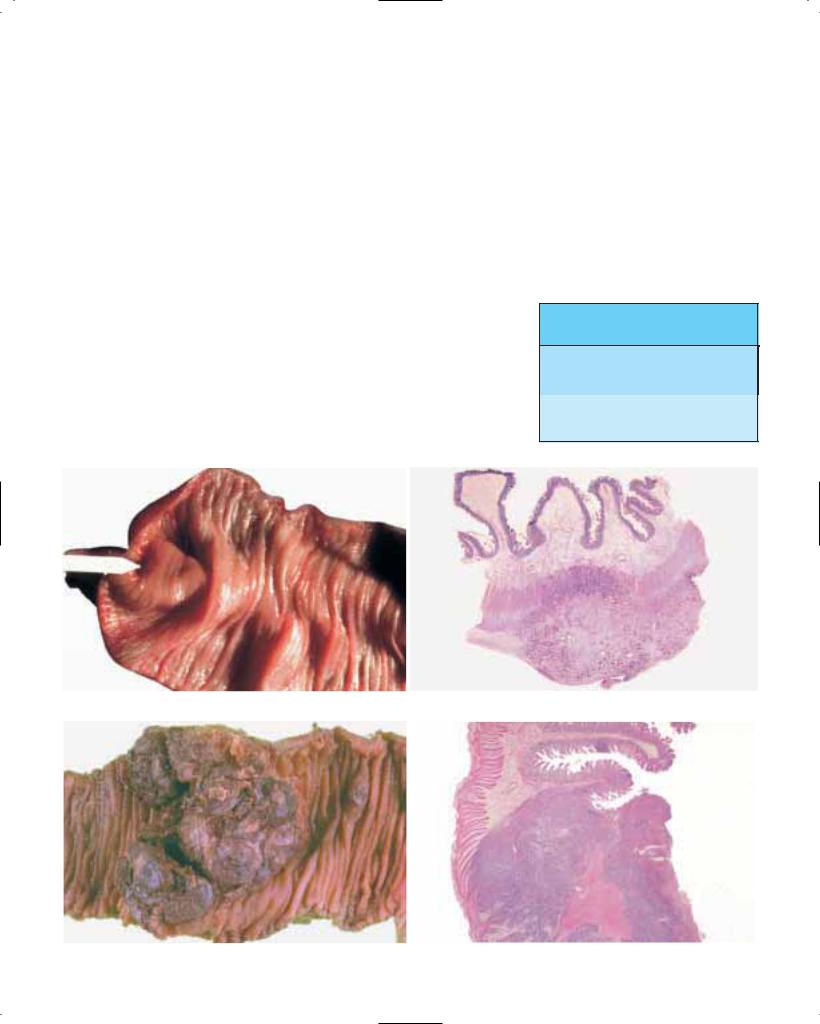
Secondary tumours of the small and large intestines
C. Niederau
L.H. Sobin
Definition
Tumours of the intestines that originate from an extra-intestinal neoplasm or which are discontinuous with a primary tumour elsewhere in the gastrointestinal tract.
Epidemiology
Metastatic spread to the small intestine is more frequent than to any other site in the gastrointestinal tract (see Table 3.02). Secondary carcinomas of the small bowel are as common as primary carcinomas at this site {1234}.
Origin
For small intestine, melanoma, lung, breast, colon and kidney are the most
frequent primary sites (see Table 3.02) {130, 1022, 1378, 1209, 1457, 458}. Metastatic spread from primary lung cancer to the small intestine is more frequent than to stomach and colon (Table 4.01). Virtually all primary cancers can occasionally lead to metastases in the small intestine and, because of the low frequency of primary small bowel cancer, a high proportion of small intestinal malignancies are metastatic.
The pathogenesis of intestinal metastasis usually involves haematogenous spread of tumour cells. Invasion from neighbouring primary tumours also occurs, e.g. pancreatic carcinoma to duodenum and prostate carcinoma to rectum.
Primary melanomas of the intestine are very rare. Although most melanomas found in the small bowel have no history
Table 4.01
Frequency of metastasis from breast (695 cases) and lung (747 cases) to gastrointestinal tract {130}.
Primary |
Stomach |
Small |
Colon |
site |
|
intestine |
|
Breast |
3.6% |
1.7% |
1.4% |
Lung |
1.3% |
4.4% |
1.9% |
Metastasis
A B
Fig. 4.27 Metastatic adenocarcinoma, small intestine. A Tumour is beneath swollen mucosa. B Tumour in muscularis propria. Submucosa is oedematous.
A B
Fig. 4.28 A, B Metastatic malignant melanoma, small intestine.
Secondary tumours 91

Fig. 4.29 Metastatic adenocarcinoma, small intestine. Muscularis propria contains tumour. Mucosa is free of neoplasia.
of a primary tumour, the general consensus is that they are virtually all secondary, usually from misdiagnosed or regressed primary melanomas {458}.
Clinical features
Small intestinal metastases can cause bleeding and obstruction as well as nonspecific symptoms such as abdominal discomfort, gas distension, and diarrhoea {1378, 580}.
Imaging
The identification of a small bowel tumour always raises the question of whether the tumour is primary or secondary. Contrast radiography shows narrowing and abnormalities of the small intestinal wall. Advanced cases result in stenosis with distension due to obstruction.
Fig. 4.30 Metastatic breast carcinoma, colon. Tumour cells expand submucosa.
Macroscopy
Typical features of intestinal metastases include intestinal wall thickening, submucosal spread, and ulcers. Melanomas may not be pigmented and may appear as nodules or polyps.
Histopathology
Metastases are typically submucosal or subserosal making the distinction between primary and secondary tumours relatively easy. Cytokeratin immunohistochemistry may help to differentiate between primary colon cancer (positive for cytokeratin 20), metastases from ovary and breast (usually positive for cytokeratin 7) and those from liver, kidney and prostate (usually negative for both cytokeratins 7 and 20) {2047, 129}. On the other hand, the distinction between
Fig. 4.31 Metastatic breast carcinoma, caecum, diastase-PAS stain. Many tumour cells are mucin positive.
multiple primary small bowel carcinoids and their metastases may not be possible. This also applies to leiomyosarcomas/stromal tumours of the small intestine.
Prognosis
Intestinal metastases usually represent a late stage of disease in which other haematogenous metastases are also frequently found. Therefore, the prognosis is poor. Exceptions are melanoma and renal cancer in which metastases confined to the bowel may be associated with prolonged survival after resection.
92 Tumours of the small intestine
CHAPTER 5
Tumours of the Appendix
The appendix is the most frequent site of carcinoids, i.e. tumours with endocrine differentiation, that span a wide range of morphological variety.
Adenocarcinomas of the appendix also show interesting morphological variations, from those that resemble the usual colorectal carcinoma to those that arise from a carcinoid and to mucinous tumours that may appear well differentiated and indistinguishable from adenoma and yet spread widely through the peritoneal cavity.

WHO histological classification of tumours of the appendix1
Epithelial tumours |
|
Non-epithelial tumours |
|
Adenoma |
8140/02 |
Neuroma |
9570/0 |
Tubular |
8211/0 |
Lipoma |
8850/0 |
Villous |
8261/0 |
Leiomyoma |
8890/0 |
Tubulovillous |
8263/0 |
Gastrointestinal stromal tumour |
8936/1 |
Serrated |
8213/0 |
Leiomyosarcoma |
8890/3 |
|
|
Kaposi sarcoma |
9140/3 |
Carcinoma |
|
Others |
|
Adenocarcinoma |
8140/3 |
|
|
Mucinous adenocarcinoma |
8480/3 |
Malignant lymphoma |
|
Signet-ring cell carcinoma |
8490/3 |
|
|
Small cell carcinoma |
8041/3 |
Secondary tumours |
|
Undifferentiated carcinoma |
8020/3 |
|
|
|
|
Hyperplastic (metaplastic) polyp |
|
Carcinoid (well differentiated endocrine neoplasm) |
8240/3 |
|
|
EC-cell, serotonin-producing neoplasm |
8241/3 |
|
|
L-cell, glucagon-like peptide |
|
|
|
and PP/PYY producing tumour |
|
|
|
Others |
|
|
|
Tubular carcinoid |
8245/1 |
|
|
Goblet cell carcinoid (mucinous carcinoid) |
8243/3 |
|
|
Mixed carcinoid-adenocarcinoma |
8244/3 |
|
|
Others |
|
|
|
|
|
|
|
______________
1This classification is modified from the previous WHO histological classification of tumours {845} taking into account changes in our understanding of these lesions. In the case of endocrine neoplasms, it is based on the recent WHO classification {1784} but has been simplified to be of more practical utility in morphological classification.
2Morphology code of the International Classification of Diseases for Oncology (ICD-O) {542} and the Systematized Nomenclature of Medicine (http://snomed.org). Behaviour is coded /0 for benign tumours, /3 for malignant tumours, and /1 for unspecified, borderline or uncertain behaviour.
TNM classification of tumours of the appendix
TNM classification1, 2
T – Primary Tumour
TX |
Primary tumour cannot be assessed |
T0 |
No evidence of primary tumour |
Tis |
Carcinoma in situ: intraepithelial or invasion of lamina propria3 |
T1 |
Tumour invades submucosa |
T2 |
Tumour invades muscularis propria |
T3 |
Tumour invades through muscularis propria into subserosa |
|
or into non-peritonealized periappendiceal tissue |
T4 |
Tumour directly invades other organs or structures |
|
and/or perforates visceral peritoneum |
N – Regional Lymph Nodes |
|
NX |
Regional lymph nodes cannot be assessed |
N0 |
No regional lymph node metastasis |
N1 |
Metastasis in 1 to 3 regional lymph nodes |
N2 |
Metastasis in 4 or more regional lymph nodes |
______________
M – Distant Metastasis
MX Distant metastasis cannot be assessed M0 No distant metastasis
M1 Distant metastasis
Stage Grouping |
|
|
|
Stage 0 |
Tis |
N0 |
M0 |
Stage I |
T1 |
N0 |
M0 |
|
T2 |
N0 |
M0 |
Stage II |
T3 |
N0 |
M0 |
|
T4 |
N0 |
M0 |
Stage III |
Any T |
N1 |
M0 |
|
Any T |
N2 |
M0 |
Stage IV |
Any T |
Any N |
M1 |
1 {1, 66}. The classification applies only to carcinomas.
2 A help desk for specific questions about the TNM classification is available at http://tnm.uicc.org.
3This includes cancer cells confined within the glandular basement membrane (intraepithelial) or lamina propria (intramucosal) with no extension through muscularis mucosae into submucosa.
94 Tumours of the appendix
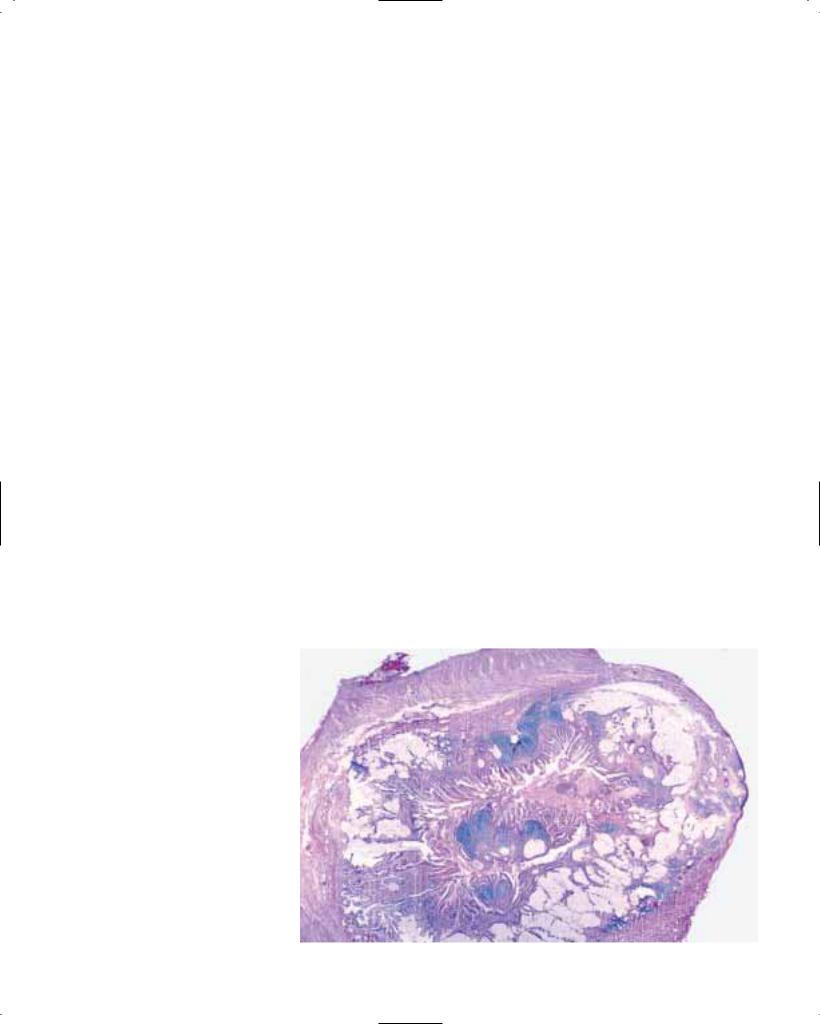
Adenocarcinoma of the appendix
N.J. Carr
M.J. Arends
G.T. Deans
L.H. Sobin
Definition
A malignant epithelial neoplasm of the appendix with invasion beyond the muscularis mucosae.
ICD-O codes |
|
Adenocarcinoma |
8140/3 |
Mucinous adenocarcinoma |
8480/3 |
Signet-ring cell carcinoma |
8490/3 |
Epidemiology
Adenocarcinoma of the appendix occurs in 0.1% of appendicectomies, corresponding to an estimated incidence of 0.2/100,000 per annum {393, 1928}. Adenocarcinomas accounted for 58% of malignant appendiceal tumours in the SEER database, the remainder being mostly carcinoids. The rates for the carcinomas stayed constant during the period 1973-1987 {1928}. The median age of patients with mucinous and non-muci- nous adenocarcinoma was about 65 years in SEER data; other studies suggest a peak age at manifestation in the sixth decade {250, 393}. Males appear to be more commonly affected than females {393}.
Aetiology
Patients with chronic ulcerative colitis (UC) have an increased susceptibility to formation of epithelial dysplasia and malignancy in affected segments of bowel; inflammatory involvement of the appendix is seen in approximately half of UC cases with pancolitis.
Both adenoma and adenocarcinoma of the appendix have been described in patients affected by long-standing ulcerative colitis {1394}.
Clinical features
Signs and symptoms
Many patients with appendiceal adenocarcinoma have clinical features indistinguishable from acute appendicitis. Most of the remaining cases present as an abdominal mass {250, 393}. Spread to the peritoneal cavity may produce large volumes of mucus, causing pseudomyxoma peritonei. Such cases may present
with abdominal distension. Rarely, exter- |
mucocoele, but this is |
descriptive not a |
nal fistulation occurs {251, 393, 707}. |
pathological diagnosis {250, 251}. |
|
Imaging
Ultrasound, computerised tomography (CT) scan or barium enema are of limited benefit in the pre-operative diagnosis of cases presenting as acute appendicitis. Ultrasound and CT scan are the preferred imaging procedures in cases presenting with abdominal mass or pseudomyxoma peritonei {393, 707}. Serial CT scanning and CEA measurements can assess the extent of peritoneal involvement and the subsequent course of the disease. Intraepithelial neoplasia of the appendix may occur concurrently with a carcinoma elsewhere in the large intestine {393}.
Macroscopy
In cases of primary adenocarcinoma, the appendix may be enlarged, deformed or completely destroyed {250, 251, 1612}. Well differentiated lesions are often cystic and may be called cystadenocarcinomas. A grossly appreciated swelling of the appendix due to the accumulation of mucus within the lumen can be termed
Tumour spread and staging
Although the TNM classification currently uses the same criteria as for colorectal tumours, appendiceal cases should be separately classified. This is particularly important because of the special nature of pseudomyxoma peritonei, where malignant cells may be scarce and acellular mucin may seem to have spread further than the malignant cells {250}.
Well differentiated mucinous appendiceal adenocarcinomas generally grow slowly, and typically produce the clinical picture of pseudomyxoma peritonei. Lymph node metastases tend to occur late. Rarely, tumour growth in the retroperitoneum may produce ‘pseudomyxoma retroperitonei’ {1194}. The behaviour of non-mucinous carcinomas resembles that of their colonic counterparts.
Pseudomyxoma peritonei
Pseudomyxoma peritonei is the presence of mucinous material on peritoneal surfaces. It is not a complete histological
Fig. 5.01 Mucinous adenocarcinoma arising in a villous adenoma. The lumen is lined by a villous adenoma.
Adenocarcinoma 95

A B
Fig. 5.02 Appendiceal mucinous adenocarcinoma. Fig. 5.03 Pseudomyxoma peritonei. A Several loops of bowel are encased in a multilocular mucinous mass. B Well differentiated mucus producing epithelium embedded in a fibrous matrix; mucus is present within the lumen and is extravasated into the stroma.
diagnosis in itself; the prognosis will depend on the nature of the causative lesion. Nevertheless, pseudomyxoma peritonei is often applied to a distinctive clinical picture produced by well differentiated mucinous adenocarcinomas in which the growth of malignant cells within the peritoneal cavity causes a slow but relentless accumulation of mucin. Cells may be very scanty within this mucinous material.
A distinctive feature of well differentiated mucinous carcinomatosis is its distribution in the abdomen. There is a tendency to spare the peritoneal surfaces of the bowel, whereas large-volume disease is found in the greater omentum, beneath the right hemidiaphragm, in the right retrohepatic space, at the ligament of Treitz, in the left abdominal gutter and in the pelvis {1854}. In these cases, tumour growth tends to remain confined to the abdomen for many years. Mucinous cysts within the spleen occur occasionally {433}.
It has been suggested that appendiceal adenomas can cause widespread pseudomyxoma peritonei with an ultimately fatal outcome, and some authors use the term ‘adenomucinosis’ for the spread of such lesions through the abdomen {1611, 1612}. It is considered more likely that
Fig. 5.04 Pseudomyxoma peritonei.
such cases are examples of well differentiated adenocarcinoma.
Although most cases of pseudomyxoma peritonei are due to spread from a primary carcinoma of the appendix, cases have been reported in association with mucinous carcinomas of other sites, including gallbladder, stomach, colorectum, pancreas, fallopian tube, urachus, lung, and breast {346, 612, 707, 981, 1199, 2199}.
Although the ovary has been thought of as a common primary site {104, 1705}, there is an accumulating body of evidence based on immunohistochemistry and molecular genetics suggesting that this is not the case, and that in most patients with low-grade mucinous tumours of the ovary and appendix with pseudomyxoma peritonei the lesions are probably metastatic from an appendiceal primary {1536, 1611, 1612, 1871, 2187}.
Histopathology
The majority of appendiceal adenocarcinomas are well differentiated and mucinous {250, 706}. If signet-ring cells account for more than 50% of the neoplasm, the term signet-ring cell carcinoma is appropriate.
The term mucinous cystadenocarcinoma may be used for well differentiated mucinous tumours with cystic structures. However, this designation is descriptive and does not constitute a separate disease entity {251, 2115}.
Diagnostic criteria
The fundamental criterion for making the diagnosis of adenocarcinoma is the presence of invasive neoplasm beyond the muscularis mucosae; this is the same criterion that is applied throughout the large intestine (see Table 5.01). However, in practice it is not always easy to deter-
mine the extent of invasion, because well differentiated carcinomas of the appendix can mimic adenomas by invading on a broad front rather than showing infiltrative or single-cell invasion. Conversely, in some adenomas, acellular mucin dissects through the wall, mimicking invasion; this feature may be especially prominent if there is inflammation. If there is acellular mucin in the appendiceal wall, the diagnosis of adenoma should only be made if the muscularis mucosae is intact since this term implies that the lesion is curable by complete excision.
It is appropriate to use the term mucinous tumour of uncertain malignant potential for neoplasms in which the histological features do not allow distinction between a lesion that is benign (an adenoma) from one that has the potential to cause metastases (an adenocarcinoma). The term low-grade mucinous cystic tumour has also been used for lesions that are histologically not frankly malignant {2187A}.
Grading
Grading is the same as in the large intestine. Some adenocarcinomas of the appendix are so well differentiated that their neoplastic features may be very subtle {250}.
Fig. 5.05 Mucocoele of appendix.
96 Tumours of the appendix

Fig. 5.06 Serrated adenoma of appendix.
Precursor lesions and benign tumours
By analogy with the rest of the large intestine, an adenoma-carcinoma sequence is assumed to occur in the appendix; the finding of a residual adenoma in some cases of adenocarcinoma supports this contention {1548}. However, some adenocarcinomas appear to arise from goblet cell carcinoid tumours {209, 250}. Compared to adenomas of the colon, adenomas of the appendix are more likely to be villous or serrated {250, 706, 1548, 2115, 2110}.
Many appendiceal serrated and villous adenomas display minimal cytological abnormalities; such lesions need to be distinguished from hyperplastic polyps or mucosal hyperplasia. Pedunculated hyperplastic polyps of the type seen in the colon are unusual in the appendix, but diffuse hyperplasia is relatively common {2184}. The diagnosis of hyperplastic polyp/diffuse hyperplasia should not be made if there are cytological abnormalities in the epithelial cells; if any are present, then the diagnosis of adenoma should be considered. The presence of villous structures is also a pointer towards adenoma.
As they grow, adenomas of the appendix typically become cystic, and the lining epithelium becomes undulating rather than villous. Such lesions may produce a mucocoele and be given the descriptive appellation of cystadenoma.
Genetic susceptibility
Familial adenomatous polyposis coli (FAP)
A review of 71 000 appendix specimens revealed 33 benign and 6 malignant appendiceal tumours in patients with familial polyposis coli {324}. Several cases of adenocarcinoma of the appendix have been reported in FAP patients, including a patient with appendiceal
Fig. 5.07 Serrated adenoma (left) and tubulovillous adenoma (right).
adenocarcinoma as the presenting feature {1464}.
Hereditary non-polyposis colorectal cancer (HNPCC)
This familial cancer syndrome confers increased susceptibility to proximal colon cancer {1936}, but it is not yet clear whether there is also an increased risk of appendiceal neoplasms.
Other polyposis syndromes
It is difficult to establish accurately the risk of genetic susceptibility to tumours of the appendix in Peutz-Jeghers and juvenile polyposis syndrome on account of the rarity of these conditions. Intussusception with an ‘inside-out’ appendix in Peutz-Jeghers syndrome has been reported, caused by a hamartomatous polyp of the appendix or an appendiceal
Fig. 5.08 Adenoma with undulating morphology.
polyp with villous adenomatous changes and focal carcinoma in situ {1243}.
Genetics
Limited data are available on molecular genetic alterations in appendiceal tumours, and these data indicate similarities to those in colorectal tumours. KRAS mutations have been identified in approximately 70% of appendiceal mucinous adenomas, mostly in codon 12 and a few in codon 13 {1871}. In addition, KRAS mutation has been identified in an appendix cystadenoma associated with a long history of ulcerative colitis {1123}. Tumour suppressor gene allelic imbalances have been found in about half of appendiceal mucinous adenomas with loss of heterozygosity (LOH) at several chromosomal loci, including 5q22, 6q, 17p13, and 18q21. LOH was most fre-
Fig. 5.09 Hyperplastic polyp of appendix. Cytological abnormalities of intraepithelial neoplasia are absent.
Adenocarcinoma 97

quent at the 5q locus linked to the APC tumour suppressor gene which in the colorectum is strongly associated with transition to adenoma {1871}. In cases of pseudomyxoma peritonei (well differentiated mucinous adenocarcinoma), LOH at one or two polymorphic microsatellite loci was seen in approximately half of the cases and was considered an indication of monoclonality.
Prognosis and predictive factors
SEER data showed the 5-year survival rates for localized adenocarcinoma to be 95%, compared with a 5-year survival of 80% for mucinous or cystadenocarcinoma. When distant metastases were present, the 5-year survival rates were 0% and 51% respectively {1928}. This reflects the low aggressive potential of mucinous tumours that spread to the peritoneum {1769}.
Features that have been associated with a poor prognosis in appendiceal adenocarcinoma include advanced stage,
high-grade, and nonmucinous histology {345, 1365, 1769}. The spread of mucus beyond the right lower quadrant of the abdomen (whether or not cells are identified within it) is an independent prognostic variable, as is the presence of neoplastic cells outside the visceral peritoneum of the appendix {250}. When pseudomyxoma peritonei is present, abdominal distension, weight loss, high histological grade, and morphological
Table 5.01
Terminology of epithelial neoplasms of the appendix.
evidence of invasion of underlying structures have been found to be poor prognostic factors, whereas complete excision of tumour is associated with prolonged disease-free survival {346, 1612, 612}.
Cytological examination of aspirated mucus and DNA flow cytometry are unhelpful in predicting prognosis {612, 707}.
Diagnosis |
Criteria |
Significance |
|
|
|
Adenoma |
Tumour confined to appendiceal mucosa |
Does not have the capacitiy to |
(Cystadenoma) |
and |
metastasize and can be cured by |
|
No histological evidence of invasion |
complete local excision. |
|
|
|
Adenocarcinoma |
Histological evidence of mural invasion |
Can spread beyond the appendix |
(Cystadenocarcinoma) |
or |
with peritoneal, lymph node or dis- |
|
Presence of metastases, |
tant metastases. |
|
including spread to peritoneal cavity |
|
|
|
|
98 Tumours of the appendix

Endocrine tumours of the appendix
Definition
Tumours with endocrine differentiation arising in the appendix.
Epidemiology
Incidence and time trends
Carcinoids account for 50-77% of all appendiceal neoplasms {1252, 1131}. Their incidence rate is 0.075 new cases per 100,000 population per year and appears to have been decreasing in the time period 1950-1991 {1251}. Approximately 19% of all carcinoids are located in the appendix.
Age and sex distribution
The mean age at presentation is 32-43 years (range, 6 to 80 years) {1251, 1252, 1607}. Tubular carcinoids occur at a significantly younger age than goblet cell carcinoids (average, 29 versus 53 years) {209}.
Appendiceal carcinoids occur more frequently in females than in males {1251}. This could reflect the greater number of incidental appendicectomies performed in women {1252} but in the SEER database, the frequency of non-carcinoid appendiceal tumours is similar among males and females, suggesting that the higher rate of appendiceal carcinoids in women may not be due solely to higher rates of appendicectomy {1251}. Furthermore, the prevalence of girls among children with appendiceal carcinoids can not be explained by differences in appendicectomy rates {866A, 1255}.
Clinical features
The majority of appendiceal endocrine tumours are found incidentally in appendicectomy specimens; the majority of these are asymptomatic and located in the distal end of the appendix. In a small number of cases, carcinoids involving the remaining portions of the appendix may obstruct the lumen and produce appendicitis {2059, 209}.
Carcinoid syndrome caused by an appendiceal carcinoid is extremely rare and almost always related to widespread
A
Fig. 5.10 A, B EC-cell carcinoid tumour.
metastases, usually to the liver and retroperitoneum {1252, 1927}.
Macroscopy
Appendiceal EC-cell carcinoids are firm, greyish-white (yellow after fixation), and fairly well circumscribed, but not encapsulated, and measure usually less than 1 cm in diameter {1252}. Tumours > 2 cm are rare; most are located at the tip of the appendix {1254}. Goblet-cell carcinoids and mixed endocrine-exocrine carcino-
Fig. 5.11 Carcinoid tumour of appendix with typical yellow colouration.
C. Capella
E. Solcia
L.H. Sobin
R. Arnold
B
mas of the appendix may be found in any portion of the appendix and appear as an area of whitish, sometimes mucoid induration without dilatation of the lumen. They range in size from 0.5 to 2.5 cm {442}.
Because of their diffusely infiltrative nature, goblet cell carcinoids tend not to form distinct tumours and their size generally cannot be assessed accurately. In a series of 33 cases {209} only two were suspected grossly; 11 involved the tip and 22 were circumferential.
Histopathology
Carcinoid (well differentiated endocrine neoplasm)
Most endocrine tumours of the appendix are serotonin-producing enterochromaffin (EC)-cell carcinoids, while only a minority are glucagon-like peptide and PP/PYY-producing L-cell carcinoids and mixed endocrine-exocrine carcinomas. They are classifiied according to the WHO histological classification of endocrine tumours {1784}.
Endocrine tumours 99

Fig. 5.12 Carcinoid tumour infiltrating mesoappendix.
EC-cell, serotonin-producing carcinoid
Argentaffin EC-cells, producing both serotonin and substance P, are arranged in rounded solid nests with some peripheral palisading (type A structure according to Soga and Tazawa {1775}). Occasionally, there may also be glandular formations (type C structures), forming a mixed (A+C) pattern. Most tumours display muscular and lymphatic invasion or perineural involvement; two thirds of the cases invade the peritoneum, possibly through endolymphatic spread {1252}. Despite these signs of apparent aggressiveness appendiceal carcinoids infrequently produce lymph node or distant metastases, in contrast to ileal carcinoids.
No relevant histologic, cytological, or cytochemical differences have been detected between ileal and appendiceal carcinoids, despite their very different clinical behaviour, with the exception of the presence of S100-positive sustentacular cells surrounding tumour nests in appendiceal lesions. In this respect, ECcell appendiceal carcinoids resemble subepithelial neuroendocrine complexes rather than intraepithelial endocrine cells
{1115, 586, 1182}. In contrast, sustentacular cells are lacking in ileal and colonic EC-cell tumours, which develop from ECcells of the mucosal crypts {1115, 1291}.
L-cell, glucagon-like peptide and PP/PYY-producing carcinoid
These are much less common. L-cell tumours are non-argentaffin, producing glucagon-like peptides (GLP-1, GLP-2, and the enteroglucagons glicentin and oxyntomodulin) and PP/PYY. They feature a characteristic tubular or trabecular pattern (type B pattern according to Soga and Tazawa {1775, 820, 1724, 1783}). These tumours generally measure only 2 to 3 mm and are the appendiceal counterpart of L-cell tumours that are most frequent in the rectum.
Mixed endocrine-exocrine neoplasms
This term is used for certain tumours of the appendix that show features of both glandular and endocrine differentiation, i.e. goblet cell carcinoid, tubular carcinoid and mixed carcinoid-adenocarcino- mas {2059, 1254}.
Goblet-cell carcinoid. This tumour is characterized by a predominant submucosal growth. It typically invades through the appendiceal wall in a concentric manner that does not produce a welldefined tumour {209}. The mucosa is characteristically spared, with the exception of areas of connection of tumour nests with the base of the crypts. The tumour is composed of small, rounded nests of signet-ring-like cells resembling normal intestinal goblet cells, except for nuclear compression. Lumens are infrequently observed. Lysozyme-positive Paneth cells as well as foci resembling
Fig. 5.13 Small cell carcinoma arising in an appendiceal tubulovillous adenoma.
Brunner glands may be present {2059, 790}. Mucin stains are intensely positive within goblet cells and extracellular mucin pools {790}. Argentaffin and argyrophil cells, sparse or forming small nests, are identified in 50% and 88% of cases, respectively {2059}.
Immunohistochemically, the endocrine cell component is positive for chromogranin A, serotonin, enteroglucagon, somatostatin, and/or PP {790, 725}. The goblet cells express CEA. On ultrastructural examination, both dense core endocrine granules and mucin droplets are found {442, 725}. Both elements are occasionally present within the cytoplasm of the same cell {442, 790}.
Tubular carcinoid. This tumour is often misinterpreted as a metastatic adenocarcinoma, because it does not resemble the typical carcinoid and shows little contact with the mucosa. It is composed of small, discrete tubules, some with inspissated mucin in their lumen. Short trabecular structures are frequent, but solid nests are generally absent. In sparse cells or in small groups of tumour cells, the argentaffin reaction is positive in 75%
A B
Fig. 5.14 EC-cell carcinoid tumour. A Chromogranin B. B S-100 immunohistochemistry demonstrating sustentacular cells.
100 Tumours of the appendix

Fig. 5.15 L-cell tumour showing trabecular pattern |
Fig. 5.16 Clear cell carcinoid. |
Fig. 5.17 Tubular carcinoid. |
and glicentin immunoexpression. |
|
|
A B C
Fig. 5.18 Goblet cell carcinoid tumour. A Typical concentric mural distribution of tumour with preservation of the appendiceal lumen. Mucin positive tumour nests (green) are seen in this Movat stain. Lumen is compressed, but intact. B Typical clusters of goblet cells. C Chromogranin A positive cells.
and the argyrophil reaction in 89% of cases {2059}. Useful criteria for diagnosing this tumour are origin from the base of the crypts, integrity of the luminal mucosa, orderly arrangements, and absence of cytological abnormalities and mitoses. Immunohistochemically, tumour cells are often positive for chromogranin A, glucagon, serotonin, and IgA, while they are unreactive for S100 protein {586, 209}.
Mixed carcinoid-adenocarcinoma. This term has been proposed to designate carcinomas of the appendix that arise by progression from a pre-existing gobletcell carcinoid. These carcinomas occur in the apparent absence of neoplastic change in the mucosal epithelium {209}.
Genetics
Loss of heterozygosity at MEN-1 gene locus in sporadic appendiceal carcinoids was reported {829}, but has not been confirmed in more recent studies {394, 1938}.
Unlike colonic adenocarcinomas, KRAS mutations have not been detected either in typical or in goblet-cell carcinoid of the appendix {1556}, while in the same study, TP53 mutations (mainly G:C to A:T transitions) were detected in 25% of gob- let-cell carcinoids.
Prognosis and predictive factors
The majority of patients with endocrine tumours of the appendix have a favourable prognosis. Clinically nonfunctioning, non-angioinvasive lesions confined to the appendiceal wall, and < 2 cm in diameter are generally cured by complete local excision, whereas invasion of the mesoappendix or beyond or metastatic spread indicates that the lesion is aggressive. The most important risk factors appear to be tumour size > 2 cm and invasion of the mesoappendix {1134}. Lesions confined to the appendiceal wall that show angioinvasion or are > 2 cm in size, carry an uncertain malignant potential.
Location of tumours at the base of the appendix with involvement of the surgical margin or of the caecum is prognostically unfavourable, requiring at least a partial caecectomy to avoid residual tumour or subsequent recurrence {1931}. The reported frequency of metastases from appendiceal carcinoids ranged from 1.4% and 8.8% in older series {1252, 1927, 1254, 1780}, while in a more recent study the frequency of regional metastases was 27%, and that of distant metastases 8.5% {1251}.
The 5-year survival of patients with appendiceal carcinoid is 94% for localized disease, 85% for regional disease, and 34% for distant metastases {1251}.
Goblet-cell carcinoids are more aggressive than conventional carcinoids, but not as malignant as adenocarcinomas of the appendix. In one study the percentage of patients dead of goblet cell carcinoids was 12.5% {442}. Tubular carcinoids, in contrast, are clinically benign {209}.
Endocrine tumours 101

Miscellaneous tumours of the appendix
Neuromas are common in the appendix. The most frequent manifestation is the axial neuroma, which causes fibrous obliteration of the appendiceal lumen. Occasionally, neuromas may be found in the mucosa or submucosa without luminal obliteration {1423, 251, 1818}. Appendiceal neuromas may be reactive lesions. Histologically, they consist of a myxoid and collagenous background within which a variety of cells is present, including nerve fibres, spindle cells that immunoexpress S-100 protein, endocrine cells, mast cells and eosinophils. In this context, the presence of endocrine cells should not be mistaken for carcinoid tumour. However, it has been suggested that some carcinoids of the appendix might develop in the same setting as appendiceal neuroma. {251}.
Stromal tumours may affect the appendix on rare occasions; they have generally been described in the literature as being of smooth muscle type {324, 865}.
Kaposi sarcoma may be found in the appendix as part of the acquired immunodeficiency syndrome {406}. Rarely, it occurs in individuals without evidence of HIV infection {295}.
Fig. 5.19 Burkitt lymphoma of appendix.
Malignant lymphomas involve the appendix usually as part of more general intestinal spread. Lymphomas presenting as primary disease of the appendix are rare; some are of Burkitt type {1295, 1761}.
Secondary tumours are unusual in the appendix. Primary sites include carcinomas of the gastrointestinal and urogenital
N.J. Carr
L.H. Sobin
C. Niederau
tract, breast, lung, and gallbladder. Metastatic thymoma and melanoma have also been reported {130, 98, 570, 607, 1051, 1407, 1615, 1822, 2129}. A common pattern is serosal involvement, presumably due to transcoelomic spread.
102 Tumours of the appendix
CHAPTER 6
Tumours of the Colon and Rectum
Colorectal carcinomas vary considerably throughout the world, being one of the leading cancer sites in the developed countries. Both environmental (diet) and genetic factors play key roles in its aetiology. Genetic susceptibility ranges from well-defined inherited syndromes, e.g. familial adenomatous polyposis, to ill-defined familial aggregations. Molecular genetic mechanisms are diverse, and recent data suggest two main pathways: a mutational pathway, which involves inactivation of tumour suppressor genes such as APC; and microsatellite instability which occurs in hereditary nonpolyposis colon cancer (HNPCC) and a proportion of sporadic carcinomas.
The main precursor lesion is the adenoma, which is readily detected and treated by endoscopic techniques. Non-neo- plastic polyps are not considered precancerous unless they occur in polyposis syndromes. Inflammatory bowel diseases, such as chronic ulcerative colitis, bear resemblance to Barrett oesophagus as a precursor lesion with a potential for control by endoscopic surveillance. Cure is strongly related to anatomic extent, which makes accurate staging very important.
Lymphomas, endocrine tumours, and mesenchymal tumours are quite uncommon at this site.

WHO histological classification of tumours of the colon and rectum1
Epithelial tumours |
|
Adenoma |
8140/0 |
Tubular |
8211/0 |
Villous |
8261/0 |
Tubulovillous |
8263/0 |
Serrated |
8213/0 |
Intraepithelial neoplasia2 (dysplasia) |
|
associated with chronic inflammatory diseases |
|
Low-grade glandular intraepithelial neoplasia |
|
High-grade glandular intraepithelial neoplasia |
|
Carcinoma |
|
Adenocarcinoma |
8140/3 |
Mucinous adenocarcinoma |
8480/3 |
Signet-ring cell carcinoma |
8490/3 |
Small cell carcinoma |
8041/3 |
Squamous cell carcinoma |
8070/3 |
Adenosquamous carcinoma |
8560/3 |
Medullary carcinoma |
8510/3 |
Undifferentiated carcinoma |
8020/3 |
Carcinoid (well differentiated endocrine neoplasm) |
8240/3 |
EC-cell, serotonin-producing neoplasm |
8241/3 |
L-cell, glucagon-like peptide and PP/PYY producing tumour |
|
Others |
|
Mixed carcinoid-adenocarcinoma |
8244/3 |
Others |
|
_________
Non-epithelial tumours |
|
Lipoma |
8850/0 |
Leiomyoma |
8890/0 |
Gastrointestinal stromal tumour |
8936/1 |
Leiomyosarcoma |
8890/3 |
Angiosarcoma |
9120/3 |
Kaposi sarcoma |
9140/3 |
Malignant melanoma |
8720/3 |
Others |
|
Malignant lymphomas |
|
Marginal zone B-cell lymphoma of MALT Type |
9699/3 |
Mantle cell lymphoma |
9673/3 |
Diffuse large B-cell lymphoma |
9680/3 |
Burkitt lymphoma |
9687/3 |
Burkitt-like /atypical Burkitt-lymphoma |
9687/3 |
Others
Secondary tumours
Polyps
Hyperplastic (metaplastic)
Peutz-Jeghers
Juvenile
1This classification is modified from the previous WHO histological classification of tumours {845} taking into account changes in our understanding of these lesions. In the case of endocrine neoplasms, it is based on the recent WHO classification {1784} but has been simplified to be of more practical utility in morphological classification.
2Morphology code of the International Classification of Diseases for Oncology (ICD-O) {542} and the Systematized Nomenclature of Medicine (http://snomed.org). Behaviour is coded /0 for benign tumours, /3 for malignant tumours, /2 for in situ carcinomas and grade III intraepithelial neoplasia, and /1 for unspecified, borderline or uncertain behaviour. Intraepithelial neoplasia does not have a generic code in ICD-O. ICD-O codes are available only for lesions categorized as glandular intraepithelial neoplasia grade III (8148/2), and adenocarcinoma in situ (8140/2).
TNM classification of tumours of the colon and rectum
TNM classification1, 2
T – Primary Tumour |
M – Distant Metastasis |
|
|||
TX |
Primary tumour cannot be assessed |
MX |
Distant metastasis cannot be assessed |
||
T0 |
No evidence of primary tumour |
M0 |
No distant metastasis |
|
|
Tis |
Carcinoma in situ: intraepithelial or invasion of lamina propria3 |
M1 |
Distant metastasis |
|
|
T1 |
Tumour invades submucosa |
|
|
|
|
T2 |
Tumour invades muscularis propria |
|
|
|
|
T3 |
Tumour invades through muscularis propria into subserosa |
Stage Grouping |
|
|
|
|
or into non-peritonealized pericolic or perirectal tissues |
|
|
|
|
T4 |
Tumour directly invades other organs or structures4 |
Stage 0 |
Tis |
N0 |
M0 |
|
and/or perforates visceral peritoneum |
Stage I |
T1 |
N0 |
M0 |
|
|
|
T2 |
N0 |
M0 |
N – Regional Lymph Nodes |
Stage II |
T3 |
N0 |
M0 |
|
NX |
Regional lymph nodes cannot be assessed |
|
T4 |
N0 |
M0 |
N0 |
No regional lymph node metastasis |
Stage III |
Any T |
N1 |
M0 |
N1 |
Metastasis in 1 to 3 regional lymph nodes |
|
Any T |
N2 |
M0 |
N2 |
Metastasis in 4 or more regional lymph nodes |
Stage IV |
Any T |
Any N |
M1 |
_________
1 {1, 66}. This classification applies only to carcinomas.
2 A help desk for specific questions about the TNM classification is available at http://tnm.uicc.org.
3This includes cancer cells confined within the glandular basement membrane (intraepithelial) or lamina propria (intramucosal) with no extension through muscularis mucosae into submucosa.
4 Direct invasion in T4 includes invasion of other segments of the colorectum by way of the serosa, e.g. invasion of sigmoid colon by a carcinoma of the cecum.
104 Tumours of the colon and rectum

Carcinoma of the colon and rectum
Definition
A malignant epithelial tumour of the colon or rectum. Only tumours that have penetrated through muscularis mucosae into submucosa are considered malignant at this site. The presence of scattered Paneth cells, neuroendocrine cells or small foci of squamous cell differentiation is compatible with the diagnosis of adenocarcinoma.
ICD-O codes |
|
Adenocarcinoma |
8140/3 |
Mucinous adenocarcinoma |
8480/3 |
Signet-ring cell carcinoma |
8490/3 |
Small cell carcinoma |
8041/3 |
Squamous cell carcinoma |
8070/3 |
Adenosquamous carcinoma |
8560/3 |
Medullary carcinoma |
8510/3 |
Undifferentiated carcinoma |
8020/3 |
Epidemiology
An estimated 875,000 cases of colorectal cancer occurred worldwide in 1996, representing about 8.5% of all new cancers {1531}. The age-standardized incidence (cases/100,000 population) varies greatly around the world, with up to 20fold differences between the high rates in developed countries of Europe, North and South America, Australia/New Zealand, and Asia and the still lower rates in some recently developed coun-
tries (Malaysia, Korea) and in developing countries of Africa, Asia and Polynesia. Significant differences also exist within continents, e.g. with higher incidences in western and northern Europe than in central and southern Europe {336}. Among immigrants and their descendants, incidence rates rapidly reach those of the adopted country, indicating that environmental factors are important. According to the U.S. SEER database, the incidence rate for adenocarcinoma of the colon is 33.7/100,000 and increased by 18% during the period from 1973 through 1987 while the incidence of rectal adenocarcinoma (12.8/100,000) and mucinous adenocarcinoma in the colon and rectum (0.3 and 0.8, respectively) remained relatively constant {1928}. During the last decade of the 20th century, incidence and mortality have decreased {566}. By contrast, the incidence in Japan, Korea and Singapore is rising rapidly {737}, probably due to the acquisition of a Western lifestyle. Incidence increases with age {2121}: carcinomas are rare before the age of 40 years except in individuals with genetic predisposition or predisposing conditions such as chronic inflammatory bowel disease.
Incidence rates in the 1973-87 SEER data for colonic and rectal adenocarci-
S.R. Hamilton |
C.A. Rubio |
B. Vogelstein |
L.H. Sobin |
S. Kudo |
F. Fogt |
E. Riboli |
S.J. Winawer |
S. Nakamura |
D.E. Goldgar |
P. Hainaut |
J.R. Jass |
Fig. 6.03 Double contrast barium enema showing adenocarcinoma of colon. Between the proximal (top) and distal (bottom) segment of the colon the lumen is narrowed with an irregular surface, due to tumour infiltration.
noma for males were higher than those for females; whites had higher rates than blacks for rectal adenocarcinoma, but blacks had higher rates for colonic adenocarcinoma {1928}. During 1975-94, a decrease in incidence in whites was evident, while the incidence of proximal colon cancers in blacks still increased {1958}.
Aetiology
Diet and lifestyle
A high incidence of colorectal carcinomas is consistently observed in populations with a Western type diet, i.e. highly caloric food rich in animal fat combined
Hungary
Australia
Japan
France
United States
Spain
Finland
Incidence
Brazil
Mortality
South America
0 |
10 |
20 |
30 |
40 |
50 |
60 |
Fig. 6.01 Worldwide annual incidence (per 100,000) of colon and rectum cancer in males. Numbers on the map indicate regional average values.
From: Globocan, IARC Press, Lyon.
Fig. 6.02 Male incidence (blue) and mortality (orange) of colorectal cancer in some selected countries.
From: Globocan, IARC Press, Lyon.
Adenocarcinoma 105

with a sedentary lifestyle. Epidemiological studies have indicate that meat consumption, smoking and alcohol consumption are risk factors. Inverse associations include vegetable consumption, prolonged use of non-steroidal antiinflammatory drugs, oestrogen replacement therapy, and physical activity {1531, 2121}. Fibre may have a protective role, but this has been questioned recently.
The molecular pathways underlying these epidemiological associations are poorly understood, but production of heterocyclic amines during cooking of meat, stimulation of higher levels of fecal bile acids and production of reactive oxygen species have been implicated as possible mechanisms {416, 1439}.
Vegetable anticarcinogens such as folate, antioxidants and inducers of detoxifying enzymes, binding of luminal carcinogens, fibre fermentation to produce protective volatile fatty acids, and reduced contact time with colorectal epithelium due to faster transit may explain some of the inverse associations.
Chronic inflammation
Chronic inflammatory bowel diseases are significant aetiological factors in the development of colorectal adenocarcinomas {1582}. The risk increases after 8-10 years and is highest in patients with early-onset and widespread manifestation (pancolitis).
Ulcerative colitis. This chronic disorder of unknown aetiology affects children and adults, with a peak incidence in the early third decade. It is considered a premalignant disorder, with duration and extent of disease being the major risk factors. Population-based studies show a 4.4-fold increase in mortality from col-
A B
Fig. 6.04 A Depressed lesion highlighted with indigo-carmine dye spray corresponding to high-grade intraepithelial neoplasia. B Flat, elevated adenoma with high-grade intraepithelial neoplasia after indigocarmine dye spray.
orectal carcinoma {1504, 448, 1835, 1214}. In clinical studies, the increase in incidence is usually higher, up to 20-fold {647, 990}. Involvement of greater than one half of the colon is associated with a risk to develop carcinoma of approximately 15%, whereas left sided disease may bear a malignancy risk of 5% {1727, 1045}. Ulcerative proctitis is not associated with an increased carcinoma risk.
Crohn disease. Development of carcinoma is seen both in the small intestine and the large intestine. The risk of colorectal malignancy appears to be 3 fold above normal {581}. Long duration and early onset of disease are risk factors for carcinoma.
Modifying factors. Non-steroidal antiinflammatory drugs and some naturally occurring compounds block the biochemical abnormalities in prostaglandin homeostasis in colorectal neoplasms. Some of these agents cause a dramatic involution of adenomas but their role in
the chemoprevention of adenocarcinoma is less clear. Polymorphisms in key enzymes can alter other metabolic pathways that modify protective or injurious compounds, e.g. methylenetetrahydrofolate reductase, N-acetyltransferases, glu- tathione-S-transferases, aldehyde dehydrogenase and cytochrome P-450 {1766, 686, 1300}. These polymorphisms may explain individual susceptibility or predisposition among populations with similar exposures {1555}.
Irradiation.
A rare but well recognized aetiological factor in colorectal neoplasia is therapeutic pelvic irradiation {1974}.
Localization
Most colorectal carcinomas are located in the sigmoid colon and rectum, but there is evidence of changing distribution in recent years, with an increasing proportion of more proximal carcinomas
A B C
Fig. 6.05 Endoscopic features of (A) polypoid, (B) flat, slightly elevated and (C) flat adenoma.
106 Tumours of the colon and rectum

{1928}. Molecular pathology has also shown site differences: tumours with high levels of microsatellite instability (MSI-H) or ras proto-oncogene mutations are more frequently located in the caecum, ascending colon and transverse colon. {842, 1563, 1897}.
Clinical features
Signs and symptoms
Some patients are asymptomatic, especially when their neoplasm is identified by screening or surveillance. Haematochezia and anaemia are common presenting features due to bleeding from the tumour. Many patients experience change in bowel habit; in the right colon, the fluid faeces can pass exophytic masses, whereas in the left colon the solid faeces are more often halted by annular tumours so that constipation is more common. There may be associated abdominal distension. Rectosigmoid lesions can produce tenesmus. Other symptoms include fever, malaise, weight loss, and abdominal pain. Some patients present with the complications of obstruction or perforation.
Imaging
Modern imaging techniques permit noninvasive detection and clinical staging. Conventional barium enema detects large tumours, while air-contrast radiography improves the visualization of less advanced lesions. Cross-sectional imaging by CT, MRI imaging and transrectal ultrasonography permit some assessment of the depth of local tumour invasion and the
A B
C D
Fig. 6.06 A Endoscopic view of two small flat adenomas highlighted with indigo-carmine to show the abnormal tubular pit pattern. B Magnifying video endoscopy of a tubulovillous adenoma highlighted with indigocarmine to show cribriform pattern. C Histological section of a flat elevated tubular adenoma showing lowgrade intraepithelial neoplasia. D Stereomicroscopic view with indigo-carmine dye spray of a depressed adenoma with high-grade intraepithelial neoplasia containing very small round pits.
presence of regional and distant metastases {2202}. Scintigraphy and positron emission tomography are also used.
Endoscopy
The development of endoscopy has had a major impact on diagnosis and treat-
ment. Colonoscopy allows observation of the mucosal surface of the entire large bowel with biopsy of identified lesions. Chromoendoscopy employing dyes to improve visualization of non-protruding lesions and magnification, have been developed. The flat neoplastic lesions
A B
Fig. 6.07 A Small adenocarcinoma invading muscularis propria, arising in a depressed adenoma. B Early adenocarcinoma invading submucosa, arising in a flat adenoma.
Adenocarcinoma 107

A B C
Fig. 6.08 Advanced colorectal carcinomas. A Small depressed invasive carcinoma (arrow) with a nearby protruding adenoma, B Advanced colorectal carcinoma, depressed type. C Cross section of adenocarcinoma with extension into the submucosa (pT1).
have been designated by Japanese gastroenterologists as ‘type II’, with three subtypes: IIa, ‘en plateau’ elevated; IIb, completely flat; and IIc, ‘en plateau’ depressed. The depressed lesions have, despite a smaller diameter, a poor prognosis with prompt penetration in the submucosa. The pit pattern of the surface at magnification 100 allows a reliable prediction of histology. Therapeutic endoscopy, including snare polypectomy and endoscopic mucosectomy, can be used to remove colorectal neoplasms, especially adenomas, and carcinomas with minimal submucosal invasion. Protruded neoplasms can usually be resected by snare polypectomy. Superficial lesions (flat and depressed) and some protruded
Fig. 6.09 Small ulcerating adenocarcinoma of colon producing a depressed lesion.
Fig. 6.10 Well differentiated adenocarcinoma arising in Crohn disease, invading wall beneath intraepithelial neoplasia.
lesions may be removed by endoscopic mucosal resection {2121, 2122, 1164}.
Macroscopy
The macroscopic features are influenced by the phase in the natural history of tumours at the time of discovery. Carcinomas may be exophytic/fungating with predominantly intraluminal growth, endophytic/ulcerative with predominantly intramural growth, diffusely infiltrative/linitis plastica with subtle endophytic growth, and annular with circumferential involvement of the colorectal wall and constriction of the lumen. Overlap among these types is common. Pedunculated exophytic lesions have a mural attachment narrower than the head of the tumour, with the stalk consisting of uninvolved mucosa and submucosa, while sessile exophytic tumours have broad attachment to the wall.
Carcinomas of the proximal colon tend to grow as exophytic masses while those in the transverse and descending colon are more often endophytic and annular. On cut section, most colorectal carcinomas have a relatively homogeneous appearance although areas of necrosis can be seen. Adenocarcinomas of the mucinous (colloid) type often have areas with grossly visible mucus. Carcinomas with high levels of microsatellite instability (MSI-H) are usually circumscribed and about 20% are mucinous {842}.
Tumour spread and staging
Following transmural extension through the muscularis propria into pericolic or perirectal soft tissue, the tumour may involve contiguous structures. The consequences of direct extension depend on the anatomic site. An advanced rectal carcinoma may extend into pelvic structures such as the vagina and urinary bladder, but cannot gain direct access to
the peritoneal cavity when it is located distal to the peritoneal reflection.
By contrast, colonic tumours can extend directly to the serosal surface. Perforation can be associated with transcoelomic spread to the peritoneal cavity (peritoneal carcinomatosis). Involvement of the peritoneal surface should only be diagnosed if the peritoneum is ulcerated or if tumour cells have clearly penetrated the mesothelium. Since the peritoneal surface infiltrated by tumour cells may become adherent to adjacent structures, direct extension into adjoining organs can also occur in colonic carcinomas that have invaded the peritoneal portion of the wall {62}. Implantation due to surgical manipulation occurs only occasionally, but has been reported after laparoscopic colectomy for cancer {1106}.
Spread via lymphatic or blood vessels can occur early in the natural history and lead to systemic disease. Despite the presence of lymphatics in the colorectal mucosa, lymphogenic spread does not
Fig. 6.11 Crohn-like lymphoid reaction associated with a colonic adenocarcinoma.
occur unless the muscularis mucosae is breached and the submucosa is invaded, This biological behaviour stands in sharp contrast to carcinomas of the stomach where metastasis occurs occa-
108 Tumours of the colon and rectum
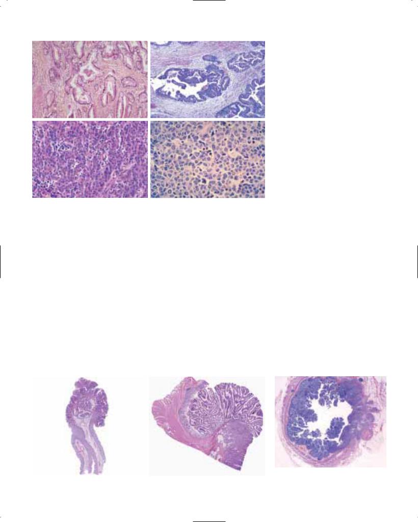
A B
C D
Fig. 6.12 A Well differentiated adenocarcinoma. B Moderately diffferentiated adenocarcinoma. C Poorly differentiated adenocarcinoma; this lesion was MSI-H and shows numerous intraepithelial lymphocytes. D Undifferentiated carcinoma.
sionally from purely intramucosal carcinomas. Invasion of portal vein tributaries in the colon and vena cava tributaries in the rectum can lead to haematogenous dissemination.
Staging
The classification proposed by C. Dukes in 1929-35 for rectal cancer serves as the template for many staging systems currently in use. This family of classifications takes into account two histopathological features: depth of penetration into the wall and the presence or absence of metastasis in regional lymph nodes. The TNM classification {66} is replacing the Dukes classification.
Histopathology
The defining feature of colorectal adenocarcinoma is invasion through the muscularis mucosae into the submucosa. Lesions with the morphological characteristics of adenocarcinoma that are confined to the epithelium or invade the lamina propria alone and lack invasion through the muscularis mucosae into the submucosa have virtually no risk of metastasis. Therefore, ‘high-grade intraepithelial neoplasia’ is a more appropriate term than ‘adenocarcinoma in-situ’, and ‘intramucosal neoplasia’ is more appropriate than ‘intramucosal adenocarcinoma’. Use of these proposed terms helps to avoid overtreatment.
Most colorectal adenocarcinomas are gland-forming, with variability in the size and configuration of the glandular structures. In well and moderately differentiated adenocarcinomas, the epithelial cells are usually large and tall, and the gland lumina often contain cellular debris.
Mucinous adenocarcinoma
This designation is used if > 50% of the lesion is composed of mucin. This variant is characterized by pools of extracellular mucin that contain malignant epithelium as acinar structures, strips of cells or single cells. Many high-frequen- cy micro-satellite instability (MSI-H) carcinomas are of this histopathological type.
Signet-ring cell carcinoma
This variant of adenocarcinoma is defined by the presence of > 50% of tumour cells with prominent intracytoplasmic mucin {1672}.
The typical signet-ring cell has a large mucin vacuole that fills the cytoplasm and displaces the nucleus. Signet-ring cells can occur in the mucin pools of mucinous adenocarcinoma or in a diffusely infiltrative process with minimal extracellular mucin. Some MSI-H carcinomas are of this type.
Adenosquamous carcinoma
These unusual tumours show features of both squamous carcinoma and adenocarcinoma, either as separate areas within the tumour or admixed. For a lesion to be classified as adenosquamous, there should be more than just occasional small foci of squamous differentiation. Pure squamous cell carcinoma is very rare in the large bowel.
A |
B |
Fig. 6.14 Villous adenoma of rectum and invasive |
Fig. 6.13 A Tubulovillous adenoma showing invasive adenocarcinoma within the core of the polyp. |
adenocarcinoma. Two of four lymph nodes in |
|
B Adenocarcinoma arising in a villous adenoma. |
|
perirectal tissue have metastasis. |
Adenocarcinoma 109

Fig. 6.15 Adenocarcinoma within lymphatic vessel.
Fig. 6.16 Metastatic adenocarcinoma in regional lymph node.
Medullary carcinoma
This rare variant is characterized by sheets of malignant cells with vesicular nuclei, prominent nucleoli and abundant pink cytoplasm exhibiting prominent infiltration by intraepithelial lymphocytes {856}. It is invariably associated with MSI-H and has a favourable prognosis when compared to other poorly differen-
tiated and undifferentiated colorectal carcinomas.
Undifferentiated carcinoma
These rare tumours lack morphological evidence of differentiation beyond that of an epithelial tumour and have variable histological features {1946}. Despite their undifferentiated appearances, these tumours are genetically distinct and typically associated with MSI-H.
Other variants
Carcinomas that include a spindle cell component are best termed spindle cell carcinoma or sarcomatoid carcinoma. The spindle cells are, at least focally, immunoreactive for cytokeratin. The term carcinosarcoma applies to malignant tumours containing both carcinomatous and heterologous mesenchymal elements. Other rare histopathological variants of colorectal carcinoma include pleomorphic (giant cell), choriocarcinoma, pigmented, clear cell, stem cell, and Paneth cell-rich (crypt cell carcinoma). Mixtures of histopathological types can be seen.
Carcinosarcoma
Carcinomas that include a spindle cell component are best termed sarcomatoid carcinoma or spindle cell carcinoma. The spindle cells are, at least focally, immuno-
A B
C D
Fig. 6.17 Mucinous adenocarcinoma. A Cut surface with glassy appearance. B Mucinous adenocarcinoma beneath high-grade intraepithelial neoplasia in ulcerative colitis. C Well-differentiated tumour with large mucin lakes. D Multilocular mucin deposits with well-differentiated adenocarcinoma.
Fig 6.18 Signet-ring cell carcinoma invading a nerve.
Fig. 6.19 Adenocarcinoma with venous invasion.
reactive for cytokeratin. The term carcinosarcoma applies to malignant tumours containing both carcinomatous and heterologous mesenchymal elements.
Grading
Adenocarcinomas are graded predominantly on the basis of the extent of glandular appearances, and should be divided into well, moderately and poorly differentiated, or into low-grade (encompassing well and moderately differentiated adenocarcinomas) and high-grade (including poorly differentiated adenocarcinomas and undifferentiated carcinomas). Poorly differentiated adenocarcinomas should show at least some gland formation or mucus production; tubules are typically irregularly folded and distorted.
When a carcinoma has heterogeneity in differentiation, grading should be based on the least differentiated component, not including the leading front of invasion. Small foci of apparent poor differentiation are common at the advancing edge of tumours, but this feature is insufficient to classify the tumour as poorly differentiated {1543}.
The percentage of the tumour showing formation of gland-like structures can be used to define the grade. Well differentiated (grade 1) lesions exhibit glandular structures in > 95% of the tumour; moderately differentiated (grade 2) adenocarci-
110 Tumours of the colon and rectum

A B C
Fig. 6.20 A Signet-ring cell carcinoma arising in an adenoma; intramucosal signet-ring cells adjacent to adenomatous glands. B Signet-ring cells infiltrating muscularis propria. C Lymph node metastasis of a signet-ring cell carcinoma.
noma has 50-95% glands; poorly differentiated (grade 3) adenocarcinoma has 5-50%; and undifferentiated (grade 4) carcinoma has < 5%. Mucinous adenocarcinoma and signet-ring cell carcinoma by convention are considered poorly differentiated (grade 3). Medullary carcinoma with MSI-H appears undifferentiated. Additional studies of the biological behaviour of MSI-H cancers are needed to relate the morphological grade and molecular subtypes of mucinous, signetring cell and medullary carcinoma to outcome since MSI-H carcinomas have an improved stage-specific survival {788, 924, 1098}.
Precursor lesions
During the past decade the natural history of colorectal carcinomas has been extensively studied in correlation with the underlying accumulation of genetic alterations.
Aberrant crypt foci (ACF)
The earliest morphological precursor of epithelial neoplasia is the aberrant crypt focus (ACF). Microscopic examination of
mucosal sheets dissected from the bowel wall and stained with methylene blue, or mucosal examination with a magnifying endoscope, reveal ACFs to have crypts of enlarged calibre and thickened epithelium with reduced mucin content. Microscopy shows two main types: ACFs with features of hyperplastic polyps and a high frequency of ras protooncogene mutations, and dysplastic ACFs (micro-adenomas) associated with a mutation of the APC gene {1375}. Progression from ACF through adenoma to carcinoma characterizes carcinogenesis in the large intestine {1326}.
Adenomas
These precursor lesions are defined by the presence of intraepithelial neoplasia, histologically characterized by hypercellularity with enlarged, hyperchromatic nuclei, varying degrees of nuclear stratification, and loss of polarity. Nuclei may be spindle-shaped, or enlarged and ovoid. Inactivation of the APC/betacatenin pathway commonly initiates the process and results in extension of epithelial proliferation in dysplastic
epithelium from the base of the crypts, where it normally occurs, toward or onto the luminal surface {851, 1528}. Polyps appear to grow as a consequence of accelerated crypt fission resulting from APC gene mutation {564}. Intraepithelial neoplasia can be low-grade or highgrade, depending on the degree of glandular or villous complexity, extent of nuclear stratification, and severity of abnormal nuclear morphology. Paneth cells, neuroendocrine cells and squamous cell aggregates may be seen in adenomas and may become a dominant constituent of the epithelium.
Macroscopy. Colorectal adenomas can be classified into three groups: elevated, flat, and depressed {973}. Elevated adenomas range from pedunculated polyps with a long stalk of non-neoplastic mucosa to those that are sessile. Flat or non-protruding adenomas and depressed adenomas are recognized macroscopically by mucosal reddening, subtle changes in texture, or highlighting by dye techniques. The term adenoma is applied even though the lesions are not polypoid because intraepithelial neopla-
|
MSI-H |
* p<0.05; ** p<0.01 |
MSS |
Fig. 6.21 Sporadic proximal colonic carcinomas. Comparison of pathology of MSI-H |
Fig. 6.22 Frequency of adenocarcinoma in adenomas relative to size and archi- |
(red) and microsatellite stable MSS (blue) carcinomas. |
tecture. |
Adenocarcinoma 111

Fig. 6.23 Clear cell carcinoma of colon.
Fig. 6.24 Tubulovillous adenoma. Pedunculated with long stalk of non-neoplastic mucosa.
A
B
Fig. 6.25 A Sessile villous adenoma. B Section through a villous adenoma.
A
Fig. 6.26 A, B Crypt adenoma in a patient with FAP.
sia (dysplasia) is the hallmark of these lesions. Depressed adenomas are usually smaller than flat or protruding ones and tend to give rise to adenocarcinoma while still relatively small (mean diameter, 11 mm) due to a greater tendency to progress {1628}. These adenomas have a lower frequency of ras mutation than polypoid adenomas {974}.
Histopathology. Tubular adenomas are usually protruding, spherical and pedunculated, or non-protruding (flat). Microscopically, dysplastic glandular structures occupy at least 80% of the luminal surface. Villous adenomas are typically sessile with a hairy-appearing surface. Microscopically, leaf-like projections lined by dysplastic glandular epithelium comprise more than 80% of the luminal surface. Distinction of villous structures from elongated separated tubules is sometimes problematical. Villous architecture is defined arbitrarily by the length of the glands exceeding twice the thickness of normal colorectal mucosa. Tubulovillous adenomas have a mixture of tubular and villous structures with a ratio between 80%/20% and 20%/80%. Serrated adenomas are characterized by the saw-tooth configuration of a hyperplastic (metaplastic) polyp on low power microscopy, but the epithelium lining the upper portion of the crypts and luminal surface is dysplastic. Serrated adenomas can also have a tubular or villous component, but low-lev- els of microsatellite instability (MSI-L) and altered mucin are characteristic of these serrated lesions {840}. By contrast, mixed hyperplastic polyp/adenoma contains separate identifiable areas of each histopathological type {1092}. Occasionally, some villous adenomas show in the slopes of the villi closely packed small glands; those adenomas have been referred to as villo-microglandular adenomas {972}.
B
Although tiny flat or depressed adenocarcinomas are well-described, it is difficult to determine if de novo adenocarcinomas without a benign histopathological precursor lesion ever occur in the large bowel, because adenocarcinoma can overgrow the precursor lesion. The prolonged time interval usually required for progression of intraepithelial to invasive neoplasia offers opportunities for prevention or interruption of the process to reduce mortality due to colorectal carcinoma.
Intraepithelial neoplasia can also occur in the absence of an adenoma, in a preexisting lesion of another type (such as a hamartomatous polyp in juvenile polyposis syndrome and Peutz-Jeghers syndrome), and in chronic inflammatory diseases.
Hyperplastic (metaplastic) polyps
The definition is a mucosal excrescence characterized by elongated, serrated crypts lined by proliferative epithelium in the bases with infolded epithelial tufts and enlarged goblet cells in the upper crypts and on the luminal surface, imparting a saw-tooth outline. In the appendix, diffuse hyperplasia may occur as a sessile mucosal proliferation.
The epithelial nuclei in the serrated region are small, regular, round and located at
Fig. 6.27 Tubular adenoma of colon.
112 Tumours of the colon and rectum
|
A |
B |
Fig. 6.28 Tubulovillous adenoma, partly sessile, |
Fig. 6.29 |
A Adenoma with low-grade dysplasia and well-maintained glandular architecture. B Low-grade |
partly pedunculated. |
dysplasia with regular but slightly elongated, hyperchromatic nuclei. Cytoplasmic mucin is retained. |
|
the base of the cells adjoining the basement membrane, which is often thickened beneath the surface epithelial cells. The cytoplasm contains prominent mucin vacuoles, which are usually larger than normal goblet cells. The proliferative zone often shows increased cellularity and mitotic activity, which can be mistaken for adenoma. Hyperplastic polyps are traditionally considered non-neoplastic, but ras mutation is common, clonality has been demonstrated, and biochemical abnormalities and epidemiological associations that occur in colorectal adenomas and carcinomas have been found {851, 663, 1178}. These lines of evidence suggest that hyperplastic polyps may be neoplastic but have a molecular pathogenesis that differs from the adenomaadenocarcinoma sequence due to absence of inactivation of the APC/betacatenin pathway.
Juvenile polyps
Sporadic juvenile polyps are typically spherical, lobulated and pedunculated and considered hamartomatous. They most commonly occur in children. The surface is often eroded and friable, and the cut surface typically shows mucincontaining cysts. On histology, the abundant stroma is composed of inflamed,
often oedematous granulation tissue that surrounds cystically dilated glands containing mucin. The glands are lined by cuboidal to columnar epithelial cells with reactive changes. The juvenile polyps in patients with juvenile polyposis syndrome may have the macroscopic and microscopic appearances of sporadic juvenile polyps, but they often have a frond-like growth pattern with less stroma, fewer dilated glands and more proliferated small glands (microtubular pattern) than their sporadic counterparts. Intraepithelial neoplasia (dysplasia) is rare in sporadic juvenile polyps. Intraepithelial neoplasia in this setting results from inactivation of the APC/beta-catenin pathway analogous to the genetic basis of adenoma formation {2145}.
Peutz-Jeghers polyps
These are discussed in the small intestine section.
Reactive lesions
Inflammatory polyps. These non-neoplas- tic polyps are composed of varying proportions of reactive epithelium, inflamed granulation tissue and fibrous tissue, often with morphological similarity to juvenile polyps; inflammatory polyps are seen in a variety of chronic inflammatory
diseases including chronic inflammatory bowel disease and diverticulitis.
Lymphoid polyps. These result from aggregates of reactive mucosa-associat- ed lymphoid tissue with conspicuous germinal centres located in the mucosa and/or submucosa.
Mucosal prolapse. On occasion, mucosal prolapse can produce morphological features that mimic neoplasia, including polyps, masses and ulcers characterized histologically by elongated, distorted, regenerative glands surrounded by a proliferation of smooth muscle fibres from the muscularis mucosae, together with superficial erosions, inflamed granulation tissue and fibrosis {159}. Widening of gland lumina at the surface is common. Examples of this phenomenon include inflammatory cloacogenic polyp {1083}, solitary rectal ulcer and cap polyp. The process can extend into the bowel wall, producing colitis cystica profunda.
Neoplasia in chronic inflammatory bowel disease
There is evidence that the natural history of colorectal carcinomas associated with chronic colitis differs from that of ordinary adenomas both morphologically and with respect to the type and sequence of genetic alterations.
A B C
Fig. 6.30 Adenomas with high-grade dysplasia. A Loss of normal glandular architecture, hyperchromatic cells with multi-layered irregular nuclei and loss of mucin, high nuclear/cytoplasmic ratio. B Marked nuclear atypia with prominent nucleoli. C Adenoma with focal cribriform pattern .
Adenocarcinoma 113

Fig. 6.31 Serrated adenoma with irregular indentation of the neoplastic epithelium.
Ulcerative colitis (UC)
Development of carcinoma is apparently metachronous to the development of intraepithelial neoplasia (classified as low-grade and high-grade) complicating chronic colitis. Because invasion can be associated with intraepithelial neoplasia exhibiting relatively mild morphological changes, high-grade intraepithelial neoplasia is diagnosed in colitis on the basis of abnormalities that are less severe than the criteria for high-grade intraepithelial neoplasia in adenomas. It may be flat or present as a 'dysplasia associated lesion or mass' (DALM); the latter is often associated with a synchronous carcinoma arising beneath the dysplastic surface. DALMs are considered high-grade lesions through their architecture alone, and both DALM of any grade of dyspla-
Fig. 6.32 Microtubular adenoma.
Fig. 6.33 Apoptotic cells in an adenoma demonstrated by M30 immunohistochemistry.
sia and high-grade flat dysplasia are associated with invasive carcinoma in about 40% of cases. The diagnosis of DALM and high-grade flat dysplasia usually leads to total colectomy {1687}. It may be difficult to distinguish a DALM from an incidental adenoma in a patient with UC.
Attempts have been made to identify early dysplastic lesions in UC with cell cycle proliferation markers. Topoisomerase II alpha and Ki-67 have been shown to increase significantly over baseline expression in UC related dysplasias. Ki-67 positive cells are found both at the surface and the base of the crypts, indicating a fundamental deregulation of the proliferative cell pool {1368}. Mutations of TP53 appear to be an early event and are already present in intraepithelial neoplasia associated with UC, in contrast to the adenoma-carcinoma sequence in sporadic colorectal carcinomas. Some TP53 mutations have even been observed in non-dysplastic mucosa of chronic inflammation {516, 1463, 2175}.
Alterations of p16 have also been identified in early UC but only very infrequently in adenomas. Both tumour tissue and multiple colorectal cancer cell lines studied showed absence of LOH in 9p 1 {2019, 878}.
Microsatellite instability and gene alterations in p16 and p53 may represent early events during the development of dysplasia and carcinoma, and these changes may lead to susceptibility for allelic loss of other genes such as APC and DCC. It has been shown that LOH of genetic areas close to the VHL locus on 3p is frequently present in DALM lesions and, less frequently, in flat dysplastic lesions. These changes are not usually seen in sporadic adenomas {515}. This may indicate that dysplasia in UC and sporadic adenomas may follow different genetic pathways.
Crohn disease
Intraepithelial neoplasia, classified as low-grade or high-grade, is associated with a high proportion of Crohn carcinomas, either adjacent to the invasive lesion or at a distance from it {1757}. Similar to UC, polypoid dysplastic lesions are diagnosed as DALM in Crohn’s disease.
Mucinous adenocarcinomas are seen in Crohn disease more frequently than in
Fig. 6.34 Tubulovillous adenoma with pseudoinvasion. Small clusters of adenomatous cells produce multilocular, large mucin deposits that expand the adenoma’s stalk. This growth pattern resembles mucinous carcinoma but is not malignant.
sporadic colorectal carcinomas {656}. There is an increased frequency of adenocarcinomas within perianal fistulas, and of squamous cell carcinomas of the anal mucosa {992}.
Similar to UC, TP53 and c-KRAS mutations are observed earlier in Crohn-asso- ciated intraepithelial neoplasia than in the adenoma-carcinoma sequence of sporadic colorectal cancer {1562}.
Genetic susceptibility
The spectrum of genetic susceptibility is broad, ranging from well-defined autosomal dominantly inherited syndromes with known germline genetic mutations to illdefined familial aggregation {1531, 1928, 642}. The diseases are traditionally divided into polyposis syndromes characterized by large numbers of polyps, e.g. familial adenomatosis coli (FAP), and non-polyposis syndromes with a small number of or absence of polyps, e.g. hereditary nonpolyposis colorectal cancer (HNPCC). They are described in the following chapters.
A non-truncating polymorphism of the APC gene that induces an unstable polyadenin repeat sequence, occurs in approximately 5% of Ashkenazi Jews
114 Tumours of the colon and rectum

and carries a modestly elevated risk of colorectal cancer. Only small numbers of adenomas occur in patients with this form of germline APC alteration {1004}.
Li-Fraumeni syndrome
MIM No: Li-Fraumeni syndrome 151623; TP53 mutations 191170
Li-Fraumeni syndrome is an autosomal dominant disorder characterized by multiple primary neoplasms in children and young adults, with a predominance of soft tissue sarcomas, osteosarcomas and breast cancer, and an increased incidence of brain tumours, leukaemia and adrenocortical carcinomas {1403}. Criteria for proband identification are: (1) occurrence of sarcoma before age 45,
(2) at least one first-degree relative with any tumour before age 45, and (3) at least one firstor second-degree relative with cancer before age 45 or with sarcoma at any age {717, 141, 1066}.
In about 70% of Li-Fraumeni kindreds, affected family members carry a germline mutation in TP53 {1151}. From 1990 to 1999, a total of 144 families with a TP53 germline mutation were identified. A database of these mutations is available at http://www.iarc.fr/p53/Germ. htm {699}. As with somatic mutations, germline mutations cluster in conserved regions of exons 4 to 9, with major hotspots at codons 175, 248 and 273. It has been suggested that cancer phenotype correlates with the position of the mutation within the coding sequence, with lower age of clinical manifestation in probands with mutations falling in the DNA-binding domain of the p53 protein {142}. The most frequent type of germline mutation is transition (GC to AT) at CpG dinucleotides 556. The molecular basis of tumour predispositions in families within TP53 germline mutations is not known. Recent studies have excluded tumour suppressor genes such as PTEN and CDKN2 {214}.
Gastrointestinal manifestations
Neoplasms of the digestive tract represent 7% of the tumours observed in LiFraumeni families. Most of these tumours are colorectal carcinoma, with a minority of stomach carcinomas. The male:female ratio is 1.5 and the mean age at clinical manifestation is 45, which correspond to a relatively long latency period as compared to other types of cancers occurring in Li-Fraumeni families {1403}. Preferential familial occurrence of stomach cancer
A B
Fig. 6.35 Proliferating cells demonstrated by immunohistochemistry for MIB1. A Hyperplastic polyp with proliferative cells restricted to the basal parts of the crypts. B Tubular adenoma with proliferating adenomatous epithelium also at the luminal surface.
Fig. 6.36 Hyperplastic polyps. Typical sessile |
Fig. 6.37 Hyperplastic polyp with deep proliferative, |
appearance. |
non-serrated zone protruding into submucosa. |
A B
Fig. 6.38 Hyperplastic polyp. A Pedunculated. B Short deep proliferative zone and superficial serrated mature zone.
Adenocarcinoma 115

Fig. 6.39 Inverted hyperplastic polyp. Endophytic growth of hyperplastic glands projects into submucosa. Proliferative zone at the periphery, maturation at the center.
(familial clustering) has been observed only in Japan, a country at high incidence for that type of tumour. Cancers of the liver and of the upper gastrointestinal tract are exceedingly rare (less than 0.5% of all Li-Fraumeni neoplasms). In these neoplasms, sporadic cases often carry somatic TP53 mutations. The low frequency of these tumours in families with germline TP53 mutations suggests that the pre-existence of a TP53 mutation is not sufficient to increase the likelihood of cancer development.
BRCA 1 and BRCA 2
In a retrospective analysis of 33 large, high-risk breast and breast/ovarian cancer families linked to the BRCA1 locus, a significantly elevated risk of colon cancer was found, with an estimated relative risk
A
B
Fig. 6.40 Juvenile polyp. A Smooth eroded surface with numerous mucous retention cysts, typical of sporadic juvenile polyps. B Expanded inflamed stroma with distorted glands showing reactive atypia.
of 4.11 (95% CI 2.36 - 7.15) {518}. This corresponds to a risk of colon cancer by age 70 of about 6%. In this study, there did not seem to be any increased relative risk at younger ages, although power to detect either sex or age effects was somewhat low in this set of data. In a similar study of BRCA2 carriers {69}, no increased risk of colorectal cancer was observed. However, there was a significantly elevated risk for both stomach and gallbladder tumours among known or likely mutation carriers with estimated relative risks associated with BRCA2 of 2.6 (95% CI 1.46 - 4.61) and 5.0 (1.50 - 16.5), respectively.
Molecular genetics
The development of most colorectal carcinomas is believed to begin in a col-
orectal epithelial cell with a mutational inactivation of the APC (adenomatous polyposis coli) suppressor gene {922, 636, 186}. This inactivation has multiple consequences, including interference with E-cadherin homeostasis and dysregulation of transcription of genes. Clonal accumulation of additional genetic alterations then occurs, including activation of proto-oncogenes such as c-myc {680} and ras, and inactivation of additional suppressor genes. The genes commonly inactivated during progression include genes on chromosome 18 {1583, 614} and the TP53 gene on the short arm of chromosome 17 {1056, 415}. The mutated TP53 gene product, in turn, fails to regulate normally a variety of genes regulated by wild-type p53, including p21WAF1/CIP1 cyclin-depend- ent kinase inhibitor which complexes with proliferating cell nuclear antigen {349}, and genes leading to apoptosis, including BAX {278}. For many suppressor genes, inactivation of one allele is often caused by loss of all or part of the chromosome where the gene resides. Various other chromosomal loci have high frequencies of loss in colorectal cancer due to chromosomal instability {1044}, but the target genes are not yet known.
Microsatellite instability (MSI)
Some colorectal cancers are distinguished by extensive nucleotide insertions or deletions in numerous, intrinsically unstable repeated sequences in tumour DNA, termed microsatellite instability (MSI), also termed ubiquitous somatic mutations, DNA replication errors (RER), or nucleotide instability {1540, 860}.
MSI is defined as a change of any length due to either insertion or deletion of repeating units, in a microsatellite within a
|
A |
B |
Fig. 6.41 Reactive epithelial changes in ulcerative |
Fig. 6.42 Low-grade intraepithelial neoplasia in ulcerative colitis. A Patchy hyperbasophilic regular glands, |
|
colitis. |
with dysplasia extending to the luminal surface. B Haphazardly arranged dysplastic glands. |
|
116 Tumours of the colon and rectum

A B C
Fig. 6.43 A – C High-grade intraepithelial neoplasia in ulcerative collitis with multilayered hyperchromatic elongated nuclei extending to the luminal surface.
tumour when compared to normal tissue. It has been recommended that a panel of five microsatellites should be used as a reference standard (BAT25, BAT26, D5S346, D2S123, D17S250) for carcinomas of the large intestine {164}. If two or more of these markers show MSI, the lesion is classified as high-frequency microsatellite instability (MSI-H); if only one marker shows MSI, it is classified as low-frequency microsatellite instability (MSI-L); if no markers show MSI it is classified as microsatellite stable (MSS). If more than five markers are used, the criteria should be modified to reflect the percentage of markers demonstrating MSI. Thus, MSI-H lesions would exhibit MSI in more than 30-40% of markers tested.
MSI-H carcinomas are characteristic of hereditary nonpolyposis colorectal cancer syndrome (HNPCC) due to germline mutation of one of a group of DNA mismatch repair genes followed by somatic inactivation of the other allele. Sporadic MSI-H tumours comprise about 15% of colorectal carcinomas. They usually follow transcriptional silencing of both alleles of the hMLH1 mismatch repair gene due to aberrant methylation of cytosine residues in the cytosine and guanine-rich promoter region {886, 696}. The alterations that accumulate during progression of both hereditary and sporadic neoplasms characterized by MSI-H include
mutations in microsatellites within the coding region of some genes, such as the type II receptor for TGF-beta1 and BAX {548}. In contrast to microsatellitestable cancers, MSI-H cancers display nucleotide rather than chromosomal instability; allelic deletions are rare {1044}.
Recent studies indicate a functional link between defective DNA mismatch repair and the Wnt-signalling pathway. Approximately 25% of sporadic colorectal carcinomas with defective mismatch repair (MSI-H) were shown to contain frameshift mutations in the AXIN2 gene, which leads to a stabilization of β-catenin and activation of β-catenin/T-cell factor (TCF). This was associated with an accumulation in tumour cell nuclei which was absent in colorectal cancer without mis-
match repair deficiency and in the absence of APC mutations. AXIN2 mutant protein appears to be more stable than the wild-type gene product, suggesting a dominant-negative effect {1079A}.
Prognosis and predictive factors
Morphology. Macroscopic and microscopic features reportedly related to prognosis are summarized in Table 6.01 {2348}.
Poor prognosis has been associated with both large and small tumour size, with sessile and ulcerated configuration as contrasted with polypoid cancer, with extensive involvement of the bowel circumference, with the presence of complete bowel obstruction, with perforation, and with serosal deposits.
A B
Fig. 6.44 Dysplasia associated lesion or mass (DALM). A Polypoid lesion. B Raised lesion simulating an adenoma.
Adenocarcinoma 117

Fig. 6.45 Rectal carcinoma arising in ulcerative colitis. Surface dysplasia overlies invasive carcinoma in this DALM.
Histopathological features related to poor prognosis include deep infiltration of the layers of the wall, extensive involvement of a particular layer, an infiltrative pattern of the invasive edge of the tumour as contrasted to an expansile pattern, and poor differentiation, including signet-ring cell and mucinous adenocarcinoma, adenosquamous carcinoma, small cell carcinoma and anaplastic carcinoma {1672, 1946, 220, 916, 266}. Mucinous adenocarcinomas of the rectum often present at a later stage and have the poorest overall prognosis {1928}, but the MSI status influences the aggressiveness of this histopathological subtype {1221}. Other studies have shown no significant difference in prognosis between mucinous and non-muci- nous varieties of adenocarcinoma {1543}.
Lymph node metastasis. Metastasis to numerous nodes, those close to the mesenteric margin, at great distance from the primary tumour, or in retrograde lymph nodes, have been associated with poor prognosis while the prognostic value of identification of micrometastasis in lymph nodes by immunohistochemical or molecular techniques is still controversial {1564, 1387, 221}.
Fig. 6.46 Immunoexpression of p53 in intraepithelial neoplasia in ulcerative collitis.
Angiogenesis. Neovascularization of tumour stroma is crucial in supporting tumour growth, and high levels of microvessel density have been interpreted as an adverse prognostic feature {2010}.
Inflammatory response. The presence of an intense inflammatory infiltrate with polymorphonuclear leukocytes (particularly eosinophils), lymphocytes, plasma cells, mast cells and histiocytes, as well as prominent desmoplasia have been associated with improved prognosis {1352}. In the regional lymph nodes, hyperplasia of the paracortical T-lym- phocyte areas and the B-cell germinal centers have also been reported as favourable, as has sinus histiocytosis.
Other features of colorectal carcinomas that have been shown to be of prognostic value in some studies include angiolymphatic invasion, perineural space involvement, extramural venous involvement, peritumoural lymphocytic response, and tumour-infiltrating lymphocytes. Some of these features are evaluated in a classification proposed by Jass {389}. A microacinar pattern of growth, defined as discrete, small, relatively regular tubules, is associated with reduced survival {559, 2100}.
Extent of resection. A short longitudinal surgical resection margin (2-5 cm), reflecting the surgical technique employed, has been associated with poor outcome. In rectal cancer, clearance from the circumferential margin is important. The circumferential margin represents the adventitial soft tissue margin closest to the deepest penetration of the tumour. For all segments of the large intestine that are incompletely enveloped by peritoneum or not enveloped, the circumferential margin is created by blunt or sharp dissection at operation. The mesocolic margin in resection specimens of colon cancer is usually well distant from the primary tumour, but the status of the circumferential margin is particularly important in rectal carcinoma due to the anatomic proximity of pelvic structures {15}.
Genetic predictive markers. Some of the genetic alterations identified in colorectal cancers are markers for prognosis {313, 1206}. Allelic loss of chromosome 18q was found to be an adverse prognostic indicator. Other studies reported that loss of chromosomes 17p, 1p, 5q, 8p or 18q, decreased DCC gene expression, p53
A
B
Fig. 6.47 Solitary rectal ulcer. A, B Two deep ulcers macroscopically simulating carcinoma.
Fig. 6.48 Solitary rectal ulcer with reactive hyperplastic polyp due to prolapse. This lesion should not be confused with a neoplasm.
Fig. 6.49 Inflammatory cap polyp in a patient with ulcerative collitis.
118 Tumours of the colon and rectum

overexpression, reduced p27KipI expression, high expression of cyclin A, ras gene mutation, expression of enzymes involved in matrix degradation and their inhibitors (cathepsin-L, urokinase, tissuetype plasminogen activator, tissue inhibitors of metalloproteinases), expression of genes involved in apoptosis (bcl2, bax, survivin), expression of cell surface molecules (CD44 and its variants, ICAM1, galectin 3) and metabolic enzymes (GLUT1 glucose transporter, manganesesuperoxide dismutase, thymidylate synthetase, ornithine decarboxylase, cyclooxygenase 2) have prognostic value.
In addition, colorectal cancers manifesting MSI-H have been reported to have a lower frequency of metastasis and improved prognosis when compared to microsatellite-stable tumours.
Response to therapy. No pathological features have been reported as predictive of therapeutic response, but some molecular alterations have potential as predictive markers. Studies in cell lines of colonic and other carcinomas have shown that in vitro, the status of TP53 is crucial {1382}. The TP53 pathway is closely linked to regulation of the cell cycle and of apoptosis. The presence of wild-type p53 in cell lines is associated with in vitro growth inhibition in response to many chemotherapeutic agents, and with radiation-induced upregulation of p21WAF1/CIP1 and cell cycle arrest. Tumours
Fig. 6.50 Solitary rectal ulcer. Smooth muscle increased between glands, distorting and displacing them.
manifesting MSI-H may respond to 5-FU- based chemotherapy {1109}, while p53 protein accumulation was associated with lack of response to postoperative adjuvant chemotherapy with 5-FU and levamisole {24}. Chromosome 18q loss was associated with an unfavourable survival rate in this setting.
Fig. 6.51 Inflammatory cloacogenic polyp with mucous extravasation.
Major problems exist in the interpretation of various pathological features as prognostic and predictive markers. Many of these features are interrelated but have been treated for statistical purposes as independent variables in studies. At present, anatomic staging is the mainstay of clinical decision-making.
Table 6.01
Prognostic factors in colorectal carcinoma.
Features of the primary tumour |
Evidence of vessel invasion |
Evidence of host response |
Consequences of surgical technique |
|
|
|
|
Anatomic extent of disease (TNM) |
Extramural venous involvement |
Angiogenesis |
Distance between resection margin |
Extent of circumferential involvement |
Lymphatic vessel or |
Local inflammatory and desmo- |
and tumour |
Bowel obstruction |
perineural space involvement |
plastic response to infiltrating |
Presence of residual tumour |
Perforation |
tumour |
|
|
Pattern of invasion |
Reactive changes in regional |
|
|
Grade of differentiation |
lymph nodes |
|
|
|
|
|
|
Adenocarcinoma 119

Familial adenomatous polyposis
I.C. Talbot
R. Burt
H. Järvinen
G. Thomas
Definition
Familial adenomatous polyposis (FAP) is an autosomal dominant disorder characterized by numerous adenomatous colorectal polyps that have an intrinsic tendency to progress to adenocarcinoma. It is caused by a germline mutation in the Adenomatous Polyposis Coli (APC) gene which is located on the long arm of chromosome 5 (5q21-22). Gardner syndrome is a variant of FAP that includes epidermoid cysts, osteomas, dental anomalies and desmoid tumours, in addition to colorectal adenomas. Turcot syndrome is a variant that is associated with a brain tumour (medulloblastoma). An attenuated FAP form has been distinguished from classic FAP, where the number of adenomas is less than 100 in the colon.
MIM No.: FAP, including Gardner syndrome, 175100; Turcot syndrome, 276300
Synonyms
Adenomatous polyposis coli, familial polyposis coli, Bussey-Gardner polypo-
sis, Gardner syndrome, familial multiple polyposis, multiple adenomatosis, familial polyposis of the colon and rectum, familial polyposis of the gastrointestinal tract, familial adenomatous polyposis coli, etc.
Incidence
Estimates of the incidence of FAP vary between 1 per 7000 and 1 per 30,000 newborns. The mean annual incidence rate has been constantly from 1 to 2 per 1,000,000 in Denmark and Finland while the prevalence has increased to more than 25 per 1,000,000 since the creation of preventive polyposis registries {205; 836}. In general, FAP underlies less than 1% of all new colorectal cancer cases. Between 30 and 50% of new FAP patients are solitary cases, probably representing new mutations of the APC gene.
Diagnostic criteria
Classical FAP is defined clinically by the finding of at least 100 colorectal adenomatous polyps {216}. Endoscopic visual-
A B
C D
Fig. 6.52 Colectomy specimens from patients with familial adenomatous polyposis. A Hundreds of polyps of different size cover the entire mucosal surface. B Multiple adenomas in different stages of development. C Lateral view of polyps. D Numerous small early (sessile) adenomas.
ization of diminutive polyps may require dye spray assisted endoscopy. Histological confirmation requires examination of several polyps. In the context of endoscopic screening on the basis of definite family history the detection of fewer adenomas is sufficient at an early age. The same applies on the attenuated disease form (AAPC). Final diagnosis may be achieved by demonstration of a mutated APC, but the detection rate of mutations has only been between 60 and 80% of all FAP families. In patients where the clinical criteria remain doubtful and genetic diagnosis is not achieved the finding of extracolonic features of FAP (epidermoid cysts, osteomas, desmoid tumour, gastric fundic gland polyps, etc.) may give additional diagnostic support.
The following diagnostic criteria have been established: (1) 100 or more colorectal adenomas or (2) germline mutation of the APC gene or (3) family history of FAP and at least one of the following: epidermoid cysts; osteomas; desmoid tumour.
Colorectal polyps
The colorectal polyps are adenomas, most often tubular, and resemble their sporadic counterparts.
Localization
Colorectal adenomas in FAP occur throughout the colon but follow the general distribution of sporadic adenomas, with greatest density in the rectum and sigmoid colon. The distribution of cancers follows that of the adenomas.
Clinical features
Age at clinical manifestation
Colorectal adenomas become detectable at endoscopic examination (sigmoidoscopy) between the age of 10 and 20 years, increasing in number and size with age. The most important clinical feature of FAP is the almost invariable progression of one or more colorectal adenomas to cancer. The mean age of development of colorectal cancer is about 40 years, but the cancer risk is 1 to 6% already at
120 Tumours of the colon and rectum

Fig. 6.53 Tubulovillous adenoma in familial adenomatous polyposis.
the age of 20 to 25 years {835}, and colorectal cancer has been reported even in children with FAP. Extracolonic manifestations such as epidermoid cysts, mandibular osteomas, desmoid tumours or congenital hypertrophy of the retinal pigment epithelium (CHRPE) may present in children and can serve as markers of FAP.
Symptoms and signs
In the early phase of FAP adenomas do not cause any symptoms. Specific symptoms due to colorectal adenomas are rectal bleeding and diarrhoea often accompanied by mucous discharge and abdominal pain. Symptoms appear gradually and may be easily overlooked; the mean age of appearance of symptoms was 33 years and the mean age of diagnosis 36 years in about 200 FAP patients who had no prophylactic screening arranged {216}.
Two thirds of patients diagnosed to have FAP on the basis of symptoms (propositi) already have colorectal cancer whereas in asymptomatic members of known FAP families cancer is very rare at the time of the detection of FAP provided that prophylactic endoscopic screening was arranged in good time, i.e. before the age of 20 years {836}.
Imaging and FAP screening
The appropriate screening method for diagnosing FAP is flexible sigmoidoscopy, which should be arranged for all children of an affected FAP parent from the age of 10 to 15 years and continued at 1 to 2 year intervals up to the age of 40 years if adenomas are not detected. Endoscopies can be replaced by genetic testing for the specific APC mutation in those families where the mutation has been identified. A positive test is diagnostic for FAP and signifies the need for prophylactic colectomy or proctocolectomy when the colorectal adenomas become detectable, at the age of 20 to 25 years at the latest.
If the operation is not performed immediately after the diagnosis of FAP, colonoscopy should be undertaken to evaluate the entire colon because large adenomas or cancer may reside beyond the reach of the flexible sigmoidoscope. Endoscopic evaluation of the upper gastrointestinal tract is recommended at the time of prophylactic colectomy or proctocolectomy, and should be repeated at 2 to 5 year intervals depending on the finding of adenomas in duodenal and gastric biopsies {688}. Double contrast barium enema and barium meal may be used to demonstrate polyps but are inferior to endoscopy because biopsies are required to provide histological evidence for a definite diagnosis of FAP.
Macroscopy
Most polyps in FAP are sessile and spherical or lobulated. Scattered larger pedunculated polyps are much less numerous {205; 835; 836; 688}. The colorectal polyps appear first in adolescence and, by the late teens, usually number thousands, typically carpeting the lining of the whole large bowel. Their number varies between families, in some being little more than 100, even in adults {1988}, whereas, in the majority of families, there are profuse polyps, numbering thousands. Typically, the polyps are scattered evenly along the whole large bowel but, in over one third of cases, their density is greatest in the proximal colon. Adult patients with rectal sparing have been described, even when adenocarcinoma was present in the right colon {1503}.
In any one patient the polyps range from barely visible mucosal nodules to pedunculated polyps of up to 1 cm or more. In some patients and families the adeno-
Fig. 6.54 Small ulcerated adenocarcinoma with rolled edges (arrowhead), accompanied by numerous adenomas in a patient with FAP. Polypectomy scars are present.
mas mostly measure only a few millimetres while in others they are larger, with polyps up to several centimetres. In contrast, in attenuated FAP, the polyps are so few that they may not be noticed at rigid sigmoidoscopy. Polyps rarely appear until late childhood {216} and are rarely larger than 1 cm until adulthood. Adenocarcinomas arise in only a small percentage of the adenomas.
Histopathology
Adenomas in FAP begin as single dysplastic crypts (‘unicryptal’ adenomas). In practice, to find more than one of these in a colon is unique to FAP. By excessive and asymmetrical crypt fission {1086; 433; 2062}, probably due to loss of APC-controlled growth and tissue organization, they develop into oligocryptal adenomas, which may not be visible as polyps before further growth into grossly visible adenomatous polyps. Most adenomas in FAP display a tubular architecture; infrequently they are tubulovillous or villous. Non-polypoid, flat adenomas account for approximately 5% of adenomas in the colon of affected family members {1181}. AF denomas and carcinomas in FAP are histologically identical to sporadic lesions.
Fig. 6.55 Adenocarcinoma and innumerable adenomas in a case of FAP.
Familial adenomatous polyposis 121

Proliferation
The histologically normal intestinal mucosa in FAP shows no increase in the rate of epithelial cell proliferation {2062}. Mitotic activity is not increased {1315} except in the adenomatous epithelium, in which cell proliferation is identical with that in sporadic adenomas.
Small intestinal polyps
Small bowel polyps, particularly duodenal polyps, are also adenomas. They develop preferentially in the periampullary region of the duodenum, probably due to a co-carcinogenic effect of bile {1679; 1805}. They become evident ten years later than the colorectal polyps. Using side-viewing endoscopy, adenomas have been found in 92% of patients with FAP at routine screening {1809}. They increase in size and number with time and carry a lifetime risk of duodenal or periampullary cancer of about 4% {688}. Ampullary and periampullary adenocarcinoma is one of the principal causes of death in patients who have undergone prophylactic proctocolectomy {1809}.
Extra-intestinal manifestations
Several other organs are involved in FAP but extra-intestinal manifestations rarely determine the clinical course of the disease.
Stomach
Gastric adenomas do occur with increased frequency {425} but the most
Fig. 6.56 Diagram of stomach and duodenum showing the distribution of fundic gland polyps (open circles) and adenomas (solid circles) in FAP {425}. Adenomas are concentrated in the second part of the duodenum.
Fig. 6.57 Intraepithelial neoplasia (dysplasia) of the common bile duct from a patient with FAP.
common abnormality is the fundic gland polyp. This is a non-neoplastic mucus retention type of polyp, grossly visible as a smooth dome-shaped nodule in the gastric body and fundus, usually multiple. Histologically, the lesion is characteristically undramatic, consisting of gastric body mucosa that is often normal apart from cystic dilatation of glands There is evidence of increased cell proliferation and dysplasia developing in these polyps {2144} but progression to adenocarcinoma is only a rare occurrence {2214}.
Liver and biliary tract
There is an increased incidence of hepatoblastoma in the male infants of families with FAP {563; 578}. Dysplasia has been demonstrated in the bile duct and gallbladder epithelium in patients with FAP {1377} and these patients are at risk of developing adenocarcinoma of the biliary tree {1806}.
Extra-gastrointestinal manifestations
Soft tissues
Tissues derived from all three germ layers are affected in FAP. As well as the endodermal lesions so far described, mesodermal lesions in the form of a fibromatosis unique to FAP, usually referred to as desmoid tumour, develop in a substantial minority of patients {315}. Desmoid tumours arise in either the retroperitoneal tissues or in the abdominal wall, often after trauma or previous surgery involving that site.
A desmoid is a mass of firm pale tissue, characteristically growing by expansion, usually rounded in shape. Desmoids begin as small scar-like foci of fibrosis in the retroperitoneal fat and, when large, typically extend around and between other structures such as the small or large bowel, ureters and major blood vessels. Histologically, these lesions are composed of sheets of elongated myofibroblasts, arranged in fascicles and whorls. The lesions have a dense, tough consistency and there is a variable amount of collagen. They are well vascularized and contain numerous small blood vessels that bleed profusely when incised.
Bones
Bone lesions include exostoses and endostoses. Endostoses of the mandible are found in the majority of patients {203}. They are almost always small and symptomless. Exostoses may be solitary or multiple and tend to arise in the long bones.
Teeth
Dental abnormalities have been described in 11 to 80% of individuals with FAP {241}. The abnormalities may be impaction, supernumerary or absent teeth, fused roots of first and second molars or unusually long and tapered roots of posterior teeth.
Eye
In 75-80% of patients, ophthalmoscopy reveals multiple patches of congenital
122 Tumours of the colon and rectum
A B
Fig. 6.58 Precursor lesion of mesenteric fibromatosis (desmoid tumour) in a patient with FAP. A The white band in the mesentery resembles a fibrous adhesion. B Histology shows a band of fibromatosis in the mesenteric fat.
hypertrophy of retinal pigment epithelium (CHRPE) {280}. Ultrastructurally, they are freckle-like plaques of enlarged melanincontaining retinal epithelial cells {1466}. Their value for diagnosis is limited by inconsistency and variation between families.
Fig. 6.59 Epidermoid cyst on the dorsal surface of the hand of an FAP patient.
Skin
Epidermal cysts, usually of the face and often multiple, were first described in FAP by Gardner {565}.
Endocrine system
There is a definite but relatively slight increase in the incidence of endocrine tumours in FAP, including neoplasia of pituitary, pancreatic islets and adrenal cortex {1160}, as well as multiple endocrine neoplasia syndrome, type 2b {1500} but these are of insufficient frequency or gravity to form part of a routine screening protocol. The best documented endocrine association is papillary carcinoma of thyroid {268}, largely restricted to women {202}.
Nervous system
The concurrent presence of a brain tumour and multiple colorectal polyps constitutes Turcot syndrome. Some individuals affected in this way are victims of FAP, with a germline defect of APC. These are infants or young children who present with medulloblastoma and colorectal polyps {658}.
Other individuals present later in life with a glioma, usually an astrocytoma or glioblastoma multiforme and are usually associated with hereditary non-polyposis colon cancer (HNPCC) rather than FAP {262}.
Genetics
FAP is an autosomal dominant disease with almost complete penetrance by 40 years of age. APC germline mutations are the only known cause of FAP.
Gene structure and expression
The APC gene was localized to chromosome 5q21-22 by Bodmer et al. {156} and Leppert et al. {1047}. It was isolated by the group of White {868; 629} and by the laboratories of Nakamura and Vogelstein {920; 1364}. It spans over a region of 120 Kb and is composed of at least 21 exons, 7 of which are alternatively expressed {1658}. 16 APC transcripts that differ in their 5’-most regions and arise by the alternative inclusion of 6 of these exons have been identified.
The APC gene is ubiquitously expressed in normal tissues, with highest levels in the central nervous system. Tissue-spe- cific differences were observed in the expression of APC transcripts without
exon 1, a coding region for a heptad repeat that supports homodimerization of the APC protein.
Gene product and function
The APC protein is a 2,843-amino acid polypeptide that is a negative regulator in the Wnt signaling pathway. The protein contains several functional domains that act as binding and degradation sites for β-catenin and control the β-catenin intracellular concentration. A protein-binding domain near the carboxy-terminal of APC mediates phosphorylation by glycogen synthase kinase 3 β (GSK3b) and stabilizes the formation of a complex between the two proteins {1627}. In an unstimulated cell, GSK3b promotes phosphorylation of the protein conductin/axin which is added to the APC GSK3b complex {2107; 124}. Phosphorylated axin recruits β-catenin, which is in turn phosphorylated and targeted for degradation through an APC-dependent ubiquitin-proteasome pathway {11}. Normal Wnt signalling inhibits GSK3b activity and dephosphorylates axin. As a result, β-catenin is released from the complex {2107}.
In the cytoplasm, β-catenin is involved in cytoskeletal organization with binding to microtubules. It also interacts with E-cad- herin, a membrane protein involved in cell adhesion. Free β-catenin shuttles to the nucleus where it binds to the transcription factors of the TCF/LEF family. The resulting complexes activate c-MYC {680} and cyclin D1 transcription {1753; 1922}. Lack of functional APC causes unregulated intracellular accumulation of β-catenin and thereby constitutive expression of c-MYC and of the cyclin D1 gene (CDD1).
Familial adenomatous polyposis 123

A B
Fig. 6.60 Mesenteric fibromatosis (desmoid tumour) in a patient with FAP. A The lesion entraps loops of small intestine. B Collagen bands and small vessels.
Gene mutations
The germline mutation rate leading to a new deleterious APC allele is estimated to be 5 to 9 per million gametes. As a result, most families exhibit unique mutations, and individuals with no previous family history of FAP are not uncommon. They may represent up to one fourth of propositi {143}.
A deleterious APC mutation may be found in about 95% of FAP patients. The vast majority of the mutant alleles lead to the synthesis of a truncated protein. About 10% of the mutations are large interstitial deletions that may involve the entire gene. Rare missense mutations, most with uncertain functional consequences, have been described. Mutations at codons 1061 and 1309 account for 20% of all identified germline mutations in the APC gene. In up to 5% of families, the genetic defect causing FAP is not yet known {1003}.
Genetics of FAP associated tumours
Consistent with the 2-hit model of carcinogenesis by tumour suppressor genes, the wild type APC allele is lost or mutated in the vast majority of FAP associated tumours, including colorectal adenomatous polyps and carcinoma, desmoid tumours {1245}, medulloblastoma {202}, gastroduodenal tumours {1949}, thyroid carcinoma {822} and hepatoblastoma {980}. Each colorectal adenomatous polyp is a premalignant lesion that may progress to carcinoma in an unpredictable fashion. In addition to APC mutations, colon carcinomas in FAP patients contain somatic mutations that are similar to those found in the most fre-
quent type of sporadic colon cancers not associated with replication errors. TP53 mutation and 17p allele loss have been observed in 40% of invasive carcinomas {910}. However, in some families TP53 may not be involved {30}. Loss of alleles on chromosome 18 and 22 were observed in 46% and 33% respectively. The KRAS mutation frequency increases from 11% inmoderately to 36% in severely dysplastic adenomas {30}. KRAS mutations may potentiate cyclin D1 transcription {680}. Interestingly, the type of APC germline mutation may influence the mode of inactivation of the second APC allele {30}.
Animal model
Heterozygous mutant mice for a defective Apc allele develop multiple intestinal neoplasia {1245}. The homozygous mutant embryos die prior to gastrulation {1811}. Expression of the secretory phos-
pholipase Pla2g2a is associated with a decreased number and size of adenoma in heterozygous mutant Apc mice {1283}. Implication of PLA2G2A polymorphism in FAP expressivity has not been demonstrated in humans.
Genotype / phenotype relationships
There are well documented relationships between the location of the mutation on the APC gene and the FAP phenotype. APC mutations in the first or last third of the gene are associated with attenuated colorectal polyposis (AAPC) characterized by the occurrence of less than 100 polyps and a late onset {1284}. Fundic gland polyposis is prevalent in the attenuated form of FAP but desmoids may be present only if the AAPC causing mutation lies in the 3’ end of the APC gene. Indeed, mutations after codon 1444 are associated with an increased susceptibility to desmoid tumours {340}. CHRPE
Fig. 6.61 Structure of the APC gene and location of somatic and germline mutations. From: P. Polakis, Biochim Biophys Acta 1332: F127-F147 (1997)
124 Tumours of the colon and rectum

lesions are a consistent feature, except if the APC mutation is located before exon 9 and after codon 1387 {1810; 340}. Mutations in the central region of the gene, including the mutational hotspot at codon 1309, correlate with a severe phenotype characterized by development of thousands of polyps at a young age {258}. In contrast to mutant APC proteins truncated at codon 386 or 1465, which interfered only weakly with wild-type APC activity in an in vitro system, a mutant APC protein truncated at codon 1309 was shown to be a strong inhibitor and may thus have dominant negative properties {1422}. These observations point to a possible mechanism that could contribute to the genotype/phenotype relationships observed in FAP. There may also be a correlation between slow acetylation genotypes and extracolonic manifestations of the disease {1308}.
Application of genetic testing in the clinical setting
In the absence of systematic, family based screening programs, the presenting features are usually those of malignancy, such as weight loss and inanition,
bowel obstruction, or bloody diarrhoea. In such cases, patient evaluation will frequently find a colorectal carcinoma. Occasionally, the extracolonic features of the condition may lead to presentation and diagnosis. Cases of new mutation still present in these ways, but in areas with well organized registers, gene carriers among relatives of affected patients are identified prior to symptoms either by DNA-based genetic tests or by bowel examination.
The most commonly used commercially available genetic testing for FAP involves identification of the mutant APC allele by in vitro detection of truncated APC protein {414}. This approach is referred to as in vitro protein synthesis (IVPS) testing. IVPS testing is able to detect mutation carriers in about 80% of families. Once evidence of a disease-causing mutation is found in an index case by this method, testing is near 100% predictive in other family members. It is imperative that genetic counselling be undertaken throughout the process of genetic testing. Without this, genetic testing and the use of the results are poorly applied in the clinical setting {1703}.
Screening in gene carriers is similar to that in families where genetic testing is not applied or does not work and usually involves sigmoidoscopy every 1 to 2 years, beginning between age 10 and 12 years. If a genetic diagnosis is made after that age, full colonoscopy should probably be done in view of the risk of lesions higher in the colon. Preventive total colectomy is proposed to gene carriers when polyposis becomes conspicuous. Genotype/phenotype correlations may be used to adapt clinical management to individual FAP patients.
A family member who has a negative DNA based genetic test can forgo screening if (1) the mutation found in other affected family members is obviously deleterious and (2) if the individual with a negative test has been unambiguously shown to be a non-gene carrier by DNA testing. Such individuals need no further screening as their risk to develop colon cancer is similar to that of the general population.
Familial adenomatous polyposis 125

Hereditary nonpolyposis colorectal cancer
P. Peltomäki
H. Vasen
J.R. Jass
Definition
Hereditary nonpolyposis colorectal cancer (HNPCC, Lynch syndrome) is an autosomal dominant disorder, characterized by the development of colorectal carcinoma, endometrial carcinoma,and cancer of the small intestine, ureter, or renal pelvis.
MIM No. |
120435-6 |
Diagnostic criteria
In 1990, the International Collaborative Group on HNPCC (ICG-HNPCC) proposed a set of selection criteria to provide a basis for uniformity in collaborative studies {2003}. These criteria, referred to as Amsterdam Criteria I (ACI), have been widely used since then. Recently, the criteria have been revised to include the extracolonic cancers that are part of the syndrome. The new set of diagnostic criteria (ACII), is shown in Table 6.02 {2004}. They identify families that are very likely to represent HNPCC. On the other hand, they are not intended to serve as a guide to exclude suspected families from genetic counselling and mutation analysis.
Clinical features
Predisposed individuals from HNPCC families have a high lifetime risk of devel-
oping colorectal carcinoma (70-85%), endometrial carcinoma (50%), as well as certain other cancers (below 15%) {5, 2071, 2005}. Colorectal lesions are often diagnosed at an early age (mean, 45 years), and are located in the proximal part of the colon in about two-thirds of the patients. Synchronous or metachronous colorectal carcinoma is present in 35% of patients. In over 90% of the cases, it shows microsatellite instability (MSI) (Table 6.04) {839, 1166, 1129}. The adenomas that occur in HNPCC tend to develop at an early age, to have villous components and to be more dysplastic than adenomas detected in the general population. Although multiple adenomas may be observed in HNPCC, florid polyposis is not a feature.
Extracolonic lesions include cancer of the endometrium, renal pelvis/ureter, stomach, small bowel, ovary, brain, hepatobiliary tract, and also sebaceous tumours. Among these tumours, carcinoma of the endometrium, ureter, renal pelvis, and small bowel have the highest relative risk, and are therefore the most specific for HNPCC (Table 6.03).
The occurrence of sebaceous gland tumours together with HNPCC type internal malignancy is referred to as the MuirTorre syndrome {322}. The association of primary brain tumours (usually glioblas-
Fig. 6.62 Mucinous adenocarcinoma from a patient with HNPCC.
tomas) with multiple colorectal adenomas is referred to as the Turcot syndrome {1979}. The latter has a shared genetic basis with HNPCC on the one hand and FAP on the other hand {658}.
Pathology
The pathology of HNPCC tumours is similar to that of sporadic colorectal carcinoma showing high levels of instability at short tandem repeat sequences, microsatellites (MSI-H). Many studies make no distinction between familial and non-familial MSI-H carcinomas. The following descriptions apply to all MSI-H carcinomas, but highlight subtle differences between HNPCC cancers and their sporadic counterparts where these are known.
A B
Fig. 6.63 Abundant lymphocytes infiltrate the neoplastic epithelium in these poorly differentiated (A) and moderately differentiated (B) adenocarcinomas from patients with HNPCC.
126 Tumours of the colon and rectum

Fig. 6.64 Immunohistochemistry for the MLH1 gene product in a patient with HNPCC. Normal expression is seen in the non-neoplastic epithelium (left). Expression is lost in the adenocarcinoma (right).
Macroscopy
HNPCC cancers show a predilection for the proximal colon including caecum, ascending colon, hepatic flexure and transverse colon {1130}. At least 60% occur in the proximal colon. The gross appearances have not been studied in detail. However, since HNPCC and MSI-H colorectal carcinomas show a consistent trend towards good circumscription {842, 1723}, they are more likely to present as polypoid growths, plaques, ulcers or bulky masses and less likely to present as diffuse growths or tight strictures.
Adenomas are not numerous but are likely to be more frequent in HNPCC subjects than age-matched controls {846}. Colonoscopic studies indicate that the distribution of adenomas in HNPCC may not mirror the proximal colonic predilection of carcinoma {846}. This could be
Table 6.02
Revised diagnostic criteria for HNPCC (Amsterdam criteria II)
There should be at least three relatives with an HNPCC-associated cancer: colorectal cancer (CRC), or cancer of the endometrium, small bowel, ureter or renal pelvis.
–One patient should be a first degree relative of the other two
–At least two successive generations should be affected.
–At least one tumour should be diagnosed before age 50.
–Familial adenomatous polyposis should be excluded in the CRC case(s) if any.
–Tumours should be verified by histopathological examination.
due to the occurrence of sporadic distal adenomas in older HNPCC subjects or because proximal adenomas are more likely to progress to cancer.
Histopathology
No individual microscopic feature is specific to HNPCC, but particular groups of features are diagnostically useful {1723}. Identical features are found in the 10 to 15% of sporadic colorectal cancers that show high levels of DNA microsatellite instability (MSI-H) {842}. However, sporadic MSI-H cancers present in older subjects lacking a family history of bowel cancer. HNPCC and sporadic MSI-H colorectal cancers fall into three groups based on site and microscopic criteria:
Proximally located mucinous adenocarcinomas. These are usually well circumscribed and well or moderately differentiated. Lymphocytic infiltration is not prominent but tumour infiltrating (intraepithelial) lymphocytes (TIL) may be evident in non-mucinous areas. Tubulo-vil- lous or villous adenomatous remnants adjacent to the cancer may be present. Mucin production may be more common in subjects with an MSH2 germline mutation {1723}.
Proximally located, poorly differentiated adenocarcinomas. Poor differentation indicates a failure of gland formation, the malignant epithelium being arranged in small clusters, irregular trabeculae or large aggregates. Tumours are well circumscribed and lack an abundant desmoplastic stroma. Some are peppered with TIL. A Crohn-like lymphocytic reaction may be present. This subtype has been described as medullary or ‘undifferentiated’, though the majority contains subclones in which glandular differentiation is evident. This subtype may be more common in subjects with an MSH2 mutation {1723}. In general, colorectal cancers showing TIL and/or a Crohn-like lymphocytic reaction appear to be more common in subjects with an MLH1 germline mutation {1723}.
Adenomas in HNPCC. These are more likely to show features indicative of increased cancer risk including villosity and high-grade intraepithelial neoplasia {846}. Immunohistochemical staining to demonstrate loss of expression of MLH1 or MSH2 may assist in pinpointing the underlying germline mutation. However, antigenicity may be retained in the case of MLH1, even if genetic changes have
resulted in a non-functioning protein {1924A: 1924B}. Virtually all sporadic MSI-H carcinomas lose MLH1 through methylation.
Immunohistochemical staining of MSI-H colorectal cancers confirms that the majority of TIL are CD3 positive T-cells and most, in turn, are cytotoxic (CD8 positive) {423}. In H&E sections, lymphocytes are difficult to discern when the percentage of CD3 positive lymphocytes (out of all epithelial nuclei) is less than about 5%. CD3 counts in excess of 5% occur in around 70% of MSI-H cancers. CD3 counts in excess of 10% are highly specific for MSI-H cancers. The nodular arrangements of lymphocytes occurring peri-tumourally or within the serosa (Crohn-like reaction) are B-lymphocytes surrounded by T-lymphocytes.
Genetics
Acquired genetic changes in HNPCC cancers
The demonstration of DNA microsatellite instability serves as an important biomarker for HNPCC cancers. Bandshifts in BAT26 are highly sensitive for both familial and sporadic MSI-H cancers {3}, though some cases may be missed {548}.
Table 6.03
Summary of clinical, pathological and genetic features of HNPCC (Lynch syndrome)
–Familial clustering of colorectal and/or endometrial cancer
–Excess risk of cancer of the ovary, ureter/renal pelvis, small bowel, stomach, brain, hepatobiliary tract, and skin (sebaceous tumours)
–Development of multiple cancers at an early age
–Features of colorectal adenoma include:
(1)variable numbers from one to a few; (2) increased proportion of adenomas with a villous growth pattern (3) a high degree of dysplasia; (4) rapid progression from adenoma to carcinoma and (5) high frequency of microsatellite instability (MSI-H)
– Features of colorectal cancer include:
(1) predilection for proximal colon; (2) improved survival; (3) multiple colorectal cancers (4) increased proportion of mucinous tumours, poorly differentiated tumours, and tumours with marked host-lymphocytic infiltration and lymphoid aggregation at the tumour margin.
Hereditary nonpolyposis colorectal cancer 127

A B
Fig. 6.65 Tubular adenoma from a patient with HNPCC immunostained for (A) MLH and (B) MSH2 . The neoplastic epithelium shows loss of MSH2 expression (upper portion of B)
A panel of five markers (BAT25, BAT26, D2S123, D5S346 and D17S250) has been recommended for screening purposes {164}. Bandshifts at two or more microsatellite loci are indicative of MSI-H. Around 60% of HNPCC adenomas are MSI-H {2}.
Most MSI-H cancers are diploid or near diploid and the frequency of loss of heterozygosity (LOH) is low for the traditional loci 5q, 17p and 18q {962, 841}. The frequency of APC, KRAS and TP53 mutation is reduced {962, 841}. Conversely, mutations are encountered in TGFRII, IGF2R, BAX, E2F-4, MSH3, MSH6 and caspase 5 {548, 1165, 1699, 1793, 2156, 1558}. In general, the driving force for colorectal cancer development and progression may be DNA instability (mutator pathway) or chromosomal instability (suppressor pathway). HNPCC cancers and sporadic MSI-H cancers share the mutator pathway.
Mode of inheritance,
chromosomal location, and structure
HNPCC is transmitted as an autosomal dominant trait. It is associated with germline mutations in five genes with verified or putative DNA mismatch repair function, namely MSH2 (MutS homologue 2), MLH1 (MutL homologue 1), PMS1 (Postmeiotic segregation 1), PMS2
(Postmeiotic segregation 2), and MSH6 (MutS homologue 6). Structural characteristics of these genes are given in Table 6.04. Homozygous MLH1 mutations confer to a neurofibromatosis 1 like phenotype {2048, 1580}.
Gene product
HNPCC genes are ubiquitously expressed in adult human tissues, and therefore, the expression pattern does not seem to explain the selective organ involvement in this syndrome. Expression is particularly prominent in the epithelium of the digestive tract as well as in testis and ovary {505, 1030, 2120}. In the intestine, expression is confined to the replicating compartment, i.e. the bottom half of the crypts. Immunohistochemical staining against these proteins is nuclear.
Function
The protein products of HNPCC genes are key players in the correction of mismatches that arise during DNA replication {957}. Two different MutS-related heterodimeric complexes are responsible for mismatch recognition: MSH2-MSH3 and MSH2-MSH6. While the presence of MSH2 in the complex is mandatory, MSH3 can replace MSH6 in the correction of insertion-deletion mismatches, but not single-base mispairs. Following mis-
Fig. 6.66 Microsatellite instability in HNPCC. Shifts of allele size are evident in dinucleotide and mononucleotide markers. N = normal tissue, T = tumour.
match binding, a heterodimeric complex of MutL-related proteins, MLH1-PMS2 (and possibly another alternative complex formed by MLH1-MLH3) is recruited, and this larger complex, together with numerous other proteins, accomplishes mismatch repair. The observed functional redundancy in the DNA mismatch repair protein family may help explain why mutations in MSH2 and MLH1 are prevalent in HNPCC families, while mutations in PMS1, PMS2, and MSH6 are much less frequent, and no germline mutations in MSH3 or MLH3 have been reported, so far (see below).
Mismatch repair deficiency gives rise to microsatellite instability, and as such may aid in the diagnosis of this syndrome {3}.
128 Tumours of the colon and rectum

Table 6.04
Characteristics of HNPCC-associated human DNA mismatch repair genes.
Gene |
Chromosomal |
Length of cDNA (kb) |
Number of exons |
Genomic size (kb) |
References |
|
location |
|
|
|
|
|
|
|
|
|
|
MSH2 |
2p21 |
2.8 |
16 |
73 |
{509, 956, 1029, 1079, 1686, 1486} |
MLH1 |
3p31-p23 |
2.3 |
19 |
58-100 |
{193, 660, 955, 1077, 1075, 1453} |
PMS1 |
2q31-q33 |
2.8 |
not known |
not known |
{1350} |
PMS2 |
7p22 |
2.6 |
15 |
16 |
{1347, 1350} |
MSH6 |
2p21 |
4.2 |
10 |
20 |
{13, 1686, 1451, 1349} |
|
|
|
|
|
|
However, microsatellite instability is not specific to HNPCC, occurring in 10 to 15% of apparently sporadic colorectal and other tumours as well {164}.
Correction of biosynthetic errors in the newly synthesized DNA is not the only function of the DNA mismatch repair system. In particular, it is also able to recognize lesions caused by exogenous mutagens, and has been shown to participate in transcription-coupled repair {134, 1215}.
Gene mutations
The International Collaborative Group on HNPCC maintains a database for HNPCC-associated mutations and polymorphisms (http://www.nfdht.nl). The great majority is found in MLH1 and MSH2, with a few mutations in MSH6, PMS1 and PMS2. These mutations occur in over 400 HNPCC families from different parts of the world {485}.
Most MSH2 and MLH1 mutations are truncating {1488}. However, one-third of MLH1 mutations is of missense type, which constitutes a diagnostic problem concerning their pathogenicity. Commonly used theoretical criteria in support of pathogenicity include the following: the
mutation leads to a nonconservative amino acid change, the involved codon is evolutionarily conserved, the change is absent in the normal population, and it segregates with the disease phenotype. A subset of such mutations was directly assessed for pathogenicity using a yeastbased functional assay, and there was a good correlation {1745}. As a rule, the mutations are scattered throughout the genes, but exon 12 in MSH2 and exon 16 in MLH1 constitute particular hot spots {1488}.
Mutations in the five DNA mismatch repair genes account for two-thirds of all classical HNPCC families meeting the Amsterdam criteria and showing MSI in tumours {1078}. Occurrence of these mutations is clearly lower (< 30%) in HNPCC kindreds not meeting the Amsterdam criteria {1379, 2103}. Moreover, clinically indistinguishable phenotype (non-polypotic colon cancer plus variable extracolonic cancers) may be associated with germline mutations in genes that are not involved in DNA mismatch repair, such as TGFβ-RII {1103} and E-Cadherin {1581}. As expected, tumours from such families do not characteristically show MSI.
Prognosis and predictive factors
HNPCC mutations generally have a high penetrance. There is no clear-cut correlation between the involved gene, mutation site within the gene, or mutation type vs. clinical features. MSH2 mutations may confer higher risk for extracolonic cancer as compared to MLH1 mutations {2005}. MSH6 mutations may be associated with atypical clinical features, including common occurence of endometrial cancer {2102} and late age of onset {29}. Finally, capability of the mutant protein to block the normal homologue by a dominant negative fashion may lead to a severe phenotype, in which even normal cells may manifest mismatch repair deficiency {1475, 1348}. Conversely, inability to do so may be associated with a milder phenotype and lack of extracolonic cancers {828}. Kindreds with the Muir-Torre phenotype {971} as well as a subset of those with Turcot syndrome {658} show mutations similar to those observed in classical HNPCC.
Hereditary nonpolyposis colorectal cancer 129

Juvenile polyposis
Definition
Juvenile polyposis (JP) is a familial cancer syndrome with autosomal dominant trait, characterized by multiple juvenile polyps of the gastrointestinal tract, involving predominantly the colorectum, but also the stomach and the small intestine. In addition to colorectal cancer, JP patients carry an increased risk for the development of tumours in the stomach, duodenum, biliary tree and pancreas.
MIM No. |
174900, 175050 |
A
B
C
Fig. 6.67 A – C Multiple polyps in juvenile polyposis. The contour of polyps is highly irregular, fronded, in contrast to solitary sporadic juvenile polyps.
L.A. Aaltonen
J.R. Jass
J.R. Howe
Synonyms
Generalized juvenile polyposis; juvenile polyposis coli; juvenile polyposis of infancy; juvenile polyposis of the stomach; familial juvenile polyposis; hamartomatous gastrointestinal polyposis.
Diagnostic criteria
Following the initial report by Stemper in 1975 {1831}, the following diagnostic criteria have been established: (1) more than 5 juvenile polyps of the colorectum, or (2) juvenile polyps throughout the gastrointestinal tract, or (3) any number of juvenile polyps with a family history of JP {847}. Other syndromes that display hamartomatous gastrointestinal polyps should be ruled out clinically or by pathological examination.
Epidemiology
Incidence
JP is ten-fold less common than familial adenomatous polyposis {838}, with an incidence of from 0.6 to 1 case per 100,000 in Western nations {297, 215}. JP may be the most common gastrointestinal polyposis syndrome in developing counties {1576, 2109}, and approximately half of cases arise in patients with no family history {316}.
Age and sex distribution
Two-thirds of patients with juvenile polyposis present within the first 2 decades of life, with a mean age at diagnosis of 18.5 years {316}. Some present in infancy, and others not until their seventh decade {749}. Though extensive epidemiological data do not exist, incomplete penetrance and approximately equal distribution between the sexes can be presumed.
Localization
Polyps occur with equal frequency throughout the colon and may range in number from one to more than a hundred. Some patients develop upper gastrointestinal tract polyps, most often in the stomach, but also in the small intestine. Generalized juvenile gastrointestinal polyposis is defined by the presence of
polyps in the stomach, small intestine and colon {1643}.
Clinical features
Signs and symptoms. Patients with juvenile polyposis usually present with gastrointestinal bleeding, manifesting as haematochezia. Melaena, prolapsed rectal polyps, passage of tissue per rectum, intussusception, abdominal pain, and anaemia are also common.
Imaging. Air contrast barium enema and upper gastrointestinal series may demonstrate filling defects, but are non-diag- nostic for juvenile polyps.
Endoscopy. Biopsy or excision of polyps by colonoscopy can be both diagnostic and therapeutic. Small juvenile polyps may resemble hyperplastic polyps, while larger polyps generally have a welldefined stalk with a bright red, rounded head, which may be eroded. In the stomach, polyps are less often pedunculated and are more commonly diffuse.
Macroscopy
Most subjects with juvenile polyposis have between 50-200 polyps throughout the colorectum. The rare and often lethal form occurring in infancy may be associated with a diffuse gastrointestinal polyposis {1643}. In cases presenting in later childhood to adulthood, completely unaffected mucosa separates the lesions. This is unlike the dense mucosal carpeting that is characteristic of familial adenomatous polyposis. The polyps are usually pedunculated, but can be sessile in the stomach. Smaller examples have the spherical head of a typical solitary juvenile polyp. They may grow up to 5 cm in diameter, with a multilobated head. The individual lobes are relatively smooth and separated by deep, well-defined clefts. The multilobated polyp therefore appears like a cluster of smaller juvenile polyps attached to a common stalk. Such multilobated or atypical juvenile polyps account for about 20% of the total number of polyps {847}.
130 Tumours of the colon and rectum

A B
Fig. 6.68 A, B Juvenile polyposis. The bizarre architecture differs from the round, uniform structure of sporadic juvenile polyps.
A B
Fig. 6.70 A, B Intraepithelial neoplasia in a juvenile polyp.
Fig. 6.71 TGF-β superfamily signaling through signal-transducing SMAD (1,2,3,4,5 and 8) and inhibitory SMAD (6 and 7) proteins. SMAD4, the protein defective in juvenile polyposis, plays a key role in the network. After type I receptor activation, SMADs 1,2,3,5 and 8 become phosphorylated, form homomeric complexes with each other, and assemble into heteromeric complexes with SMAD4. The complexes translocate into the nucleus, where they regulate transcription of target genes. Inhibitory Smads act opposite from R-Smads by competing with them for interaction with activated type I receptors or by directly competing with SMADs 1,2,3,5 and 8 for heteromeric complex formation with SMAD4. From: E. Piek et al. FASEB J 13: 2105 (1999).
Fig. 6.69 Juvenile polyp with intraepithelial neoplasia and early adenocarcinoma.
Histopathology
Smaller polyps are indistinguishable from their sporadic counterparts. In the multilobated or atypical variety the lobes may be either rounded or finger-like. There is a relative increase in the amount of epithelium versus stroma. Glands show more budding and branching but less cystic change than the classical solitary polyp {847}.
Cancer in juvenile polyposis
There are two histogenetic explanations for the well documented association between colorectal cancer and juvenile polyposis. Cancers could arise in coexisting adenomas. Alternatively, they may develop through dysplastic change within a juvenile polyp. While both mechanisms may apply, pure adenomas are uncommon in juvenile polyposis. By contrast, foci of low-grade dysplasia may be demonstrated in 50% of atypical or multilobated juvenile polyps. The dysplastic areas may increase in size, generating a mixed juvenile polyp/adenoma. The adenomatous component may be tubular, tubulovillous or villous. Carcinomas are more likely to be poorly differentiated and/or mucinous {847}.
Extraintestinal manifestations
Congenital anomalies have been reported in 11 to 15% of JP patients {316, 727}, with the majority occurring in sporadic cases {217}. These anomalies most commonly involve the heart, central nervous system, soft tissues, gastrointestinal tract and genitourinary system {316, 1202}. Several patients have been reported with ganglioneuromatous proliferation within juvenile polyps {428, 1218, 1513, 2081}, and others with pulmonary arteriovenous malformations and hypertrophic osteoarthropathy {348, 1760, 101, 333}.
Juvenile polyposis 131

Genetics
JP is autosomal dominant. Germline mutations in SMAD4/DPC4 tumour suppressor gene account for some of the cases {748, 751}. SMAD4 maps to chromosome 18q21.1 {651}, i.e. a region that is often deleted in colorectal carcinomas.
Gene structure and product
SMAD4 has 11 exons, encoding 552 amino acids. It is expressed ubiquitously in different human organ systems, as well as during murine embryogenesis. The gene product is an important cellular mediator of TGF-β signals relevant for development and control of cell growth and an obligate partner for SMAD2 and SMAD3 proteins in the signalling pathway from the TGF-β receptor complex to the nucleus {2099}.
Gene mutations
While relatively few germline mutations
Cowden syndrome
Definition
Cowden syndrome (CS) is an autosomal dominant disorder characterized by multiple hamartomas involving organs derived from all three germ cell layers. The classical hamartoma associated with CS is the trichilemmoma. Affected family members have a high risk of developing breast and non-medullary thyroid carcinomas. Clinical manifestaions further include mucocutaneous lesions, thyroid abnormalities, fibrocystic disease of the breast, gastrointestinal hamartomas, early-onset uterine leiomyomas, macrocephaly, mental retardation and dysplastic gangliocytoma of the cerebellum (Lhermitte-Duclos). The syndrome is caused by germline mutations of the
PTEN / MMAC1 gene.
MIM No. |
158350 |
Synonyms
Cowden disease; multiple hamartoma syndrome.
have been described thus far, three studies have confirmed, in different white populations, the frequent occurrence of a four base pair deletion in SMAD4 exon 9 {531, 751, 1622}. Haplotype analyses indicate that this is due to a mutation hotspot, rather than an ancient founder mutation {531, 751}. The families segregating this particular mutation tend to be large, perhaps indicating high penetrance.
It seems likely that SMAD4 is not the only gene underlying JP since only a subset of the families have SMAD4 germline mutations {531, 748, 751, 1622}, and many families are not compatible with 18q linkage {748, 751, 1622}. The PTEN gene has also been proposed as underlying JP {1421}, but this report has not been confirmed by other studies and the present notion is that individuals with PTEN mutations should be considered having Cowden syndrome, with a risk of breast and thyroid cancer {469}.
Prognostic factors
The most severe form of juvenile polyposis presents in infancy, with diarrhoea, anemia, and hypoalbuminemia; these patients rarely survive past 2 years of age. Although polyps in juvenile polyposis patients have classically been described as hamartomas, they do have malignant potential. The risk of colorectal carcinoma is approximately 30-40% and that of upper gastrointestinal carcinoma is 10-15% {749}. Typical age of colon carcinoma diagnosis is between 34 and 43 years (range 15-68 years), and upper gastrointestinal carcinoma 58 years (range 21-73 years) {749, 847, 834}. Most cases occur in patients who have not been screened radiologically or endoscopically, suggesting that cancers may be preventable through close surveillance.
C. Eng
I.C. Talbot
R. Burt
Diagnostic criteria
Because of the variable and broad expression of CS and the lack of uniform diagnostic criteria prior to 1996, the International Cowden Consortium {1334} compiled operational diagnostic criteria for CS (Table 6.05), based on the published literature and their own clinical experience {467}. Trichilemmomas and papillomatous papules are particularly important to recognize. CS usually presents by the late 20s. It has variable expression and an age-related penetrance although the exact penetrance is unknown. By the third decade, 99% of affected individuals have developed the mucocutaneous stigmata although any of the other features could be present already (see Table 6.05). Because the clinical literature on CS consists mostly of reports of the most florid and unusual families or case reports by subspecialists interested in their respective organ systems, the spectrum of component signs is unknown. Despite this, the most com-
monly reported manifestations are mucocutaneous lesions, thyroid abnormalities, fibrocystic disease and carcinoma of the breast, gastrointestinal hamartomas, multiple, early-onset uterine leiomyoma, macrocephaly (specifically, megencephaly) and mental retardation {1819, 665, 1152, 1096}.
Epidemiology
The single most comprehensive clinical epidemiological study estimated the prevalence to be 1 per million population {1819, 1334}. Once the gene was identified {1071}, a molecular-based estimate of prevalence in the same population was 1:200 000 {1333}. Because of the difficulty in recognizing this syndrome, prevalence figures are likely underestimates.
Intestinal neoplasms
Hamartomatous polyps. In a small but systematic study comprising 9 well documented CS individuals, 7 of whom had a
132 Tumours of the colon and rectum
A B
Fig. 6.72 A, B Colonic polyps in Cowden syndrome. Distorted glands and fibrous proliferation in lamina propria.
germline PTEN mutation, all 9 had hamartomatous polyps {2075}. Several varieties of hamartomatous polyps are seen in this syndrome, including lipomatous and ganglioneuromatous lesions {2075}. Presumably, these polyps can occur anywhere in the gastrointestinal tract. Those in the colon and rectum usually measure from 3 to 10 millimetres but can reach 2 centimetres in diameter. Some of the polyps are no more than tags of mucosa but others have a more definite structure. Most are composed of a mixture of connective tissues normally present in the mucosa, principally smooth muscle in continuity with the muscularis mucosae {242}. Examples containing adipose tissue have been described. The mucosal glands within the lesion are normal or elongated and irregularly formed but the epithelium is normal and includes goblet cells and columnar cells {242}. Lesions in which autonomic nerves are predominant, giving a ganglioneuroma-like appearance, have been described but seem to be exceptional {1017}. The vast majority of CS hamartomatous polyps are asymptomatic. In a study of 9 CS individuals, glycogenic acanthosis of the oesophagus was found in 6 of the 7 with PTEN mutation {2075}.
Gastrointestinal malignancies are generally not increased in CS {1819, 468} although rare individual CS families appear to have an increased prevalence of colon cancer (Eng, unpublished observations).
Extraintestinal manifestations
Breast cancer. The two most commonly recognized cancers in CS are carcinoma of the breast and thyroid {1819}. In the
general population, lifetime risks for breast and thyroid cancers are approximately 11% (in women), and 1%, respectively. Breast cancer has been rarely observed in men with CS {1167}. In women with CS, lifetime risk estimates for the development of breast cancer range from 25 to 50% {1819, 665, 1096, 467}. The mean age at diagnosis is likely 10 years earlier than breast cancer occurring in the general population {1819, 1096}. Although Rachel Cowden died of breast cancer at the age of 31 {196, 1081} and the earliest recorded age at diagnosis of breast cancer is 14 {1819}, the great majority of breast cancers are diagnosed after the age of 30-35 (range 14 – 65) {1096}. The predominant histology is ductal adenocarcinoma. Most CS breast carcinomas occur in the context of DCIS, atypical ductal hyperplasia, adenosis and sclerosis {1691}.
Thyroid cancer. The lifetime risk for thyroid cancer can be as high as 10% in males and females with CS. Because of small numbers, it is unclear if the age of onset is truly earlier than that of the general population. Histologically, the thyroid cancer is predominantly follicular carcinoma although papillary histology has also been rarely observed {1819, 665, 1152} (Eng, unpublished observations). Medullary thyroid carcinoma has not been observed in patients with CS.
Benign tumours. The most important benign tumours are trichilemmomas and papillomatous papules of the skin. Apart from those of the skin, benign tumours or disorders of breast and thyroid are the most frequently noted and probably represent true component features of this syndrome (Table 6.05). Fibroadenomas
and fibrocystic disease of the breast are common signs in CS, as are follicular adenomas and multinodular goitre of the thyroid. An unusual central nervous system tumour, cerebellar dysplastic gangliocytoma or Lhermitte-Duclos disease, has recently been associated with CS {1445, 468, 932}.
Other malignancies and benign tumours have been reported in patients or families with CS. Some authors believe that endometrial carcinoma could be a component tumour of CS as well. It remains to be shown whether other tumours (sarcomas, lymphomas, leukaemia, meningiomas) are true components of CS.
Genetics
Chromosomal location and mode of transmission
CS is an autosomal dominant disorder, with age related penetrance and variable expression {468}. The CS susceptibility gene, PTEN, resides on 10q23.3 {1071, 1334, 1068}.
Gene structure
PTEN/MMAC1/TEP1 consists of 9 exons spanning 120-150 kb of genomic distance {1167, 1820, 1068}. It is believed that intron 1 occupies much of this (approximately 100 kb). PTEN is predicted to encode a 403-amino acid phosphatase. Similar to other phosphatase genes, PTEN exon 5 specifically encodes a phosphatase core motif. Exons 1 through 6 encode amino acid sequence that is homologous to tensin and auxilin {1065, 1820, 1068}.
Gene product
PTEN is virtually ubiquitously expressed {1820}. Detailed expression studies in
Cowden syndrome 133

development have not been performed. However, early embryonic death in pten -/- mice would imply a crucial role for PTEN in early development {1526, 1868, 407}.
PTEN is a tumour suppressor and is a dual specificity phosphatase {1304}. It is a lipid phosphatase whose major substrate is phosphtidylinositol-3,4,5- triphosphate (PIP3) which lies in the PI3 kinase pathway {553, 1814, 1142, 364, 1067}. When PTEN is ample, PIP3 is converted to 4,5-PIP2, which results in hypophosphorylated Akt/PKB, a known cell survival factor. Hypophosphorylated Akt is apoptotic. Transient transfection studies have shown that ectopic expression of PTEN results in apoptosis in breast cancer lines mediated by Akt {1067} and G1 arrest in glioma lines {553, 554}. The G1 arrest is not fully explained by the PTEN-PI3K-Akt pathway. It is also believed that PTEN can dephosphorylate FAK and inhibit integrin and MAP kinase signalling {637, 1892}.
Gene mutations
Approximately 70-80% of CS cases, as strictly defined by the Consortium criteria, have a germline PTEN mutation {1167, 1071}. If the diagnostic criteria are relaxed, then mutation frequencies drop
Table 6.05
to 10-50% {1335, 1964, 1124}. A formal study which ascertained 64 unrelated CS-like cases revealed a mutation frequency of 2% if the criteria are not met, even if the diagnosis is made short of one criterion {1168}.
A single research centre study involving 37 unrelated CS families, ascertained according to the strict diagnostic criteria of the Consortium, revealed a mutation frequency of 80% {1167}. Exploratory genotype-phenotype analyses revealed that the presence of a germline mutation was associated with a familial risk of developing malignant breast disease {1167}. Further, missense mutations and/or mutations 5’ of the phosphatase core motif seem to be associated with a surrogate for disease severity (multiorgan involvement). A small study comprising 13 families with 8 PTEN mutationpositive members could not find any genotype-phenotype associations {1333} but this may be due to the small sample size.
Bannayan-Riley-Ruvalcaba syndrome (BRR). Previously thought to be clinically distinct, BRR (MIM 153480), characterized by macrocephaly, lipomatosis, haemangiomatosis and speckled penis, is likely allelic to CS {1169}. Approximately
International Cowden Consortium diagnostic criteria for CS.
60% of BRR families and isolated cases combined carry a germline PTEN mutation {1170}. There were 11 cases classified as true CS-BRR overlap families in this cohort, and 10 of these had a PTEN mutation. The overlapping mutation spectrum, the existence of true overlap families and the genotype-phenotype associations which suggest that the presence of germline PTEN mutation is associated with cancer strongly suggest that CS and BRR are allelic and part of a single spectrum at the molecular level. The aggregate term of PTEN hamartoma tumour syndrome (PHTS) has been suggested {1170}.
The identification of a germline PTEN mutation in a patient previously thought to have juvenile polyposis {1421} excludes that diagnosis, and points to the correct designation as CS or BRR {469, 751, 983, 750, 1171}.
Prognosis
There have been no systematic studies to indicate if CS patients who have cancer have a prognosis different from that of their sporadic counterparts.
Diagnostic criteria |
Operational diagnosis in an individual |
Operational diagnosis in a family where one |
|
|
individual is diagnostic for Cowden |
|
|
|
Pathognomonic Criteria |
1. Mucocutanous lesions alone if: |
1. At least one pathognomonic criterion |
Mucocutanous lesions: |
a) there are 6 or more facial papules, of |
|
Trichilemmomas, facial |
which 3 or more must be trichilemmoma, or |
2. Any one major criterion with or without |
Acral keratoses |
b) cutaneous facial papules and oral mucos- |
minor criteria |
Papillomatous papules |
al papillomatosis, or |
|
Mucosal lesions |
c) oral mucosal papillomatosis and acral ker- |
3. Two minor criteria |
|
atoses, or |
|
Major Criteria |
d) palmoplantar keratoses, 6 or more |
|
Breast CA |
|
|
Thyroid CA, esp. follicular carcinoma |
2. Two major criteria but one must include |
|
Macrocephaly (Megencephaly) ( 97%ile) |
macrocephaly or LDD |
|
Lhermitte-Duclos disease (LDD) |
|
|
|
3. One major and three minor criteria |
|
Minor Criteria |
|
|
Other thyroid lesions |
4. Four minor criteria |
|
(e.g. adenoma or multinodular goiter) |
|
|
Mental retardation (IQ )75) |
|
|
Gastro-intestinal hamartomas |
|
|
Fibrocystic disease of the breast |
|
|
Lipomas |
|
|
Fibromas |
|
|
Genitourinary tumours (e.g. uterine fibroids) or |
|
|
malformation |
|
|
|
|
|
134 Tumours of the colon and rectum

Hyperplastic polyposis
R. Burt
J.R. Jass
Definition
Multiple or large hyperplastic (metaplastic) polyps of the large intestine, typically located proximally, and often exhibiting familial clustering.
Synonyms and historical annotation
The term metaplastic polyposis has been used synonymously. Early descriptions emphasized a multiplicity of hyperplastic polyps throughout the colorectum and caused diagnostic confusion with familial adenomatous polyposis (FAP) {2114}. The condition was also reported to occur in young male subjects. These descriptions (predating the colonoscopic era) were biased towards cases mimicking FAP or showing unusual aspects such as young age of onset. In the colonoscopic era, the features of large polyp size and/or distribution throughout the colorectum serve to distinguish hyperplastic polyposis from the far more common occurrence of small hyperplastic polyps in the distal colon and rectum. Hyperplastic polyposis should be distinguished from sporadic hyperplastic polyps in view of its association with colorectal neoplasia {1198, 126} and reports of familial clustering {849}.
Diagnostic criteria
In the absence of generally accepted guidelines on what would constitute the minimum number of polyps or polyp size to warrant a diagnosis of hyperplastic polyposis, the following criteria are recommended: (1) At least five histological-
Fig. 6.74 Colectomy specimen, hyperplastic polyposis.
ly diagnosed hyperplastic polyps proximal to the sigmoid colon of which two are greater than 10 mm in diameter, or (2) any number of hyperplastic polyps occurring proximal to the sigmoid colon in an individual who has a first degree relative with hyperplastic polyposis, or
(3) more than 30 hyperplastic polyps of any size, but distributed throughout the colon.
Clinical features
Unless there is associated malignancy, hyperplastic polyposis is generally asymptomatic. Larger hyperplastic polyps may occasionally present with rectal bleeding. The condition may be diagnosed in adults of all ages. Although considered as rare, the condition is probably under-reported.
Firm management guidelines have not been developed. The rather frequently observed association with adenomatous polyps and colon carcinomas suggests that some surveillance of patients is required, with generous biopsy sampling and polypectomy as appropriate, particularly of larger polyps, to determine if neoplasia is present. Subtotal colectomy is occasionally necessary in patients with multiple adenomatous polyps if there are numerous and rapidly growing hyperplastic polyps that make it nearly impossible to selectively eliminate neoplastic lesions.
Imaging
Small polyps may be indistinguishable from diminutive adenomas. High resolution videoendoscopy, combined with dye spraying, will demonstrate the diagnostic star-shaped crypt opening {1191}. Larger hyperplastic polyps may either present as pale flat lesions on the crest of a mucosal fold or may become protuberant. The head may darken and become lobulated, simulating an adenoma. The colonoscopic phenotype in some patients simulates FAP with scores to hundreds of 1mm to 5mm in diameter polyps, while others exhibit a smaller number of centimeter sized darker ses-
Fig. 6.73 Hyperplastic polyp in a patient with hyperplastic polyposis.
sile lesions that grossly may be confused with multiple villous adenomas. With either phenotype, one or several adenomas may be found in addition to the hyperplastic polyps. High resolution videoendoscopy suggests that a mixed hyperplastic and cerebriform pattern may be indicative of serrated adenoma {1191}.
Histopathology
Most hyperplastic polyps are indistinguishable from their common counterparts, apart from their large size. As in the sporadic hyperplastic polyp, the proliferative zone is increased but remains confined to the lower crypt. There is abnormal retention of cells in the upper maturation zone associated with the characteristic appearance of serration. A small proportion contains foci of intraepithelial neoplasia (dysplasia) that may
Fig. 6.75 Immunohistochemistry for the hMLH1 gene product in a mixed hyperplastic polyp / adenoma in a case of hyperplastic polyposis. Normal expression (right) is lost in the glands with intraepithelial neoplasia (left).
Hyperplastic polyposis 135

A B
Fig. 6.76 A Serrated adenoma in a patient with hyperplastic polyposis. B Mixed hyperplastic polyp / adenoma in a patient with hyperplastic polyposis.
either resemble a tubular, tubulovillous, or villous adenoma, or retain a serrated architecture supporting a diagnosis of serrated adenoma {1987, 1092, 337}.
Hyperplastic polyps and serrated adenomas show a similar mucinous phenotype exemplified by upregulation of the goblet cell mucin MUC2, reduction of the intestinal mucin MUC4 and neo-expression of the gastric mucin MUC5AC. This suggests that hyperplastic polyps and serrated adenomas represent a histogenetic continuum {139}.
Unusual growth patterns, including inversion and pseudoinvasion, with associated disorganization of the muscularis mucosae, are more characteristic of large polyps {1729, 1773} and will there-
fore be over-represented in hyperplastic polyposis.
It has been suggested that hyperplastic polyposis be distinguished from ‘serrated adenomatous polyposis’ {1944}. However, the histological distinction between a large hyperplastic polyp and a serrated adenoma is not straightforward and there is probably no sharp division between hyperplastic polyposis and ‘serrated adenomatous polyposis’.
Genetics
Despite being regarded as non-neoplas- tic, hyperplastic polyps may show clonal genetic changes, including chromosomal rearrangements at 1p, KRAS mutation and low levels of DNA microsatellite insta-
bility {775}. Mutations of TP53 and increased immunoexpression of p53 are limited to areas of high-grade intraepithelial neoplasia in serrated adenomas {720}. In hyperplastic polyposis, microsatellite instability is seen in areas of intraepithelial neoplasia. High levels of microsatellite instability (MSI-H) are associated with loss of expression of the DNA mismatch repair protein hMLH1 in these lesions {844}. This observation fits with the suggestion that DNA microsatellite instability may be caused by the silencing of DNA mismatch repair genes by methylation of the promoter region {361}. A mutation affecting a gene that controls methylation might account for familial and non-familial cases of hyperplastic polyposis, placing this condition within the spectrum of colorectal lesions showing mismatch repair deficiency {1950}. An epigenetic mechanism involving disordered methylation would explain polyp multiplicity and the tendency for hyperplastic polyps to regress spontaneously {986}.
Prognosis
Sporadic hyperplastic polyps are generally believed not to be associated with an increased cancer risk. Evidence for hyperplastic polyposis being a precancerous lesion includes the observation of mixed hyperplastic/adenomatous polyps in this condition and the synchronicity of hyperplastic polyposis and colorectal cancer {1198, 126}. The genetic changes noted above offer further evidence for a direct relationship between hyperplastic polyposis and colorectal carcinoma, and support the concept of a hyperplastic polyp-adenoma-carcinoma sequence {775}.
136 Tumours of the colon and rectum

Endocrine tumours
of the colon and rectum
C. Capella
E. Solcia
L.H. Sobin
R. Arnold
Definition
Endocrine tumours of the large intestine are defined as in the small intestine.
Epidemiology
Incidence and time trends
Endocrine tumours of the colon have an incidence of 0.07-0.11 up to 0.21 cases per 100,000 population per year {1251}. In a recent series, carcinoids from caecum to transverse colon (midgut) represented about 8% and descending colon and rectosigmoid (hindgut) carcinoids about 20% of 5973 gastrointestinal carcinoids {1251}. Rectal carcinoids had a reported incidence of 0.14-0.76 cases per 100,000 population per year. In the 40-year time period (from 1950 to 1991) the percentage of caecal carcinoids, among carcinoids of all sites, nearly doubled, as did the percentage of rectosigmoid lesions {1251}.
Age and sex distribution
The reported average age at diagnosis is 58 years, for rectal, and 66 years, for colonic carcinoids, and the M/F ratio is 1.06, for rectal, and 0.66, for colonic carcinoids {1251}.
Aetiology
Some colorectal carcinoids have been reported in the large bowel of patients with ulcerative colitis {584, 622} or Crohn disease {722, 622}. In association with these conditions, the tumours tend to be multiple {1208}. However, there appears to be no evidence to substantiate a direct association between inflammatory bowel
Fig. 6.77 Endoscopically resected carcinoid tumour of rectum.
disease and carcinoid tumours, because almost all cases were found incidentally after surgery for inflammatory bowel disease {622}.
Localization
Endocrine tumours are more common in the rectum (54% of the cases), followed by the caecum (20%), sigmoid colon (7.5%), rectosigmoid colon (5.5%) and ascending colon (5%) {1251, 1784}.
Clinical features
Patients with colonic carcinoid tumours most commonly present in the seventh decade with symptoms of abdominal pain and weight loss, though some present late with liver metastases {1616}. Less than 5% of patients present with the carcinoid syndrome {1616, 128}. Carcinoids of the colon are associated with metachronous or synchronous non-carcinoid neoplasms in 13% of cases {1251}.
Half of rectal endocrine tumours are asymptomatic and are discovered at rou-
A B
Fig. 6.78 A, B Carcinoid tumour of rectum. Trabecular pattern, typical of L-cell tumour.
tine rectal examination or endoscopy, while the other half give rise to symptoms, typically rectal bleeding, pain or constipation {857, 1836}. Rectal carcinoids are practically never associated with the carcinoid syndrome {857, 1836, 212}.
Small cell carcinomas are aggressive neoplasms and can present with symptoms due to local disease or to widespread metastases.
Macroscopy
The majority of colonic carcinoids are detected in the right colon {1616, 128} and are larger than carcinoids of the small intestine, appendix, and rectum. The average size was 4.9 cm in cases reviewed by Berardi {128}.
Rectal carcinoids appear as submucosal nodules, sometimes polypoid, often with apparently intact overlying epithelium {968}. Larger lesions tend to be somewhat fixed to the rectal wall. In the great majority of cases the tumour is found 4 to 13 cm above the dentate line and on the anterior or lateral rectal walls {222}. The majority of rectal endocrine tumours are solitary and measure less than 1 cm in diameter {222}. Reviewing 356 cases reported in the literature, Caldarola et al. {222} found that only 13% of rectal carcinoids measured more than 2 cm in diameter.
Histopathology
Carcinoid – well differentiated endocrine neoplasm
Colonic serotonin-producing EC-cell tumours show histological, cytological, cytochemical, and ultrastructural features that are identical to those of jejunoileal EC-cell tumours, including the absence of S100 protein positive sustentacular cells {1784}.
L-cell, glucagon-like peptide and PP/PYYproducing tumours are characterized histologically by a predominance of a type B {1775} ribbon pattern, often admixed with type C (tubuloacini or broad, irregular trabeculae with rosettes) and only occasionally with areas of type A solid nest structures. These patterns are different from
Endocrine tumours 137

Fig. 6.79 Rectal carcinoid showing prostatic acid phosphatase immunoreactive cells.
those of EC-cell tumours, in which type A structures prevail. The argentaffin reaction is usually negative {146}, while consistently positive results are obtained with Grimelius stain {488}. Immunohistochemically, they stain for panendocrine markers (neuron-specific enolase, synaptophysin, chromogranins) and for a variety of peptide hormones {488}. Among 62 rectal carcinoids derived from surgical pathology files, about 80% displayed more or less abundant glucagon-like peptide (GLP-1, GLP-2, glicentin) and/or PP/PYY immunoreactivities typical of intestinal L-cells, whereas only 30% showed serotonin immunoreactivity and 20% somatostatin immunoreactivity, usually in only few cells {1780, 507}. Although there is a prevalence of L-cells in these tumours, minority populations of substance P, insulin, enkephalin, betaendorphin, neurotensin, and motilin immunoreactive cells have also been identified {1780, 488, 212}. The vast majority (82%) of colorectal carcinoids tested in one series of 84 cases showed immunoreactivity for prostatic acid phos-
phatase, a finding that is unusual in other gut carcinoids and possibly is related to the common origin of the rectum and prostate from cloacal hindgut {488}. Ultrastructurally, rectal L-cells show round to slightly angular secretory granules similar to those of L-cells of the normal human intestine {506}.
Small () 2 cm) benign L-cell rectal carcinoids show an immunohistochemical Ki-67 index ) 1%, while large (> 2 cm) L-cell carcinomas show a Ki-67 index5% (La Rosa S, Capella C, Solcia E, unpublished observations, 1999).
Small cell carcinoma (poorly differentiated neuroendocrine neoplasm)
These are morphologically identical to small cell carcinomas of the lung, and correspond to grade 3 tumours according to Rindi et al. {1589}. They are usually found in the right colon, and are frequently associated with an overlying adenoma or adjacent adenocarcinoma {2085}, but are not associated with carcinoid tumours. Small cell carcinomas typically express neuroendocrine markers (e.g. chromogranin, synaptophysin) by immunohistochemistry. Patients usually have liver metastases at the time of original surgery, and the prognosis is poor {207}.
Large cell neuroendocrine carcinoma is a malignant neoplasm composed of large cells having organoid, nesting, trabecular, rosette-like and palisading patterns that suggest endocrine differentiation, which can be confirmed by immunohistochemistry and electron microscopy. In contrast to small cell carcinoma, cyto-
Fig. 6.80 Small cell carcinoma arising in a tubulovillous adenoma of the sigmoid colon.
plasm is more abundant, nuclei are more vesicular and nucleoli are prominent {1954}. These tumours have not been well described in the gastrointestinal tract because of their apparent low frequency.
Genetics
Loss of heterozygosity at MEN-1 locus has been reported in two sporadic colonic and two sporadic rectosigmoidal carcinoids {829}. However, this finding has not been confirmed by more recent studies {394, 1938}. Colorectal carcinoids do not represent an integral part of MEN-1 {1444}. A case of rectal carcinoid tumour associated with Peutz-Jeghers syndrome has been reported {2032}.
Prognosis
Colonic EC-cell carcinoids are frequently malignant, local spread of the tumours was found in 36-44% of patients and distant metastases in 38% {1251, 1616}. The reported 5-year survival rate was 25-42% and the 10-year survival rate was 10% {1251, 1616}. Modlin found malig-
A B
Fig. 6.81 Small cell carcinoma. A Typical oval or moulded nuclei with diffuse chromatin, scant cytoplasm and little stroma. B Neuroendocrine granules in electronmicrograph.
138 Tumours of the colon and rectum

nant (non-localized) tumours represent- |
For rectal carcinoids, an overall malig- |
ed 71% of the cases among colonic car- |
nancy rate of 11% to 14% has been cal- |
cinoids, and 14% of cases among rectal |
culated in some studies {1251, 488}. |
carcinoids {1251}. The alleged poor |
Recognised malignancy criteria include: |
prognosis of colonic carcinoids has been |
a size of the tumour greater than 2 cm |
questioned as possibly the result of a |
{857, 1328, 930}, invasion of the muscu- |
proportion actually being poorly differen- |
laris propria {857, 212, 1328}, atypical |
tiated adenocarcinomas with carcinoid- |
histology {964}, presence of more than 2 |
like growth patterns {1928}. |
mitoses per 10 high power (X 400) micro- |
scopic fields, and DNA aneuploidy {1963}. Patients with rectal carcinoids generally have a good prognosis, showing a 5-year survival rate of 72%-89% {1251, 1931}, which is better than the 5-year survival rate of 60% for patients with jejuno-ileal carcinoids {211}. The prognosis is excellent if the tumour diameter is 1 cm or less {294}.
B-cell lymphoma
of the colon and rectum
H.K. Müller-Hermelink
A. Chott
R.D. Gascoyne
A. Wotherspoon
Definition
Primary lymphoma of the colorectum is defined as an extranodal lymphoma arising in either the colon or rectum with the bulk of disease localized to this site {796}. Contiguous lymph node involvement and distal spread may be seen, but the primary clinical presentation is the colon and/or rectum.
Epidemiology
Primary lymphomas arising in the large intestine are less frequent than either gastric or small bowel lymphomas {792}. Primary colorectal lymphomas account for about 0.2% of all neoplasms at this site. The lymphoma subtypes that present in the colorectum are similar to those that involve the small intestine, with the exception of immunoproliferative small intestinal disease (IPSID). Mucosa-asso- ciated lymphoid tissue (MALT) lymphomas of both small and large cell type account for the majority of lymphomas of the colorectum {1733}. Mantle cell lymphoma (MCL), often in the form of multiple lymphomatous polyposis, is less frequent but accounts for a larger propor-
tion of primary lymphomas in the colorectum than in the small bowel {1733}.
Most colorectal lymphomas occur in older patients without a clear sex predominance. Amongst aquired immunodeficiency syndrome (AIDS) patients, the median age is lower and the majority of cases occur in homosexual men. Involvement of the colorectum by Burkitt lymphoma is distinctly uncommon in immunocompetent individuals.
Aetiology
The factors involved in the aetiology of colorectal lymphomas are similar to those in the small intestine. Inflammatory bowel disease, particularly ulcerative colitis, is a recognized predisposing factor {1733}. Diverticular disease does not appear to be a risk factor for the development of lymphoma.
Immunodeficiency disorders giving rise to lymphoma have a predilection for the gastrointestinal tract. The frequency of colorectal lymphomas has significantly increased, partly due to the AIDS epidemic.
Localization
Most colorectal lymphomas involve the distal large bowel, rectum and anus. There is a preference for rectal lymphoma in patients infected with the human immunodeficiency virus (HIV) {787, 1057}. Multifocal involvement is uncommon with the exception of multiple lymphomatous polyposis {1733}.
Clinical features
The presenting features are very similar to epithelial neoplasms at this site. Rectal bleeding is the most common symptom, followed by diarrhoea, abdominal pain, passage of mucus per rectum, constipation, abdominal mass, weight loss, irregular bowel habit, anal pain and worsening of ulcerative colitis symptoms. Occasional cases are found incidentally, while an acute presentation with rupture of the colon is distinctly uncommon {1733, 611}.
Similar to gastric lymphomas, colorectal lymphomas can be diagnosed using endoscopy and biopsy. Computerized tomography and barium enema have a role in diagnosis and determining the
B-cell lymphoma 139
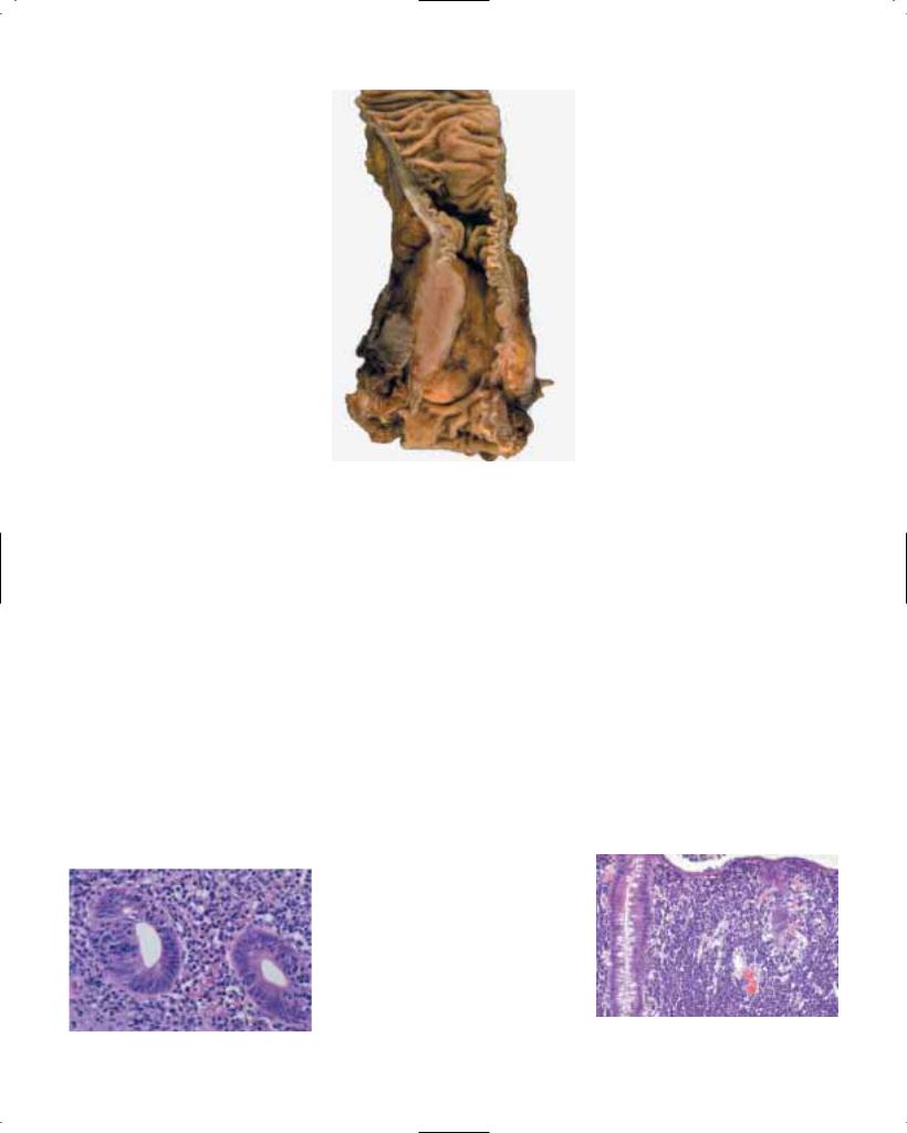
extent of disease. Multiple lymphomatous polyposis has a characteristic radiological picture with numerous polyps of variable size throughout the colon. Transrectal ultrasonography may also be a useful adjunct for diagnosis.
Macroscopy
Most low-grade lymphomas present as well defined protuberant growths that deeply invade the bowel wall. Diffuse large B-cell lymphoma (DLBCL) and Burkitt lymphoma tend to form larger masses with stricture and ulcer formation involving long segments of the colorectum. Low-grade and aggressive MALT lymphomas typically remain localized for prolonged periods, but may spread to involve loco-regional lymph nodes.
Mantle cell lymphoma (MCL) may present as an isolated mass or as multiple polyps producing the clinical picture of multiple lymphomatous polyposis {2084}. In most cases, the colon is more significantly involved than the small bowel. Importantly, other histological subtypes of lymphoma can produce this clinicopathological entity (see below). The polyps range in size from 0.5 cm to 2 cm with much larger polyps found in the ileocaecal region {791, 1292}. MCL frequently spreads to involve the spleen, extra-abdominal lymph nodes, bone marrow and peripheral blood.
Histopathology
MALT lymphoma
The majority of intestinal lymphomas involving the large bowel are B-cell lymphomas of MALT type, including both low-grade and aggressive histologies {796}. The histological and immunophenotypic features are discussed in detail in the section describing lymphomas of the stomach. Colorectal low-grade MALT
Fig. 6.82 MALT lymphoma of rectum with lymphoepithelial lesions.
Fig. 6.83 Malignant lymphoma of rectum.
lymphomas resemble those of the small |
|
|
intestine in that lymphoepithelial lesions |
|
|
are less prominent than in the stomach. |
|
|
Precise criteria for defining a MALT lym- |
Burkitt lymphoma |
|
phoma of large cell type are lacking, as |
The details of the histology, immunophe- |
|
are the criteria for distinguishing transfor- |
notype, |
cytogenetics and molecular |
mation within a low-grade MALT lym- |
genetics are described in detail in the |
|
phoma. When both histologies are evi- |
small |
intestinal lymphoma section |
dent, the neoplasm is best described as |
(Chapter 4). |
|
composite. When small foci of large |
|
|
transformed cells or early sheeting-out of |
Burkitt-like lymphoma |
|
large cells are detected within a back- |
The histological and cytogenetic features |
|
ground of low-grade intestinal MALT lym- |
have been previously described in the |
|
phoma, their presence should be noted |
small intestinal lymphoma section. AIDS |
|
{383}. Currently, the prognostic impact of |
patients have a preponderance of cases |
|
these findings and their effect on treat- |
with this histology. Many are of small non- |
|
ment are undetermined. DLBCLs arising |
cleaved cell type with the typical molec- |
|
in the large bowel that lack a background |
ular and cytogenetic changes associat- |
|
of low-grade MALT lymphoma are best |
ed with classical Burkitt lymphoma, and |
|
classified as extranodal diffuse large |
|
|
B-cell lymphoma, not otherwise specified, until such time as confirmatory tests can be established to clearly determine the histogenesis of these neoplasms from the mucosal immune system.
Mantle cell lymphoma
The morphology of MCL involving the large bowel is identical to MCL at nodal sites {110}. The architecture is most frequently diffuse, but a nodular pattern and a less common true mantle-zone pattern are also seen. Reactive germinal centers may be found and are usually
Fig. 6.84 Burkitt lymphoma of colon. The malignant cells infiltrate the lamina propria and produce lymphoepithelial lesions.
140 Tumours of the colon and rectum

are best considered to be part of the same biological entity {236}. However, patients with AIDS have also been recognized to have another lymphoma, with features intermediate between small non-cleaved cell lymphoma with plasmablastic differentiation and immunoblastic lymphoma, plasmacytoid type. This latter lymphoma subtype is strongly associated with EBV infection and TP53 mutations {236}.
Other B-cell lymphomas
Any subtype of B-cell lymphoma can arise in a colorectal site, including those thought to arise from peripheral lymph node equivalents. De novo DLBCLs are equal in frequency to low-grade MALT lymphomas in the colorectum {1733}, and are particularly common in the setting of HIV infection. Rectal involvement in AIDS patients typically demonstrates DLBCL with either centroblastic or immunoblastic cytomorphology. These lymphoma subtypes can be distinguished using phenotypic markers including Bcl-6, CD138 (syndecan-1) and EBV-related protein, latent membrane protein (LMP-1). Small noncleaved and centroblastic lymphomas express Bcl-6, but fail to express CD138 or LMP-1 in the majority of cases. Immunoblastic lymphomas in the HIV setting do not express Bcl-6, but are positive for both CD138 and LMP-1, in keeping with a non-germinal center histogenesis {237}.
Genetics
MALT lymphoma
Cytogenetic and molecular features of intestinal low-grade MALT lymphomas are incompletely understood. The presence of either t(1;14)(p22;q32) or t(11;18)(q21;q21) and the corresponding molecular abnormalities, rearrangement of bcl-10 or AP12-MLT, have not been described at this site, thus the relationship of these lesions to gastric MALT lymphomas is unclear {2116, 412}.
Fig. 6.85 Mantle cell lymphoma infiltrating the submucosa predominantly , thereby causing a polypoid lesion.
Furthermore, trisomy 3 is common in gastric MALT lymphomas, but the frequency of this cytogenetic abnormality in primary intestinal lymphoma is unknown. Some of these DLBCLs may have a low-grade MALT component evident, providing compelling evidence that their histogenesis is related to the mucosal immune system.
Mantle cell lymphoma
MCL is characterized by a recurrent cytogenetic abnormality, the t(11;14) (q13;q32). This translocation deregulates expression of the bcl-1 oncogene on chromosome 11. Rearrangement can be detected using Southern blot analysis, PCR or fluorescent in situ hybridization (FISH).
Prognosis
The relevant prognostic factors in colorectal lymphomas are similar to those for the small intestine, and have been described in detail in that section. MCL is
an aggressive lymphoma, which typically presents in advanced stage; there is often involvement of mesenteric and peripheral lymph nodes, spleen, bone marrow and peripheral blood {670}.
Fig. 6.86 Mantle cell lymphoma.
B-cell lymphoma 141

Mesenchymal tumours of the colon and rectum
M. Miettinen
J.Y. Blay
L.G. Kindblom
L.H. Sobin
Definition
A variety of benign and malignant mesenchymal tumours that arise in the large intestine as a primary site.
Classification
The morphological definitions of these lesions follow the WHO histological classification of soft tissue tumours {2086}. Stromal tumours are described in detail in the chapter on gastric mesenchymal tumours.
Epidemiology
Sarcomas accounted for 0.1% of malignant large intestinal tumours in SEER data {1928}. Males were affected slightly less than females. Adults between the 6th and 8th decades were primarily affected.
Aetiology
Aetiological factors are poorly understood for most colorectal mesenchymal tumours. Kaposi sarcoma usually occurs in association with AIDS, but it has also been described in connection with inflammatory bowel disease, in one case following immunosuppressive therapy {1930, 1584}. Human herpesvirus 8 is usually demonstrable by PCR in Kaposi sarcoma cells. An angiosarcoma has been reported in the colon, related to a persistent foreign body {149}.
Fig. 6.87 Leiomyoma of rectum.
Pathological features
Lipomas are composed of mature adipose tissue and are surrounded by a fibrotic capsule. They usually arise in the submucosal layer of the caecum or the sigmoid colon. When ulcerated, the lipocytes may become irregular and hyperchromatic. Lipomas should be distinguished from lipohyperplasia of the ileocaecal valve {1726}.
Neurofibromas and schwannomas occur in the colorectum. Most patients with the former have neurofibromatosis, and in
these cases plexiform neurofibromas are common. Ganglioneuromas occur rarely in the mucosa.
Vascular tumours are classified into benign (such as haemangiomas, lymphangiomas and angiomatosis) and malignant (such as haemangioendotheliomas and angiosarcomas). Kaposi sarcoma is mostly asymptomatic; a few present with GI-bleeding {319}. Intestinal lesions may be observed without cutaneous disease {114}. The tumours are often multiple mucosal or submucosal nodules. Histologically typical are sheets of spindle cells interspersed by clusters of extravasated erythrocytes. Cytoplasmic hyaline PAS-positive globules are usually seen. The spindle cells are generally positive for CD31 and CD34 and are negative for actin, desmin and c-kit.
Leiomyomas usually are detected in the rectum and colon as small polyps arising from the muscularis mucosae, and consist of well-differentiated smooth muscle cells with a similar immunohistochemical profile as observed in oesophageal leiomyomas {1227}. Leiomyomatosis has been described in the colon with a circumferential semiconstrictive growth in a 35 cm long segment {529}. It is not known whether colorectal leiomyomas and leiomyomatosis have the same colla-
A B
Fig. 6.88 Leiomyosarcoma. A Cigar-shaped nuclei. B Pleomorphic cells with atypical mitosis.
142 Tumours of the colon and rectum
Fig. 6.89 Colonic lipoma. |
Fig. 6.90 Malignant stromal tumour. |
A B
Fig. 6.91 Kaposi sarcoma. A Submucosal infiltrate. B Vascular slit pattern.
gen type IV deletions as the oesophageal leiomyomas.
Gastrointestinal stromal tumours (GISTs) of the colorectum are similar to those in the stomach and small intestine and are discussed in the section on gastric mesenchymal neoplasms. Most reports antedate the separation of GISTs and leiomyosarcoma. GISTs occur mainly between the 6th and 8th decades, and most are malignant {89}. Many tumours grow beyond the rectal wall making radical surgery difficult and recurrences common. Histologically, the examples reviewed by us have all been of the spindle cell variety, all have been c-kit positive, and most of them CD34-positive. Actin-positivity occurs, but the tumours are desmin-negative. C-kit mutations have been shown in rectal GISTs {1018}. The survival from large bowel stromal/ smooth muscle sarcomas appears to be slightly higher than that of the small bowel and lower than that of the stomach and oesophagus {461}.
Mesenchymal tumours 143


CHAPTER 7
Tumours of the Anal Canal
Although incidence rates are still low, there has been a significant increase in squamous cell carcinoma over the last 50 years. HIV infected homosexual men appear particularly at risk. HPV DNA is detectable in most anal squamous cell carcinomas.
Despite its short length, the anal canal produces a variety of tumour types reflecting its complex anatomic and histological structure. Squamous, glandular, transitional, and melanocytic components occur at this site, either alone, or in combination.

WHO histological classification of tumours of the anal canal
Epithelial tumours |
|
Undifferentiated carcinoma |
8020/3 |
|
|
Others |
|
Intraepithelial neoplasia1 (dysplasia) |
|
|
|
Squamous or transitional epithelium |
|
Carcinoid tumour |
8240/3 |
Glandular |
|
|
|
Paget disease |
8542/32 |
Malignant melanoma |
8720/3 |
Carcinoma |
|
Non-epithelial tumours |
|
Squamous cell carcinoma |
8070/3 |
|
|
Adenocarcinoma |
8140/3 |
Secondary tumours |
|
Mucinous adenocarcinoma |
8480/3 |
|
|
Small cell carcinoma |
8041/3 |
|
|
_________
1Behaviour is coded /0 for benign tumours, /3 for malignant tumours, /2 for in situ carcinomas and grade III intraepithelial neoplasia, and /1 for unspecified, borderline or uncertain behaviour. Intraepithelial neoplasia does not have a generic code in ICD-O. ICD-O codes are available only for lesions categorized as squamous intraepithelial neoplasia, grade III
(8077/2), squamous cell carcinoma in situ (8070/2), transitional cell carcinoma in situ (8120/2), glandular intraepithelial neoplasia, grade III (8148/2), and adenocarcinoma in situ (8140/2). 2 Morphology code of the International Classification of Diseases for Oncology (ICD-O) {542} and the Systematized Nomenclature of Medicine (http://snomed.org).
TNM classification of tumours of the anal canal
TNM classification1, 2
T – Primary Tumour
TX |
Primary tumour cannot be assessed |
T0 |
No evidence of primary tumour |
Tis |
Carcinoma in situ |
T1 |
Tumour 2 cm or less in greatest dimension |
T2 |
Tumour more than 2 cm but not more than 5 cm in greatest |
|
dimension |
T3 |
Tumour more than 5 cm in greatest dimension |
T4 |
Tumour of any size invades adjacent organ(s), e.g., vagina, ure- |
|
thra, bladder (involvement of sphincter muscle(s) alone is not |
|
classified as T4) |
N – Regional Lymph Nodes |
|
NX |
Regional lymph nodes cannot be assessed |
N0 |
No regional lymph node metastasis |
N1 |
Metastasis in perirectal lymph node(s) |
N2 |
Metastasis in unilateral internal iliac and/or inguinal lymph |
|
node(s) |
N3 |
Metastasis in perirectal and inguinal lymph nodes and/or bilater- |
|
al internal iliac and/or inguinal lymph nodes |
M – Distant Metastasis
MX Distant metastasis cannot be assessed M0 No distant metastasis
M1 Distant metastasis
Stage Grouping |
|
|
|
Stage 0 |
Tis |
N0 |
M0 |
Stage I |
T1 |
N0 |
M0 |
Stage II |
T2 |
N0 |
M0 |
|
T3 |
N0 |
M0 |
Stage IIIA T1 |
N1 |
M0 |
|
|
T2 |
N1 |
M0 |
|
T3 |
N1 |
M0 |
|
T4 |
N0 |
M0 |
Stage IIIB T4 |
N1 |
M0 |
|
|
Any T |
N2, N3 |
M0 |
Stage IV |
Any T |
Any N |
M1 |
_________
1 {1, 66}. This classification applies only to carcinomas.
2 A help desk for specific questions about the TNM classification is available at http://tnm.uicc.org.
3This includes cancer cells confined within the glandular basement membrane (intraepithelial) or lamina propria (intramucosal) with no extension through muscularis mucosae into submucosa.
146 Tumours of the anal canal and anal glands

Tumours of the anal canal
C. Fenger
M. Frisch
M.C. Marti
R. Parc
Definition
Tumours that arise from or are predominantly located in the anal canal. The most frequent neoplams of this region are human papilloma virus (HPV-)associ- ated squamous cell carcinomas and adenocarcinomas.
Topographic definition of anal canal and anal margin
The anal canal is defined as the terminal part of the large intestine, beginning at the upper surface of the anorectal ring and passing through the pelvic floor to end at the anus {68}. The most important macroscopic landmark in the mucosa is the dentate (pectinate) line composed of the anal valves and the bases of the anal columns. Histologically, the mucosa can be divided into three zones. The upper part is covered with colorectal type mucosa. The middle part is the anal transitional zone (ATZ), which is covered by a specialized epithelium with varying appearances; it extends from the dentate line and on average 0.5-1.0 cm upwards {490, 1929}. The lower part extends from the dentate line and downwards to the anal verge and has formerly been called the pecten. It is covered by squamous epithelium, which may be partly keratinized, particularly in case of mucosal prolapse.
The perianal skin (the anal margin) is defined by the appearance of skin
Anorectal ring
Anal columns
Anal valves and sinuses
DENTATE LINE
Intersphincteric groove
appendages. There exists no generally accepted definition of its outer limit {62, 66, 845}. The term anus refers to the distal external aperture of the alimentary tract. Anal margin tumours are classified according to the WHO histological typing of skin tumours {682}.
Squamous cell carcinoma
Definition
Squamous cell carcinoma (SCC) of the anal canal is a malignant epithelial neoplasm that is frequently associated with chronic HPV infection.
ICD-O code |
8070/3 |
Epidemiology
SCC of the anal canal and anal margin typically occurs among patients in their 6th or 7th decade of life {540}. However, anal SCCs may occur in young adults, particularly in patients with cellular immune incompetence {1212}. Unselected, population-based studies show an approximate 2:1 female predominance among patients with anal SCC {540, 600, 1213}.
There are few published, histologically verified incidence rates of anal cancer {540, 600, 1213}. Data from most popula- tion-based cancer registries worldwide show age standardized incidence rates of anal SCC of between 0.5 and 1.0 per
Surgical anal canal
Histological anal canal
Anatomical anal canal
Anal verge, ‘anus’
Fig. 7.01 Anatomy of the anal canal. Printed with permission from ref 490.
100,000 in women and between 0.3 and 0.8 per 100,000 in men {1471}. Still a relatively rare disease, anal SCC has shown a remarkable increase in incidence during the past half century {540, 600, 1213}. From being similar in the two sexes until approximately 1960 at 0.2 per 100,000, annual age-adjusted incidence rates in Denmark rose 2.5-fold in men and 5-fold in women during the period 1943-1994. For both men and women, urban populations are at higher risk than rural populations {540, 600, 1213}, and there are considerable racial differences in incidence. In the United States, blacks tend to have higher incidence rates than whites {1213}, while Asians and Pacific Islanders appear to be at very low risk {70}.
Homosexual men appear to constitute a group at particular risk {368, 538, 140, 96, 369, 540, 1213, 1690, 730}. In the United States, the incidence of anal SCC in homosexual men has been estimated to be 11 to 34 times higher than in the general male population and approximately as high as the incidence of cervical cancer before the introduction of cervical cytology screening {369, 1447}. HIV infected homosexual men appear to be at particularly risk {1212, 1449, 598}. Other sexual factors strongly associated with anal SCC include number of sexual partner, receptive anal intercourse, and co-existence of sexually transmitted diseases {368, 538, 730, 733}.
Aetiology
Sexually transmittable human papillomaviruses (HPVs) are detected by PCR in the majority of anal SCC {355, 367, 538, 704, 732, 1448}. One large study showed that SCCs involving the anal canal are more often high-risk HPV positive (92%) than lesions confined to the perianal skin (64%) {536}, suggesting that HPV-unrelated pathways may apply particularly to cancers of the perianal skin. A strong association with tobacco smoking has been established in women, but the role of smoking in men is less clear {367, 539, 730, 733}. States of cellular immunosuppression are associated
Squamous cell carcinoma 147

Fig. 7.02 Normal histology of the anal transition zone.
with increased risk of anal squamous cell carcinoma. This has been observed for renal transplant recipients {150, 1494} and for patients with HIV infection and AIDS {1212}.
Haemorrhoids and fissures, fistulae and abscesses in the anal region were long considered predisposing factors {192, 198, 1618}. However, three case-control studies {368, 537, 733} and two cohort studies {541, 1074} failed to support the association. Crohn’s disease of long duration, which has been implicated in the aetiology of anal SCC based on case reports {992, 1765}, was not associated with risk in the only controlled study addressing the issue {537}.
Oestrogen and androgen receptors have been found in the anal mucosa and its supportive tissue {1396}, suggesting a physiological role of sex hormones in their maintenance. Women who reach menarche late and women with short fertile periods may be at elevated risk of anal SCC {539}.
Clinical features
Symptoms and signs
Anal intraepithelial neoplasia is often an unexpected finding in minor surgical specimens. Clinical manifestations of anal cancer are often late and non-spe-
Fig. 7.03 In situ hybridisation for HPV 16/18 is positive in this anal carcinoma.
cific and are mainly related to tumour size and extent of infiltration. They include anal pruritus, discomfort in sitting position, sensation of a pelvic mass, pain, change in bowel habit, incontinence due to sphincter infiltration, discharge, bleeding, fissure, or fistula. The initial nonspecificity of clinical features explains why diagnosis can be delayed {855, 1621, 1719, 1835}.
The clinical diagnosis of an anal tumour should always be confirmed by histological examination. A forceps or needle biopsy is usually sufficient to establish the diagnosis. The biopsy should be accompanied by an exact description of location and appearance of the biopsy site. An excisional biopsy is inadvisable, because wound healing delay would postpone optimal chemo-radiotherapy treatment. Enlarged lymph nodes may be excised or biopsied with needle aspiration under radiological control.
Imaging
Computerised tomography (CT) scan, magnetic resonance imaging (MRI), and needle aspiration are used to detect inguinal and pararectal node involvement. Endoanal ultrasonography (EUS) enables assessment of spread in terms of proximal and circumferential extension and infiltration of deep layers. Furthermore, EUS enables the follow-up of irradiated carcinomas {703}. CT scan and MRI allow detection of involved lymph nodes and distant metastases {1835}.
Exfoliative cytology
In patients with increased risk such as individuals with HIV or women with genital tract SCC, the use of anal smears taken with a cytology brush from the area below the dentate line is recommended {1689}.
Macroscopy
The tumour may present as a small ulceration or fissure with slightly exophytic and indurated margins, and irregular thickening of the anoderm and anal margin with chronic dermatitis. The lesion may have a different colour from the surrounding tissue.
If ulceration and infiltration develop, the lesion becomes fixed to the underlying structures and may bleed. In advanced stages, the sphincteric muscles are deeply infiltrated although there may be little mucosal ulceration.
Fig. 7.04 Ulcerating nodular squamous cell carcinoma of anus.
Tumour spread and staging
Anal SCC should be staged according to the TNM system {66}. Treatment for anal SCC has now changed from surgery alone to sphincter preserving procedures including radiation and chemotherapy, sometimes in combination with local excision. Large surgical specimens are therefore rare. The examination should include resection lines in all directions and a careful search for lymph nodes. Clinical results of the combined treatment regimes are comparable or even better than those for surgery alone, but detection of residual disease can be more difficult by imaging techniques due to local fibrosis. In such cases a transanal full thickness tru-cut needle biopsy may be helpful {785}. Identification of residual
Fig. 7.05 Squamous cell carcinoma arising at dentate line.
148 Tumours of the anal canal

Fig. 7.06 Well differentiated squamous cell carcinoma composed of large cells showing keratinization.
tumour cells may be facilitated by immunostaining for high molecular weight cytokeratins (CKs).
In 15-20% of cases, the lesion may infiltrate the lower rectum and the neighbouring organs including the rectovaginal septum, bladder, prostate and posterior urethra, sometimes with suppuration and fistulas. The vulva is usually spared. Lymphatic spread occurs in up to 40 percent of cases {165, 1174, 1621, 1719, 2033}. Tumours proximal to the pectinate line drain into the pelvis along the middle rectal vessels to the pelvic side walls and internal iliac chains and superiorly via the superior rectal vessels to the periaortic nodes. Tumours distal to the dentate line drain along cutaneous pathways to the inguinal and the femoral nodal chains. Inguinal nodes are involved in about 10-20% of cases {230, 575, 1174, 1650, 1692}. Inguinal lymph nodes can be involved bilaterally in a small number of cases at time of presentation. Retrograde lymphatic drainage occurs in advanced cases when the lymphatics are obstructed by malignant spread {1621, 1719}.
Histopathology
Squamous cell carcinoma of anal canal
Anal SCC may show a single predominant line of differentiation, but most exhibit a mixture of areas with different histological features. One pattern is that of large, pale eosinophilic cells and keratinization of either lamellar or single cell type. Another is that of small cells with palisading of the nuclei in the periphery of tumour cell islands. The latter often contain necrotic eosinophilic centres. Intermediate stages between these two extremes are often present. Differentiation into tubular or spindle cell configuration may be found. The invasive margin can vary from well circumscribed to irreg-
ular, and a lymphocytic infiltrate may be pronounced or absent. None of these features have been shown to have any prognostic significance, but poor keratinization, prominent basaloid features and small tumour cell size are related to infection with ‘high risk’ HPV {536}. The keratin profile of anal SCC is complex and variable {2112, 2113}. The usual immunoexpression pattern is shown in Table 7.01.
The second edition of the WHO classification of SCC in the anal canal included the large-cell keratinizing subtype, the large-cell non-keratinizing subtype, and the basaloid subtype {845}. The value of this classification of anal SCC has been questioned in recent years. Many tumours show more than one subtype. Thus in a study of 100 cases of anal carcinomas, 99 showed some features of squamous differentiation (keratinisation, stratification and prickles), 65 showed basaloid features (small cell change, palisading, retraction artefact and central eosinophilic necrosis) and 26 showed focal evidence of ductal proliferation and occasionally positive staining for PAS after diastase digestion {2111}. Furthermore, the diagnostic reproducibility of these subtypes is low {492}. This is probably the reason that the proportion of basaloid carcinoma in larger series has varied from 10 to almost 70 %, and that no significant correlation between histological subtype and prognosis has been established. In addition, the histological diagnosis is nowadays nearly always performed on small biopsies, that may not be representative for the whole tumour {492}. Therefore, it is recommended that the generic term ‘squamous carcinoma’ be used for these tumours, accompanied by a comment describing those histopathological features that may possibly affect the prognosis or reflect different aetiolo-
N
Fig. 7.07 Squamous cell carcinoma composed of basaloid cells. Central necrosis (N) of tumour nests is typical.
gies, i.e. size of predominant neoplastic cell, basaloid features, degree of keratinisation, adjacent squamous intraepithelial neoplasia, or presence of mucinous microcysts.
Apart from the verrucous carcinoma mentioned below, only two rare histological subtypes seem to have a different biological course, both having a less favourable prognosis {1734}. One is characterized by areas with well formed acinar or cystic spaces containing mucin that reacts with Alcian dyes or PAS after diastase digestion. This is termed squamous cell carcinoma with mucinous microcysts. The other is characterized by a rather uniform pattern of small tumour cells with nuclear moulding, high mitotic rate, extensive apoptosis and diffuse infiltration in the surrounding stroma. This has been called small cell (anaplastic) carcinoma, but should not be confused with small cell carcinoma (poorly differentiated neuroendocrine carcinoma).
Fig. 7.08 Squamous cell carcinoma showing a combination of basaloid and squamous features.
Squamous cell carcinoma of anal margin
The distinction between anal canal and anal margin SCC may be difficult, as tumours often involve both areas at the time of diagnosis. This may account for the varying data on prognosis, but this is generally better for anal margin SCC than for anal canal SCC, in particular if local resection is possible {392, 530, 1484}. Anal margin SCC is often of the large cell variant {536, 1484}.
Verrucous carcinoma
In the anogenital area, this tumour is also called giant (malignant) condyloma or Buschke-Lowenstein tumour. It has a cauliflower-like appearance, is larger than the usual condyloma with a diameter up to 12 cm, and fails to respond to
Squamous cell carcinoma 149

A B
Fig. 7.09 Squamous cell carcinoma of anus. A Combination of basaloid features and keratinization. B Large cells, poorly differentiated.
conservative treatment. In contrast to an ordinary condyloma, it is characterized by a combination of exophytic and endophytic growth. Histologically, it shows acanthosis and papillomatosis with orderly arrangement of the epithelial layers and an intact but often irregular base with blunt downward projections and ker- atin-filled cysts. The endophytic growth is accompanied by destruction of the underlying tissues. Cytologically, the epithelial cells appear benign. Large nuclei with prominent nucleoli may be present, but dysplasia is usually minimal and mitoses are restricted to the basal layers {162}.
Some verrucous carcinomas contain HPV, the most common types being 6 and 11. They are regarded as an intermediate state between the ordinary condyloma and SCC, and the clinical course is typically that of local destructive invasion without metastases. Among
33 published anorectal cases, 42 per cent have shown malignant transformation {133}. The presence of severe cytological changes, unequivocal invasion or metastases should lead to the diagnosis of SCC and to the appropriate therapy.
Grading
Poor prognosis has been related to poor differentiation {165}, especially if this was defined only by the degree of dissociation of tumour cells {599}. However, such differences may be related to tumour stage in multivariate analysis {1734}. Grading on biopsies is not recommended, as these may not be representative for the tumour as a whole.
Precursor lesions and benign tumours
Chronic HPV infection
Warts in the perianal skin and lower anal canal (condyloma acuminatum) show the same histology as their genital counter-
Fig. 7.10 Mucinous carcinoma of anus. Tumour extends to anal sphincter.
parts. Flat koilocytic lesions also occur. They should always be totally embedded and examined histologically for possible presence of intraepithelial neoplasia.
Intraepithelial neoplasia
Precancerous anal intraepithelial neoplasia (AIN) in the anal transition zone (ATZ) and the squamous zone, has also been termed dysplasia, carcinoma in-situ and anal squamous intraepithelial lesion (ASIL) {494, 1449}. The corresponding lesions in the perianal skin are commonly referred to as Bowen disease. This terminology is complicated by the fact that the precancerous changes are not always restricted to one area. Leukoplakia is a clinical term and should not be used as a histological diagnosis.
Anal intraepithelial neoplasia (squamous cell dysplasia in the anal canal). Most cases of AIN are incidental findings in minor surgical specimens for benign conditions. When macroscopically detected, AIN may present as an eczematoid or papillomatous area, or as papules or plaques. The latter may be irregular, raised, scaly, white, pigmented or erythematous and occasionally fissured. Induration or ulceration may indicate invasion. Histologically, AIN is characterized by varying degrees of loss of stratification and nuclear polarity, nuclear pleomorphism and hyperchromatism, and increased mitotic activity with presence of mitoses high in the epithelium. The surface may or may not be keratinized, and koilocytic changes may be present.
AIN has been graded into I, II or III, or into mild, moderate and severe dysplasia {494}. Reproducibility studies have shown considerable observer variation {254}. A two grade system (lowand high-grade) may be more appropriate.
Squamous dysplasia at the anal margin - Bowen disease. Clinically, this presents as a white or red area in the perianal skin that may be in continuity with dysplastic lesions in the anal canal. HPV DNA is sometimes identified, including types 16 and 18, among others. Histologically it shows full thickness dysplasia of the squamous and sometimes the pilosebaceous epithelium, with disorderly maturation, mitoses at all levels and dyskeratosis. Occasionally, atypical keratinocytes may resemble Paget cells, but are negative for low molecular weight CKs and for mucin. In pigmented Bowen disease the neoplastic cells are invariably negative
150 Tumours of the anal canal

Fig. 7.11 Giant condyloma.
for S-100 protein and HMB-45.
Bowen disease has a strong tendency to recurrence after local treatment but only a few percent will progress to SCC. It is often associated with genital neoplasia but not with internal malignancies {1161, 1668}.
Bowenoid papulosis. This condition presents as multiple 2-10 mm reddish brown papules or plaques, most commonly in sexually active young adults. Aetiologically it is related to HPV infection, usually HPV 16. Bowenoid papulosis is similar histologically to Bowen disease, and the distinction is made on a combination of clinical and pathological observations. Bowenoid papulosis tends to resolve spontaneously, but can recur {635}. It does not progress to carcinoma.
Genetic susceptibility
Human leukocyte antigens (HLAs) are involved in the presentation of viral antigens to the immune system. Since the aetiology of most anal SCCs involves HPV infection {536}, susceptibility to cancer development might be HLA type dependent. However, no study has addressed the association between specific HLA class I or II alleles and the risk, and attempts to identify other genetic susceptibility markers for anal SCC have so far been unsuccessful {286, 287}.
Genetics
HPV DNA is detectable in most anal SCCs; in a large population-based series of anal SCCs in Denmark and Sweden, 84% contained HPV DNA, with higher proportions of HPV-DNA positive cancers among women and homosexual men than among non-homosexual men {536}.
Loss of functional tumour suppressor protein p53 appears to be centrally involved in the development of anal and
anogenital SCCs {355, 356, 704, 1040}. Inactivation of p53 may occur at the gene level through point mutations leading to the production of inactive p53 or, less frequently, by means of deletions in the relevant area of chromosome 17p {704}. More typically, p53 inactivation occurs at the protein level through formation of a complex between the viral protein E6 (expressed by ‘high-risk’ HPV types) and a cellular protein, the E6-associated protein, which when bound to p53 leads to rapid proteolytic degradation of p53 {2092}. The level of p53 expression does not correlate with HPV status {704}. The E7 protein of ‘high risk’ HPV types binds to the retinoblastoma protein, pRb {440}, disrupting signals that normally restrict proliferation to the basal epithelial layer. The resulting increased proliferation increases the risk of malignant transformation on exposure to DNA damaging stimuli. The combination of increased cell proliferation (pRb inactivation) and impaired ability to induce cell cycle arrest or apoptosis following DNA damage (p53 inactivation) are two central mechanisms through which ‘high risk’ types of HPV increase the risk of anogenital cancer.
Additional gene alterations appear to be involved in malignant progression and invasion. In one study, the c-myc gene
Fig. 7.12 High-grade intraepithelial neoplasia adjacent to normal rectal epithelium.
Fig. 7.13 In situ hybridisation (black stain) for HPV 6/11 in an anal condyloma.
Fig. 7.14 Carcinoma of anal canal. Small neoplastic glands simulate anal glands.
was found to be amplified in 30% of anal SCCs {355}, while other cellular oncogenes, including ras and cyclin D, do not seem to be centrally involved {708, 1737}. Several chromosomal aberrations have been observed in anal SCCs {704, 1294}. Using comparative genomic hybridization, one study identified consistent gains in chromosomes 3q, 17, and 19 as well as losses in chromosomes 4p, 11q, 13q, and 18q {704}.
Prognosis and predictive factors
The most important prognostic factors in recent larger series of anal canal SCC are tumour stage and nodal status {530, 1483, 1734}. SCC of the anal margin has a slightly better prognosis, which depends only on inguinal node involvement {1484}. DNA ploidy status has only been shown to be of independent prognostic significance in one of three larger series {599, 1702, 1734}. Expression of p53, cathepsin D, c-erb B2 and retinoblastoma gene protein are not predictive factors {169, 731, 784, 1901}.
Adenocarcinoma
Definition
Anal canal adenocarcinoma is an adenocarcinoma arising in the anal canal epithelium, including the mucosal surface, the anal glands and the lining of fistulous tracts.
ICD-O code |
8140/3 |
Clinical features
The clinical features of anal adenocarcinoma of colorectal type do not differ from those of anal SCC. Perianal adenocarcinomas may present as submucosal tumours, sometimes in combination with fistulas. Occasionally, there may be
Squamous cell carcinoma / Adenocarcimoma 151

Fig. 7.15 Low-grade squamous intraepithelial neoplasia with koilocytosis.
associated Paget disease of the anus (see below). Tumour spread and staging largely correspond to anal SCC.
Histopathology
Adenocarcinoma arising in anal mucosa
Most adenocarcinomas found in the anal canal represent downward spread from an adenocarcinoma in the rectum or arise in colorectal type mucosa above the dentate line. Macroscopically and histologically, they are indistinguishable
Fig. 7.18 Adenocarcinoma arising in a fistula.
Fig. 7.16 High-grade squamous intraepithelial neoplasia with hyperkeratosis.
from ordinary colorectal type adenocarcinoma, and do not seem to represent a special entity except for their low location. Adenocarcinoma in the anal transitional zone (ATZ) may develop after restorative proctocolectomy for ulcerative colitis {1711}.
Extramucosal (perianal) adenocarcinoma
Approximately two hundred cases of extramucosal adenocarcinoma have been reported, the largest series unfortunately with insufficient histological data {9}. A minimum criterion for the diagnosis is an overlying non-neoplastic mucosa, which may be ulcerated. Recent reports indicate that about two thirds of these tumours manifest in men with a mean age about 60 years. Reliable data for the prognosis for such patients have not been identified. Difficulties in establishing the correct diagnosis may delay proper treatment.
Extramucosal adenocarcinoma seem to fall into two groups, based on their association with either fistulae or remnants of anal glands. At present, no laboratory methods can distinguish between these two.
The epithelium of persistent anal fistulae is most often of the same type as found in the anal glands and ATZ {1117}, and the epithelium in these two locations show the same profile with regard to
A
B
Fig. 7.17 A, B Inflammatory cloacogenic polyp. Dilated elongated hyperplastic glands showing regenerative atypia. Surface erosion is a constant feature.
mucin composition {491} and keratin expression {2113}.
Adenocarcinoma within anorectal fistula.
These tumours develop in pre-existing anal sinuses or in fistulae {74}. Some are associated with Crohn disease {992}. Others may contain epithelioid granulomas, often related to foci of inflammation or extravasated mucin but without other signs of inflammatory bowel disease {863}.
Rarely, the tumours may be related to fistulae lined by normal rectal mucosa including muscularis mucosae, most likely representing adenocarcinomas arising in congenital duplications {863}. Histologically, carcinomas arising in fistulae usually are of the mucinous type, but tubular adenocarcinomas and squamous neoplasia can also be found {992, 2173}.
Adenocarcinoma of anal glands. Only a few cases have been reported in which convincing evidence for origin in an anal gland has been demonstrated by continuity between anal gland epithelium and tumour {118, 650, 1472, 2087, 2131}. With a single exception {650}, these patients have had no history of previous or concomitant fistula. The tumours were all characterized by a combination of ductular and mucinous areas. Pagetoid spread was present in at least one case {2131}.
152 Tumours of the anal canal
Grading
Anal adenocarcinomas are graded as colorectal adenocarcinomas.
Precursor lesions
Anal adenocarcinomas are thought to arise from glandular intraepithelial neoplasia, which can be graded as in the colorectum.
Prognosis and predictive factors
The prognosis for anal adenocarcinoma seems to be related only to the stage at diagnosis and is poorer than that for SCC {118, 930, 1305}.
Basal cell carcinoma of the anal margin
Basal cell carcinoma, the most common skin cancer, is primarily found on sunexposed areas, and only a few more than a hundred cases have been reported in the anal area. {1353}. The aetiology is unknown and there is no evidence of HPV infection {1332}. The tumour commonly presents as an indurated area with raised edges and central ulceration, located in the perianal skin but occasionally involving the squamous zone below the dentate line. Histologically, it can show the same variability in morphology as basal cell carcinoma elsewhere, most reported cases having had a solid or adenoid pattern.
Basal cell carcinoma is sufficiently treated by local excision and metastases are extremely rare. It is therefore important to distinguish it from squamous carcinoma, and this may be particularly difficult on small biopsies. Both tumours can be found in the squamous zone, and both can show a combination of basaloid, squamous and adenoid features and an inflammatory infiltrate in the stroma {50}. Numerous and even atypical mitoses may be present in basal cell carcinomas {1538}. However, basaloid areas in squa-
Fig. 7.19 Paget disease of the anal canal.
mous carcinoma usually show less conspicuous peripheral palisading, more cellular pleomorphism, and often large, eosinophilic necrotic areas. Immunohistochemistry may be helpful in establishing the diagnosis. Basal cell carcinoma is positive for Ber-EP4 and negative for CKs 13, 19 and 22, and for CEA, EMA, AE 1 and UEA 1, while basaloid variants of squamous cell carcinoma usually show the opposite pattern {50, 1061}.
Paget Disease
Extramammary Paget disease usually affects sites with a high density of apocrine glands, such as the anogenital region, where it presents as a slowly spreading, erythematous eczematoid plaque that may extend up to the dentate line {1667}. Histologically, the basal part or whole thickness of the squamous epithelium is infiltrated by large cells with abundant pale cytoplasm and large nuclei. Occasional cells have the appearance of signet-rings. Paget cells invariably react positively for mucin stains and nearly always for CK 7, but Merkel cells and Toker cells may also be positive for the latter {120, 1112}.
Paget disease of the anus appears to represent two entities. About half of the cases are associated with a synchronous or metachronous malignancy, most often a colorectal adenocarcinoma. Such cases can be regarded as a pagetoid extension of the tumour. They usually react positively for CK 20 and negatively for gross cystic disease fluid protein-15, a marker for apocrine cells. This is in contrast to the other half, which are not associated with internal malignancies but have a high local recurrence rate and may become invasive {1162}. Only this latter entity can be regarded as a true epidermotrophic apocrine neoplasm {85, 120, 595, 1374}.
Other lesions
Squamous cell papilloma of the anal canal
Rarely, papillomatous processes covered by normal, more or less keratinized squamous epithelium can be found in the anus. Such lesions should be tested for the presence of HPV. Negative cases are commonly regarded as ‘burned-out’ condylomas.
Fig. 7.20 Paget disease of the anal canal. Large Paget cells are distributed throughout the non-neo- plastic squamous epithelium.
Papillary hidradenoma
This rare tumour arises in the perianal apocrine glands, typically in middle aged women and only exceedingly rarely in men {1082}. It presents as a circumscribed nodule approximately 1 cm in diameter and may resemble a haemorrhoid.
Histologically, it consists of a papillary mass with a cyst-like capsule. The papillae are lined by a double layer of epithelial cells, the outer layer being composed of cells containing mucin. The tumour does not express the eccrine marker IKH-4, but it must be remembered that adenocarcinoma metastases also are negative {811}. Convincing examples of anal apocrine adenocarcinoma have not been published.
Keratoacanthoma
There are a few reports on keratoacanthoma arising in the perianal skin {454}.
Neuroendocrine tumours
Neuroendocrine tumours may arise in the anus {493, 744}. They are, however, conventionally classified as rectal. An immunohistochemical study of 17 rectal neuroendocrine tumours showed that most were of L-cell type {294}. For details, see in chapter 5 the section on endocrine tumours of the colon and rectum.
Adenocarcinoma 153

A B
Fig. 7.21 Secondary Paget disease of the anus. A The underlying adenocarcinoma is present beneath the squamous epithelium. High molecular weight keratin immunostain is largely restricted to normal squamous epithelium. B Low molecular weight keratins 8 and 13 immunostaining of tumour cells.
Malignant melanoma
Anal melanoma is rare. It is a disease of adults with a wide age range; most patients are white {339, 182}. Presentation is usually with mass and rectal bleeding, but tenesmus, pain and change in bowel habit also occur {339}. Macroscopy. Lesions may be sessile or polypoid. Pigmentation of the lesion is often appreciated. Satellite nodules may occur.
Histopathology. The features resemble those of cutaneous melanomas. The majority shows a junctional component adjacent to the invasive tumour, and this finding is evidence that the lesion is primary rather than metastatic. The tumour cells express S-100 and HMB-45.
Prognosis. Anal melanomas spread by lymphatics to regional nodes, and
haematogenously to the liver and thence to other organs. Metastases are frequent at time of presentation, and the prognosis is poor; the 5-year survival is less than 10% {339, 157}. The chances of longterm survival are increased if the lesion is small.
Mesenchymal and neurogenic tumours
These are all rare and the exact point of origin may be difficult to establish. Recent reports on tumours in the anorectal and perianal area include haemangioma, lymphangioma {372}, haemangiopericytoma {478}, leiomyoma, malignant fibrous histiocytoma and leiomyosarcoma {1110}, rhabdomyoma in a newborn {1014}, and rhabdomyosarcoma in childhood {1560} and adulthood
Fig. 7.22 Malignant melanoma of anus with typical polypoid appearance.
{902}, fibrosarcoma, neurilemmoma and neurofibroma {571}, granular cell tumour (myoblastoma) {862}, spindle cell lipoma and aggressive angiomyxoma {503} and extraspinal ependymoma in a newborn {2074}. HIV infected persons may, in addition to the increased risk of squamous neoplasia, develop Kaposi sarcoma in the perianal area {113}.
Malignant lymphoma
Primary lymphomas of the anorectal region are rare in the general population, but much more common in patients with AIDS, particularly homosexual men. All are of B-cell type, the most common types being large cell immunoblastic or pleomorphic {687, 786}. Langerhans cell histiocytosis has been described in children {617, 874} and an adult {329}.
A B C
Fig. 7.23 Malignant melanoma of anus. A Polypoid growth is frequent. B Scattered tumour cells contain melanin. C Epitheloid melanoma cells with prominent nucleoli.
154 Tumours of the anal canal

Table 7.01
Anal tumours, immunoreactivity profile (exceptions occur, especially among CK and mucin)1
|
|
CK |
CK |
CK |
|
|
|
|
|
|
8+18 |
7/20 |
5+14 |
Mucin |
CEA |
Vim |
Special |
|
|
|
|
|
|
|
|
|
|
Colorectal adenocarcinoma |
+ |
–/+ |
– |
+ |
+ |
– |
|
|
Squamous cell variants |
– |
–/– |
+ |
– |
– |
– |
CK 13/19 |
|
Basal cell carcinoma |
– |
–/– |
+ |
– |
– |
– |
Ber EP4 |
|
Neuroendocrine tumour |
+ |
–/– |
– |
– |
– |
– |
Chrom/Synap |
|
Malignant melanoma |
– |
–/– |
– |
– |
– |
+ |
S–100, HMB–45 |
|
Bowen (also pigmented) |
– |
–/– |
+ |
– |
– |
– |
|
|
Paget cells local Paget |
+ |
+/– |
– |
+ |
+ |
– |
GCDFP–15 |
|
Paget cells, from CRC |
+ |
+/+ |
– |
+ |
+ |
– |
|
|
Prostatic carcinoma |
+ |
–/– |
– |
– |
– |
– |
PSA, PSAP |
|
Malignant lymphoma |
– |
–/– |
– |
– |
– |
+ |
LCA and others |
––––––––––
1 Chrom |
= |
Chromogranin A |
PSA |
= |
Prostate specific antigen |
CK |
= |
Cytokeratin |
PSAP |
= |
Prostate specific acid phosphatase |
CRC |
= |
Colorectal carcinoma |
Synap |
= |
Synaptophysin |
GCDFP |
= |
Gross cystic disease fluid protein |
Vim |
= |
Vimentin |
Secondary tumours
Metastases to the anal canal and perianal skin are rare. Most primaries are found in the rectum or colon, but occasionally also in the respiratory tract, breast and pancreas {157, 182, 379, 888, 1767, 489}. There are few reports of metastatic squamous cell carcinoma {574}. Malignant lymphoma, leukaemia and myeloma may infiltrate the anal canal, and eosinophilic granuloma has also been described {489}.
Clinically, anal metastases cause similar symptoms to primary tumours at this site, including pain, bleeding and incontinence.
Neoplasia-like lesions Fibroepithelial polyp
Also called fibrous polyp or anal tag, this is one of the most frequent anal lesions. It may be found in the squamous zone or the perianal skin in up to half of all individuals {2101}. Grossly, the polyp is spherical or elongated with a greater
diameter ranging from a few mm up to 4 cm. The surface is white or grey and may show superficial ulceration. Histologically, it consists of a fibrous stroma covered by squamous epithelium, which usually is slightly hyperplastic and may be keratinized. The stroma may be more or less dense and often contains fibroblastic cells with two or more nuclei and a considerable number of mast cells {630}. Neuronal hyperplasia is a common feature {495}.
Fibroepithelial polyps may be associated with local inflammation such as fissure or fistula {1084}.
Granulomas can be found in about one third of skin tags in cases of Crohn’s disease {1905}. Others may represent the end stage of a thrombosed haemorrhoid, but remnants of haemorrhoidal vessels or signs of previous bleeding are rarely found. Most are probably of idiopathic nature as the incidence is rather similar in patients with or without anal diseases {2101}.
Inflammatory cloacogenic polyp
This polyp was first described in 1981 {1083}. It arises in the ATZ and forms a rounded or irregular mass measuring from 1 to 5 cm in diameter. Histologically, it consists of hyperplastic rectal mucosa, partly covered with ATZ type or squamous epithelium. The surface is typically eroded and the stroma shows oedema, vascular ectasia, inflammatory cells and granulation tissue. Vertically oriented smooth muscle fibres are found between the elongated and tortuous crypts. The inflammatory cloacogenic polyp is commonly associated with mucosal prolapse, sometimes in company with haemorrhoids {296, 1052}.
Malacoplakia
Cutaneous malacoplakia may arise in immunocompromised patients and present as perianal nodules {1102}.
Miscellaneous tumours 155


CHAPTER 8
Tumours of the Liver
and Intrahepatic Bile Ducts
The most frequent and important hepatic neoplasm is the primary hepatocellular carcinoma (HCC). In many parts of the world, in particular Africa and Asia, it poses a significant disease burden. In these high incidence regions, chronic infection with hepatitis B virus (HBV) is the principal underlying cause, with the exception of Japan which has a high prevalence of hepatitis C infection. HBV vaccination has become a powerful tool in reducing cirrhosis and HCC, but implementation is still suboptimal in several high risk regions. In Western countries, chronic alcohol abuse is a major aetiological factor.
Hepatic cholangiocarcinoma has a different geographical distribution, with peak incidences in Northern Thailand. Here, it is caused by chronic infection with the liver fluke, Opisthorchis Viverrini, which is ingested through infected raw fish.

WHO histological classification of tumours of the liver and intrahepatic bile ducts
Epithelial tumours |
|
Benign |
|
Hepatocellular adenoma (liver cell adenoma) |
8170/01 |
Focal nodular hyperplasia |
|
Intrahepatic bile duct adenoma |
8160/0 |
Intrahepatic bile duct cystadenoma |
8161/0 |
Biliary papillomatosis |
8264/0 |
Malignant |
|
Hepatocellular carcinoma (liver cell carcinoma) |
8170/3 |
Intrahepatic cholangiocarcinoma |
8160/3 |
(peripheral bile duct carcinoma) |
|
Bile duct cystadenocarcinoma |
8161/3 |
Combined hepatocellular and cholangiocarcinoma |
8180/3 |
Hepatoblastoma |
8970/3 |
Undifferentiated carcinoma |
8020/3 |
Non-epithelial tumours |
|
Benign |
|
Angiomyolipoma |
8860/0 |
Lymphangioma and lymphangiomatosis |
9170/0 |
Haemangioma |
9120/0 |
Infantile haemangioendothelioma |
9130/0 |
Malignant |
|
Epithelioid haemangioendothelioma |
9133/1 |
Angiosarcoma |
9120/3 |
Embryonal sarcoma (undifferentiated sarcoma) |
8991/3 |
Rhabdomyosarcoma |
8900/3 |
_________
Others
Miscellaneous Tumours |
|
Solitary fibrous tumour |
8815/0 |
Teratoma |
9080/1 |
Yolk sac tumour (endodermal sinus tumour) |
9071/3 |
Carcinosarcoma |
8980/3 |
Kaposi sarcoma |
9140/3 |
Rhabdoid tumour |
8963/3 |
Others
Haemopoietic and lymphoid tumours
Secondary tumours
Epithelial abnormalities
Liver cell dysplasia (liver cell change) Large cell type (large cell change) Small cell type (small cell change)
Dysplastic nodules (adenomatous hyperplasia) Low-grade
High-grade (atypical adenomatous hyperplasia) Bile duct abnormalities
Hyperplasia (bile duct epithelium and peribiliary glands) Dysplasia (bile duct epithelium and peribiliary glands) Intraepithelial carcinoma (carcinoma in situ) 8500/211
Miscellaneous lesions
Mesenchymal hamartoma Nodular transformation
(nodular regenerative hyperplasia) Inflammatory pseudotumour
1Morphology code of the International Classification of Diseases for Oncology (ICD-O) {542} and the Systematized Nomenclature of Medicine (http://snomed.org). Behaviour is coded /0 for benign tumours, /1 for unspecified, borderline or uncertain behaviour, /2 for in situ carcinomas and grade III intraepithelial neoplasia and /3 for malignant tumours.
TNM classification of tumours of the liver and intrahepatic bile ducts
TNM classification1, 2, 3
TPrimary Tumour
TX |
Primary tumour cannot be assessed |
T0 |
No evidence of primary tumour |
T1 |
Solitary tumour 2 cm or less in greatest dimension without vas- |
|
cular invasion |
T2 |
Solitary tumour 2 cm or less in greatest dimension with vascular |
|
invasion; or multiple tumours limited to one lobe, none more than |
|
2 cm in greatest dimension without vascular invasion; or solitary |
|
tumour more than 2 cm in greatest dimension without vascular |
|
invasion. |
T3 |
Solitary tumour more than 2 cm in greatest dimension with vas- |
|
cular invasion; or multiple tumours limited to one lobe, none more |
|
than 2 cm in greatest dimension with vascular invasion; or multi- |
|
ple tumours limited to one lobe, any more than 2 cm in greatest |
|
dimension with or without vascular invasion. |
T4 |
Multiple tumours in more than one lobe; or tumour(s) involve(s) a |
|
major branch of the portal or hepatic vein(s); or tumour(s) with |
|
direct invasion of adjacent organs other than gallbladder; or |
|
tumour(s) with perforation of visceral peritoneum. |
N – Regional Lymph Nodes
NX |
Regional lymph nodes cannot be assessed |
N0 |
No regional lymph node metastasis |
N1 |
Regional lymph node metastasis |
M – Distant Metastasis
MX Distant metastasis cannot be assessed M0 No distant metastasis
M1 Distant metastasis
Stage Grouping |
|
|
|
Stage I |
T1 |
N0 |
M0 |
Stage II |
T2 |
N0 |
M0 |
Stage IIIA T3 |
N0 |
M0 |
|
Stage IIIB T1 |
N1 |
M0 |
|
|
T2 |
N1 |
M0 |
|
T3 |
N1 |
M0 |
Stage IVA T4 |
Any N |
M0 |
|
Stage IVB Any T |
Any N |
M1 |
|
_________
1 {1, 66}. This classification applies only to primary hepatocellular and cholangio-(intrahepatic bile duct) carcinomas of the liver. 2 A help desk for specific questions about the TNM classification is available at http://tnm.uicc.org.
3 For classification, the plane projecting between the bed of the gallbladder and the inferior vena cava divides the liver in two lobes.
158 Tumours of the liver and intrahepatic bile ducts

Hepatocellular carcinoma
Definition
A malignant tumour derived from hepatocytes. Most common aetiological factors are viral infections (HBV, HCV), dietary aflatoxin B1 ingestion and chronic alcohol abuse.
Epidemiology
Primary liver cancer (PLC) is a major public health problem worldwide. In 1990, the global number of new cases was estimated at 316,300 for males and 121,100 for females, accounting for 7.4% (males) and 3.2% (females) of all malignancies, excluding skin cancer {1469}. Hepatocellular carcinoma (HCC) is the most common histological type of PLC. Population-based cancer registries show that HCC as a percentage of histologically specified PLCs varies considerably {1471} but in over half of the registries, the fraction is above 70%.
Regions with percentages less than 40% are exceptional, e.g., Khon Kaen (Thailand), where intrahepatic cholangiocarcinoma is predominant, due to endemic infection with liver flukes (Opisthorchis viverrini) {1470}. Owing to the limited availability of histological data, the following epidemiological survey is based on PLC but it can be assumed that it largely reflects HCC incidence and mortality.
Geographical distribution
The estimated PLC incidence in 1990 for 23 areas of the world is shown in Figure 8.01 {1469}. High-risk areas with an agestandardized incidence rate (ASIR, standardized to world population) of more than 20.1 per 100,000 for males are SubSaharan and South Africa, East Asia, and Melanesia. Low-risk areas with an ASR < 3.2 are North and South America, South-Central Asia, Northern Europe, Australia and New Zealand. Thus, developing countries carry the greatest disease burden, with more than 80% of accounted global cases. The geographical distribution of PLC is similar for males and females, although males have a considerably higher risk of developing PLC. Geographical variations in PLC risk are present even in relatively homogenous populations and environments {1471, 176}.
Geographical variations in HCC incidence and mortality can be ascribed to different levels of exposure to HCC risk factors: chronic infections with hepatitis B virus (HBV) and aflatoxin exposure in developing countries, and smoking and alcohol abuse in developed countries {1545, 1482, 1417}. In Japan, local differences in the age-standardized mortality rate (ASMR, standardized to
S. Hirohashi |
H.E. Blum |
K.G. Ishak |
Y. Deugnier |
M. Kojiro |
P. Laurent Puig |
I.R. Wanless |
H.P. Fischer |
N.D. Theise |
M. Sakamoto |
H. Tsukuma |
|
Fig. 8.03 Age-specific incidence rates of liver cancer in males for selected populations 1992.
From: M. Parkin et al. {1471}.
world population) reflect the sero-preva- lence of anti-hepatitis C virus (anti-HCV) antibodies among blood donors {1973, 1893, 1471, 67}.
Time trends
In most countries, the incidence rates stayed largely constant or have decreased over the past two decades. However, they have increased in Japan and Italy, especially for males {982, 1522}. A changing prevalence of risk factors among populations as well as changes in diagnostic techniques and in classification of the disease and appreciably affected the disease incidence.
3.6 |
|
|
|
|
|
|
6.6 |
|
35.8 |
|
|
28.4 |
|
|
4.1 |
|
|
|
|
|
|
|
|
3.1 |
< 3.6 |
< 5.4 |
< 10.8 |
< 20.9 |
< 48.9 |
Fig. 8.01 Worldwide annual incidence (per 100,000) of liver cancer in males (1995). Numbers on the map indicate regional average values.
> 8% = high
2-7% = intermediate < 2% = low
Fig. 8.02 Geographic distribution of the prevalence of chronic HBV infection, based on HBs Ag serology.
Hepatocellular carcinoma 159

Age and sex distribution
Regional age-specific incidence rates differ significantly (Fig. 8.03). Qidong and Hong Kong (China) are high-risk populations for HBV-related HCC. Characteristics of their curves are a steep increase in the ages 20-34 years; in Qidong the curve levels off already at the age of 40. Osaka (Japan) is a highrisk area, but Varese (Italy) is a low to intermediate risk area; approximately 70% of HCC in these populations is related to chronic HCV infection {1417}. Their rates increase at older ages and show relatively high rates over age 55-59. The curve for whites in the USA (SEER data) is representative of both low-risk populations. Males are always more frequently affected than females but high male to female ratios of > 3 in the age-specific rates occur particularly in populations with a high incidence of HCC {1534, 402, 1906, 391, 452}.
Aetiology
Chronic infection with HBV, HCV or both is the most common cause of HCC worldwide {889}. Among Western populations, alcohol-induced liver injury is a leading cause of liver cirrhosis and constitutes the most important HCC risk {426}. In Southern China and subSaharan Africa, dietary ingestion of high levels of aflatoxin may present a special environmental hazard, particularly in individuals chronically infected with HBV. Other exogenous factors have also been incriminated, including iron overload {1155}, long-term use of oral contraceptives {1158, 2034}, and high-dose anabolic steroids. The development of liver cirrhosis, particularly in association with inherited genetic diseases such as alpha-1-antitrypsin deficiency or haemochromatosis, place the individual at a greatly increased risk of HCC development.
HCC risk is increased if aetiological risk factors exist in combination, e.g., HCV infection and alcohol use {341} or HBV infection and exposure to aflatoxin {1864}.
Liver cirrhosis
The major clinical HCC risk factor is liver cirrhosis, largely independent of its aetiology (Fig. 8.04). Approximately 70–90% of HCCs develop in patients with macronodular cirrhosis which is characterised by the presence of large nodules
|
|
Infection |
|
|
Symptomatic |
|
Asymptomatic |
|
acute hepatitis B |
|
acute hepatitis B |
Fulminant hepatitis |
|
Chronic infection |
“Healthy” carrier |
(Death) |
Cirrhosis |
|
Hepatocellular carcinoma |
|
(Death) |
|
Death |
Fig. 8.04 Overview of outcome of HBV infection.
of varying size (up to several centimeters in diameter), containing portal fields and efferent veins, separated by broad, irregularly shaped connective tissue septae and scars. Macronodular and mixed macro-micro-nodular cirrhosis are typically caused by or associated with viral hepatitis, metabolic disorders, and toxic liver injury. Micronodular cirrhosis is characterised by uniform nodules of approximately 3 mm that lack the typical liver architecture and do not contain a central vein. They are typically observed as a consequence of alcoholic liver disease, haemochromatosis, and biliary cirrhosis.
Hepatitis B virus (HBV)
HBV is a small DNA virus belonging to the group of hepatotropic DNA viruses known as hepadnaviruses. HBV consists of an outer envelope, composed mainly of hepatitis B surface antigen (HBsAg), and an internal core (nucleocapsid), which contains hepatitis B core antigen (HBcAg), a DNA polymerase/reverse transcriptase, and the viral genome. The genome consists of a partly doublestranded circular DNA molecule of about 3200 base pairs with known sequence and genetic organisation. In recent years, HBV variants with mutations in viral genes and in some regulatory genetic elements have been detected in patients with HBV infection; these mutations can have biological consequences. Epidemiological studies have convincingly shown that HCC development is closely associated with chronic HBV infection. The incidence of HCC in chron-
ically HBV-infected individuals is approximately 100 times higher than in the uninfected population, and the lifetime HCC risk of males infected at birth approaches 50%. In the absence of a common molecular mechanism for HBV-induced hepatocarcinogenesis, definitive proof for a direct oncogenic role of HBV is still lacking. Nevertheless, at least three lines of evidence support a direct oncogenic role for HBV in the development of HCC:
(1) integration of HBV DNA into the chromosomal DNA of HCCs, (2) the role of the HBV X gene in the pathogenesis of HBV-associated HCCs, in particular its binding to and inactivation of p53, and
(3) HCC development in animal models of chronic hepadnavirus infection. In addition, the declining HCC incidence following HBV vaccination clearly supports the aetiological contribution {275). Chronic hepatitis D virus (HDV) infection does not increase the risk of HCC development over that of HBV infection alone, but the latency period between HDV infection and HCC development is 30-40
Fig. 8.05 Interactions between aflatoxin B1 (AFB1) and HBV infection in liver cancer.
160 Tumours of the liver and intrahepatic bile ducts

years, compared with 30-60 years for HBV infection alone.
Hepatitis C virus (HCV)
HCV has a single-stranded RNA genome of positive polarity, around 10 kb in length, that codes for a single polyprotein consisting of 3010-3033 amino acids. Post-translational processing in the 5'-3' direction yields the structural protein C (RNA-binding nucleocapsid protein) and the E1 and E2 envelope proteins, and the non-structural proteins NS1-NS5, including RNA-dependent RNA polymerase {321}.
As soon as the HCV genome was cloned, it became evident that viruses isolated from various geographic regions have marked genetic heterogeneity. Sequence comparison shows at least 6 different HCV genotypes. Although mutations have been identified in all regions of the HCV genome, the genes for the envelope proteins E1 and E2 appear to be particularly variable. A mutation rate of 1 or 2 nucleotides per 1000 bases per infectionyear appears to be characteristic of chronic HCV infection. This mutation rate is about 10 times higher than that of HBV. Some HCV genotypes may be more frequently associated with HCC development than others {321}.
Anti-HCV antibodies are found in 15–80% of HCC patients, depending on the patient population studied. HCV appears to be a major cause of HCC in Japan, Italy, and Spain, but it seems to play a less important role in South Africa and Taiwan {321}. HCV-associated HCCs typically develop after 20-30 years of infection and are generally preceded by liver cirrhosis. Thus far, there is no evidence to suggest that HCV integrates into the cellular genome or has another direct role in the molecular pathogenesis of HCC. Rather, HCC develops via HCVinduced chronic liver injury, progressing to fibrosis and cirrhosis.
Alcohol
Among Western populations, alcoholinduced liver injury is the leading cause of chronic liver disease and liver cirrhosis and constitutes the most important HCC risk factor {426}. Regular daily consumption of > 50g ethanol in females or > 80g in males is generally considered sufficient to induce liver cirrhosis, although individual susceptibility can vary considerably. Patients who abuse
A B
C D
Fig. 8.06 Hepatocellular carcinoma. A Nodular type. B Massive type. C Diffuse type. D Multifocal type.
alcohol and have coexisting liver disease from other causes (such as chronic HCV infection) have the highest risk for HCC development {341, 1432, 1508, 2106}.
Aflatoxin B1 (AFB1)
AFB1 is a potent liver carcinogen in several animal species as well as in humans {2128}. It is produced by the moulds
Aspergillus parasiticus and Aspergillus flavus which under hot and humid conditions in tropical countries typically contaminate grain, particularly ground nuts
(peanuts). Dietary ingestion of high levels of aflatoxins presents a significant environmental hazard, particularly in the context of coexisting chronic HBV infection {1864, 1265} which leads to a more than 50-fold increase in the risk of developing HCC (Fig. 8.05).
AFB1 is metabolized by cytochrome P450 enzymes to its reactive form, AFB1-5,9-oxide, which covalently binds to cellular macromolecules. Reaction with DNA at the N7 position of guanine preferentially causes a G:C > T:A muta-
Fig. 8.07 Hepatocellular carcinoma, trabecular.
Hepatocellular carcinoma 161

A B C
D E F
H G I
Fig. 8.08 Histological subtypes of hepatocellular carcinoma. A Pseudoglandular. B Clear cell. C Fatty change. D Spindle cell. E Scirrhous type. F Scirrhous type, Masson trichrome stain. G Poorly differentiated, with numerous mitotic figures. H Pleomorphic. I Multinucleated giant cell.
tion in codon 249 of the TP53 tumour suppressor gene, leading to an amino acid substitution of arginine to serine {188}. In Southern China and Subsaharan Africa, the two world regions with the highest levels of food contamination with AFB1, this mutation is present in > 40% of HCC {1265} and can be detected in serum DNA of patients with preneoplastic lesions and HCC {924}. In regions where AFB1 levels in food are very low or undetectable, codon 249 transversion mutations are either very rare or absent.
Clinical features
Symptoms and signs
Most HCC patients have a past or current history of chronic liver disease from different causes {1681}. The major clinical risk factor for HCC development is liver cirrhosis; 70–90% of HCCs develop in a macronodular cirrhosis {452}.
The presenting symptoms in patients with HCC include abdominal pain, gen-
eral malaise, anorexia or weight loss, and nausea or vomiting. The symptoms are caused by the underlying chronic liver disease or cirrhosis and its clinical complications, or by the HCC itself. The most common clinical signs in HCC patients are hepatomegaly, ascites, fever, jaundice, and splenomegaly.
The laboratory findings are in part determined by the underlying liver disease, which results in elevations of various liver enzymes, such as aspartate amino transferase (AST), alanine aminotransferase (ALT), alkaline phosphatase (AP), gamma-glutamyl-transpeptidase (GGT), and bilirubin. These laboratory parameters are not HCC-specific, however. A significantly raised level of alpha-fetopro- tein (AFP) of > 500 ng/ml, or continuously rising values even if less than 100 ng/ml, strongly suggest HCC. However, not all cases of HCC are associated with AFP elevation, and raised AFP may also be found in liver disease without HCC. Furthermore, in the early stages of HCC
development, AFP levels do not closely correlate with clinical HCC stage. AFP levels, therefore, have to be interpreted individually in the context of other clinical symptoms and signs as well as imaging studies. Another HCC-specific marker is des-gamma-carboxyprothrombin (DCP), which is roughly equivalent to AFP.
Occasionally, HCC patients develop a paraneoplastic syndrome, with erythrocytosis, hypoglycaemia or hypercalcaemia.
Imaging
Imaging studies are important in patient management for the identification and localization of HCC. Useful techniques include ultrasonography of the liver and the abdomen, colour Doppler ultrasonography, computed tomography (CT), lipiodol CT, magnetic resonance imaging, angiography, and possibly positron emission tomography. The standard imaging techniques are ultrasonography and CT. In most cases, these allow HCC detection and staging. In patients
162 Tumours of the liver and intrahepatic bile ducts
A B
Fig. 8.09 A Numerous Mallory bodies in a hepatocellular carcinoma (two examples indicated by arrows). B Hyaline inclusions in a hepatocellular carcinoma.
A B
Fig. 8.10 Pale bodies in hepatocellular carcinoma. A Haematoxylin and eosin. B Immunoreactivity for fibrinogen.
with suspected HCC metastases, a chest X-ray, bone scan, or other imaging modalities may be indicated.
Liver biopsy
The definitive diagnosis of HCC depends on the histological examination of the lesion, especially in AFP-negative patients. Ultrasoundor CT-guided percutaneous biopsy with a 22-gauge needle usually provides sufficient tissue for diagnosis with minimum risk of bleeding or seeding of tumour cells along the needle tract. However, in patients with significantly elevated AFP levels who are potentially eligible for HCC resection or liver transplantation, liver biopsy is not recommended to eliminate the residual risk of tumour cells spreading before surgery.
Macroscopy
Macroscopic features of HCCs vary depending on the size of the tumour and the presence or absence of liver cirrhosis. In general, most HCCs associated
with liver cirrhosis tend to present as an expansile tumour with a fibrous capsule and intratumoural septa, while those without cirrhosis tend to be massive and non-encapsulated. Varying degrees of infiltrative growth, tumour thrombi in the portal veins, and intrahepatic metastases, which are common in advanced tumours, modify the gross appearance. Occasionally, numerous minute tumour
nodules are distributed throughout the liver and may be difficult to be distinguished from regenerative nodules in liver cirrhosis.
Hepatocellular carcinomas are occasionally pedunculated. Patients are usually females and the tumours are thought to arise in accessory lobes of the liver. Following surgical resection, the prognosis is excellent.
Table 8.01
Immunohistochemistry of HCC.
Antigen |
Result |
|
|
Hepatocyte (Dako) |
Positive (most useful in diagnosis) |
Polyclonal carcinoembryonic antigen |
Positive (canalicular pattern) |
Alpha fetoprotein |
Positive or negative |
Fibrinogen |
Positive or negative |
Cytokeratins 8 and 18 |
Usually positive |
Cytokeratins 7 and 19 |
Usually negative |
Cytokeratin 20 |
Usually negative |
Epithelial membrane antigen |
Negative |
BER EP4 |
Negative |
|
|
Hepatocellular carcinoma 163

A B
Fig. 8.11 Hepatocellular carcinoma in a 17-year old patient with Fanconi anaemia. A Green bile staining and extensive necrosis and haemorrhage. B Trabecular and pseudoglandular pattern with bile plugs.
Tumour spread
Invasion into the blood vessels, in particular into the portal vein, is a characteristic of HCC. Tumour thrombi in the portal veins are present in more than 70% of autopsies of advanced HCCs. Intrahepatic metastases is caused mostly by tumour spread through the portal vein branches. Tumour invasion into the major bile ducts is infrequent clinically, but found in about 6% of autopsy cases. Extrahepatic metastasis is mostly haematogenous, the lungs being the most common target. Regional lymphatic metastasis is frequent though distant lymph nodes are rarely involved.
Histopathology
HCCs consist of tumour cells that resemble hepatocytes. The stroma is composed of sinusoid-like blood spaces lined by a single layer of endothelial cells. Unlike the sinusoidal endothelial cells in normal liver tissue, those of HCC are immunohistochemically positive for CD34 and factor-VIII-related antigen. Ultrastructural observation shows a base- ment-membrane-like structure between the endothelial cells and tumour cell trabeculae, and basement-membrane-like materials are immunohistochemically positive with antibodies for laminin and type IV collagen. Thus, the sinusoid-like blood spaces resemble capillary vessels. This phenotypic change of sinusoids is called 'capillarization' {472, 919, 917}.
In the sinusoidal blood spaces, varying numbers of macrophages, which show immunohistochemical positivity with antilysozyme and CD68, are also present and resemble Kupffer cells in well differ-
entiated tumours {1894}. HCCs vary architecturally and cytologically. The different architectural patterns and cytological variants frequently occur in combination. Immunohistochemical features of HCC are summarized in Table 8.01.
Architectural patterns
Trabecular (plate-like). This pattern is the most common in well and moderately differentiated HCCs. Tumour cells grow in cords of variable thickness that are separated by sinusoid-like blood spaces. Well-differentiated tumours have a thin trabecular pattern and trabeculae become thicker with de-differentiation. Sinusoid-like blood spaces often show varying degrees of dilatation, and peliosis hepatis-like change are occasionally observed in advanced HCCs.
Pseudoglandular and acinar. HCC frequently has a glandular pattern, usually admixed with the trabecular pattern. The glandular structure is formed mostly by a single layer of tumour cells, and some glandular or acinar structures are formed by dilatation of the bile canaliculus-like structure between cancer cells. Pseudoglands frequently contain proteinaceous fluids, which often stain with PAS but do not stain with mucicarmine or Alcian blue. Bile may be present. Cystic dilatation of the pseudoglands sometimes occurs, such dilated glands are occasionally formed by degeneration of thick trabeculae. Generally, the glandular structure is smaller in well differentiated tumours than in moderately differentiated tumours.
Compact. Sinusoid-like blood spaces are inconspicuous and slit-like, giving the tumour a solid appearance.
Scirrhous. This uncommon type is characterised by marked fibrosis along the sinusoid-like blood spaces with varying degrees of atrophy of tumour trabeculae. It is observed even in small tumours. The scirrhous type should not be confused with cholangiocarcinoma or fibrolamellar carcinoma. Similar fibrotic changes occur following chemotherapy, radiation, and transchemo arterial embolization. Such post-therapeutic fibrosis should be distinguished from the scirrhous variant. The term 'sclerosing hepatic carcinoma'
Fig. 8.12 Immunostaining for polyclonal CEA demonstrates canaliculi in a hepatocellular carcinoma.
Fig. 8.13 Bile production in a hepatocellular carcinoma.
164 Tumours of the liver and intrahepatic bile ducts

{1424}, which has been used to designate a variety of tumours arising in noncirrhotic livers and associated with hypercalcemia, does not constitute a distinct histopathological entity {806}, some of these tumours appear to be hepatocellular, but others are intrahepatic (peripheral) cholangiocarcinomas.
Cytological variants
Pleomorphic cell. Tumour cells show marked variation in cellular and nuclear size, shape, and staining. Bizarre multinucleated or mononuclear giant cells are often present, and osteoclastlike giant cells may be seen rarely. Generally, pleomorphic tumour cells lack cohesiveness and do not show a distinct trabecular pattern. Pleomorphic cells are common in poorly differentiated tumours. Clear cell. The tumour consists predominantly of cells with clear cytoplasm due to the presence of abundant glycogen. This type is sometimes difficult to distinguish from metastatic renal cell carcinoma of clear cell type.
Sarcomatous change. HCC occasionally appears sarcomatous, characterised by the proliferation of spindle cells or bizarre giant cells. When the tumour consists solely of sarcomatous cells, it is difficult to distinguish from sarcomas such as fibrosarcoma and myogenic sarcoma. When sarcomatous features are predominant, the tumour is called sarcomatoid HCC or sarcomatous HCC. In many cases, however, the sarcomatous change is present in a part of the tumour, and transitional features between trabecular HCC and sarcomatous components are frequent. Sarcomatous change is more frequent in cases with repeated chemo-therapy or transchemo arterial embolization {953}, but it is also seen in small tumours. Most sarcomatous cells are positive for vimentin or desmin but negative for albumin and
alpha-fetoprotein. Some are also positive for cytokeratin.
Fatty change. Diffuse fatty change is most frequent in small, early-stage tumours less than 2 cm in diameter. Its frequency declines as tumour size increases, and fatty changes are rather infrequent in advanced tumours. Metabolic disorders related to hepatocarcinogenesis and insufficient blood supply in the early neoplastic stage have been suggested as a possible mechanism for the development of fatty change in small tumours, but a definite mechanism has not yet been determined.
Bile production. Bile is occasionally observed, usually as plugs in dilated canaliculi or pseudoglands. When bile production is prominent, the tumour is yellowish in color and turns green after formalin fixation.
Mallory hyaline bodies are intracytoplasmic, irregular in shape, eosinophilic and PAS-negative. They consist of aggregated intermediate filaments and show immunohistochemical positivity with antiubiquitin antibodies.
Globular hyaline bodies are small, round, homogeneous, and strongly acidophilic intracytoplasmic bodies. They are PAS-positive and stain orange to red with Masson trichrome stain. Immunohistochemically, they are often positive for alpha-1-antitrypsin.
Pale bodies are intracytoplasmic, round to ovoid, amorphous and lightly eosinophilic. They represent an accumulation of amorphous materials in cystically dilated endoplasmic reticulum, and show distinct immunohistochemical positivity with anti-fibrinogen {1846}. They are commonly seen in the fibrolamellar variant of HCC but are also found in the common types of HCC, especially in scirrhous HCC.
Ground glass inclusions are rarely observed in tumours of HBsAg-positive
patients. They stain with modified orcein, Victoria blue, or aldehyde fuchsin, and show immunohistochemical positivity with anti-HBsAg antibody. They are not seen in tumour casts in the portal vein or in extrahepatic metastases, and most are thought to be HBsAg-positive hepatocytes entrapped in a tumour.
Fibrolamellar HCC
This variant usually arises in non-cirrhotic livers of adolescents or young adults {353}. It is rare in Asian and African countries but not so rare in Western countries. The tumour cells grow in sheets or small trabeculae that are separated by hyalinized collagen bundles with a characteristic lamellar pattern. They are large and polygonal and have a deeply eosinophilic and coarsely granular cytoplasm and distinct nucleoli. The eosinophilic granularity is due to the presence of a large number of mitochondria. Pale bodies are frequently present, and stainable copper, usually in association with bile, can occasionally be shown.
Undifferentiated carcinoma
Undifferentiated carcinoma is rare, accounting for less than 2% of epithelial liver tumours. There is male preponderance but data on geographical distribution are not available. Localization, clinical features, symptoms and signs, and diagnostic procedures display no difference as compared to hepatocellular carcinoma. Undifferentiated carcinomas are postulated to have a worse prognosis (compared to HCC), although greater case numbers to support this are not available {351, 806}.
Grading
According to histological grade, HCC is classified into well differentiated, moderately differentiated, poorly differentiated, and undifferentiated types.
A B C
Fig. 8.14 Nodule-in-nodule type of hepatocellular carcinoma. The border between early and advanced components is shown in C.
Hepatocellular carcinoma 165

A B
Fig. 8.15 A, B Fibrolamellar type of hepatocellular carcinoma.
Well differentiated HCC. This is most commonly seen in small, early-stage tumours less than 2 cm in diameter and is rare in advanced tumours. The lesions are composed of cells with minimal atypia and increased nuclear/cytoplasmic ratio in a thin trabecular pattern, with frequent pseudoglandular or acinar structures and frequent fatty change. In most tumours larger than 3 cm in diameter, well-differentiated carcinoma is observed only in the periphery if at all.
Moderately differentiated HCC. The moderately differentiated type is the commonest in tumours larger than 3 cm in diameter and is characterized by tumour cells arranged in trabeculae of three or more cells in thickness. Tumour cells have
A
B
Fig. 8.16 Early hepatocellular carcinoma showing well differentiated histological features.
abundant eosinophilic cytoplasm and round nuclei with distinct nucleoli. A pseudoglandular pattern is also frequent, and pseudoglands frequently contain bile or proteinaceous fluid.
Poorly differentiated HCC. This proliferates in a solid pattern without distinct sinusoid-like blood spaces, and only - slitlike blood vessels are observed in large tumour nests. Neoplastic cells show an increased nuclear/cytoplasmic ratio and frequent pleomorphism, including bizarre giant cells. Poorly differentiated HCC is extremely rare in small earlystage tumours.
Malignant progression of HCC. HCC is known to vary histologically even within a single nodule. From the viewpoint of histological grade, most cancer nodules less than 1 cm in diameter have a uniform distribution of well differentiated cancerous tissues, whereas approximately 40% of cancer nodules 1.0-3.0 cm in diameter consist of more than 2 types of tissue of different histological grades {900}. Less differentiated tissues are always located inside, surrounded by well differentiated tumour on the outside. The area of well differentiated neoplasm diminishes as the tumour size increases, and they are completely replaced by less-well-differentiat- ed cancerous tissues when the tumour size reaches a diameter of around 3 cm. When less-well-differentiated areas within a well differentiated tumour nodule are growing expansively, the nodule often has a 'nodule-in-nodule' appearance {1275}.
HBV integration pattern, chromosomal allele loss, and mutational inactivation of tumour suppressor genes has indicated multicentric independent development of these nodules {1647, 1392}. These studies have shown that nodules apparently growing from portal vein tumour thrombi or satellite nodules surrounding a large main tumour represent intrahepatic metastases, whereas other nodules can be considered multicentric HCCs if they satisfy any of the following three criteria:
(1) multiple, small early-stage HCCs or concurrent small early-stage HCCs and classical HCCs; (2) presence of peripheral areas of well differentiated HCC in both lesions or in the smaller ones; and
(3) multiple HCCs of obviously different histology.
Multicentric HCCs are associated with a high rate of tumour recurrence, even after curative resection, making treatment difficult and the prognosis poor. The presence of hyperplastic foci, smallcell dysplasia, an increase in the proliferative activity of non-tumourous liver tissue, or the progression of background liver disease are risk factors for multicentric HCC development {1902, 1859}.
Multicentric development of HCC |
|
HCCs frequently occur as multiple intra- |
Fig. 8.17 Adenomatous hyperplasia with minimal |
hepatic nodules. Genetic analysis of |
nuclear atypia and without features of malignancy. |
166 Tumours of the liver and intrahepatic bile ducts

Precursor and benign lesions
Early stage HCC and precancerous lesions
Because of remarkable advances in imaging techniques and their widespread availability, increased numbers of small HCCs are detected clinically. Liver transplantation has become common treatment for liver cirrhosis and HCC in highly selected cases. Studies of resected and explant livers have revealed new information about the morphological characteristics of small early-stage HCC and equivocal nodular lesions. The most striking information is that HCC associated with cirrhosis probably evolves from precancerous lesions, and well differentiated HCC further progresses to a less differentiated form {952, 1646, 1882, 1645, 81}.
Histological features of small early-stage HCC
Although some small HCCs show features of classical HCCs, most less than 1.5 cm in diameter are vaguely nodular with indistinct margins macroscopically and have a uniform distribution of well differentiated cancerous tissues. They are characterized by increased cell density with increased nuclear/cytoplasmic ratio, increased staining intensity (eosinophilic or basophilic), irregular thin trabecular pattern with a frequent acinar or pseudoglandular pattern, and fatty change {959, 1324}. Diffuse fatty change of tumour cells is present in approximately 40% of tumours less than 2 cm in diameter. Many portal tracts are present within the tumour nodule, and tumour cell invasion into some portal tracts can be seen. At the tumour boundary, neoplastic
cells proliferate as though they are replacing normal hepatocytes ('replacing growth'), and there is no capsule formation. These small tumours may correspond to 'carcinoma in-situ' or 'microinvasive carcinoma' of the liver. They tend to preserve the underlying liver structures, including portal tracts, receive portal blood supply, and do not show tumour blushing in angiographic examinations. In contrast, classical HCCs, even if small and well differentiated, show tumour blushing without portal flow {1883}. Invasion into the stromal tissue can be sometimes identified, but vascular invasion and intrahepatic metastases are exceptional {1942}. Moreover, these lesions are locally curable, have a favorable long-term outcome, and can be defined clinically as 'early HCC'.
Adenomatous hyperplasia (dysplastic nodules)
This lesion is characterized by marked enlargement of individual cirrhotic nodules that show thick liver cell plates. Small nodular lesions, most of which are below 1.5 cm in size, have been noticed in the livers of patients with HCCs that have been resected surgically and in explant cirrhotic livers. The nodules show variable atypia but lack features of definite malignancy. Macroscopically, most lesions are vaguely nodular and are not much different from small, well differentiated HCC with indistinct margins; it is almost impossible to distinguish them from cancer on the one hand or from large regenerative nodules on the other hand. Microscopically, they are characterized by a moderate increase in cell density with a slightly irregular trabecular
A1 cm
B
C
Fig. 8.19 A–C Atypical adenomatous hyperplasia with mild atypia and extensive fatty change.
pattern. There are many portal tracts within the nodules but no invasion into the portal tracts. These nodules sometimes contain distinct, well differentiated cancer foci. Many of them gave rise to distinct HCC in clinical follow-up studies {1882, 1645} and are, therefore, considered precancerous lesions. Some of
A B
Fig. 8.18 Adenoma. A Extensive central haemorrhage. B Benign appearing hepatocytes arranged in plates, one or two cells thick.
Hepatocellular carcinoma 167

A B
Fig. 8.20 Focal nodular hyperplasia. A Solitary lobulated nodule with typical central stellate scar. B Masson trichrome stain shows extensive blue connective tissue component.
these nodules contain areas with a marked increase in cell density, a more irregular trabecular pattern, and frequent fatty change, characteristic of well differentiated HCC but insufficient in extent to warrant such a diagnosis.
These foci have been designated adenomatous hyperplasia {1080, 806} or dysplastic nodule {64}. Additional terms used for these lesions include macroregenerative nodule, hyperplastic nodule and borderline lesions.
Morphological criteria for the differential diagnosis of adenomatous hyperplasia (dysplastic nodule, low grade), atypical adenomatous hyperplasia (dysplastic
A
B
Fig. 8.21 Nodular regenerative hyperplasia. A Multiple pale nodules of varying size. B Reticulin stain showing mild distortion of liver architecture.
nodule, high grade) and early-stage HCC are still under discussion, mainly due to the lack of objective phenotypic or genotypic markers {1080, 64, 805}.
Focal liver cell dysplasia (LCD)
Large cell dysplasia. The term liver cell dysplasia (LCD) was first coined by Anthony et al. {73} to describe a change characterized by cellular enlargement, nuclear pleomorphism and multinucleation of liver cells occurring in groups or occupying whole cirrhotic nodules. The change was found in only 1% of patients with normal livers, in 7% of patients with cirrhosis and in 65% of patients with cirrhosis and HCC. There was a strong relationship between LCD and HBsAg seropositivity {73}. They concluded that the presence of LCD identified a group of patients at high risk for development of HCC, and that such patients should be followed by serial alpha-fetoprotein determinations.
Small cell dysplasia. Watanabe et al. {2068} have expanded the original definition of LCD to include a 'small cell' variant. The nuclear/cytoplasmic ratio is increased in small cell dysplasia, the ratio being between that of liver cancer and normal hepatocytes. This is in contrast to large cell dysplasia that has normal nuclear/cytoplasmic ratio. Also, multinucleation and large nucleoli are characteristic of large cell dysplasia but not small cell dysplasia. The small dysplastic cells have more of a tendency to form small round foci than large dysplastic cells. On the basis of their morphological and morphometric studies Watanabe et al. {2068} proposed the
hypothesis that small cell dysplasia, rather than large cell dysplasia, is the precancerous lesion in man.
Hepatocellular adenoma
A benign tumour composed of cells closely resembling normal hepatocytes, which are arranged in plates separated by sinusoids. On gross examination, adenomas are soft, rounded, yellow or tan masses, often with areas of necrosis, haemorrhage, and fibrosis. A fibrous capsule is uncommon. Lesions are solitary in two-thirds of cases {511}. When more than 10 lesions are encountered, a diagnosis of 'adenomatosis' has been recommended {511}.
Adenoma is histologically composed of benign-appearing hepatocytes arranged in plates one or two cells in thickness {64, 803, 351, 71}. Portal tracts are absent; the lesion is supplied by arteries and veins. In most cases, the tumour cells are uniform in size and shape, but occasionally, mild to moderate cytological variation may be seen. Mitotic activity is almost never found. Lipofuscin, fat and clear cell change (due to water or glycogen accumulation) are often present in the cytoplasm. Haemorrhage, infarction, fibrosis, and peliosis hepatis may be seen.
The differential diagnosis may be difficult with small biopsies. Features suggesting hepatocellular carcinoma include mitoses, high nuclear/cytoplasmic ratio, and plates more than 2 cells in thickness. Loss of a normal reticulin pattern is common in HCC whereas it is preserved in hepatocellular adenoma. HCC typically also shows diffuse capillarization using
168 Tumours of the liver and intrahepatic bile ducts

CD34 immunostain in comparison with hepatocellular adenoma, which is either negative or shows only focal staining. Any evidence of ductular differentiation suggests a regenerative lesion such as focal nodular hyperplasia (FNH). Portal tracts within the peripheral portion of an adenoma may cause confusion.
The clinical setting is an important consideration in differential diagnosis. Most patients have a known risk factor, especially the use of contraceptive or anabolic steroids. Glycogen storage disease has also been associated. A diagnosis of adenoma should be made with caution in the absence of a known cause or in the presence of cirrhosis, where dysplastic nodules, carcinoma, and large regenerative nodules are far more frequent.
Focal nodular hyperplasia (FNH)
A lesion composed of hyperplastic hepatic parenchyma, subdivided into nodules by fibrous septa which may form stellate scars. The majority of FNH lesions are asymptomatic. Infarction may lead to abdominal pain but rupture is rare. When more than one FNH lesion is present the patient often has other features suggesting a systemic abnormality of angiogenesis, including hepatic haemangioma, intra-cranial lesions (vascular malformations, meningeoma, astrocytoma), and dysplasia of large muscular arteries {2054, 2055}.
Most FNH lesions are solitary, firm, and lobulated nodules (Fig. 8.20). Lesions on the surface of the liver may protrude above the capsule. On cut section, they are circumscribed but not encapsulated, and paler than the surrounding liver. They typically consist of a central stellate scar surrounded by parenchymal nodules. Although most lesions are paler than the surrounding liver, a less common telangiectatic type has prominent blood-filled vascular spaces {64, 2055}.
Histologically, FNH has a regular hierarchical structure defined by the arterial supply, which is usually a single artery with several orders of branching. Each terminal branch is located in the center of a 1 mm nodule. The large arteries often have degenerative changes in the media and eccentric intimal fibrosis. The arteries are found in a fibrous stroma without portal veins and usually without ducts. Proliferating ductules are usually present and may be prominent, com-
|
T1 |
|
|
|
|
HindIII |
BamHI |
||
|
|
|
|
|
|||||
|
|
|
|
|
|
T1 T2 N |
T1 T2 N |
||
|
|
|
|
|
(kb) |
|
|
|
|
|
|
|
|
|
23.1 |
|
|
|
|
|
|
|
|
|
9.4 |
|
|
|
|
|
|
|
|
|
6.6 |
|
|
|
|
|
|
|
|
|
4.4 |
|
|
|
|
|
|
|
|
|
|
||||
|
|
|
|
2.3 |
|
|
|
|
|
|
T2 |
|
|
|
2.0 |
|
|
|
|
|
|
|
|
|
|
|
|
|
|
|
|
|
|
|
|
|
|
|
|
Fig. 8.22 Multicentric, independent development of two HCC (T1 and T2), indicated by differences in their HBV DNA integration pattern. (N, normal tissue)
monly with visible features of chronic cholestasis (cholate stasis, copper accumulation) and neutrophil infiltration. Nascent FNH is a small region of hyperplasia or dilated sinusoids, recognised in the context of more definite FNH lesions. The rare telangiectatic type of FNH has a similar arterial supply but with markedly dilated sinusoids comprising at least a quarter of the lesion.
The histological differential diagnosis of FNH includes cirrhosis, in which septa contain portal areas, and hepatocellular adenoma. If the ductular component is not sampled, an unequivocal diagnosis may not be possible.
Nodular regenerative hyperplasia (NRH)
This condition is characterized by small regenerative nodules dispersed throughout the liver, associated with acinar atrophy with occlusive portal vascular lesions.
The liver has a normal weight and shape with a fine granularity of the capsular surface. The cut surface demonstrates a diffuse nodularity with most nodules measuring 1-2 mm. Occasionally, there are clusters of nodules up to several cm in diameter {64, 2056, 2053}. The nodules are paler than the atrophic hepatic parenchyma which surrounds them.
Microscopically, the normal architecture is mildly distorted by widespread atrophy admixed with numerous monoacinar regenerative nodules. The nodules are composed of normal-appearing hepatocytes in plates 1-2 cells wide centered
on portal tracts. The atrophic regions have small hepatocytes in thin trabeculae with dilated sinusoids. No significant paren-chymal fibrosis is present but numerous small portal veins are obliterated.
Histological diagnosis of NRH depends on the recognition of a nodular architecture in the absence of parenchymal fibrosis. Nodularity may be suspected when there are two adjacent populations of hepatocytes that are normal and atrophic, respectively. This pattern is best appreciated on a reticulin stain. Macro-nodular, incomplete septal, or regressed cirrhosis commonly have regions with this configuration, especially in livers with healed portal vein thrombosis {1742}. These forms of cirrhosis are difficult to exclude in a small biopsy.
Genetic susceptibility
Several rare inherited disorders of metabolism are associated with an increased risk of developing HCC.
Carbohydrate metabolism disorders
In glycogen storage disease (GSD), especially type 1 {323}, HCC can develop within preexisting adenomatous lesions {137}. Distinction between benign and malignant tumours is difficult, since GSD-associated HCCs are well differentiated, and atypical lesions ('nodule within nodule' pattern and Mallory bodies) are found commonly in GSD-related adenomas {137, 1527}. Cirrhosis is never present.
Hepatocellular carcinoma 169

C1 |
|
|
C2 |
|
|
H99N |
|
H99T |
|
H103N |
H103T |
|
|
EB |
EBM |
EBH |
EB |
EBM |
EBH |
EBM |
EBH |
EBM |
EBH |
EBM |
EBH |
EBM |
EBH |
2.6 kb
E, EcoRI; B; BamHI; M, Mspl; H, Hpall; C1 and 2, cases with liver metastatic lesions of primary colonic cancer; H99 and 103, HCC cases.
= normal liver tissue |
= cirrhotic liver tissue |
= HCC |
Chronic hepatitis |
HCC |
HCC |
Fig. 8.23 CpG methylation around E-cadherin promoter in HCCs and non-tumorous liver showing chronic hepatitis or cirrhosis. CpG methylation was detected in 46% of liver tissues showing chronic hepatitis or cirrhosis and 67% of HCCs. Heterogenous E-cadherin expression was detected in hepatocytes in 7 (41%) of the 17 liver tissues showing chronic hepatitis or cirrhosis; small focal areas of hepatocytes showed only slight E-cadherin immunoreactivity. Reduced E-cadherin expression was observed in 10 (59%), in which over 50% of the HCC cells in each patient lacked or showed only slight E-cadherin immunoreactivity, of the 17 HCCs.
Protein metabolism disorders
In alpha-1-antitrypsin deficiency (A1ATD) {1501}, only male A1ATD homozygotes are at high risk for HCC, even in the absence of cirrhosis {473}. Further-more, cholangiocarcinomas and combined hepatocellular and cholangiocarcinomas in non-cirrhotic livers of adult patients with heterozygous A1ATD of PiZ type are well documented {2207}. HCC occurs in 18%-35% of patients with hereditary tyrosinaemia {2082, 1996}. The nontumourous liver is cirrhotic and often dysplastic {808}. HCC has further been reported in 14% of adult-onset cases of hypercitrullinaemia in the absence of cirrhosis {1324A}.
Disorders of porphyrin metabolism. The prevalence of HCC in porphyria cutanea tarda (PCT) ranges from 7% to 47% {1755, 1073}. Almost all HCCs occur in male patients older than 50 years with preexisting cirrhosis and a long-standing history of symptomatic PCT. The involvement of additional risk factors is likely {396}. Rarely, PCT evolves as a paraneoplastic syndrome associated with HCC {1389}. Other hepatic porphyrias
are occasionally associated with HCC {1073, 53}.
Chronic cholestatic syndromes.
HCC may complicate paucity of intrahepatic bile ducts {1028, 99, 898}, biliary atresia {2082}, congenital hepatic fibrosis {2082}, and Byler syndrome {1550}.
Metal-storage diseases.
The relative risk for the development of primary liver cancer in inherited haemochromatosis has been calculated as being greater than 200 {181, 1351, 487}. HCC develops usually in patients with cirrhosis {403, 951}, even after iron depletion {403}. Iron-free foci (defined as clear-cut, sublobular, hepatocytic nodules free of iron or having significantly less iron than the surrounding parenchyma) may represent an early step of HCC in genetic haemochromatosis {403}. In Wilson’s disease, HCC is present only exceptionally {293}.
Hepatic vascular anomalies.
Cases of HCC have been occasionally reported in hereditary haemorrhagic
telangiectasia {831} and ataxia-telang- iectasia {2083}.
Extrahepatic inherited conditions.
Several cases of HCC have been reported in familial adenomatous polyposis of the colon {1000}. Occasional cases have also been described in neurofibromatosis, Soto syndrome, and situs inversus {2082}. Cases of hepatocellular adenomas and HCC in young patients with Fanconi anaemia have been also described {1033}.
Genetics
Clonal expansion and subclonal progression during multistage carcinogenesis
Most HCCs are associated with HBV or HCV infection. Clonal expansion of hepatocytes is initiated during regeneration in damaged livers; a clonal integration pattern of HBV was identified in cirrhotic nodules {2170}. Advanced HCCs often emerge as ‘nodule-in-nodule’ HCCs; the early and advanced HCC components of a ‘nodule-in-nodule’ type HCC showed identical integration patterns of HBV {1968, 1647}. Ordinary HCCs with increased cell proliferation and neovascularization are subsequently formed.
TP53 mutations
Point and frameshift mutations of the TP53 tumour suppressor gene are frequent in areas with low exposure to aflatoxin B1 {1393}. TP53 mutations were most frequent and were clustered in domains IV and V in poorly differentiated HCCs, but were less frequent and equally distributed in domains II to V in well or moderately differentiated HCCs in one study {1393}. Analysis of ‘nodule-in-nod- ule’ type HCC shows that TP53 mutation is associated with the progression of HCC from an early to a more advanced stage {1392, 1391}.
In areas with high exposure to AFB1, mutation of the third nucleotide in codon 249 of TP53 is frequent {758, 188}, suggesting that some TP53 mutations can be fingerprints of past exposure to a given carcinogen (see 'Aetiology', above).
HBV X
The HBV X open reading frame is frequently integrated and expressed. HBV X [MLS1] can bind to the C terminus of p53, inhibits its sequence-specific DNA binding and transcriptional activation and suppresses p53-induced apoptosis
170 Tumours of the liver and intrahepatic bile ducts

{2050, 2051, 457}. HBV X may affect a wide range of p53 functions and thereby contribute to the molecular pathogenesis of HCCs. HBV X further inhibits nucleotide excision repair {858}.
Oncogenes
Mutational activation of known oncogenes is rare. Point mutations of the c-KRAS gene and coamplification of the cyclin D1 gene were detected in only 3% {1967} and 11% {1355} of HCCs, respectively.
Recent findings, obtained by comparative genomic hybridization of amplified sequences mapped to 11q12, 12p11, and 14q12, may lead to the characterization of new genes involved in hepatocarcinogenesis {1163}.
Wnt pathway and beta-catenin
In the wingless/Wnt pathway, mutations of the β-catenin gene were detected in 26-41% of HCCs {386, 760}. Nuclear accumulation of β-catenin was observed by immunohistochemistry in all HCCs with β-catenin mutations {760}. No mutation was detected in mutation cluster region of the APC gene in any of 22 HCCs analysed {760}. Deletions on chromosomes 1p, 4q, and 16p were significantly associated with the absence of β-catenin mutation, which suggests that a β-catenin-activating mutation is involved in cases without chromosomal instability {1041}.
Genetic instability and allelic loss
Frequent allelic losses have been found at loci on 1p, 4q, 5q, 8p, 11p, 13q, 16p, 16q, and 17p by restriction fragment length polymorphism analysis {2046, 200, 1970, 2203, 546, 1759, 459, 460}. Loss of heterozygosity (LOH) on chromosome 16 was detected in 52% of informative cases {1970}. The common deleted region lay between HP (16q22.1) and CTRB (16q22.3-q23.3) loci {1970}. These losses occurred more frequently in HCCs with poor differentiation, of large size, and with metastasis, and were not detected in early-stage HCCs {1970}. LOH on chromosome 16 may be involved in enhancement of tumour aggressiveness. Recent development of microsatellite markers allows an extensive allelotypic analysis {2171, 163, 1307, 1515, 659, 108}. Detailed deletion mapping revealed that allelic loss at a 1-cM-interval flanked by D4S2921 and
Prevalence of TP53 mutation |
70 |
(codon 249; AGG > AGT); |
|
% of total HCC |
60 |
|
50 |
|
40 |
|
30 |
|
20 |
|
10 |
|
0 |
Thailand (1/15) |
Japan (1/274) |
|
||||||
Europe/USA (0/107) |
Sth. Africa (Transkei, Natal; 3/48) |
|
||||||
|
|
|
|
|
|
|||
|
|
|
|
China (Shanghai; 3/52) |
|
|||
20 |
|
|
|
|
|
|
|
|
|
40 |
|
|
|
|
|
|
|
|
|
|
60 |
|
|
|
|
|
Age standardized incidence |
|
|
|
|
|
|||
50 |
||||||||
|
|
|
80 |
|
||||
|
|
|
|
|||||
of hepatocellular carcinoma |
|
|
|
100 |
|
|||
|
|
|
|
|
||||
|
|
|||||||
(males/105)
Senegal (10/15)
Mozambique (8/15)
China (Quidong, Quanxi; 38/73)
200
150
100Average range of aflatoxin
B1 intake (ng/kg body weight per day)
Fig. 8.24 Correlation between TP53 mutation at codon 249, dietary exposure to aflatoxin B1, and regional incidence of hepatocellular carcinoma (HCC).
D4S2930 loci on 4q35 was frequent in HCCs with poor differentiation and of large size {108}. Inactivation of unidentified tumour suppressor genes within this region may contribute to progression of HCCs.
Microsatellite instability is another pathway for genetic instability other than chromosomal instability. Only 11% of HCCs had replication errors in one study, and the incidence of replication errors correlated significantly with poor differentiation and portal vein involvement of HCCs {961}.
Cell cycle regulators
The gene product of p16INK4 binds to cyclin-dependent kinase (CDK) 4 and prevents CDK4 from forming an active complex with cyclin D. p16 protein loss may contribute to both earlyand latestage hepatocarcinogenesis, because it
was observed in 22% of early-stage HCCs and occurred approximately twice as often in advanced HCCs as in earlystage HCCs {763}. Neither p16 homozygous deletion/mutation nor loss of p16 mRNA expression was observed in HCCs lacking p16 protein {763}, suggesting post-transcriptional inactivation. DNA methylation around the promoter region of the p16 gene has been observed in HCC {1187}.
Expression of p21WAF1/CIP1 mRNA, a universal CDK inhibitor, was reduced markedly in 38% of HCCs {762}. p21 mRNA expression of HCCs with TP53 mutations was significantly lower than that of HCCs with wild-type TP533 {762}. p21 expression is regulated predominantly by dependence on TP53 in HCCs. mRNA expression of p27Kip1, another universal CDK inhibitor, was reduced in 52% of HCCs {764}.
Fig. 8.25 DNA sequencing autoradiographs of |
Fig. 8.26 Nuclear accumulation of β-catenin protein |
β-catenin mutations in HCC {760}. |
in neoplastic hepatocytes in a HCC associated with |
|
HCV infection {760}. |
Hepatocellular carcinoma 171

TP53 mutations |
Angiogenesis |
PCR-SSCP analysis
Fig. 8.27 Malignant progression in a HCC (T1) with new tumour clones (T2, T3). Only T3 shows a mutation in TP53. Macroscopy shows typical ‘nodule-in-nodule’ pattern. Neo-angiogenesis (arrow) is restricted to one of the nodules.
Growth factors
Transforming growth factor-beta (TGF-β) was expressed at a high level in 82% of HCCs and was associated with HBV infection {756}. TGF-β expression could be part of a chain of events by which HBV contributes to the development of HCCs. TGF-β1, TGF-β2, and TGF-β3 showed marked mRNA overexpression in HCCs {818, 12}. TGF-β was expressed in both tumour and stroma cells; this suggests that TGF-β may play a role in hepatocarcinogenesis through both autocrine and paracrine pathways {12}. The mannose- 6-phosphate / insulin-like growth factor-II receptor (M6P/IGF2R) regulates cell proliferation through interactions with TGF-β and IGF II. A study from the U.S.A. reported LOH at the M6P/IGF2R locus and mutations of the remaining allele were identified in 61% and 55% of HCCs, respectively {2149}, while no M6P/IGF2R mutations were detected in HCCs from Japanese patients {2031}.
Angiogenic growth factors. mRNA expression of basic fibroblast growth factor (bFGF) was high in HCCs {1746}. Strong immunoreactivity for bFGF was
localised in the progressed HCC component but not in the early-stage component of a nodule-in-nodule HCC {712}. Acquisition by cancer cells of the capacity to produce bFGF could be an important event in the stepwise progression of HCC. Greater mRNA expression of vascular endothelial growth factor (VEGF) was found in 60% of HCCs and was significantly correlated with the intensity of tumour staining in angio-grams. This suggests that VEGF contributes significantly to angiogenesis during hepatocarcinogenesis {1239, 1869}.
DNA methylation
DNA methyltransferase (DNMT1) mRNA expression was significantly higher in chronic hepatitis and cirrhotic nodules than in normal livers, and was even higher in HCCs {1863}. Indeed, DNA hypermethylation at D16S32, TAT, and D16S7 loci on chromosome 16 is frequently present even in chronic hepatitis and cirrhotic nodules {885}. The incidence and degree of aberrant DNA methylation increased in HCCs compared with chronic hepatitis and cirrhotic nodules
{885}. Aberrant DNA methylation may participate even in the early developmental stages of HCCs by predisposing some loci to allelic loss or silencing specific genes {885}.
DNA methylation around the promoter region of the E-cadherin tumour suppressor gene, which is located on 16q22.1, was detected in 46% of chronic hepatitis and cirrhotic nodules and in 67% of HCCs {884}. DNA hypermethylation around the promoter region correlated significantly with reduced E-cadherin expression in HCCs {884}. The HIC-1 (hypermethylated-in-cancer) tumour suppressor gene was identified at the D17S5 locus. DNA hypermethylation at the D17S5 locus was detected in 44% of chronic hepatitis and cirrhotic nodules and in 90% of HCCs {883}. LOH at this locus, which was preceded by DNA hypermethylation, was detected in 54% of HCCs {883}. The HIC-1 mRNA expression level of chronic hepatitis and cirrhotic nodules was significantly lower than that of normal livers, and that of HCCs was even lower {883}. Thus, silencing of tumour suppressor genes by aberrant DNA methylation is a significant event during hepatocarcinogenesis.
Prognosis and predictive factors
The prognosis of patients with HCC is generally very poor, particularly in cases with AFP levels greater than 100 ng/ml at the time of diagnosis, partial or complete portal vein thrombosis, and presence of a TP53 mutation {45, 1861}. Spontaneous regression has been reported rarely. Most studies report a five-year survival rate of less than 5% in symptomatic HCC patients. HCCs are largely resistant to radioand chemotherapy. Long-term survival is likely only in patients with small, asymptomatic HCC that can be treated by surgical resection, including liver transplantation, or non-surgical methods, including percutaneous ethanol or acetic acid injection and percutaneous radiofrequency thermal ablation.
172 Tumours of the liver and intrahepatic bile ducts
Intrahepatic cholangiocarcinoma
Y. Nakanuma |
A.S.-Y Leong |
B. Sripa |
T. Ponchon |
V. Vatanasapt |
K.G. Ishak |
Definition
An intrahepatic malignant tumour composed of cells resembling those of bile ducts. Intrahepatic (or peripheral) cholangiocarcinoma (ICC) arises from any portion of the intrahepatic bile duct epithelium, i.e. from intrahepatic large bile ducts (the segmental and area ducts and their finer branches) or intrahepatic small bile ducts. Cholangiocarcinoma arising from the right and left hepatic ducts at or near their junction is called hilar cholangiocarcinoma and is considered an extrahepatic lesion.
Epidemiology
Incidence and geographical distribution
ICC is a relatively rare tumour in most populations but second among primary malignant liver tumours; about 15% of liver cancers are estimated to be ICC {61, 2162, 1467}. The frequency of ICC among all liver cancers ranges from 5% in males and 12% in females in Osaka, Japan, to 90% in males and 94% in females in Khon Kaen, Thailand {1467, 1471} (Fig. 8.29).
The highest incidence of ICC is found in areas of Laos and North and Northeast Thailand suffering from endemic infection with the liver fluke, Opisthorchis viverrini. In 1997, the age standardized incidence of ICC in Khon Kaen (Thailand) was 88 per 100,000 in males and 37/105 in females {1467, 1471}. About 90% of the histologically confirmed cases of liver cancer in Khon Kaen are ICC, and almost all the ICC cases were found to be related to chronic O. viverrini infection {2006, 2007}. In the Clonorchis sinensis endemic area in Korea, there is also a high incidence of liver cancer with truncate incidence rates (35-64 years group) of 75 per 100,000 in males and 16 per 100,000 in females {23}. About 20% of liver cancers in Pusan, Korea, are ICC {871}.
Time trends
In both endemic and non-endemic areas, there have been no significant
changes in the incidence of ICC in recent years {61}. It is less than 10 years since O. viverrini drug therapy was initiated; since it probably takes 30 years for ICC to complicate opisthorchiasis, the trends of ICC are probably not likely to change in the next decade {2007, 2009}.
Age and sex distribution
Patients with ICC are elderly, with no clear sex differences. ICC occurs at rather older ages than hepatocellular carcinoma (HCC) in most clinical series {1419}.
Aetiology
Although many aetiological factors have been characterized, the cause of ICC remains speculative in many cases.
Parasites
Clonorchis sinensis parasitizes the bile ducts of millions of individuals in the Far East, particularly China and Korea
Fig. 8.28 Histology of the liver fluke, Opisthorchis viverrini, in a hepatic bile duct.
{1467}. Early reports from Hong Kong have shown that 65% of patients with ICC were infected by C. sinensis {747}. However, the incidence of C. sinensis infection in the general population was also similarly high at that time {308}. ICC from this cause appears to less frequent in recent years.
By contrast, infection of O. viverrini is continuing in Northeast Thailand, and
Fig. 8.29 Age-standardized incidence of liver cancer, per 100,000, in males, 1992 . Rates for cholangiocarcinoma and hepatocellular carcinoma are estimates. From: M. Parkin et al. {1468}
0 |
10 |
|
20 |
|
30 |
40 |
85 |
|
95 CCA |
HCC |
All |
|||||||
|
|
|
|
|
|
|
|
|
|
|
|
|
|
|
|
|
6.8 |
94.2 |
Thailand, Khon Kaen |
|
|
|
|
|
|
|
|
|
|
|
|
|
|
84.6 |
|||
|
|
|
|
|
|
|
|
|
|
|
|
|
|
|||||
Japan, Osaka |
|
|
|
|
|
|
|
|
|
|
|
|
|
2.8 |
37.9 |
41.6 |
||
Hong Kong |
|
|
|
|
|
|
|
|
|
|
|
|
|
5.4 |
27.9 |
39.2 |
||
|
|
|
|
|
|
|
|
|
|
|
|
|
||||||
Japan, Hiroshima |
|
|
|
|
|
|
|
|
|
|
|
|
|
1.0 |
24.6 |
28.5 |
||
|
|
|
|
|
|
|
|
|
|
|
|
|
||||||
Singapore, Chinese |
|
|
|
|
|
|
|
|
|
|
|
|
|
1.0 |
24.2 |
27.2 |
||
Philippines, Manila |
|
|
|
|
|
|
|
|
|
|
|
|
|
1.4 |
18.2 |
23.5 |
||
|
|
|
|
|
|
|
|
|
|
|
|
|
||||||
Thailand, Chiang Mai |
|
|
|
|
|
|
|
|
|
|
|
|
|
6.1 |
11.3 |
19.7 |
||
|
|
|
|
|
|
|
|
|
|
|
|
|
||||||
Singapore, Malay |
|
|
|
|
|
|
|
|
|
|
|
|
|
1.9 |
11.5 |
14.3 |
||
Italy, Varese |
|
|
|
|
|
|
|
|
|
|
|
|
|
0.9 |
8.6 |
10.6 |
||
|
|
|
|
|
|
|
|
|
|
|
|
|
||||||
France, Bas-Rhin |
|
|
|
|
|
|
|
|
|
|
|
|
|
1.0 |
7.8 |
9.1 |
||
Switzerland, Zurich |
|
|
|
|
|
|
|
|
|
|
|
|
|
0.4 |
5.1 |
6.0 |
||
Spain (6 registries) |
|
|
|
|
|
|
|
|
|
|
|
|
|
0.5 |
4.1 |
5.8 |
||
|
|
|
|
|
|
|
|
|
|
|
|
|
||||||
Finland |
|
|
|
|
|
|
|
|
|
|
|
|
|
1.2 |
2.9 |
4.7 |
||
USA, SEER, Black |
|
|
|
|
|
|
|
|
|
|
|
|
|
0.6 |
3.3 |
4.2 |
||
|
|
|
|
|
|
|
|
|
|
|
|
|
||||||
Denmark |
|
|
|
|
|
|
|
|
|
|
|
|
|
1.1 |
2.1 |
4.0 |
||
Slovakia |
|
|
|
|
|
|
|
|
|
|
|
|
|
0.8 |
1.9 |
2.9 |
||
Scotland |
|
|
|
|
|
|
|
|
|
|
|
|
|
0.7 |
1.7 |
2.9 |
||
Israel, Jews |
|
|
|
|
|
|
|
|
|
|
|
|
|
0.3 |
2.2 |
2.8 |
||
Canada |
|
|
|
|
|
|
|
|
|
= CCA |
|
|
0.4 |
1.8 |
2.5 |
|||
USA, SEER, White |
|
|
|
|
|
|
|
|
|
|
|
0.4 |
1.6 |
2.4 |
||||
|
|
|
|
|
|
|
|
|
= HCC |
|
|
|||||||
New Zealand, non Maori |
|
|
|
|
|
|
|
|
|
|
|
0.4 |
1.3 |
2.1 |
||||
Australia (4 states) |
|
|
|
|
|
|
|
|
|
= All |
|
|
0.2 |
1.7 |
2.0 |
|||
|
|
|
|
|
|
|
|
|
|
|
|
|
|
|
|
|
|
|
Intrahepatic cholangiocarcinoma 173

the evidence for the role of opisthorchiasis in the induction of ICC is compelling {2009, 2008}. Carcinogenesis is probably related to the length and severity of infection, the host’s immune response, and other variables such as ingestion of dietary carcinogens, for example nitrosamines. In northeast Thailand, several carcinogenic N-nitroso compounds and their precursors exist at low levels in the daily diet {1230}. In addition, endogenous nitrosamine formation by liver fluke infection has been reported {1673}. Both exogeneous and in situ nitrosamine formation may lead to DNA alkylation and deamination {1346}. It seems that the presence of parasites induces DNA damage and mutations as a consequence of the formation of carcinogens/free radicals and of cellular proliferation of the intrahepatic bile duct epithelium.
Hepatolithiasis
Hepatolithiasis (recurrent pyogenic cholangitis), which is not uncommon in the Far East, is also associated with ICC {1857, 1321}. It is frequently observed in clonorchiasis {746} but not in opisthorchiasis. Most of these cases are associated with calcium bilirubinate stones; a few cases with cholesterol stones have also been reported. Patients with intrahepatic stones and ICC have a significantly longer duration of symptoms and a higher frequency of previous biliary surgery.
Inflammatory bowel disease and primary sclerosing cholangitis
Patients with primary sclerosing cholangitis (PSC) and ulcerative colitis (UC) have a predisposition to develop colorectal neoplasia and also bile duct carcinoma, including ICC {672, 1993, 194, 2078}.
Epstein-Barr virus (EBV) infection
Rare examples of ICC have a lymphoepitheliomatous, undifferentiated pattern. Clonal EBV has been found in such cases {757, 2025}.
Non-biliary cirrhosis
There are several reports of ICC arising in non-biliary cirrhosis, particularly hepatitis virus-related liver cirrhosis {2159, 1940}. HCV is frequent in such cases and ICC is usually of a smaller, mass-forming type. Such ICC and combined hepatocellularcholangiocarcinomas share apomucin profiles {1669}, suggesting that these two tumours have a similar or common histogenesis, or that ICC associated with cirrhosis might be the result of exclusive proliferation of the cholangiocellular component of the combined type. Genotypes of hepatitis B and C viruses have been shown in cholangiocarcinoma cells {2049, 1787}.
Deposition of Thorotrast
Thorotrast is a radioactive α-particle emitter that was widely used as a radioopaque intra-arterial contrast medium between 1930 and 1955. ICC has been recorded in many patients with prior exposure to Thorotrast. The data suggest that the chronic alpha-irradiation may be the causative factor, with latent periods ranging from 25 to 48 years.
Biliary malformations and other lesions
ICC may arise rarely in solitary unilocular or multiple liver cysts, congenital segmental or multiple dilatation of the bile ducts (Caroli disease), congenital hepatic fibrosis, and von Meyenburg complexes {736, 2165}.
Clinical features
The site of the tumour, its growth pattern and the presence or absence of stricture
A B
Fig. 8.31 Cholangiocarcinoma, CT images. A The right lobe contains a mass and shows peripheral bile duct dilation. B Arrows indicate a peribiliary spreading type.
Fig. 8.30 Ultrasonography of an intrahepatic cholangiocarcinoma. A hyperechoic mass is present in a dilated bile duct.
or obstruction of the biliary tree are responsible for the variable clinical features of ICC.
Symptoms and signs
General malaise, mild abdominal pain and weight loss are frequent clinical symptoms. When the carcinoma infiltrates the hilar region, jaundice and cholangitis become manifest. ICCs, particularly those arising from the small bile ducts, may go unnoticed until they have attained a large size. The liver is enlarged to a lesser extent, ascites is less common, and signs of portal hypertension are absent or minimal. Patients with unrelieved obstruction of the intrahepatic large bile ducts may die from complications, e.g. liver failure or sepsis.
Imaging
Advanced cases of ICC show mixed growth and spreading patterns with intrahepatic metastases. Computerized tomography (CT) images of ICC usually show a lobulated or fused hypodense space-occupying lesion with peripheral enhancement, probably due to central hypocellular dense fibrosis. Secondary dilated ducts around the tumour are detectable by CT and ultrasonography. A focal area of carcinoma involving the bile duct wall is identifiable by spiral CT. Endoscopic retrograde, transhepatic or magnetic resonance cholangiography is a useful adjunct for the identification of the level of biliary obstruction and secondary bile duct dilatation.
ICCs at relatively early and surgically resectable stages are classifiable into three representative types of growth patterns {1080}, and these patterns, which are evaluable by imaging studies, can be useful for the preoperative staging of
174 Tumours of the liver and intrahepatic bile ducts

A B C
Fig. 8.32 Macroscopic features of intrahepatic cholangiocarcinoma. A Cut surface shows massive tumour and multiple intrahepatic metastatic nodules. Surrounding liver is non-cirrhotic. B White, scar-like mass in a normal liver (mass forming types) together with dilated peripheral bile ducts. C Intraductal growth type of intrahepatic cholangiocarcinoma.
tumour extent and for designing the surgical procedure. The mass forming type is an expansile nodule and is the most common. The tumour borders between the cancerous and noncancerous portions are relatively clear. The contrast enhanced CT scan shows a lowdensity tumour with peripheral ring-like increased density. The periductal-infiltrat- ing type, which is usually associated with biliary stricture, is relatively common. The tumour exhibits diffuse infiltration along the portal pedicle. This type resembles hilar or extrahepatic bile duct carcinoma. The contrast enhanced CT demonstrates a small cancerous enlargement of the portal pedicle, or a mass central to the dilated peripheral ducts. The anatomical location of the involved ducts can be evaluated by caliber changes or the rigidity of the bile duct on high-quality cholangiographic images. The intraductal growth type (intraductal papillary cholangiocarcinoma) is less common {351}. These tumours are confined within the dilated part of an intrahepatic large bile duct, with no or mild extension
beyond the bile duct walls. Some tumours of this type of ICC might have arisen from biliary papillomatosis after malignant transformation. Marked localized dilatation of the affected duct is detectable by ultrasound or CT. Cholangiography shows filling defects in the biliary tract, due to polypoid tumours and mucin.
Macroscopy
ICC can arise from any portion of the intrahepatic bile duct epithelium {61, 1418}. Lesions are gray to gray-white, firm and solid, although some tumours show intraductal growth, sometimes with polyp formation. Typical tumours consist of variably sized nodules, usually coalescent. Portal tract infiltration is also seen. Central necrosis or scarring are common, and mucin may be visible on the cut surfaces. ICC cases involving the hepatic hilum are hardly distinguishable from hilar cholangiocarcinoma, and such cases show cholestasis, biliary fibrosis, and cholangitis with abscess formation. ICC is not often noted in a non-cirrhotic liver.
ICC in endemic areas of liver fluke infection is similar to that described in nonendemic regions; liver flukes are rarely seen nowadays due to mass treatment. In hepatolithiasis-associated ICC, the tumour tends to proliferate and spread along the stone-containing ducts. The liver lobe or segments containing stones involved by ICC are atrophic in some cases.
Tumour spread
ICC shows direct spread into the surrounding hepatic parenchyma, portal pedicle and bile duct. Intrahepatic metastases develop in nearly all cases at a relatively advanced stage.
Vascular invasion is a frequent histological finding relatively early, suggesting the development of early metastasis. The incidence of metastases in regional lymph nodes is higher than in HCC. Blood-borne spread occurs later, to the lungs in particular; other sites include bone, adrenals, kidneys, spleen, and pancreas.
A B
Fig. 8.33 Intrahepatic cholangiocarcinoma. A Well differentiated tubular adenocarcinoma. B Moderately differentiated tubular adenocarcinoma.
Intrahepatic cholangiocarcinoma 175

A B
Fig. 8.34 A Intrahepatic cholangiocarcinoma showing papillary growth pattern involving the peribiliary glands . The bile duct lumen (top and center) is free of carcinoma. B Papillary cholangiocarcinoma invading the bile duct wall.
On rare occasions, the tumour shows extensive intraluminal spread of bile ducts throughout the liver. The tumour cells can also infiltrate into the peribiliary glands of the intrahepatic large bile ducts and their conduits. It may be difficult to distinguish this lesion from reactive proliferated peribiliary glands histologically.
Histopathology
Most ICCs are adenocarcinomas showing tubular and/or papillary structures with a variable fibrous stroma {326}. There is no dominant histological type of ICC in cases associated with liver flukes or hepatolithiasis when compared to those in non-endemic areas.
Adenocarcinoma
This common type of ICC growing in the hepatic parenchyma and portal pedicle reveals a significant heterogeneity of histological features and degree of differentiation. At an early stage, a tubular pattern with a relatively uniform histological picture is frequent. Cord-like or micropapillary patterns are also seen. The cells are small or large, cuboidal or columnar, and can be pleomorphic. The nucleus is small and the nucleolus is usually less prominent than that of HCC. The majority of cells have a pale, eosinophilic or vacuolated cytoplasm; sometimes, the cells have a clear and abundant cytoplasm or resemble goblet cells.
Fig. 8.35 High-grade intraepithelial neoplasia of a peribiliary gland in a patient with hepatolithiasis.
ICC arising from the large intrahepatic bile ducts shows intraductal micropapillary carcinoma and in situ like spread along the biliary lumen. Once there is invasion through the periductal tissue, the lesion may be well, moderately, or poorly differentiated adenocarcinoma, with considerable desmoplasia and stenosis or obliteration of the bile duct lumen.
Infrequently, a papillary tumour growing in the duct lumen is supported by fine fibrovascular cores. Cholangio-carcino- ma arising from the intrahepatic peribiliary glands {1914} mainly involves these glands, sparing the lining epithelial cells at an early stage.
An abundant fibrous stroma is an important characteristic of ICC. Activated perisinusoidal cells (myofibroblasts) are incorporated into the tumour, producing extracellular matrix proteins that lead to fibrosis {1913}. Usually, the central parts of the tumour are more sclerotic and hypocellular, while the peripheral parts show more actively proliferating carcinoma cells. On rare occasions, the tumour cells are lost in a massive hyaline stroma, which may be focally calcified.
The secretion of mucus in one form or another can be demonstrated in the majority of tumours by mucicarmine, dia- stase-PAS and Alcian blue staining. Mucus core (MUC) proteins 1, 2, and 3 are detectable in the carcinoma cells {1264, 1670}. ICC cells can immunoexpress cytokeratins 7 and 19, CEA, epithelial membrane antigen, and blood group antigens. Bile may be present occasionally in ICC as a result of destruction of the bile ducts or entrapment of non-neoplas-
176 Tumours of the liver and intrahepatic bile ducts

tic hepatocytes or bile ductules containing bile. It is always seen at the periphery of the tumour. Bile production by tumour cells is never found.
Carcinoma cell nests with small tubular or cord-like patterns extend by compressing the hepatocytes or infiltrating along the sinusoids. Occasionally, carcinoma cells abut directly on to hepatocytes. As a result, the portal tracts are incorporated within the tumour and appear as tracts of elastic fibre-rich connective tissue. Fibrous encapsulation is not seen.
ICC frequently infiltrates portal tracts, and invades portal vessels (lymphatics, portal venules); there is also perineural invasion, particularly in the large portal tracts. Infiltrating, well-differentiated tubular carcinoma must be differentiated from the non-neoplastic pre-existing small bile ducts. The carcinoma cells infiltrate around nerve fibres and have variablysized cancerous lumens.
Adenosquamous and squamous carcinoma. The former is an adenocarcinoma containing significant amounts of unequivocal squamous carcinomatous elements, i.e. keratin and/or intercellular bridges. The latter is entirely composed of squamous cell carcinoma. They are occasionally seen at advanced stages of ICC.
Cholangiolocellular carcinoma. The carcinoma cells are arranged as small, regular, narrow tubular structures resembling ductules or canals of Hering {1828}. The cells are larger than the usual ICC.
Mucinous carcinoma. A predominant component of extracellular mucus (mucus lakes), usually visible to the naked eye, is present in the stroma. Carcinoma cells distended with mucus are seen floating in the mucus lakes. The histology is similar to that seen in other organs. These tumours show rapid progression clinically {1671}.
Signet-ring cell carcinoma. A malignant tumour in which there is a predominance of discrete cells distended with mucus. ICC composed only of signet ring cells is extremely rare.
Sarcomatous ICC. A cholangiocarcinoma with spindle cell areas resembling spindle cell sarcoma or fibrosarcoma or with features of malignant fibrous histiocytoma. This variant may have a more aggressive behaviour. Carcinomatous foci, including squamous cell carcinoma, are scattered focally.
A B
C D
Fig. 8.36 Intrahepatic cholangiocarcinoma. A Clear cell type. B Mucinous type. C Pleomorphic type. D Spindle cell type.
Lymphoepithelioma-like carcinoma. Two cases of undifferentiated lymphoepitheliomatous lesions with adenocarcinoma have been reported {757, 2025}. In these cases, EBV-coded nuclear RNAs were demonstrable.
Clear cell variant. This lesion is characterized by distinct overgrowth of clear cells in an acinar or tubular pattern. The tumour cells are PAS reactive and diastase resistant, indicating the presence of mucin.
Mucoepidermoid carcinoma. This variant resembles the tumour arising in salivary glands.
Differential diagnosis
Hepatocellular carcinoma. Some ICCs grow in a cord-like pattern reminiscent of the trabeculae of HCC. The cords are always separated by a connective tissue stroma rather than by sinusoids; canaliculi and bile are also absent. Almost all
Fig. 8.37 Intrahepatic cholangiocarcinoma. In situ hybridisation for human telomerase mRNA shows signal in carcinoma cells (left). Non-neoplastic bile duct is negative (right).
Intrahepatic cholangiocarcinoma 177

A B
Fig. 8.38 Intrahepatic cholangiocarcinoma, in a patient with heterozygous alpha-1 antitrypsin deficiency of the Piz type. A Tubular adenocarcinoma. B Cytokeratin 7 immunohistochemistry demonstrates tumour cells spreading along bile ducts and infiltrating liver tissue.
ICCs are diffusely positive for cytokeratin 7 and 19, whereas only a few cases of HCC are positive. The hepatocyte antigen (Dako) is expressed by HCC but not by ICC.
Metastatic carcinoma. ICC cannot be distinguished histologically from metastatic adenocarcinoma of biliary tract or pancreatic origin. Occasionally, dysplastic changes in neighbouring bile ducts suggest intrahepatic origin. In addition, diffuse expression of cytokeratin 20 favours metastatic adenocarcinoma, particularly from colon {1141}. While cytokeratin 7 is common in ICC, it is not so common in metastatic carcinoma.
Sclerosing cholangitis. Periductal spread of ICC may be difficult to distinguish from sclerosing cholangitis, particularly when only biopsy material is available. The most important criteria for the diagnosis of malignancy are severe cytological atypia, random and diffuse infiltration of the duct wall by the neoplastic cells, and perineural invasion.
Fig. 8.39 Immunoexpression of atz11 demonstrates alpha-1 antitrypsin deficiency of piz type.
Grading
ICCs can be graded into well, moderately, and poorly differentiated adenocarcinoma according to their morphology. In the case of the common type of adenocarcinoma, well-differentiated lesions form relatively uniform tubular or papillary structures, moderately differentiated tumours show moderately distorted tubular patterns with cribriform formations and/or a cord-like pattern, while the poorly differentiated show severely distorted tubular structures with marked cellular pleomorphism.
Precursor and benign lesions
Biliary intraepithelial neoplasia (dysplasia)
This is characterized by abnormal epithelial cells with multilayering of nuclei and micropapillary projections into the duct lumen {2078, 1322}. The abnormal cells have an increased nuclear/cytoplasmic ratio, a partial loss of nuclear polarity, and nuclear hyperchromasia. They are divisible into low-grade and high-grade lesions. Some peribiliary glands may also be dysplastic.
Cell kinetic studies have disclosed proliferative activity of intraepithelial neoplasia between that of hyperplasia and ICC, and telomerase activity is demonstrable in both intraepithelial and invasive carcinoma {1915, 1440}. Carcinoembryonic antigen (CEA) is focally detectable in biliary intraepithelial neoplasia and more so in carcinoma {1322}. These findings support the concept of a hyperplasia-dys- plasia-carcinoma sequence in the biliary tree {1989}.
In liver fluke infestations, the bile ducts first show desquamation of the epithelial
lining with subsequent hyperplasia, periductal fibrosis, inflammation and goblet cell metaplasia {2008, 913}. The neoplastic transformation from hyperplasia in bile ducts to ICC through dysplastic changes is demonstrable in opisthorchiasis. In hepatolithiasis, the findings are those of cholangitis, with proliferation of the biliary epithelial lining and peribiliary glandular cells, and multiple foci of biliary intraepithelial neoplasia {1323}. Hyperplasia and intraepithelial neoplasia of the duct epithelium in livers with Thorotrast-deposition and congenital biliary anomalies may be also related to the development of ICC {1626, 2165}.
It has been reported in patients with PSC that biliary intraepithelial neoplasia could evolve from papillary hyperplasia {2078, 1107}. However, recent experience at orthotopic liver transplantation of PSC has detected hardly any in situ or invasive neoplastic foci.
Biliary papillomatosis
Dilated intrahepatic and extrahepatic bile ducts are filled with papillary or villous excrescences, which microscopically are papillary or villous adenomas with delicate fibrovascular stalks covered with a columnar or glandular epithelium {806, 351}. They are soft and white, red or tan. In some cases, there are variable degrees of cellular atypia and multilayering of nuclei. Occasionally, foci of in situ or invasive carcinoma are encountered {1340}.
Von Meyenburg complex (biliary microhamartoma)
The lesions are small, up to several mm in diameter. They are usually multiple and
178 Tumours of the liver and intrahepatic bile ducts

A B
Fig. 8.40 Bile duct adenoma. A Frozen section. B Cytokeratin immunostain showing characteristic branching pattern of bile ducts.
are adjacent to a portal area. Within a fibrous or hyalinized stroma, they present as irregular or round ductal structures that appear somewhat dilated and have a flattened or cuboidal epithelium. The lumina contain proteinaceous or bilestained secretion. These lesions carry little or no malignant potential {736, 673}.
Bile duct adenoma (BDA)
BDA is usually single and subcapsular, and is white and well circumscribed but non-encapsulated. BDA is usually less than 1 cm in size, and is composed of a proliferation of small, normal appearing ducts with cuboidal cells that have regular nuclei and lack dysplasia {44}. These ducts have no or little lumen and can elaborate mucin. Their fibrous stroma shows varying degrees of chronic inflammation and collagenization. Enclosed in the lesion are normally spaced portal tracts. They are considered to be a focal reaction to injury.
BDA and peribiliary glands share common antigens, suggesting a common line of differentiation {136}. Occasionally, BDA contains periductular endocrine cell clusters {1384}.
In addition, there are several atypical BDA with a neoplastic nature. Biliary adenofibroma is characterized by a complex tubulocystic biliary epithelium without mucin production, together with abundant fibroblastic stromal components {1972}. Its expansive growth, and foci of epithelial tufting, cellular atypia and mitoses favor a neoplastic process.
biliary anomalies, and also in normal livers, multiple cysts may be seen around the intrahepatic large bile ducts {1319, 1320}. They are visible by ultrasound or CT. These cysts are derived from peribiliary glands and should be differentiated from ICC clinically and histologically.
Diffuse and multifocal hyperplasia of peribiliary glands
Diffuse, severe, macroscopically recognizable dilatation and hyperplasia of the peribiliary glands of intrahepatic and extrahepatic bile ducts is a rare condition {1319, 437}. Some ducts may be cystically dilated. Lack of familiarity with this lesion could lead to an erroneous diag-
nosis of a well-differentiated cholangiocarcinoma. It occurs in apparently normal livers and also in acquired liver diseases.
Molecular genetics and genetic susceptibility
Mutations of the RAS and TP53 genes are the most common genetic abnormalities identified in ICC. The incidence of KRAS mutations ranges from 100% and 60% among British {1054} and Japanese patients respectively {1878, 1402}, to 4% among Thai patients {1510}. Taiwanese and Korean patients show an intermediate frequency {1037, 887}. The most frequently mutated position in the KRAS
Intrahepatic peribiliary cysts |
Fig. 8.41 Bile duct adenoma. Small, normal appearing proliferating bile ducts associated with a small con- |
In chronic advanced liver disease and |
nective tissue component and lymphocytic infiltration. |
Intrahepatic cholangiocarcinoma 179

gene is codon 12 involving GGT (glycine) to GAT (aspartic acid). Less frequent mutations have been identified in codon 13, involving GGT (glycine) to GAT (aspartic acid) and codon 61, involving CAA (glutamine) to CAC (histidine) {1402, 1969, 1511}.
TP53 mutations occur between exons 5 to 8, the most common change being G to A transitions {887, 1511, 907, 1848}. The mutations are random with no specific hot spot, being mostly missense mutations and less frequently nonsense mutations {887}. p53 protein is immunohistochemically detectable in carcinoma cells in more that 70% of ICC cases.
KRAS and TP53 mutations correlate with the gross morphology of ICC {1969, 1401}; a higher prevalence of KRAS gene alterations is found in the periductal and spicular forming infiltrating subtype compared to the slower growing, non-invasive mass-forming type. TP53 mutations are prominent in the massforming type of ICC.
The variable incidence of KRAS mutations in different populations of ICC may reflect different aetiologies. O. viverrini infection and increased consumption of nitrates and nitrites are contributing factors in Thailand where the incidence of KRAS abnormalities is low {2025, 1446}. Overexpression of c-erbB-2 occurs in one fourth to about two thirds of carcinoma of the biliary tract, and may be used as a phenotypic marker for neoplastic transformation {1912}. Membranous expression
of E-cadherin, alpha-catenin, and betacatenin is reduced in a majority of ICC and this down-regulation correlates with ICC at high-grade {91}.
Overexpession of MET, the receptor for hepatocytes growth factor, occurs in ICC and correlates with tumour differentiation, being poorly expressed in poorly differentiated tumours {1912}. It also correlates with the markedly increased proliferation indices seen in precancerous glands and cholangiocarcinoma. Biliary epithelial cells are continuously exposed to genotoxic insults such as chronic inflammation and hydrophobic bile acids, predisposing to oncogenic mutations. Progression to malignancy may be due, in part, to failure in activating apoptosis and deleting cells with genetic damages {263}. The anti-apoptotic protein bcl-2, is overexpressed in ICC {281} and telomerase activity is detectable in carcinoma cells of almost all ICC cases.
Prognosis and predictive factors
Early detection of ICC is difficult, and the overall prognosis after resection is poor compared with that of HCC. Lymph node spread, vascular invasion, positive margins and bilobar distribution are associated with a high recurrence rate and a poor prognosis. One study found the 5-year survival rate was 39% in patients with mass-forming tumours and 69% for intraductal tumours while no patients with mass-forming plus periductal-infiltrating tumours survived > 5 years {2161}.
Histologically, squamous cell or sarcomatous elements and mucinous variants confer a poor prognosis {1312, 1313}. Patients with well differentiated ICC seem to survive longer than those with moderately or poorly differentiated ones. A few cases of well differentiated ICC with bland features resembling bile duct adenoma show a good prognosis {522}. MUC 2 protein expression is relatively frequent in well differentiated ICC, suggesting a somewhat more favourable prognosis {1915}.
Lymph node metastasis is a significant prognostic factor {2160}. The 5-year survival rate in patients with lymph node metastases is significantly lower than that in patients without lymph node metastasis (51%).
In liver fluke-associated ICC, survival after right hepatectomy is better than after left hepatectomy, and is not associated with tumour size {1990}. In addition, multiple tumour masses have a poor prognosis. Concomitant hepatolithiasis prevents precise diagnosis preoperatively, and precipitates biliary sepsis. Longterm post-surgical survival of patients with stone-containing ICC compared to ICC alone is controversial {291, 1849}. ICC found in non-biliary cirrhosis is usually detectable as a small nodule during fol- low-up of hepatitis virus-related cirrhosis, and is treatable with hepatectomy {2159}.
180 Tumours of the liver and intrahepatic bile ducts

Combined hepatocellular and cholangiocarcinoma
C. Wittekind
H.P. Fischer
T. Ponchon
Definition
A rare tumour containing unequivocal elements of both hepatocellular and cholangiocarcinoma that are intimately admixed.
This tumour should be distinguished from separate hepatocellular carcinoma and cholangiocarcinoma arising in the same liver {605}. Such tumours may be widely separated or close to each other (‘collision tumour’).
A
B
Fig. 8.42 Combined hepatocellular carcinoma and cholangiocarcinoma arising in non-cirrhotic liver tissue in a patient with heterozygous Piz type alpha- 1 antitrypsin deficiency. A Pale, homogeneous cut surface. B Microscopic, showing glandular areas.
Epidemiology
This tumour type comprises less than 1% of all liver carcinomas. There are similar geographical distribution differences as for hepatocellular carcinoma and a similar age and sex distribution.
Tumour spread and staging
Some studies have found a higher frequency of lymph node metastasis compared with HCC.
Macroscopy
Gross inspection does not show significantly different morphology compared to hepatocellular carcinoma. In tumours with a major cholangiocarcinomatous component with fibrous stroma, the cut surface is firm.
Histopathology
Combined hepatocellular and cholangiocarcinoma is the term preferred for a tumour containing both hepatocellular and distinct or separate cholangiocarcinoma. The presence of both bile and mucus should be sought in the combined tumour. This category should not be used for tumours in which either form of growth is insufficiently differentiated for positive identification.
Hepatocytes preferentially express cytokeratins 8 and 18 and, like duct epithelial cells, cytokeratins 7 and 19. However, the different patterns of expression are not as clear-cut in these tumours. For practical purposes, demonstration of bile canaliculi by polyclonal CEA (mixed biliary glycoproteins) combined with Hep Par immu-
noexpression is sufficient for the diagnosis of a hepatocellular carcinomatous component, and that of neutral epithelial mucin by the PAS-diastase reaction for the diagnosis of a cholangiocarcinomatous component {1046, 1456, 667}.
Prognostic factors
Some authors have reported patients with combined hepatocellular and cholangiocarcinoma having a worse prognosis as compared with patients with HCC.
A
B
Fig. 8.43 Combined hepatocellular and cholangiocellular carcinoma. A Microtrabecular HCC and cholangiocarcinoma with desmoplastic response. B Border zone between HCC and cholangiocarcinoma.
Combined hepatocellular and cholangiocarcinoma 181

Bile duct cystadenoma and cystadenocarcinoma
C. Wittekind
H.P. Fischer
T. Ponchon
Definition
A cystic tumour either benign (cystadenoma) or malignant (cystadenocarcinoma), lined by epithelium with papillary infoldings that may be mucus-secreting or, less frequently, serous. Lesions arise from ducts proximal to the hilum of the liver. They differ from tumours that arise in cystic congenital malformation and in parasitic infections and hepatolithiasis.
Epidemiology
Bile duct cystadenoma and cystadenocarcinoma are rare {809}. Cystadenoma is seen almost exclusively in females, with cystadenocarcinoma appearing equally in males and females. The average age of patients is 50-60 years.
Clinical features
Patients often present with abdominal pain and mass. A few patients have jaundice. Elevated serum levels of tumour marker CA 19-9 may occur. Imaging techniques show multilocular cystic tumour(s), occasionally with tiny papillary folds in the cystic wall.
Macroscopy
The cysts are usually multilocular and typically range from 5 to 15 cm diameter {809}. In cystadenocarcinoma, a large papillary mass may occur as well as
solid areas of grey-white tumour in a thickened wall.
Tumour spread and staging
Cystadenocarcinomas show intrahepatic spread and metastasis to regional lymph nodes in the hepatoduodenal ligament. Distant metastases occur most frequent in the lungs, the pleura and the peritoneum. Staging is performed according to the TNM Classification of liver tumours {66}.
Histopathology
Cystadenomas are usually multilocular and are well defined by a fibrous capsule, which may contain smooth muscle fibres. The contents of the locules are either thin, opalescent or glairy fluid, or mucinous semisolid material.
Two histological variants are recognized. The mucinous type is more common and is lined by columnar, cuboidal, or flattened mucus-secreting epithelial cells resting on a basement membrane; polypoid or papillary projections may be present. About 5% of the tumours reveal neuroendocrine differentiation, as identified by expression of chromogranin and synaptophysin. Subjacent to the basement membrane is a cellular, compacted mesenchymal stroma, which in turn is surrounded by looser fibrous tissue. This mesenchymal component is seen only in
females and has been likened to ovarian stroma. The stromal cells express vimentin, and there are many cells that express smooth muscle actin. A xan-
Fig. 8.44 Biliary cystadenoma. The lining epithelium is cuboidal and lies on ovarian-like stroma, beneath which is a band of dense tissue.
Fig. 8.45 Severe dysplasia in the epithelium of an intrahepatic large bile duct in a case of hepatolithiasis.
A B
Fig. 8.46 Bile duct cystadenoma. A Large peribiliary cysts in the connective tissue of the hilus; the background liver shows advanced cirrhosis. B Variably sized cysts are intermingled with peribiliary glands.
182 Tumours of the liver and intrahepatic bile ducts
thogranulomatous reaction, with foam cells, cholesterol clefts and pigmented lipofuscin-containing macrophages, may be present in the cyst wall. The serous type consists of multiple, small locules lined by a single layer of cuboidal cells with clear cytoplasm containing glycogen. The cells rest on a basement membrane but are not surrounded by the mesenchymal stroma typical of the mucinous variety. Squamous metaplasia may also occur.
Cystadenocarcinomas are usually multilocular and contain mucoid fluid. Malignant change may not involve all of the epithelium lining the cyst; it is usually multifocal. The tumours are so well defined that complete removal can usually be
achieved with good prognosis. Differentiation from intrahepatic bile duct cystadenoma depends on the demonstration of cytological (particularly nuclear) atypia, mitosis, and invasion of the underlying stroma.
Some bile duct cystadenocarcinomas may be misdiagnosed as bile duct cystadenomas because insufficient sampling results in tumour morphology showing no cytological features of malignancy or invasion of the underlying stroma {351, 809, 1268, 2096}.
Prognostic factors
The prognosis of patients with biliary duct cystadenocarcinomas is good if a curative resection is possible. The
Fig. 8.47 Bile duct cystadenocarcinoma. Papillary folding with serous and mucinous neoplastic epithelium.
course of patients with unresectable tumours seems to be better than of patients with cholangiocarcinoma {71}.
Bile duct cystadenoma and cystadenocarcinoma 183

Hepatoblastoma
Definition
A malignant embryonal tumour with divergent patterns of differentiation, ranging from cells resembling fetal epithelial hepatocytes, to embryonal cells, and differentiated tissues including osteoid-like material, fibrous connective tissue and striated muscle fibers.
Epidemiology
Hepatoblastoma is the most frequent liver tumour in children. Four percent of hepatoblastomas are present at birth, 68% in the first two years of life and 90% by five years of age. Only 3% are seen in patients over 15 years of age. A recent increase in the incidence of tumours in infants with birth weights below 1500 grams has been reported {776, 777, 1899}. There is a male predominance of 1.5:1 to 2:1, but no racial predilection.
Localization
Hepatoblastomas occur as a single mass in 80% of cases, involving the right lobe in 57%, the left lobe in 15% and both lobes in 27% of patients {1838}. Multiple masses, seen in the other 20% of cases, may occur in either or both lobes.
Clinical features
Hepatoblastomas are often noted by a parent or physician as an enlarging abdomen in the infant that may be
accompanied by weight loss or anorexia. Less frequently nausea, vomiting, and abdominal pain are present. Jaundice is seen in 5% of cases. Rarely, tumour cells may produce human chorionic gonadotrophin, leading to precocious puberty with pubic hair, genital enlargement and deepening voice, noted most prominently in young boys.
Hepatoblastoma is accompanied by anemia in 70% of cases and by thrombocytosis in 50%, with platelet counts exceeding 800 x 109/L in nearly 30% of cases {1717}. Alpha fetoprotein (AFP) is elevated in about 90% of patients at the time of diagnosis. The levels of AFP parallel the course of the disease, falling to normal levels after complete removal of the tumour and rising with recurrence of the lesion. AFP levels may be normal or only slightly elevated with small cell undifferentiated hepatoblastoma. Caution must be taken in evaluating the levels of AFP in younger infants since the ‘adult’ level of AFP (< 25ng/mL) is not reached until approximately six months of age.
Other laboratory abnormalities can include elevated levels of serum cholesterol, bilirubin, alkaline phosphatase, and aspartate aminotransferase {10}.
Imaging
Computed tomography (CT) shows single or multiple masses within the liver, which in 50% of cases display calcifica-
Table 8.02
Age distribution of hepatic tumours in young patients. Data are from the Armed Forces Institute of Pathology (AFIP), Washington, DC (U.S.A.)
|
Birth to |
Birth to |
|
2 years (%) |
20 years (%) |
Type of Tumour |
(285 cases) |
(716 cases) |
|
|
|
Hepatoblastoma |
43.5 |
27.6 |
Infantile haemangioendothelioma |
36.1 |
16.5 |
Mesenchymal hamartoma |
13.3 |
8.0 |
Hepatocellular carcinoma |
1.4 |
18.9 |
Focal nodular hyperplasia |
1.1 |
10.1 |
Undifferentiated "embryonal" sarcoma |
1.1 |
7.2 |
Nodular regenerative hyperplasia |
2.1 |
4.5 |
Hepatocellular adenoma |
0.0 |
3.8 |
Angiosarcoma |
1.4 |
2.4 |
Embryonal rhabdomyosarcoma |
0.0 |
1.0 |
|
|
|
J.T. Stocker
D. Schmidt
Fig. 8.48 Hepatoblastoma in a patient 3 years of age. The T2 weighted MRI shows a liver mass that histologically corresponds to fetal epithelial hepatoblastoma.
tion {1233}. Magnetic resonance imaging (MRI) along with CT can help differentiate hepatoblastoma from infantile haemangioendothelioma, mesenchymal hamartoma, and hepatocellular carcinoma by demonstrating cystic or vascular features peculiar to each lesion {1999}. MRI may also be used to characterize epithelial and mesenchymal components of hepatoblastoma {1533}.
Macroscopy
Hepatoblastomas vary in size from 5 to 22 cm in diameter and from 150 to 1,400 g in weight. Single and multiple lesions may be well circumscribed, the edge of the lesion being separated from the normal liver by an irregular pseudocapsule. Pure fetal hepatoblastomas have the tanbrown colour of normal liver, while mixed hepatoblastomas display a variety of colours from brown to green to white. The lesions are often nodular and bulge from the cut surface. Areas of necrosis and haemorrhage are usually present and may appear as soft or gelatinous, brown to red tissue {1837}.
Tumour spread
At clinical manifestation, 40-60% of hepatoblastomas are either very large or involve both lobes to the extent that they are considered unresectable {1839}. Preoperative chemotherapy, however, reduces the size of the lesion in nearly 85% of these patients to a size that renders it resectable. Tumour spread includes local extension into the hepatic
184 Tumours of the liver and intrahepatic bile ducts

Fig. 8.49 Epithelial hepatoblastoma presenting as a large, well demarcated lesion with central haemorrhage.
veins and inferior vena cava. The lung is the most frequent site of metastases; approximately 10-20% of patients have pulmonary metastases when first diagnosed. Hepatoblastomas also spread to bone, brain, ovaries, and the eye {179, 1600, 619, 463}.
Histopathology
Hepatoblastomas display a distinct variety of histological patterns that may be present in varying proportions. Some tumours are composed entirely of uniform fetal epithelial cells or small undifferentiated cells, while others contain a variety of tissue types including hepatic fetal epithelial and embryonal cells, fibrous connective tissue, osteoid-like material, skeletal muscle fibers, nests of squamous epithelial cells, and cells with melanin pigment.
Pure fetal epithelial differentiation
Accounting for nearly one third of cases, the fetal epithelial pattern is composed of thin trabeculae of small cuboidal cells resembling the hepatocytes of the devel-
Table 8.03
Staging of Hepatoblastoma according to the Children's Cancer Study Group (CCSG) classification.
Stage I |
Complete resection |
Stage II |
Microscopic residual |
|
Negative nodal |
|
involvement |
|
No spilled tumour |
Stage III |
Gross residual or |
|
Nodal involvement or |
|
Spilled tumour |
Stage IV |
Metastatic disease |
|
|
oping fetal liver. These cells contain a small round nucleus with fine nuclear chromatin and an indistinct nucleolus. The cytoplasm varies from finely granular to clear, reflecting variable amounts of glycogen and lipid which can impart a 'light and dark' pattern to the lesion when viewed at lower magnifications. Canaliculi may be seen between hepatocytes of the 2-3 cell layer trabeculae, but only rarely is bile stasis present. In biopsies taken before preoperative chemotherapy, foci of extramedullary haematopoiesis (EMH) composed of clusters of erythroid and myeloid precursors may be present in the sinusoids {2023}. Sinusoids are lined by endothelial and Kupffer cells which show a more diffuse staining with UEA-1 and anti-CD34 than the focal staining of the sinusoidal endothelial cells of normal liver {1630}.
The fetal phenotype has been significantly associated with both diploid DNA nuclear content and low proliferative activity assessed by flow cytometry and PCNA labeling index {1640}.
Combined fetal and embryonal epithelial
Approximately 20% of cases display a pattern combining fetal epithelial cells and sheets or clusters of small, ovoid to angulated cells with scant amounts of dark granular cytoplasm surrounding a nucleus with increased nuclear chromatin. The cells display little cohesiveness but may cluster into pseudorosette, glandular or acinar structures. These small, round, blue cells resemble the blastemal cells seen in nephroblastomas, neuroblastomas and other 'embryonal' tumours in children. While often intermixed with the fetal epithelial cells, the foci of embryonal cells, which are devoid of glycogen and lipid, can be identified by their absence of staining with PAS or oil red-O stains. Mitotic activity is more pronounced in the embryonal areas, and associated with a low TGF-alpha expression. EMH, in the absence of preoperative chemotherapy, may also be noted {925}.
Macrotrabecular
In about 3% of cases of fetal or fetal and embryonal epithelial hepatoblastomas, areas containing broad trabeculae (6-12 or more cells in thickness) are present. These macrotrabeculae are composed of fetal and embryonal epithelial cells and a third, larger cell type characterized
by more abundant cytoplasm and larger nuclei. Although the trabeculae resemble those seen in the pseudoglandular type of hepatocellular carcinoma, the cells display only mild hyperchromasia and anisocytosis, and mitotic activity is low. The term 'macrotrabecular' is applied to only those cases in which macrotrabeculae are a prominent feature of the lesion. If only an isolated focus is present, the
Table 8.04
Clinical syndromes, congenital malformations and other conditions that have been associated with hepatoblastoma.
Absence of left adrenal gland Acardia syndrome
Alcohol embryopathy Beckwith-Wiedemann syndrome
Beckwith-Wiedemann syndrome with opsoclonus, myoclonus
Bilateral talipes Budd-Chiari syndrome
Cleft palate, macroglossia, dysplasia of ear lobes
Cystothioninuria
Down syndrome, malrotation of colon, Meckel diverticulum, pectum excavatum, intrathoracic kidney, single coronary artery
Duplicated ureters Fetal hydrops Gardner syndrome
Goldenhar syndrome – oculoauriculovertebral dysplasia, absence of portal vein Hemihypertrophy
Heterotopic lung tissue
Heterozygous α1-antitrypsin deficiency HIV or HBV infection
Horseshoe kidney Hypoglycemia Inguinal hernia Isosexual precocity
Maternal clomiphene citrate and Pergonal Meckel diverticulum
Oral contraceptive, mother Oral contraceptive, patient Osteoporosis
Persistent ductus arteriosus Polyposis coli families Prader-Willi syndrome Renal dysplasia
Right-sided diaphragmatic hernia Schinzel-Geidion syndrome Synchronous Wilms tumour Trisomy 18
Type 1a glycogen storage disease Umbilical hernia
Very low birth weight
Hepatoblastoma 185

A B
Fig. 8.50 Pure fetal epithelial hepatoblastoma. Variable concentrations of glycogen and lipid within tumour cells create dark and light areas.
A B
Fig. 8.51 Pure fetal hepatoblastoma. A Cuboidal cells form trabeculae. B Immunoreactivity for alpha-fetoprotein is present in most tumour cells. A cluster of hematopoietic cells is present at lower center.
classification is based on the epithelial or mixed epithelial/mesenchymal components present.
Small cell undifferentiated
Hepatoblastomas composed entirely of noncohesive sheets of small cells resem-
Fig. 8.52 Fetal and embryonal epithelial hepatoblastoma. Fetal epithelial cells with a high cytoplasmic lipid concentration are separated by a band of fibrous connective tissue from a vascular mass of embryonal cells.
bling the small blue cells of neuroblastoma, Ewing sarcoma, lymphoma, and rhabdomyosarcoma are called small cell undifferentiated hepatoblastomas and amount to about 3% of the tumours. This type is believed to represent the least differentiated form of hepatoblastoma {602}.
While often difficult to identify as hepatic in origin, the presence of small amounts of glycogen, lipid and bile pigment, along with cytoplasmic cytokeratin, helps separate this lesion from metastatic small cell tumours. The cells are arranged as solid masses with areas of cellular pyknosis and necrosis and high mitotic activity. Sinusoids are present but decreased in amount compared to the fetal epithelial pattern, and there is pronounced intracellular expression of extracellular matrix proteins and large numbers of fibers immunoreactive for collagen type III {1629}.
Mixed epithelial and mesenchymal
The largest number of hepatoblastomas (44%) display a pattern combining fetal and embryonal epithelial elements with primitive mesenchyme and mesenchymally derived tissues. Of these mixed tumours, 80% have only immature and
Fig. 8.53 Fetal and embryonal hepatoblastoma. Embryonal epithelial cells occur singly and in glandlike structures.
186 Tumours of the liver and intrahepatic bile ducts

mature fibrous tissue, osteoid-like tissue and cartilaginous tissue, in addition to the epithelial cells. The other 20% contain additional elements.
The mesenchymal elements of the 'simple' mixed tumour are interspersed with the fetal and embryonal epithelial elements. The primitive mesenchymal tissue consists of a light myxomatous stroma containing large numbers of spindleshaped cells with elongate nuclei. The cells may display a parallel orientation with collagen fibers and cells resembling young fibroblasts. More mature fibrous septa with well differentiated fibroblasts and collagen may also be seen.
Islands of osteoid-like tissue composed of a smooth eosinophilic matrix containing lacunae filled with one or more cells are the hallmark of the mixed lesion. Rarely, they are the only 'mesenchymal' component noted in a predominantly fetal epithelial hepatoblastoma. In fact, the 'osteoid' material is positive for alpha 1-antitrypsin, alpha 1-antichymotrypsin, alpha fetoprotein, carcinoembryonic antigen, chromogranin A, epithelial membrane antigen, vimentin and S-100 protein, suggesting an origin from epithelial cells {10, 2058, 1629}. The cells within the lacunae, while 'osteoblast-like' with angulated borders, abundant eosino-philic cytoplasm and one or more round or oval nuclei, may in some areas blend with adjacent areas of embryonal epithelial cells, further supporting their epithelial origin. Cartilaginous material may also be present.
Mixed with teratoid features
In addition to the features noted in the 'simple' mixed epithelial/mesenchymal hepatoblastoma, about 20% of lesions will display additional features, including striated muscle, bone, mucinous epithelium, stratified squamous epithelium, and melanin pigment {1839}. These tissues may occur separately or be admixed with others. It is important to differentiate these teratoid features from a true teratoma, which does not contain fetal and embryonal epithelial hepatoblastoma areas. There is, however, a single case report of a discrete cystic teratoma contiguous to a hepatoblastoma {331}.
Staging
These is no official TNM classification for hepatoblastoma but a TNM-type system has been proposed {332}. The Children's
Fig. 8.54 Pure fetal epithelial hepatoblastoma. Clusters of small, dark haematopoietic cells are present.
Cancer Study Group (CCSG) classification is widely used. While 40-60% of patients are considered inoperable at the time they are first seen and 10-20% have pulmonary metastases, preoperative chemotherapy and transplantation for the more extensive lesions have resulted in resectability for nearly 90% of cases.
Precursor lesions and benign tumours
Precursor lesions of hepatoblastoma have not been identified, but hepatoblastoma must be differentiated from other
liver tumours and pseudotumours that occur in the same age period. Infantile haemangioendothelioma, the most commonly occurring benign tumour of the liver, is seen almost exclusively in the first year of life and presents as an asymptomatic mass or, less frequently, as congestive heart failure due to rapid shunting of blood through the liver {1708}. MRI and arteriography are helpful in establishing the diagnosis.
Mesenchymal hamartoma, another benign lesion, occurs during the first 2-3
Fig. 8.55 Fetal and embryonal hepatoblastoma. The embryonal cells may resemble other blastemal cells, e.g. those encountered in nephroblastoma or neuroblastoma.
Hepatoblastoma 187

Fig. 8.56 Macrotrabecular hepatoblastoma. On the left, the tumour consists of |
Fig. 8.57 Mixed epithelial and mesenchymal hepatoblastoma. Areas showing |
macrotrabeculae. The one to two-cell thick trabeculae of fetal epithelial hepa- |
mesenchymal tissue and foci of osteoid-like material are present, together with |
toblastoma pattern are seen on the right. |
areas of epithelial hepatoblastoma. |
years of life and presents as a rapidly enlarging mass due to accumulation of fluid within cysts formed in the mesenchymal portion of the lesion {1841}. CT and MRI are useful in defining the cystic nature of the lesion. Focal nodular hyperplasia and nodular regenerative hyperplasia may be seen in the first few years of life but are more common in older children {1839}. Hepatocellular adenoma is rarely seen in the first 5-10 years of life, but may be difficult to differentiate from a pure fetal epithelial hepatoblastoma.
Genetic susceptibility
Congenital anomalies are noted in approximately 5% of patients (Table 8.04) and include renal malformations such as horseshoe kidney, renal dysplasia and duplicated ureters, gastrointestinal malformations such as Meckel diverticulum, inguinal hernia and diaphragmatic her-
nia, and other disparate malformations such as absent adrenal gland and heterotopic lung tissue. Other syndromes with an increased incidence of hepatoblastoma include Beckwith-Wiedemann syndrome, trisomy 18, trisomy 21, Acardia syndrome, Goldenhar syndrome, Prader Willi syndrome, and type 1a glycogen storage disease {1585}.
Hepatoblastoma and familial adenomatous polyposis (FAP) are associated due to germline mutation of the adenomatous polyposis coli (APC) gene. FAP kindreds include patients with hepatoblastoma who have an APC gene mutation at the 5' end of the gene {267, 578}. Alterations in APC have also been noted in cases of hepatoblastoma in non-familial adenomatous polyposis patients {1390}.
Molecular genetics
Cytogenetic abnormalities include tri-
somy for all or parts of chromosome 2, trisomy for chromosome 20 and loss of heterozygosity (LOH) for the telomeric portion of 11p (11p15.5). The material lost on 11p is always of maternal origin {43}. LOH has also been observed on the short and long arms of chromosome 1 with a random distribution of parental origin for chromosome arm 1p and a paternal origin for chromosome arm 1q {970}. TP53 overexpression has been described in several cases, but TP53 mutations in exons 5 to 9 are infrequent {1406}. Increased copy numbers of c-met and K-sam proto-oncogenes and cyclin D1 genes have been described in a case of hepatoblastoma in an adult patient {977}.
The presence of oval cell antigen has been demonstrated in hepatoblastomas, which supports the stem cell origin of these tumours {1631}.
A B
Fig. 8.58 Mixed epithelial and mesenchymal hepatoblastoma with teratoid features. A Squamous differentiation. B Skeletal muscle fibres.
188 Tumours of the liver and intrahepatic bile ducts

Fig. 8.59 Mixed epithelial and mesenchymal hepatoblastoma. Fetal epithelial cells upper left and embryonal epithelial cells upper right lie adjacent to a focus of osteoid-like material.
Fig. 8.60 Mixed epithelial and mesenchymal hepatoblastoma with teratoid features. This area resembling fetal hepatoblastoma contains black melanin pigment.
Prognosis and predictive factors
Prognosis is directly affected by the ability to resect the lesion entirely, i.e. to attain Stage I or II following the initial surgery {332, 446, 648, 2024}. Chemotherapy and transplantation have allowed resectability in 90% of cases, increasing the overall survival to 65-70%.
Survival in Stage I is nearly 100% and Stage II survival approaches 80%.
AFP levels are useful in predicting outcome by observing their response to surgery and chemotherapy {1997}. AFP levels of 100 to 1,000,000 ng/mL at initial diagnosis are associated with a better prognosis than if they are < 100 or
> 1,000,000ng/mL. Other factors positively influencing prognosis include tumour confined to one lobe, fetal epithelial growth pattern, and multifocal dissemination (rather than unifocal growth pattern in the liver with distant metastases and vascular invasion) {2022}.
Hepatoblastoma 189

Lymphoma of the liver
A. Wotherspoon
Definition
Primary lymphoma of the liver is defined as an extranodal lymphoma arising in the liver with the bulk of the disease localized to this site. Contiguous lymph node involvement and distant spread may be seen but the primary clinical presentation is in the liver, with therapy directed to this site.
Epidemiology
Primary lymphoma of the liver is rare {796}. It is mainly a disease of white middle aged males {1043, 1217} although an occasional case has been reported in childhood {1557}. Most are B-cell lymphomas. Primary hepatosplenic T-cell lymphomas have a different distribution. Patients are almost always male (M:F approximately 5:1) but are usually younger with a mean age of 20 years (range 8-68 years) {334}.
In contrast to primary lymphoma, secondary liver infiltration is a frequent occurrence, being present in 80-100% of cases of chronic leukaemia, 50-60% of cases of non-Hodgkin lymphoma and approximately 30% of cases of multiple myeloma {2042, 261}.
Aetiology
A proportion of cases are associated with hepatitis C virus infection with and without mixed cryoglobulinaemia {390, 56, 1257, 90, 371, 1625, 311}. Other lymphomas have been reported arising within a background of hepatitis B virus infection {1441, 1183}, HIV infection {1680, 1516} and primary biliary cirrhosis {1535}.
Clinical features
The most frequent presenting symptoms are right upper abdominal/epigastric pain or discomfort, weight loss and fever {1043, 1217}. Most cases are solitary or multiple masses within the liver which may be misdiagnosed as a primary liver tumour or metastatic cancer {1043, 1217}. Some cases have been reported with diffuse infiltration of the liver associated with hepatomegaly but without a
discrete mass, simulating hepatic inflammation {668}.
Hepatosplenic T-cell lymphomas present with hepatosplenomegaly, usually without peripheral lymphadenopathy and without lymphocytosis. There is almost always thrombocytopenia and most patients are anaemic. Liver function tests are usually abnormal with moderate elevation of levels of transaminases and alkaline phosphatase. Serum lactate dehydrogenase level may be very high {334}.
Histopathology
B-cell lymphoma
The majority of primary hepatic lymphomas are of diffuse large B-cell type with sheets of large cells with large nuclei and prominent nucleoli. Phenotypically these characteristically express the pan B-cell markers CD20 and CD79a.
Occasional cases of Burkitt lymphoma have been described {759} in which the morphology is typical of Burkitt lymphoma encountered elsewhere in the digestive tract. Immunophenotypically the cells express CD20, CD79a and CD10. They are generally negative with antibodies to bcl-2 protein.
Low-grade B-cell lymphomas of MALT type have also been described. These are characterized by a dense lymphoid infiltrate within the portal tracts. The atypical lymphoid cells have centrocyte-like cell morphology and surround reactive germinal centres. Lymphoepithelial lesions are formed by the centrocyte-like cells and the bile duct epithelium, and these may be highlighted by staining with anti-cytokeratin antibodies. Nodules of normal liver may be entrapped within the tumour. The cells express pan-B-cell markers CD20 and CD79a and are negative for CD5, CD10 and CD23. There is no expression of cyclinD1 {797, 1143, 923}. Secondary involvement of the liver by chronic lymphocytic leukaemia and B-cell non-Hodgkin lymphoma tends to show a distribution involving the portal triads although nodular infiltration may also be seen with non-Hodgkin lymphoma and multiple myeloma {2042}.
Hepatosplenic T-cell lymphoma
This is characterized by infiltration of the sinusoids by a monomorphic population of medium sized cells with a moderate amount of eosinophilic cytoplasm. The nuclei are round or slightly indented with moderately dispersed chromatin and contain small, usually basophilic, nucleoli. There may be mild sinusoidal dilation and there are occasional pseudo-peliotic lesions. Perisinusoidal fibrosis may be present. Portal infiltration is variable. A similar sinusoidal pattern of infiltration is seen in the spleen and bone marrow both of which are usually involved by the lymphoma at diagnosis {486, 334}.
The cells are usually immunoreactive for CD2, CD3, CD7 and the cytotoxic granule related protein TIA-1. There is usually no expression of CD5. The majority of cases are CD4-/CD8+ although some are CD4-/CD8- {486, 334}. A CD4+ variant has been described very infrequently {771}. There is variable expression of CD16 and CD56. All cases are negative for βF1 and positive with antibodies for the T-cell receptor δ.
Genetics
Hepatosplenic T-cell lymphoma exhibits rearrangement of the T-cell receptor γ gene. EBV sequences have not been detected {334}. Cytogenetic studies have shown isochromosome 7q in a number of cases and in some this has been present as the sole cytogenetic abnormality {524, 48}
Prognosis
The prognosis of primary hepatic lymphoma is generally poor. Chemotherapy or radiotherapy alone has been reported to be ineffective but combination modalities, including surgery in resectable cases, can give relatively good results. {1043, 1217}. Hepatosplenic T-cell lymphomas are very aggressive, with a mean survival of 1 year {334} although the CD4+ subtype may be associated with a slightly longer survival {771}.
190 Tumours of the liver and intrahepatic bile ducts

Mesenchymal tumours of the liver
Definition
Benign and malignant tumours arising in the liver, with vascular, fibrous, adipose and other mesenchymal tissue differentiation.
K.G. Ishak
P.P. Anthony
C. Niederau
Y. Nakanuma
ICD-O codes
ICD-O codes, terminology, and definitions largely follow the WHO ‘Histological Typing of Soft Tissue Tumours’ {2086}.
Imaging
Imaging studies establish the presence of a space-occupying lesion or lesions in the liver, and may provide a diagnosis or differential diagnosis {1565}. Biopsy of a mass is, however, needed for a definitive diagnosis {806}.
Mesenchymal hamartoma
Mesenchymal hamartoma is a ‘tumour malformation’ that develops in utero. It accounts for 8% of all liver tumours and pseudotumours from birth to 21 years of age, but during the first two years of life it represents 12% of all hepatic tumours and pseudotumours, and for 22% of the benign neoplasms {1839}. It usually manifests in the first two years of life and there is a slight male predominance. Lesions involve the right lobe in 75% of cases, the left lobe in 22% and both lobes in 3%.
Presentation is typically with abdominal swelling, but rapid accumulation of fluid in the tumour can cause sudden enlargement of the abdomen {1841}. Macroscopically, it is usually a single mass that can attain a large size (up to 30 cm or more). Mesenchymal hamartoma has an excellent prognosis after resection. The fate of untreated lesions is not known but there is no convincing evidence of malignant transformation.
Histopathology. This tumour-like lesion is composed of loose connective tissue and epithelial ductal elements in varying proportions. Grossly, the cut surfaces exhibit solid, pink-tan areas and cysts containing a clear fluid. Histologically, the connective tissue is typically loose and oedematous with a matrix of acid mucopoly-
A B
C D
Fig. 8.61 Mesenchymal hamartoma. A Cut surface shows cysts and tan-white tissue. B Mixture of bile ducts, mesenchymal tissue and blood vessels. C Bile ducts display a ductal plate malformation; the primitive mesenchymal tissue consists of loosely arranged stellate cells. In addition to blood vessels, the tumour also contains liver cells (top). D Fluid accumulation in the mesenchyme mimics lymphangioma, but the spaces lack an endothelial lining.
Table 8.05
Presentation of mesenchymal tumours of the liver.
Mode of Presentation |
Examples |
|
|
Asymptomatic (incidental finding) |
Any |
Upper abdominal mass +/- hepatomegaly |
Any |
Sudden increase in size of tumour |
Mesenchymal hamartoma, |
|
cavernous haemangioma |
Febrile illness with weight loss |
Inflammatory pseudotumour, |
|
embryonal sarcoma, angiosarcoma |
Acute abdominal crisis from rupture |
Cavernous haemangioma, angiosarcoma, |
|
epithelioid haemangioendothelioma |
Budd-Chiari syndrome |
Epithelioid haemangioendothelioma |
Congestive heart failure |
Infantile haemangioendothelioma |
Cardiac tumour syndrome |
Embryonal sarcoma |
Consumption coagulopathy |
Cavernous haemangioma, |
|
infantile haemangioendothelioma |
Hypoglycaemia |
Solitary fibrous tumour |
Portal hypertension |
Epithelioid haemangioendothelioma, |
|
inflammatory pseudotumour |
Liver failure |
Epithelioid haemangioendothelioma, |
|
angiosarcoma |
Obstructive jaundice |
Inflammatory pseudotumour |
Lung metastases |
Epithelioid haemangioendothelioma, |
|
angiosarcoma |
|
|
Mesenchymal tumours 191

|
|
|
|
lined by plump endothelial cells usually |
||
|
|
|
|
arranged in a single layer, but multilayer- |
||
|
|
|
|
ing and tufting can occur. The vessels |
||
|
|
|
|
are supported by a scanty fibrous stroma |
||
|
|
|
|
that may be loose or compact. Larger |
||
|
|
|
|
cavernous vessels with a single layer of |
||
|
|
|
|
flat endothelial cells are often present in |
||
|
|
|
|
the centre of the larger lesions; these |
||
|
|
|
|
vessels may undergo thrombosis with |
||
A |
|
|
B |
infarction, secondary fibrosis and calcifi- |
||
|
|
|
|
cation. Other characteristic features of |
||
|
|
|
|
infantile haemangioendothelioma are |
||
|
|
|
|
small bile ducts scattered between the |
||
|
|
|
|
vessels, and foci of extramedullary |
||
|
|
|
|
haematopoiesis. Endothelial cells in the |
||
|
|
|
|
tumour express Factor VIII-related anti- |
||
|
|
|
|
gen and CD34. |
|
|
|
|
|
|
Prognosis. Infantile haemangioendothe- |
||
|
|
|
|
lioma has an overall survival of 70%; |
||
C |
|
|
D |
adverse risk factors include congestive |
||
|
|
heart failure, jaundice and the presence |
||||
Fig. 8.62 Infantile haemangioendothelioma. A Red and brown tumour with focal hemorrhage. B Multiple |
of multiple tumours {1708}. Single |
|||||
brown cavitary lesions. C The tumour is well circumscribed but not encapsulated, and consists of small ves- |
tumours are generally resected although |
|||||
sels. D Masson trichrome stain shows vessels lined by a single layer of plump endothelial cells surrounded |
some 5-10% undergo spontaneous re- |
|||||
by a scant fibrous stroma. Note the scattered bile ducts. |
gression. Hepatic artery ligation or trans- |
|||||
|
|
|
|
arterial embolization are other therapeu- |
||
|
|
|
|
tic modalities. There are occasional |
||
saccharide, or it is collagenous and |
and Cornelia de Lange syndrome. |
reports of transformation of infantile hae- |
||||
arranged |
concentrically around |
the |
Patients may develop congestive heart |
mangioendothelioma to |
angiosarcoma |
|
ducts. Fluid accumulation leads to sepa- |
failure or consumption coagulopathy, with |
{1708}. |
|
|
||
ration of the fibres with formation of lym- |
or without an abdominal mass {397, |
|
|
|
||
phangioma-like areas and larger cavities. |
1708}, and about 10% have haeman- |
Cavernous haemangioma |
||||
The epithelial component consists of bile |
giomas of the skin. |
This is the most frequently occurring |
||||
ducts that may be tortuous and occa- |
Grossly, infantile haemangioendothe- |
benign tumour of the liver. The reported |
||||
sionally dilated. The ducts often are |
lioma forms a single large mass (55%) or |
incidence varies from 0.4 to 20%, the |
||||
arranged in a ductal-plate-malformation |
involves the entire liver by multiple |
highest figure being the result of a thor- |
||||
pattern. Islets of liver cells without an aci- |
lesions (45%). The single tumours have a |
ough prospective search {892}. It is more |
||||
nar architecture may be present. |
maximum diameter up to 14 cm while the |
frequent in females, and occurs at all |
||||
Numerous arteries and veins are scat- |
multiple lesions are often less than a cen- |
ages but is least common in the paedi- |
||||
tered throughout, as are foci of extra- |
timeter. The large, single lesions are red- |
atric age group. Although it usually pres- |
||||
medullary haematopoiesis. |
|
brown or red-tan, often with haemorrhag- |
ents in adults, it is thought to be a hamar- |
|||
|
|
|
ic or fibrotic centers and focal calcifica- |
tomatous lesion. It is known to increase in |
||
Infantile haemangioendothelioma |
|
tion. The small lesions appear spongy |
size or even rupture during pregnancy, |
|||
This lesion is defined as a benign tumour |
and red-brown on sectioning. |
and also may enlarge or recur in patients |
||||
composed of vessels lined by plump |
Histopathology. Lesions are composed |
on oestrogen |
therapy. |
Consumption |
||
endothelial cells, intermingled with bile |
of numerous small vascular channels |
coagulopathy |
may occur. Cavernous |
|||
ducts, that are set in a fibrous stroma. |
|
|
|
|
||
Infantile |
haemangioendothelioma |
ac- |
|
|
|
|
counts for about one fifth of all liver |
|
|
|
|
||
tumours and pseudotumours from birth to |
|
|
|
|
||
21 years of age. It usually presents in the |
|
|
|
|
||
first two years of life, when it represents |
|
|
|
|
||
40% of all tumours and pseudotumours |
|
|
|
|
||
and 70% of the benign ones {1839}. It |
|
|
|
|
||
occurs more frequently in females (63%) |
|
|
|
|
||
than in males. Infantile haemangioen- |
|
|
|
|
||
dothelioma is a localized ‘tumour malfor- |
A |
B |
|
|
||
mation’ that develops in utero. There may |
|
|
||||
be a variety of associated congenital |
Fig. 8.63 Cavernous haemangioma. A Multilocular blood-filled structures with pale solid areas. B Large thin- |
|||||
anomalies, including hemihypertrophy |
walled vascular spaces. |
|
|
|
||
192 Tumours of the liver and intrahepatic bile ducts
haemangiomas are not known to undergo malignant change. Only large symptomatic tumours require surgical excision.
Macroscopically, cavernous haemangiomas vary from a few millimeters to huge tumours (‘giant’ haemangiomas) that can replace most of the liver. They are usually single, and soft or fluctuant. When sectioned they partially collapse due to the escape of blood and have a spongy appearance. Recent haemorrhages, organized thrombi, fibrosis and calcification may be seen.
Histopathology. Lesions are typically composed of blood-filled vascular channels of varied size lined by a single layer of flat endothelial cells supported by fibrous tissue. Thrombi in various stages of organization with areas of infarction may be present, and older lesions show dense fibrosis and calcification. In sclerosed haemangiomas, most or all of the vessels are occluded and sometimes are only demonstrable by stains for elastic tissue.
Angiomyolipoma
The lesion is defined as a benign tumour composed of variable admixtures of adipose tissue, smooth muscle (spindled or epithelioid), and thick-walled blood vessels. The age range of angiomyolipoma is from 30-72 years, with a mean of 50 years {1373}. It is seen equally in males and females {604}. A small number are associated with tuberous sclerosis.
Angiomyolipomas are usually single, with 60% located in the right lobe, 30% in the left lobe, 20% in both lobes and 8% in the caudate lobe {1373}. They are sharply demarcated but not encapsulated, fleshy or firm and, when sectioned, with a homogeneous yellow, yellow-tan or tan appearance, depending on their content of fat.
Histopathology. Angiomyolipomas are composed of adipose tissue, smooth muscle and thick-walled, sometimes hyalinized blood vessels in varying proportions. Morphologically and phenotypically they are believed to belong to a family of lesions characterized by proliferation of perivascular epithelioid cells {2197}. The smooth muscle is composed of spindle-shaped cells arranged in bundles, or larger more rounded cells with an ‘empty’ (glycogen-rich) cytoplasm or an eosinophilic, epithelioid appearance. The nuclei of the spindle cells are elon-
Fig. 8.64 Sclerosed haemangioma. Pale hyalinized nodule with remnants of obliterated vessels.
gated with blunt ends, but the larger smooth muscle cells can have large, hyperchromatic nuclei with prominent nucleoli. The microscopic appearances are extensively varied and may imitate several malignant tumours, e.g. leiomyosarcoma, malignant fibrous histiocytoma and hepatocellular carcinoma
Fig. 8.65 Lymphangioma. Lymphatic channels of variable size contain clear pink fluid.
{1971}. A characteristic feature of angiomyolipoma is the presence of extramedullary haematopoiesis. The smooth muscle cells contain variable quantities of melanin and express the melanoma markers HMB-45 and Melan-A. They also express muscle specific actin and smooth muscle actin.
A B
C D
E F
Fig. 8.66 Angiomyolipoma. A Fat within the tumour imparts a yellow colour. B Fat and smooth muscle are present. C Two characteristically thick-walled arteries are surrounded by fat. D This tumour is composed predominantly of smooth muscle. The clear cytoplasm is due to glycogen that was lost during processing. E Large smooth muscle cells show perinuclear condensation of cytoplasm. F Marked extramedullary haematopoiesis.
Mesenchymal tumours 193

Solitary fibrous tumour
Solitary fibrous tumour has an age range from 32-83 years (mean, 57 years) {1270}. Its aetiology is unknown. Lesions vary considerably in size, from 2-20 cm in diameter {1270}. They arise in either lobe and are occasionally pedunculated. The external surface is smooth and the consistency firm. They are sharply demarcated but not encapsulated. Gross sections show a light tan to almost white colour with a whorled texture.
Histopathology. Solitary fibrous tumour often shows alternating cellular and relatively acellular areas. The cellular areas consist of bundles of spindle cells arranged haphazardly or in a storiform pattern. There is a well-developed reticulin network. In some cases the cells are arranged around ectatic vessels in a hae- mangiopericytoma-like pattern. Nuclei of the spindle cells are uniform and lack pleomorphism, but these tumours may undergo malignant change as evidenced by the presence of foci of necrosis, prominent cellular atypia, and mitotic activity in the range of 2-4 mitoses/10 hpf {1270, 514}. The relatively acellular areas of solitary fibrous tumour contain abundant collagen bundles with thin, stretched-out tumour cells. The tumour cells characteristically express CD34.
Inflammatory pseudotumour
This lesion is defined as a benign, nonneoplastic, non-metastasizing mass composed of fibrous tissue and proliferated myofibroblasts, with a marked inflammatory infiltration, predominantly plasma cells {318}.
The mean age at presentation of inflammatory pseudotumour of the liver is 56 years (range, 3-77) {438}; it is commoner in males (70%) than in females {1270}. Inflammatory pseudotumours are solitary (81%) or less often multiple (19%) {1275} and usually intrahepatic, but some can involve the hepatic hilum. About half of the solitary tumours are located in the right lobe. They vary in size from 1 cm to large masses involving an entire lobe, and are firm, tan, yellow-white or white. Some inflammatory pseudotumours are probably the residuum of a resolved bacterial abscess, while others may be related to Epstein-Barr virus infection {82, 318}.
Histopathology. The lesions are similar to those occurring in other sites. They are composed of inflammatory cells in a stroma of interlacing bundles of myofibro-
blasts, fibroblasts, and collagen bundles. The majority of inflammatory cells are mature plasma cells, but lymphocytes (and occasional lymphoid aggregates or follicles), as well as eosinophils and neutrophils, may be present. Macrophages, sometimes showing xanthomatous changes, occasional granulomas and, rarely, phlebitis involving portal vein branches or outflow veins, may be seen.
Lymphangioma and lymphangiomatosis
Lymphangioma is a benign tumour characterized by multiple endothelial-lined spaces that vary in size from capillary channels to large, cystic spaces containing lymph. The vascular spaces are lined by a single layer of endothelial cells, though papillary projections or tufting may be seen.
The cells rest on a basement membrane and the supporting stroma is usually scanty. Clear, pink-staining lymph fills the lymphatic channels.
Hepatic lymphangiomatosis, often accompanied by lymphangiomatosis of the spleen, skeleton, and other tissues, may represent a malformation syndrome. Diffuse lymphangiomatosis involving the liver and multiple organs is associated with a poor prognosis. Single lesions have been successfully resected.
Pseudolipoma
Pseudolipoma is believed to represent an appendix epiploica attached to the Glisson capsule after becoming detached from the large bowel {1609}. Lesions are usually a small, encapsulated mass of fat located in a concavity on the surface of the liver, the fat typically showing necrosis and calcification {891}.
Focal fatty change
Focal fatty change of the liver is characterized by multiple, contiguous acini showing macrovesicular steatosis of hepatocytes, with preservation of acinar architecture {804}. About 45% of cases of a series of focal fatty change occurred in patients with diabetes mellitus {632}.
Embryonal sarcoma
A malignant tumour composed of mesenchymal cells that, by light microscopy, are undifferentiated.
Embryonal sarcoma (‘undifferentiated’ sarcoma) comprises 6% of all primary hepatic tumours in childhood {2082}. It usually occurs between 5 and 20 years of age {1840}. Rarely, cases have occurred in middle and even old age. The incidence in males and females is equal {1840}. Embryonal sarcoma is of unknown aetiology, although one patient had a past history of prenatal exposure
A B
C D
Fig. 8.67 Embryonal sarcoma. A Yellow and brown tumour with necrotic and haemorrhagic areas. B Small nodule in the pseudocapsule and a tumour thrombus in a vessel. Multiple bile ducts are entrapped in the sarcomatous tissue. C Spindle and stellate cells together with giant cells in a loose myxoid stroma. D Pleomorphic cells with eosinophillic cytoplasmic globules.
194 Tumours of the liver and intrahepatic bile ducts
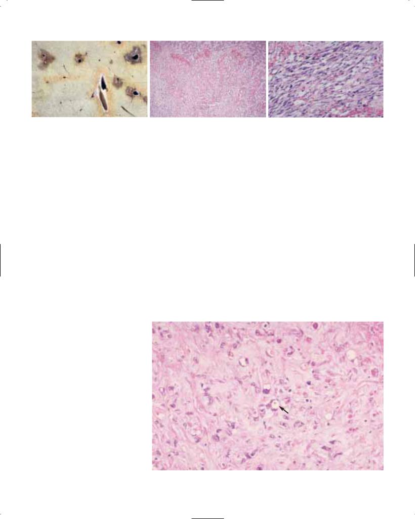
A B C
Fig. 8.68 Kaposi sarcoma. A Multiple dark brown lesions centered in large portal areas. B, C Spindle cells and slit-like vascular spaces.
to phenytoin {148}. Symptoms include abdominal enlargement, fever, weight loss, and nonspecific gastrointestinal complaints {1840}. Rarely, the tumour invades the vena cava and grows into the right atrium, mimicking a cardiac tumour {561}.
Macroscopy. Embryonal sarcoma is usually located in the right lobe of the liver, and varies from 10-20 cm in diameter. It is typically well-demarcated but not encapsulated. Gross sections reveal a variegated surface with glistening, solid, grey-white tumour tissue alternating with cystic, gelatinous areas and/or red and yellow foci of haemorrhage or necrosis.
Histopathology. Embryonal sarcoma is composed of malignant stellate or spindle cells that are compactly or loosely arranged in a myxoid stroma. Tumour cells often show prominent anisonucleosis with hyperchromasia; giant cells that may be multinucleated are seen in many cases. A characteristic feature is the presence of eosinophilic globules of varied size, sometimes many per cell, in the cytoplasm. They are PAS-positive, resist diastase digestion, and express alpha-1 antitrypsin, though the larger globules may only be immunoreactive at the periphery. Entrapped bile ducts and hepatocellular elements are often present in the peripheral areas of these tumours. The spindle, stellate and giant cells typically show no morphological evidence of differentiation, but immunohistochemical studies in a few cases have demonstrated widely divergent differentiation into both mesenchymal and epithelial phenotypes, probably from a primitive stem cell {1460}.
Prognosis. Until recently the prognosis of embryonal sarcoma has been very poor, with a median survival of less than one year after diagnosis {1840}. The survival has greatly improved in the last sev-
eral years with some patients living five or more years after combined modality therapy (surgical resection, radiotherapy, and chemotherapy).
Kaposi sarcoma
This lesion is defined as a tumour composed of slit-like vascular channels, spindle cells, mononuclear inflammatory cells, with an admixture of haemosiderinladen macrophages.
Kaposi sarcoma involves the liver in 12-25% of fatal cases of the acquired immunodeficiency syndrome (AIDS), but is not known to contribute significantly to its morbidity and mortality. In patients with AIDS, it is aetiologically related to HHV-8 infection {276, 1367}. It involves portal areas but can infiltrate the adjacent parenchyma for short distances, and is characterized grossly by irregular, variably-sized, red-brown lesions scattered throughout the liver.
Histologically, lesions resemble those occurring in other sites with spindle cells showing elongated or ovoid, vesicular nuclei with rounded ends and inconspicuous nucleoli. Eosinophilic, PAS-positive globules may be seen in the cytoplasm. The tumour cells are separated by slitlike vascular spaces . Aggregates of haemosiderin granules may be present. The spindle cells express endothelial cell markers (CD31, CD34).
Epithelioid haemangioendothelioma
A tumour of variable malignant potential that is composed of epithelioid or spindle cells growing along preformed vessels or forming new vessels.
Epithelioid haemangioendothelioma presents between 12 and 86 years (mean 47 years) {807, 1150}. Its overall incidence is unknown, but more are reported in females (61%) than in males (39%) {807, 1150}. Risk factors are not known; the
Fig. 8.69 Epithelioid haemangioendothelioma. There is extensive destruction of liver cell plates. Note the intracellular vascular lumina (arrow).
Mesenchymal tumours 195

A B
C D
Fig. 8.70 Epithelioid haemangioendothelioma. A, B Tumour cells form polypoid projections in dilated periportal sinusoids. C Dendritic tumour cells, some having intracellular vascular lumina appearing as small vacuoles. A terminal hepatic venule is infiltrated by tumour. D Tumour cells express factor VIII-related antigen.
suggestion of a relationship to oral contraceptive use has not been validated {1270}. Epithelioid haemangioendothelioma causes systemic symptoms (weakness, malaise, anorexia, episodic vomiting, upper abdominal pain, and weight loss) and hepato-splenomegaly {807, 1150}. Some patients develop jaundice and liver failure. Uncommon modes of presentation include the Budd-Chiari syndrome {2040} or portal hypertension.
Macroscopy. Macroscopically, lesions are usually multifocal; ill-defined lesions scattered throughout the liver vary from a few millimeters to several centimeters in greatest dimension. They are firm, tan to white on sectioning, and often have a hyperaemic periphery; calcification may be evident grossly.
Histopathology. The tumour nodules are ill-defined, and often involve multiple contiguous acini. In actively proliferating lesions the acinar landmarks, such as terminal hepatic venules (THV) and portal areas, can be recognized despite extensive infiltration by the tumour. The cells grow along preexisting sinusoids, THV, and portal vein branches, and often invade Glisson capsule. Growth within the acini is associated with gradual atrophy and eventual disappearance of liver cell plates. Intravascular growth may be in the form of a solid plug, or a polypoid or tuft-like projection.
Neoplastic cell are either ‘dendritic’, with spindle or irregular shapes and multiple interdigitating processes, ‘epithelioid’, with a more rounded shape and an abundant cytoplasm, or ‘intermediate’. Nuclear atypia and mitoses are mainly observed in the epithelioid cells. Cytoplasmic vacuoles, representing intracellular vascular lumens, are often identified and may contain erythrocytes. The tumour cells synthesize factor VIII-related antigen (von Willebrand factor), which can be demonstrated in the cytoplasm or in the neoplastic vascular lumens. Other endothelial cell markers, such as CD31 and CD34, are also positive.
The stroma can have a myxoid appearance due to an abundance of sulphated mucopolysaccharide. Reticulin fibres surround nests of tumour cells. Basement membrane can be demonstrated around the cells by the PAS stain, as well as ultrastructurally and immunohistochemically. Variable numbers of smooth muscle cells surround the basement membrane.
As the lesions evolve they are associated with progressive fibrosis and calcification. Eventually, tumour cells (and indeed, the vascular nature of the lesion) may be difficult if not impossible to recognize in the densely sclerosed areas. Needle biopsy specimens taken from such areas often pose diagnostic problems. The histopathological differential diagnosis includes
angiosarcoma and cholangiocarcinoma. Angiosarcoma is much more destructive than epithelioid haemangioendothelioma, obliterates acinar landmarks and results in cavity formation. Cells of cholangiocarcinoma are arranged in a tubular or glandular pattern, and often produce mucin; the cells are cytokeratin positive and do not express endothelial cell markers.
Prognosis. The clinical outcome of epithelioid haemangioendothelioma is unpredictable, with some patients having a fulminant course and others surviving many years with no therapy. A recent study {1150} showed a correlation between high cellularity of the tumour with a poor clinical outcome. Successful treatment includes resection, when feasible, and liver transplantation.
Angiosarcoma
A malignant tumour composed of spindle or pleomorphic cells that line, or grow into, the lumina of preexisting vascular spaces, such as liver sinusoids and small veins.
Worldwide, about 200 cases of angiosarcoma are diagnosed annually {848, 59}. During the period 1973-87, the SEER database of the US National Cancer Institute contained 6,391 histologicallyconfirmed primary liver cancers; of these only 65 (1%) were angiosarcomas {252}. The peak incidence is in the 6th and 7th decades of life. The male to female ratio is 3:1 {1085}.
75% of angiosarcomas of the liver have no known aetiology {484}. The remainder have been linked to prior administration of Thorotrast (a radioactive material containing thorium dioxide, that was used as an angiography contrast medium from the 1930s to the early 1950s), exposure to vinyl chloride monomer (VCM) or inorganic arsenic, and the use of andro- genic-anabolic steroids {484}.
Patients with angiosarcoma present in one of several ways: 61% have symptoms referable to the liver (e.g. hepatomegaly, abdominal pain, ascites); 15% have an acute abdominal crisis due to haemoperitoneum from rupture of the tumour; 15% have splenomegaly, often with pancytopenia; and 9% present due to distant metastases {804}. The prognosis of angiosarcoma is very poor, with most patients dying within 6 months of diagnosis.
Macroscopy. Angiosarcoma typically affects the entire liver. Grayish-white
196 Tumours of the liver and intrahepatic bile ducts

A B
C D
Fig. 8.71 Angiosarcoma. A Multiple dark brown tumour foci scattered throughout the liver. B Solid portion showing spindle cells and numerous small vascular channels. C Intravascular papillary structure covered by neoplastic endothelial cells. D Tumour cells express CD34.
A B
Fig. 8.72 Angiosarcoma. A Sinusoidal spread of tumour cells with destruction of hepatocyte plates. B Disrupted liver cells act as scaffolding for the tumour cells.
tumour alternates with red-brown haemorrhagic areas. Large cavities with ragged edges, filled with liquid or clotted blood, may be present. A reticular pattern of fibrosis is seen in cases related to prior exposure to Thorotrast.
Histopathology. Tumour cells grow along preformed vascular channels (sinusoids, THV and portal vein branches). Sinusoidal growth is associated with progressive atrophy of liver cells and disruption of the plates, with formation of larger vas-
cular channels and eventually the development of cavities of varied size. These cavities have ragged walls lined by tumour cells, sometimes with polypoid or papillary projections, and are filled with clotted blood and tumour debris. Reti-
Mesenchymal tumours 197

A B
Fig. 8.73 Angiosarcoma. A Closely packed elongated tumour cells. B Pink-brown granular deposits of Thorotrast in a portal area adjacent to an angiosarcoma.
culin fibres and, less often, collagen fibres support the tumour cells. Perithelial cells, reactive for alpha-smooth muscle actin, may also be present. The tumour cells are sometimes packed solidly in nodules that resemble fibrosarcoma.
The cells of angiosarcoma are spindleshaped, rounded or irregular in outline, and often have ill-defined borders. The cytoplasm is lightly eosinophilic, and nuclei are hyperchromatic and elongated or irregular in shape. Nucleoli can be small, or large and eosinophilic. Large, bizarre nuclei and multinucleated cells may be seen, and mitotic figures are frequently identified. The spindled cells have ill-defined outlines, a lightly eosinophilic cytoplasm, and vesicular nuclei with blunt ends. Factor VIII-related antigen can be identified in tumour cells immunohistochemically. Other useful markers include CD31 and CD34; the former is believed to be the most sensitive immunostain {1224}.
Invasion of THV and portal vein branches leads to progressive obstruction of the
lumen, and readily explains the frequently encountered areas of haemorrhage, infarction, and necrosis. Haematopoietic activity is observed in the majority of tumours.
Cases related to Thorotrast and vinyl chloride monomer are often associated with considerable periportal and subcapsular fibrosis. Thorotrast deposits are readily recognized in reticuloendothelial cells, in connective tissue of portal areas, in Glisson capsule, or in the walls of THV. The deposits are coarsely granular and refractile, and in an H&E-stained section they have a pink-brown hue. They are readily visualized by scanning electron microscopy, and thorium can be definitively identified by energy dispersive X-ray microanalysis {804}.
Genetics. Analysis of six hepatic angiosarcomas associated with VCM exposure found three TP53 mutations, all A:T→T:A transversions, which are otherwise uncommon in human cancers {728}. Another study of 21 sporadic angiosarcomas not associated with vinyl chloride
exposure found TP53 mutations to be uncommon, thus supporting previous evidence of the carcinogenic potential of chloroethylene oxide, a metabolite of VCM {1776}. A high rate of KRAS-2 mutations has been found in both sporadic and Thorotrast-induced angiosarcomas of the liver {1542}.
Malignant mesenchymal tumours other than angiosarcoma may have cytogenetic aberrations similar to those of soft tissue tumours {513, 1812}.
Carcinosarcoma
This neoplasm is defined as a malignant tumour containing an intimate mixture of carcinomatous (either hepatocellular or cholangiocellular) and sarcomatous elements; such lesions have also been called ‘malignant mixed tumour’ of the liver. Carcinosarcoma should be distinguished from carcinomas with foci of spindled epithelial cells and from the rare true ‘collision’ tumours.
198 Tumours of the liver and intrahepatic bile ducts

Secondary tumours of the liver |
P.P. Anthony |
P. DeMatos |
Definition
Malignant neoplasms metastasized to the liver from extrahepatic primary tumours.
Epidemiology
In Europe and North America, metastases predominate over primary hepatic tumours in a ratio of 40:1 {130, 1517}. In Japan the ratio is 2.6:1 {1517}. In SouthEast Asia and sub-Saharan Africa, primary hepatic tumours are more common than metastases {1909} owing to the high incidence of hepatocellular carcinoma, a shorter life span (common extrahepatic carcinomas affect older age groups) and the low incidence of certain tumour types (e.g. carcinomas of the lung and colorectum). Autopsy studies in the USA and Japan have shown that about 40% of patients with extrahepatic cancer have hepatic metastases {351, 1517}.
Aetiopathogenesis
The liver has a rich systemic (arterial) and portal (venous) blood supply, providing a potentially abundant source of circulating neoplastic cells. Circulating tumour cell arrest is controlled by Kupffer cells in the sinusoids {881, 121} and may be enhanced by growth factors such as transforming growth factor alpha (TGFα) {385}, tumour necrosis factor (TNF) {1431}, and insulin-like growth factor-1 (IGF-1) {1091}. As tumour deposits enlarge, they induce angiogenesis using native sinusoidal endothelium; this enhances their chances of survival and is often macroscopically evident {1919}.
Most metastases from unpaired abdominal organs reach the liver via the portal vein, and from other sites via the systemic arterial circulation. Lymphatic spread is less common and extension to the liver via the peritoneal fluid is rare {351}.
Cirrhosis provides some relative protection against seeding by secondary tumours {1983, 1211}. It has also been suggested that metastasis is rare in fatty livers {676}, but excess alcohol consumption apparently enhances hepatic metastases {1140}.
A B
C D
E F
Fig. 8.74 Secondary tumours in the liver. A Metastatic colon carcinoma showing umbilication and hyperemic borders. B Metastatic small cell carcinoma of lung forming inumerable small nodules. C, D Metastatic large intestinal carcinoma, cut surfaces. E Metastatic gastric adenocarcinoma, cut surface. F A metastasis lies adjacent to a Zahn infarct.
In the majority of cases, metastases to the liver are a manifestation of systemic, disseminated disease. Colorectal carcinoma, neuroendocrine tumours, and renal cell carcinoma are exceptions as these neoplasms sometimes produce isolated, even solitary deposits {1517}.
Origin of metastases
The majority of secondary liver neoplasms are carcinomas, involvement by lymphomas is next and sarcomas are uncommon. The order of frequency by primary site in Western populations is: upper gastrointestinal tract (stomach,
gallbladder, pancreas): 44-78%; colon: 56-58%; lung 42-43%; breast 52-53%; oesophagus 30-32% and genito-urinary organs 24-38% {130, 1517, 351}. Carcinomas of the prostate and the ovaries preferentially spread to the lymph nodes and the spine, and to the peritoneal cavity, respectively.
Hodgkin and non-Hodgkin lymphomas may involve the liver in up to 20% of cases on presentation and 55% at autopsy {1620, 826}. Sarcomas are much less common but 6% had hepatic metastases at presentation (mostly intra-abdominal leiomyosarcomas) in one study {833},
Secondary tumours 199

A B
Fig. 8.75 Metastatic tubular adenocarcinoma from the stomach. A Haematoxylin and eosin. B Intraluminal diastase-resistant PAS positive mucin.
A B
Fig. 8.76 Metastatic colorectal carcinoma. A Tumour is necrotic and cell type is typically columnar. B Necrosis may result in calcification.
while 34% had hepatic metastases at autopsy in another {1517}.
In a study of randomly selected liver biopsies from England and Wales {852}, the commonest histological type of metastasis was adenocarcinoma (39%), followed by carcinoma not otherwise specified (36%); the rest were undifferentiated small cell carcinoma, other special types of carcinoma, and lymphomas.
Clinical features
Symptoms and signs
Hepatic metastases produce clinical manifestations in about two-thirds of cases and they generally reveal themselves through symptoms referable to the liver. Afflicted patients often present with ascites, hepatomegaly or abdominal full-
Fig. 8.77 Metastatic breast carcinoma.
ness, hepatic pain, jaundice, anorexia, and weight loss. Constitutional symptoms, such as malaise, fatigue, and fever may be present. On examination, nodules or a mass are felt in up to 50% of the cases, and a friction bruit may be heard on auscultation. Unfortunately, symptomatic presentation is associated with bulky, rapidly progressive tumours with a poor prognosis {2035}.
Rarely, patients present with fulminant hepatic failure, obstructive jaundice, or intraperitoneal haemorrhage. Functioning neuroendocrine tumours produce syndromes of hormonal excess. ‘Carcinomatous cirrhosis’ with jaundice, ascites, and bleeding varices due to diffuse infiltration of the liver, usually by metastatic breast carcinoma, has been described {174}.
Laboratory studies
The alkaline phosphatase (ALP) and serum glutamic-oxaloacetic transaminase (SGOT) levels, although non-spe- cific, are elevated in approximately 80% and 67% of patients respectively, and most likely represent the effects of hepatic parenchymal infiltration by tumour and of generalized wasting. Elevated lactic dehydrogenase (LDH) levels are relatively specific for the presence of metastatic
melanoma. Tests of synthetic function, e.g. serum albumin levels and the prothrombin time, may be normal despite extensive metastatic involvement. Alphafetoprotein (AFP) levels may be slightly to moderately elevated but very high concentrations are more consistent with a diagnosis of hepatocellular carcinoma {904}. Carcinoembryonic antigen (CEA) levels, which are raised in as many as 90% of patients with metastases from colorectal carcinoma, can be useful in monitoring patients after primary tumour resection. However, CEA levels do not correlate well with prognosis {2043, 1821}.
Imaging
Ultrasound (US) can identify tumours measuring 1-2 cm in size, can differentiate solid from cystic lesions, and provide guidance for percutaneous needle biopsy. However, it provides poor anatomical definition and frequently misses smaller lesions.
Computed tomography (CT), using both contrasted and non-contrasted images, can also serve as a screening tool. The administration of intravenous contrast permits the detection of tumours as small as 0.5 cm in diameter {1763}. Most metastases display decreased vascularity in comparison to the surrounding hepatic parenchyma and appear as hypodense defects. Tumours that are hypervascular (e.g. melanoma, carcinoids and some breast cancers) or calcified (e.g. colorectal carcinoma) are better delineated by noncontrast views.
Magnetic resonance imaging (MRI) is more sensitive than CT in the detection of hepatic tumours and can demonstrate additional lesions, too small to be seen on CT.
Positron emission tomography (PET) can detect metastatic disease in the liver and elsewhere. Using 2-(18)fluoro-2-deoxy- D-glucose (F-18 FDG), a radiolabeled glucose analogue, PET highlights metabolically active tissues. Through co-reg- istration with anatomical studies like CT or MRI, viable malignant tumours can be differentiated from benign or necrotic lesions {54}.
CT arterial portography performed preoperatively, and intraoperative ultrasound are associated with the highest sensitivities {1796}. The former is capable of detecting lesions as small as 15 mm, although a false positive rate of
200 Tumours of the liver and intrahepatic bile ducts

A B
Fig. 8.78 Metastatic islet cell carcinoma of pancreas. A Haematoxylin and eosin. B Somatostatin immunoreactivity.
17% has been reported {1795}. Its success relies on the fact that tumours are not fed by portal vein blood, so that metastases appear as filling defects. The latter, capable of detecting lesions 2-4 mm in diameter delineates the anatomical location of tumours in relationship to major vascular and biliary structures and provides guidance for intraoperative needle biopsies. It is the definitive step in determining resectability at the time of exploratory laparotomy or laparoscopy.
Angiography use has declined in recent years. It remains useful for defining vascular anatomy for planned hepatic resections, selective chemotherapy, chemoembolization, or devascularization procedures, for assessing whether there is metastatic involvement of the portal venous system and/or hepatic veins, and for differentiating between benign vascular lesions, such as haemangiomas and metastases, when other imaging studies have yielded equivocal results.
Macroscopy
The distribution of metastases from colorectal carcinoma was found to be homogenous, regardless of the primary site of origin {1695} but in another study, it was suggested that right sided cancers predominantly metastasize to the right lobe of the liver and left sided cancers to both lobes {1749}.
Metastases are nearly always multinodular or diffusely infiltrative, but may rarely be solitary and massive (e.g. from colorectal and renal cell carcinomas). Umbilication (a central depression on the surface of a metastatic deposit) is due to necrosis or scarring and is typical of an adenocarcinoma from stomach, pan-
creas or colorectum. A vascular rim around the periphery is often seen. Highly mucin secreting adenocarcinomas appear as glistening, gelatinous masses whilst well differentiated keratinizing squamous cell carcinomas are granular. Metastatic carcinoid tumours can form pseudocysts {401}. Haemorrhagic secondary deposits suggest angiosarcoma, choriocarcinoma, carcinoma of thyroid or kidney, neuroendocrine tumour, or vascular leiomyosarcoma. Some diffusely infiltrating carcinomas (e.g. small cell carcinoma), lymphomas and sarcomas may have a soft, opaque ‘fish flesh’ appearance. Metastatic breast carcinoma in particular can produce an intensely fibrous, granular liver (‘carcinomatous cirrhosis’) either before {174} or after {1693} treatment.
Calcification of secondary deposits is a feature of colorectal carcinoma but it is seldom excessive and has no effect on prognosis {653}. Metastatic melanoma is often, but not always, of a brown-black colour. Secondary tumours may appear in the liver long after the removal of the primary.
Fig. 8.79 Systemic non-Hodgkin lymphoma involving the liver.
Histopathology
Liver biopsy samples can be obtained by percutaneous or transjugular routes with or without imaging techniques for guidance, as a wedge during laparotomy, or a fine needle can be used to aspirate material for cytology. Each of these methods has advantages and drawbacks but a guided percutaneous needle biopsy producing a core of liver for histology is the one most frequently used. It produces a tissue sample that is usually adequate for all purposes, including the use of special stains, immunohistochemistry and molecular biological techniques. Touch preparations for cytology can also be prepared from needle cores before fixation and may provide an instant diagnosis {1523}.
Differential diagnosis
Hepatocellular carcinoma can usually be distinguished from metastatic tumours by its trabecular structure, sinusoids, lack of stroma, bile production, absence of mucin secretion, and the demonstration of bile canaliculi by polyclonal CEA antisera, which is specific for a liver cell origin. Other useful immunophenotypic features in this differentiation are the presence of liver export proteins (albumin, fibrinogen, alpha-1-antitrypsin), the cytokeratin pattern, and the expression of Hep Par 1 antigen {1046}. Metastatic tumours that often mimic hepatocellular carcinoma are adrenal cortical and renal cell carcinomas. Amelanotic melanoma may also cause difficulties but it is easily identified by positive immunostaining for S100 protein and HMB45 .
The distinction between primary cholangiocarcinoma and metastatic adenocar-
Secondary tumours 201

Fig. 8.80 Typical histological changes adjacent to space occupying liver lesions: sinusoidal dilatation, leukocyte infiltration, and bile-ductular proliferation.
cinomas is much more difficult and may be impossible {351}. Cholangiocarcinoma may take on any of the histological patterns of an adenocarcinoma; it is usually tubular but may be mucinous, signetring, papillary, cystic, or undifferentiated. Mucin secretion and production of CEA are nearly always demonstrable in both primary and secondary adenocarcinomas. Metastases from many sites form similar patterns. However, small tubular or tubulo-papillary glands frequently derive from the stomach, gallbladder and extrahepatic biliary tree, and a signet-ring cell appearance suggests a gastric primary. Perhaps the easiest pattern to recognize as metastatic in origin is that exhibited by adenocarcinomas of the colon and rectum, which nearly always show glands of variable size and shape that are lined by tall columnar cells and contain debris within the lumina. Metastases from the colorectum frequently have well defined edges whereas those from other glandular sites tend to be more diffuse. Colorectal metastases are also frequently necrotic and may show calcification {653}.
The presence of carcinoma-in-situ in intrahepatic bile ducts in the vicinity of an adenocarcinoma is evidence that it is a cholangiocarcinoma. However, this may be mimicked by intrabiliary ductal growth of metastatic colonic adenocarcinoma {1593}. Analysis of cytokeratin expression may be useful in the distinction of
primary and metastatic gastrointestinal adenocarcinomas. The former express cytokeratins 7 and 19 but not 20, whereas the latter are negative for 7 and positive for 20 {1141}.
Carcinoma of the breast often produces a diffuse sinusoidal infiltrate that on imaging studies may mimic cirrhosis and, indeed, may be associated with splenomegaly, ascites and oesophageal varices {174}; sclerosis following systemic chemotherapy may exaggerate this effect. Metastases from the breast may be identified by the combined use of zinc-α2-glycoprotein, gross cystic disease fluid protein 15 and oestrogen receptor {283}. However, occult breast carcinoma presenting with metastases is rare and most patients with liver involvement have a past history of a primary tumour.
Most hepatic metastases from the lung in clinical practice are undifferentiated small cell carcinomas, characteristically producing an enlarged liver due to diffuse or miliary spread. The primary tumour may still be small, asymptomatic and undetected. Squamous cell and adenocarcinomas will metastasize to the liver but their existence is usually known already. The same applies to squamous cell carcinomas of the oesophagus and cervix. Squamous cell carcinomas of the head and neck seldom involve the liver.
Neuroendocrine/islet cell/carcinoid tumours are easily identified by their
organoid nesting pattern, uniform cytology and vascularity, and positive immunostaining for chromogranin, synaptophysin and neuron specific enolase; islet cell tumours also produce specific hormones such as insulin, glucagon, gastrin, vasoactive intestinal peptide and somatostatin, which either give rise to clinical syndromes or can be demonstrated in the blood or tumour tissue.
Most sarcomas that metastasize to the liver are gastrointestinal stromal tumours that are positive for CD34 and c-kit, or leiomyosarcomas of the uterus that may be positive for desmin or muscle-specif- ic actin. Some carcinomas, notably of the kidney, may be sarcomatoid in their morphology.
Many haematological malignancies, e.g. leukaemias, myeloproliferative disorders and both Hodgkin and non-Hodgkin lymphomas, involve the liver. Leukaemias tend to produce diffuse sinusoidal infiltrates. Hodgkin and high-grade nonHodgkin lymphomas produce tumourlike masses, while low-grade nonHodgkin lymphomas produce diffuse portal infiltrates.
Rare secondary tumours include those from the thyroid, prostate, and gonads. The diagnosis can be confirmed by the immunohistochemical demonstration of thyroglobulin, prostate specific antigen and AFP and βHCG, respectively.
A triad of histological features, namely proliferating bile ducts, leukocytes and focal sinusoidal dilatation, is found in the liver adjacent to space-occupying lesions. Their presence in a core biopsy suggests the possibility of a metastatic deposit missed by the biopsy needle.
Three lesions, bile duct adenoma, sclerosed haemangioma, and larval granuloma may resemble metastatic tumours at laparotomy.
Prognosis
In most cases, disseminated disease is present which precludes surgical intervention. Due to recent improvements in imaging techniques, more metastatic carcinomas are being diagnosed early, providing the possibility of surgical resection in a greater number of patients. When curative resection is feasible, 5-year survival can be as high as 40%; without surgical therapy, median survivals of less than 12 months should be expected {1817}.
202 Tumours of the liver and intrahepatic bile ducts
CHAPTER 9
Tumours of the Gallbladder
and Extrahepatic Bile Ducts
These two closely related tumour sites show remarkable differences in terms of epidemiology, aetiology, and clinical presentation. The incidence of gallbladder carcinoma shows prominent geographic, gender, and racial differences, while extrahepatic bile duct carcinomas show none of these variations. Aetiologic associations include gall stones, sclerosing cholangitis, ulcerative colitis, abnormal choledochopancreatic junction, choledochal cysts, and infestation with liver flukes.

WHO histological classification
of tumours of the gallbladder and extrahepatic bile ducts
Epithelial tumours |
|
Small cell carcinoma |
8041/3 |
|
|
Large cell neuroendocrine carcinoma |
8013/3 |
Benign |
|
Undifferentiated carcinoma |
8020/3 |
Adenoma |
8140/01 |
Biliary cystadenocarcinoma |
8161/3 |
Tubular |
8211/0 |
|
|
Papillary |
8260/0 |
Carcinoid tumour |
8240/3 |
Tubulopapillary |
8263/0 |
Goblet cell carcinoid |
8243/3 |
Biliary cystadenoma |
8161/0 |
Tubular carcinoid |
8245/1 |
Papillomatosis (adenomatosis) |
8264/0 |
Mixed carcinoid-adenocarcinoma |
8244/3 |
|
|
Others |
|
Intraepithelial neoplasia (dysplasia and carcinoma in situ) |
|
|
|
|
|
Non-epithelial tumours |
|
Malignant |
|
|
|
Carcinoma |
|
Granular cell tumour |
9580/0 |
Adenocarcinoma |
8140/3 |
Leiomyoma |
8890/0 |
Papillary adenocarcinoma |
8260/3 |
Leiomyosarcoma |
8890/3 |
Adenocarcinoma, intestinal type |
8144/3 |
Rhabdomyosarcoma |
8900/3 |
Adenocarcinoma, gastric foveolar type |
|
Kaposi sarcoma |
9140/3 |
Mucinous adenocarcinoma |
8480/3 |
Others |
|
Clear cell adenocarcinoma |
8310/3 |
|
|
Signet-ring cell carcinoma |
8490/3 |
Malignant lymphoma |
|
Adenosquamous carcinoma |
8560/3 |
|
|
Squamous cell carcinoma |
8070/3 |
Secondary tumours |
|
|
|
|
|
_________
1Morphology code of the International Classification of Diseases for Oncology (ICD-O) {542} and the Systematized Nomenclature of Medicine (http://snomed.org). Behaviour is coded /0 for benign tumours, /1 for unspecified, borderline, or uncertain behaviour, /2 for in situ carcinomas and grade III intraepithelial neoplasia and /3 for malignant tumours.
TNM classification of tumours of the gallbladder
TNM classification1, 2
T – Primary Tumour |
M – Distant Metastasis |
|
|
||
TX |
Primary tumour cannot be assessed |
MX |
Distant metastasis cannot be assessed |
|
|
T0 |
No evidence of primary tumour |
M0 |
No distant metastasis |
|
|
Tis |
Carcinoma in situ |
M1 |
Distant metastasis |
|
|
T1 |
Tumour invades lamina propria or muscle layer |
|
|
|
|
T1a |
Tumour invades lamina propria |
|
|
|
|
T1b |
Tumour invades muscle layer |
|
|
|
|
T2 |
Tumour invades perimuscular connective tissue, no extension |
|
|
|
|
|
beyond serosa or into liver |
|
|
|
|
T3 |
Tumour perforates serosa (visceral peritoneum) or directly |
|
|
|
|
|
invades into one adjacent organ or both (extension 2 cm or less |
|
|
|
|
|
into liver) |
|
|
|
|
T4 |
Tumour extends more than 2 cm into liver and/or into two or |
Stage Grouping |
|
|
|
|
more adjacent organs (stomach, duodenum, colon, pancreas, |
|
|
|
|
|
omentum, extrahepatic bile ducts, any involvement of liver) |
Stage 0 |
Tis |
N0 |
M0 |
|
|
Stage I |
T1 |
N0 |
M0 |
N – Regional Lymph Nodes |
Stage II |
T2 |
N0 |
M0 |
|
NX |
Regional lymph nodes cannot be assessed |
Stage III |
T1 |
N1 |
M0 |
N0 |
No regional lymph node metastasis |
|
T2 |
N1 |
M0 |
N1 |
Metastasis in cystic duct, pericholedochal, and/or hilar lymph |
|
T3 |
N0, N1 |
M0 |
|
nodes (i.e., in the hepatoduodenal ligament) |
Stage IVA |
T4 |
N0, N1 |
M0 |
N2 |
Metastasis in peripancreatic (head only), periduodenal, peripor- |
Stage IVB |
Any T |
N2 |
M0 |
|
tal, coeliac, and/or superior mesenteric lymph nodes |
|
Any T |
Any N |
M1 |
_________
1 {1, 66}. The classification applies only to carcinomas.
2 A help desk for specific questions about the TNM classification is available at http://tnm.uicc.org.
204 Tumours of the gallbladder and extrahepatic bile ducts

TNM classification of tumours of the extrahepatic bile ducts
TNM classification1, 2
T – Primary Tumour |
M – Distant Metastasis |
|
|||
TX |
Primary tumour cannot be assessed |
MX |
Distant metastasis cannot be assessed |
||
T0 |
No evidence of primary tumour |
M0 |
No distant metastasis |
|
|
Tis |
Carcinoma in situ |
M1 |
Distant metastasis |
|
|
T1 |
Tumour invades subepithelial connective tissue or fibromuscular |
|
|
|
|
|
layer |
|
|
|
|
T1a |
Tumour invades subepithelial connective tissue |
|
|
|
|
T1b |
Tumour invades fibromuscular layer |
|
|
|
|
T2 |
Tumour invades perifibromuscular connective tissue |
|
|
|
|
T3 |
Tumour invades adjacent structures: liver, pancreas, duodenum, |
|
|
|
|
|
gallbladder, colon, stomach |
|
|
|
|
|
|
Stage Grouping |
|
|
|
N – Regional Lymph Nodes |
|
|
|
|
|
NX |
Regional lymph nodes cannot be assessed |
Stage 0 |
Tis |
N0 |
M0 |
N0 |
No regional lymph node metastasis |
Stage I |
T1 |
N0 |
M0 |
N1 |
Metastasis in cystic duct, pericholedochal, and/or hilar lymph |
Stage II |
T2 |
N0 |
M0 |
|
nodes (i.e., in the hepatoduodenal ligament) |
Stage III |
T1 |
N1, N2 |
M0 |
N2 |
Metastasis in peripancreatic (head only), periduodenal, peripor- |
|
T2 |
N1, N2 |
M0 |
|
tal, coeliac, superior mesenteric, posterior peripancreatico-duo- |
Stage IVA |
T3 |
Any N |
M0 |
|
denal lymph nodes |
Stage IVB |
Any T |
Any N |
M1 |
_________
1 |
{1, 66}. The classification applies to carcinomas of extrahepatic bile ducts and those of choledochal cysts. |
2 |
A help desk for specific questions about the TNM classification is available at http://tnm.uicc.org. |
TNM classification of tumours of the Ampulla of Vater
TNM classification1, 2
T – Primary Tumour
TX |
Primary tumour cannot be assessed |
T0 |
No evidence of primary tumour |
Tis |
Carcinoma in situ |
T1 |
Tumour limited to ampulla of Vater or sphincter of Oddi |
T2 |
Tumour invades duodenal wall |
T3 |
Tumour invades 2 cm or less into pancreas |
T4 |
Tumour invades more than 2 cm into pancreas and/or |
|
into other adjacent organs |
N – Regional Lymph Nodes |
|
NX |
Regional lymph nodes cannot be assessed |
N0 |
No regional lymph node metastasis |
N1 |
Regional lymph node metastasis |
M – Distant Metastasis
MX Distant metastasis cannot be assessed M0 No distant metastasis
M1 Distant metastasis
Stage Grouping |
|
|
|
Stage 0 |
Tis |
N0 |
M0 |
Stage I |
T1 |
N0 |
M0 |
Stage II |
T2 |
N0 |
M0 |
|
T3 |
N0 |
M0 |
Stage III |
T1 |
N1 |
M0 |
|
T2 |
N1 |
M0 |
|
T3 |
N1 |
M0 |
Stage IV |
T4 |
Any N |
M0 |
|
Any T |
Any N |
M1 |
_________
1 { 1, 66}. The classification applies only to carcinomas.
2 A help desk for specific questions about the TNM classification is available at http://tnm.uicc.org.
205

Carcinoma of the gallbladder and extrahepatic bile ducts
J. Albores-Saavedra |
H.R. Menck |
J.C. Scoazec |
N. Soehendra |
C. Wittekind |
P.V.J. Sriram |
B. Sripa |
|
Definition
A malignant epithelial tumour with glandular differentiation, arising in the gallbladder or extrahepatic biliary system.
Epidemiology
Most tumours of the gallbladder and extrahepatic bile ducts are carcinomas. Only a small proportion are adenomas, carcinoid and stromal tumours {35}.
Geographic distribution
The incidence of carcinoma of the gallbladder varies in different parts of the world and also differs among different ethnic groups within the same country. In the United States, carcinoma of the gallbladder is more common in Native Americans and Hispanic Americans than in whites or blacks; the rate among female Native Americans is 21 per 100,000 compared with 1.4 per 100,000 among white females. In Latin American countries, the highest rates are found in Chile, Mexico and Bolivia. In Japan, the incidence rates are intermediate. In the general population of the United States cancer of the gallbladder accounts for 0.17% for all cancers in males and 0.49% in females.
There are no geographic variations in the incidence of extrahepatic bile duct carcinoma which accounts for 0.16% of all invasive cancers in males and 0.15% in females in the general population of the United States {35}.
Fig. 9.01 Gallbladder carcinoma with a white, irregular cut surface next to a large gall stone.
Age and sex distribution
Carcinomas of the gallbladder and extrahepatic bile ducts are diseases of older age groups. Most patients are in the 6th or 7th decades of life. Gallbladder carcinomas have a strong female predominance, whereas extrahepatic bile duct carcinomas occur more frequently in males.
Aetiology
Unlike carcinoma of the extrahepatic bile ducts, gallbladder carcinomas are not associated with primary sclerosing cholangitis or ulcerative colitis.
Gallbladder carcinoma
Gallstones. The incidence of gallbladder cancer is higher in patients with gallstones than in patients without stones {35}, and stones are present in over 80% of gallbladder carcinomas. The incidence of gallbladder carcinoma parallels that of gallstones, being more frequent in females and in certain ethnic groups, e.g. Native Americans, who have a high incidence of stones. Nevertheless, although gall stones are considered a risk factor, the overall incidence of carcinoma of the gallbladder in patients with cholelithiasis is less than 0.2%; this percentage varies with race, sex, and length of exposure to the stones {35}. While some authors have reported a correlation between gallstone size and the risk of cancer, others have not found such a correlation {35}.
Abnormal choledochopancreatic junction. Data largely reported from Japan indicate an association between gallbladder cancer and an abnormal junction of the pancreatic and common bile ducts {1248}. Normally, the main pancreatic duct and the common bile duct unite within the sphincter to form the pancreaticobiliary duct. The abnormal junction is defined as the union of the pancreatic and common bile ducts outside the wall of the duodenum beyond the influence of the sphincter of Oddi. As a result, pancreatic juice can reflux into the common bile duct, resulting in hyperplastic, meta-
Fig. 9.02 Carcinoma of the gallbladder involving the fundus (arrow). Bile ducts are normal.
Fig. 9.03 Hilar cholangiocarcinoma extending beyond both the right and left hepatic bile ducts (Klatskin type III) (arrows).
206 Tumours of the gallbladder and extrahepatic bile ducts

Fig. 9.04 Papillary adenocarcinoma, non-invasive. The tumour projects into the lumen, but does not invade the wall of the gallbladder.
plastic, and neoplastic changes in the gallbladder epithelium.
Porcelain gallbladder. Diffuse calcification of the gallbladder wall (porcelain gallbladder) is associated with carcinoma in 10-25% of cases.
Genetic susceptibility. As discussed above, carcinoma of the gallbladder is concentrated in certain racial and ethnic groups. Familial aggregation of gallbladder cancer has been recorded in the US and in other countries {35}.
Carcinoma of extrahepatic bile ducts
Well established risk factors for carcinomas of the extrahepatic bile ducts are sclerosing cholangitis, ulcerative colitis, abnormal choledochopancreatic junction, choledochal cysts and infestation with the liver flukes C. sinensis and O. viverrini. Choledocholithiasis does not seem to play a role in the pathogenesis of carcinomas of the extrahepatic bile ducts.
Clinical features
Cancer of the gallbladder usually presents late in its course. The signs and symptoms are not specific, often resembling those of chronic cholecystitis. Right upper quadrant pain is common.
Computed tomography and ultrasonography can be used to demonstrate the lesion.
Carcinomas of the extrahepatic bile ducts usually present relatively early with obstructive jaundice, which can rapidly progress or fluctuate. Jaundice usually appears while the tumour is relatively small before widespread dissemination has occurred. Other symptoms include right upper quadrant pain, malaise, weight loss, pruritus, anorexia, nausea, and vomiting. If cholangitis develops, chills and fever appear. In patients with carcinoma of the proximal bile ducts (right and left hepatic ducts, common hepatic duct), the intrahepatic bile ducts are dilated, the gallbladder is not palpable and the common duct often collapses. Patients with carcinoma in the common or cystic ducts have a distended and palpable gallbladder as well as a markedly dilated proximal duct system, as may be shown by ultrasonography and computerised tomography. Transhepatic cholangiograms and endoscopic retrograde cholangiopancreatography are essential for exact localization of carcinomas of the extrahepatic bile ducts.
Macroscopy
Carcinoma of the gallbladder appears as an infiltrating grey white mass. Some carcinomas may cause diffuse thickening and induration of the entire gallbladder wall. The gallbladder may be distended by the tumour, or collapsed due to obstruction of the neck or cystic duct. It can also assume an hourglass deformity when the tumour arises in the body and constricts the lateral walls. Papillary carcinomas are usually sessile and exhibit a polypoid or cauliflower-like appearance. Mucinous and signet ring cell carcinomas have a mucoid or gelatinous cut surface. Although any type of gallbladder
cancer may show necrosis, undifferentiated giant cell and small cell carcinomas are usually the most necrotic. Submucosal growth is an important feature of signet ring and small cell carcinomas.
Carcinomas of the extrahepatic bile ducts have been divided into polypoid, nodular, scirrhous constricting, and diffusely infiltrating types. This separation can provide a guide to the operative procedure, extent of resection, and prognosis. However, except for the polypoid tumours, this separation is rarely possible in practice because of overlapping gross features. The nodular and scirrhous types tend to infiltrate surrounding tissues and are difficult to resect. The diffusely infiltrating types tend to spread linearly along the ducts.
Tumour staging
There are separate TNM classifications for carcinomas of the gallbladder, extrahepatic bile ducts, and the ampulla of Vater.
Histopathology
The histological classification of tumours of the gallbladder and extrahepatic bile ducts is essentially similar to the previous WHO classification published in 1991 {1774} and to the classification adopted by the AFIP fascicle published in 2000 {35}.
Adenocarcinoma
Well to moderately differentiated adenocarcinomas are the most common malignant epithelial tumours of the gallbladder and extrahepatic bile ducts. They are composed of short or long tubular glands lined by cells that vary in height from low cuboidal to tall columnar, superficially resembling biliary epithelium. Mucin is frequently present in the cells and glands. Rarely, the extracellular mucin may
A B C
Fig. 9.05 Intestinal type adenocarcinoma. A Tubular glands similar to colonic adenocarcinoma. B Goblet cell type of adenocarcinoma. C Numerous serotonin containing cells in a neoplastic gland.
Carcinoma 207
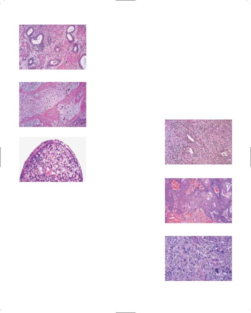
Fig. 9.06 Well differentiated adenocarcinoma infiltrating gallbladder wall.
Fig. 9.07 Mucinous adenocarcinoma of gallbladder.
Fig. 9.08 Signet-ring cell carcinoma of gallbladder.
become calcified {1465, 1606}. About one-third of the well differentiated tumours show focal intestinal differentiation and contain goblet and endocrine cells {36, 2152, 2158}. The endocrine cells may be numerous and show immunoreactivity for serotonin and peptide hormones, but a diagnosis of neuroendocrine neoplasm is not warranted. Paneth cells may rarely be seen. An extremely well differentiated adenocarcinoma with gastric foveolar phenotype that simulates adenoma has been described in the extrahepatic bile ducts {39}. Adenocarcinomas may show cribriform or angiosarcomatous patterns. They may also contain cytoand synctiotrophoblast cells.
Extrahepatic bile duct adenocarcinomas tend to be better differentiated than their gallbladder counterparts. Many gallbladder carcinomas are immunoreactive for TP53 {1907, 2125}
Histological variants of adenocarcinoma Papillary adenocarcinoma. This malignant tumour is composed predominantly of papillary structures lined by cuboidal or columnar epithelial cells often containing variable amounts of mucin. Some tumours show intestinal differentiation with collections of goblet, endocrine, and Paneth cells. Papillary adenocarcinomas may fill the lumen before invading the wall. Papillary adenocarcinomas appear to be more frequent in the gallbladder than in the extrahepatic biliary tree {2150}. In addition, skip lesions may be observed in approximately 10% of cases {1989}.
Adenocarcinoma, intestinal type. This unusual variant of adenocarcinoma is composed of tubular glands or papillary structures lined predominantly by cells with an intestinal phenotype, namely goblet cells or colonic-type epithelium or both, with or without a variable number of endocrine and Paneth cells {41}.
Mucinous adenocarcinoma. Mucinous adenocarcinomas of the biliary tree are similar to those that arise in other anatomic sites. By definition, more than 50% of the tumour contains extracellular mucin {1774}. There are two histological variants of mucinous adenocarcinomas of the gallbladder and extrahepatic bile ducts: one variant is characterized by neoplastic glands distended with mucin and lined by columnar cells with mild to moderate nuclear atypia, and the second variant is characterized by small groups or clusters of cells surrounded by abundant mucin. Some tumours show both growth patterns. The abundant mucin makes the tumour appear hypocellular.
Cystadenocarcinoma refers to a unilocular or multilocular glandular tumour that may be the result of malignant transformation of a cystadenoma.
Clear cell adenocarcinoma. This rare malignant tumour is composed predominantly of glycogen-rich clear cells having well-defined cytoplasmic borders and hyperchromatic nuclei. In addition to clear cells, a variable number of cells contain eosinophilic granular cytoplasm. The clear cells line glands or are arranged in nests, sheets, cords, trabeculae or papillary structures {40, 145, 1856}. Foci of conventional adenocarci-
noma with focal mucin production are usually found and are useful in separating primary from metastatic clear cell carcinomas. In some clear cell adenocarcinomas of the biliary tree the columnar cells contain subnuclear and supranuclear vacuoles similar to those seen in secretory endometrium. Focal hepatoid differentiation with production of alpha-fetopro- tein has been documented in clear cell carcinomas of the gallbladder {2000}.
Signet-ring cell carcinoma. Cells containing intracytoplasmic mucin displacing the nuclei toward the periphery predominate in this variant of adenocarcinoma. A variable amount of extracellular mucin is usually present. Lateral spread through the lamina propria is a common feature.
Fig. 9.09 Adenosquamous carcinoma of gallbladder.
Fig. 9.10 Squamous cell carcinoma of gallbladder.
Fig. 9.11 Undifferentiated carcinoma of gallbladder, spindle and giant cell type. No glandular differentiation.
208 Tumours of the gallbladder and extrahepatic bile ducts


CHAPTER 10
Tumours of the Exocrine Pancreas
Pancreatic carcinoma is a highly malignant neoplasm that still carries a very poor prognosis. Ductal adenocarcinoma is the most frequent type. Although cigarette smoking has been established as a causative factor, the risk attributable to tobacco abuse amounting to approximately 30%. An increased risk is also associated with hereditary pancreatitis, but additional aetiological factors remain to be identified.
Significant progress has been made in the understanding of the molecular basis of ductal carcinomas. KRAS point mutations and inactivation of the tumour suppressor genes p16, TP53 and DPC4 have been identified as most frequent genetic alterations.
Non-ductal pancreatic neoplasms span a wide range of histological features that need to be recognized by pathologists as several entities are associated with distinct opportunities for therapy.