

MusculoSkeletal Exam
.pdfChapter 9 The Elbow
2
4
1 7
6
3
8
5
2
7
4
6 |
8 |
3
5
1. Intercostobrachial
2. Upper lateral cutaneous
3. Medial cutaneous of arm
4. Lower lateral cutaneous
5. Medial cutaneous of forearm
6. Lateral cutaneous of forearm
7. Posterior cutaneous of arm
Figure 9.49 Posterior view of the sensory nerves to the arm and forearm, along with their distributions.
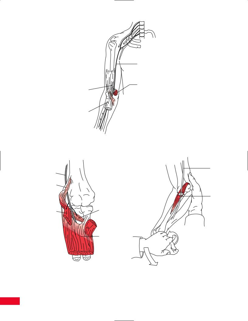
compression at the flexor digitorum superficialis arch (Figure 9.54).
Anterior Interosseous Nerve
This nerve is a motor nerve to the long flexors of the thumb and index and middle fingers, and the pronator quadratus muscle. There are no cutaneous sensory
fibers. However, this nerve does provide some sensation to the joints of the wrist. Examination of a patient with anterior interosseous nerve syndrome characteristically reveals weakness of the long flexor muscles. This can be tested for by asking the patient to make the “OK” sign. An inability to do tip-to-tip pinch of the thumb and index finger results from damage to the anterior interosseous nerve (Figure 9.55).
225
Median nerve
|
Proximal |
Ligament of Struthers |
pronator teres |
|
Lacertus fibrosis 
Pronator teres 
Flexor digitorum superficialis arch
Figure 9.50 Common sites of entrapment of the median nerve near the elbow include the ligament of Struthers, the lacertus fibrosus, pronator teres muscle, and flexor digitorum superficialis arch.
Median
Nerve
Median nerve
Ligament
of Struthers
Pronator
Teres
Superficial head |
Deep head |
|
of pronator teres |
||
of pronator teres |
||
|
Flexor digitorum superficialis
Figure 9.51 In pronator teres syndrome, the median nerve is compressed after its branches are given off to the pronator teres muscles. This muscle is intact. If the nerve is compressed by the ligament of Struthers more proximally, the pronator teres will not be functional. The median nerve is shown coursing between the two heads of the pronator teres muscle.
Figure 9.52 Pain that results from resistance of pronation
of the forearm and flexion of the wrist may be due to median nerve compression at the level of the pronator teres muscle. This maneuver squeezes the median nerve within the pronator teres muscle.
226

Chapter 9 The Elbow
Figure 9.53 The presence of pain in the forearm increased by resistance of forearm supination and elbow flexion is positive for compression of the median nerve at the lacertus fibrosus.
Figure 9.54 Pain in the proximal part of the forearm that is made worse by resisted flexion of the long finger may be due to compression of the median nerve at the level of the flexor digitorum superficialis arch.
Ulnar Nerve
The ulnar nerve is susceptible to compression at three sites in the region of the elbow. These include the arcade of Struthers, which is proximal to the elbow; the retrocondylar groove of the humerus at the elbow; and the cubital tunnel just distal to the elbow joint (Figure 9.56). The localization of ulnar neuropathy in the elbow region is best elucidated with electrodiagnostic studies.
Ulnar neuropathy at the elbow results in weakness of the intrinsic muscles of the hand. This can be tested for by having the patient attempt to adduct the little finger to the ring finger. An inability to do this is called a Wartenberg’s sign. Atrophy may also be noted in the intrinsic muscles of the hand.
Tinel’s sign may be elicited at the elbow between the olecranon process and medial epicondyle (Figure 9.57). This is obtained by tapping the ulnar nerve in its groove with your finger. The test result is positive when a tingling sensation is noted in the forearm and medial aspect of the hand. As the nerve regenerates, Tinel’s sign is felt more distally by the patient. Beware that false-positive Tinel signs are common at the elbow.
Normal |
Abnormal |
Figure 9.55 The patient with anterior interosseous nerve compression is unable to form the “OK” sign. This is due to weakness of the flexor digitorum profundus to the index finger and the flexor pollicis longus.
The patient may have increased symptoms of parasthesias and tingling in the ulnar nerve distribution when asked to flex the elbow for 5 minutes. This is called the elbow flexion test.
227
The Elbow Chapter 9
Lower cervical roots
Brachial plexus
Ulnar nerve
Medial cutaneous nerve of forearm
Arcade of Struthers
Retrocondylar groove
Cubital tunnel
Flexor carpi ulnaris
Figure 9.56 Common sites of entrapment of the ulnar nerve include the arcade of Struthers, the retrocondylar groove of the humerus, and the cubital tunnel.
Figure 9.57 Tinel’s sign is produced in the ulnar nerve by tapping in the groove between the medial epicondyle of the humerus and the ulna. Similarly, pain may be felt in the medial aspects of the hand and forearm.
Radial Nerve
The radial nerve may be compressed within the spiral groove in the proximal part of the humerus, as in Saturday night palsy (Figure 9.58).
The radial nerve (posterior interosseous branch) may also be compressed at the arcade of Frohse (Figure 9.59), which is the proximal tendinous arch of the supinator muscle.
Saturday Night Palsy
The patient will be unable to extend the wrist or fingers. Sensory loss in the distribution of the radial nerve will also be noted. The triceps muscle will be normal because the branch to it arises proximal to the damage to the radial nerve. Therefore, elbow extension will be strong.
Posterior Interosseous Nerve or Supinator Syndrome
The patient will have weakness of wrist and finger extension (see Figure 9.59). Sensation to the posterior lateral hand, brachioradialis and supinator muscle function will all be normal. On wrist extension, the
patient may deviate radially due to some sparing of the extensor carpi radialis longus and brevis muscle function, with complete loss of the extensor carpi ulnaris muscle. Note that some interphalangeal joint extension will be present due to preservation of the median and ulnar intrinsic muscles of the hand. A radial nerve lesion should always be ruled out in the presence of lateral elbow pain (tennis elbow).
Special Tests
Tennis Elbow (Lateral Epicondylitis) Test
The various maneuvers used to test for lateral epicondylitis attempt to stress the tendinous attachment of the extensor carpi radialis brevis and longus muscles at the lateral epicondyle of the humerus (Figure 9.60). These muscles can be stretched by having the patient make a fist, flex the wrist, pronate the forearm, and extend the elbow. Resisted extension of the third
228

Radial nerve
Spiral groove of humerus
Chapter 9 The Elbow
metacarpophalangeal joint or of the wrist may also be performed to stress this common attachment site of the extensor muscles.
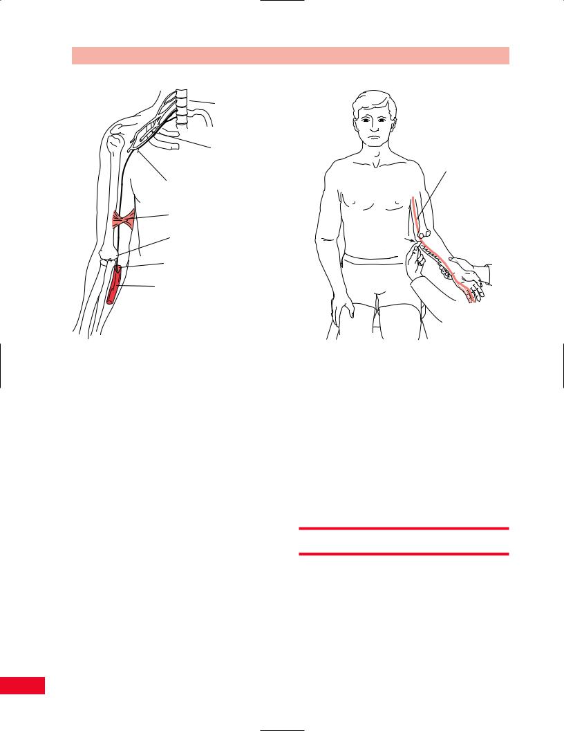
Golfer’s Elbow (Medial Epicondylitis)
Test
The patient’s forearm is supinated and the elbow and wrist are extended by the examiner (Figure 9.61). Pain is felt by the patient in the region of the medial epicondyle due to overuse of the wrist flexors. Putting the patient in this position stretches these overused muscles at their tendinous attachment to the medial epicondyle of the humerus. This test will also cause pain in the proximal medial area of the forearm in patients with overuse syndromes due to typing and playing instruments (i.e., strings, piano, and woodwinds).
Figure 9.58 The radial nerve may be compressed at the spiral groove of the humerus due to pressure, as in Saturday night palsy.
Radial nerve
Posterior interosseous nerve
Superficial radial nerve
Supinator
Referred Pain Patterns
Pain in the region of the elbow may be referred from the lower cervical spinal segments, the shoulder, and the wrist (Figure 9.62).
Medial nerve
 Biceps
Arcade
of Frohse
Figure 9.59 The posterior interosseous branch of the radial nerve may be compressed at the level of the arcade of Frohse, which is part of the supinator muscle. Note that the superficial radial nerve, which is a sensory nerve to the hand, is not affected by this compression.
229

Examiner
Extension
Flexion
Patient
Stabilizer
A
B
Figure 9.60 Tennis elbow (lateral epicondylitis) can be tested for by resisting wrist extension, as in (A), or by passively extending the elbow and flexing the wrist to stretch the tendons of the wrist extensors, as in (B).
Site of tenderness
Figure 9.61 Golfer’s elbow (medial epicondylitis) will be noted by palpating at the site of the x in the medial epicondylar region. The pain is intensified by resisting wrist flexion and forearm pronation with the elbow extended.
230
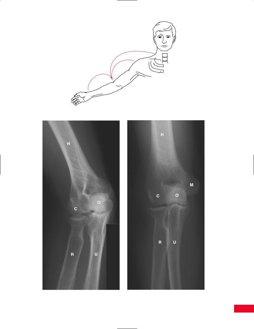
Figure 9.62 Pain may be referred to the elbow from the neck, shoulder, or wrist.
Figure 9.63 Anteroposterior view of the elbow. Figure 9.64 View of the elbow with the forearm pronated.
231
The Elbow Chapter 9
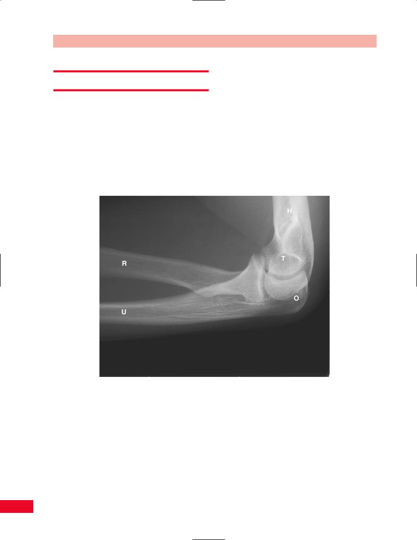
Radiological Views
Radiological views are presented in Figures 9.63, 9.64, and 9.65.
H = Humerus
O = Olecranon process of ulna R = Radius
U = Ulna
T = Trochlea of humerus
M = Medial epicondyle of humerus C = Capitellum of humerus
Figure 9.65 Lateral view of the elbow.
232

Chapter 10
The Wrist and Hand
The Wrist and Hand Chapter 10
Please refer to Chapter 2 for an overview of the sequence of a physical examination. For purposes of length and to avoid having to repeat anatomy more than once, the palpation section appears directly after the section on subjective examination and before any section on testing, rather than at the end of each chapter. The order in which the examination is performed should be based on your experience and personal preference as well as the presentation of the patient.
Functional Anatomy
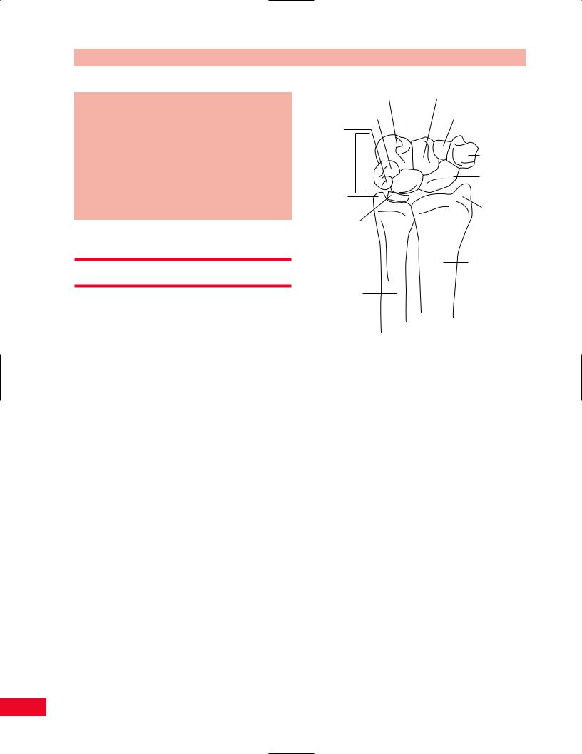
The hand can be divided into two major parts: the wrist and five digits. The carpus, or wrist, is composed of eight small bones. As a unit, the carpus can be thought of as an egg lying on its side, resting in a shallow cup. In this way, it can accommodate movement in three planes, although in unequal amounts. The greatest degree of freedom is in the flexion–extension plane. Next is ulnar–radial deviation. The least movement occurs in rotation about the long axis of the forearm.
The shallow cup is formed by both bony and soft tissues. There are laterally, the distal end of the radius and its styloid, and medially, the distal ulnar styloid and the triangular fibrocartilaginous meniscus. The meniscal soft tissue is interposed between the distal end of the ulna and the wrist bones. The “egg” of the wrist is composed of two rows of small, irregularly shaped bones called the carpals (Figure 10.1). These two rows are linked together by many interosseous ligamentous structures and are also connected through the navicular bone, which acts as a linkage between the proximal and distal carpal rows. The carpal bones, because of their shape, permit varying amounts of movement.
Together, they facilitate and modify placement of the digits in space. Because of its position as linkage between carpal rows, the navicular (meaning “boat shaped”) can be stressed across its midsection or waist, creating a fracture of that bone. Because of its vascular supply following the unusual distal-to-proximal direction, fracture of the navicular can lead to avascular necrosis and collapse of the proximal half of that bone. This damage leads to impairment of wrist function and progressive osteoarthritis of the wrist joint.
The five digits can be divided into three groups. The index and long fingers represent a stable central
Hamate Capitate
Triquetrum Lunate Trapezoid
Pisiform
Carpus |
Trapezium |
|
|
||
|
Scaphoid |
|
|
(Navicular) |
|
Styloid |
|
|
process |
Styloid |
|
of ulna |
||
process |
||
Triangular |
||
of radius |
||
fibrocartilaginous |
||
|
||
meniscus |
|
|
|
Radius |
Ulna
Figure 10.1 The eight small carpal bones of the hand form an “egg”, which rests in a shallow dish formed by the distal radius and ulnar meniscus.
column about which the medial ring and small fingers, and lateral thumb wrap.
The basal joint of the thumb is the most mobile of the hand articulations. Shaped like a saddle, the basal joint permits flexion and extension in two planes. The saddle shape, however, is quite unstable, and possibly the reason for the greater propensity for this joint to undergo osteoarthritic degeneration compared to the other joints of the hand.
Each of the digits of the hand have joints that permit flexion and extension. They can each be thought of as simple hinge joints stabilized by collateral ligaments on their medial and lateral aspects.
Movement of the wrist and digits is performed by the flow of the long, sinewy tendons passing from their origins in the forearm across the palmar and dorsal aspects of the wrist. These tendons, along with the major neurovascular structures of the hand, pass through well-defined tunnels or compartments. Two of these tunnels on the palmar side of the wrist are particularly rigid in their cross-sectional dimension. As such, neurovascular structures passing through them are particularly vulnerable to compression injuries should any space-occupying lesion invade the space of this tunnel (i.e., edema from injury or thyroid dysfunction, or adipose tissue due to obesity). These tunnels are so often
234