


290 11 Nanoparticles for Cancer Drug Delivery
micelles, or dendrimers is beyond the scope of this review, as is the application of nanoparticles in cancer diagnosis.[FettU]
11.2
Cancer: A Fatal Disease and Current Approaches to Its Cure
Cancer is the second leading cause of death in the USA<<1, claiming a total of 553,768 lives per year. Despite new discoveries of drugs and treatment combinations for cancer, the mortality rate has not changed in the past 53 years. However, the overall survival rate has been increased by 13% since 1974, due to more sensitive diagnostic techniques and early detection methods. Of the 699,560 men and 668,470 women predicted to be diagnosed with cancer in the year 2004, 64% are expected to survive [6].
The second leading cause of cancer death is prostatic adenocarcinoma in men and mammary carcinoma in women. Both these cancers develop mixed populations of hormone (estrogen/androgen)-dependent and hormone-independent cells, which are poorly to well differentiated and have variable proliferation rates. In cases of organ-confined disease, radical prostatectomy is the preferred treatment in men and mastectomy in women. Initially patients are treated with radiation and/or chemotherapy (cyclophosphamide, doxorubicin, 5-fluorouracil, [7, 8], in addition to hormone ablation. Although androgen ablation in prostate cancer patients (LHRH agonists, leuprolide [9]) leads to a reduction of the primary tumor and to its partial regression, within 2 years the disease can re-emerge in a poorly differentiated, androgen-independent form, after which no therapy is available to prolong the life of the patient [10]. In breast cancer patients estrogen ablation is achieved by tamoxifen treatment [11] or ovariectomy. Surgical removal of the primary tumor is invasive and has side effects depending on the area of resection and on the tumor (whether encapsulated or invasive). Often, removal of the primary tumor can lead to increased proliferation of metastases, single metastatic cells, and dormant cancer cells.
Up to 70% of prostate cancer patients [12] and 40% of breast cancer patients have occult metastases at the time of diagnosis [13]. Bone and lymph node metastases occur in 26% of patients with prostate adenocarcinoma [14] and in 23% of breast cancer patients [13]. More than 70% of patients die from skeletal metastases [15] (Fig. 11.1).
Current treatments can only prolong the life of the patients, but do not cure. Because of the high toxicity and poor specificity of currently used drugs, increasing the chemotherapeutic dosages is not possible. Often, patients in relapse do not respond to chemotherapy due to an acquired drug resistance which decreases the efficacy of chemotherapy. Dormant metastatic disease cannot be targeted by chemotherapy, and proliferation of occult breast cancer cells in the bone marrow is responsible for a relapse of at least one-third of the patients [13, 16, 17].
Currently used chemotherapeutic drugs are systemically active and cannot target single dormant cancer cells, tumors, or slow-growing tumors [13, 16, 18], as most

11.2 Cancer: A Fatal Disease and Current Approaches to Its Cure 291
A
B
Figure 11.1. Single cell seeding from primary tumors leads to lymph node and bone metastases. Histological sections from
(A) lymph node metastasis from primary breast cancer tumor and (B) primary breast cancer tumor.
chemotherapeutic drugs are not selective for cancer cells. To reach therapeutically effective concentration at the tumor site, therefore, high systemic dosages are required, which cause severe side effects and peripheral tissue damage. Among these side effects are destruction of bone marrow cells (which impairs the production of erythrocytes), cardiotoxicity, nephrotoxicity, hepatotoxicity, and hematotoxicity. Bone marrow cells produce erythrocytes for oxygen transport, so patients become anemic as a result of bone marrow destruction. Hematotoxicity includes the destruction of platelets needed for blood clotting and leukocytes to fight infections. Immediate side effects are nausea, alopecia, and fatigue. Longer-lasting effects are the increased susceptibility to infections, which persists until the immune system has recovered from the chemotherapy (4–6 weeks).
Most of the chemotherapeutic drugs interfere with the proliferation machinery of the cancer cells. Cyclophosphamide (Cytoxan) destroys genetic material which controls tumor cell growth. Methotrexate and 5-fluorouracil (5FU) are antimetabolites which interfere with cancer cell division. Antimicrotubule reagents prevent cell division by acting on the microtubules; among these are paclitaxel (Taxol), docetaxel (Taxotere), vincristine (Oncovin), and vinblastine (Velban). Doxorubicin (Adriamycin) is a tumor antibiotic. In order to be effective these drugs need to accumulate in the tumor cells.
High-dose chemotherapy does not necessarily cure the patient. Many patients relapse. Recurrence of the cancer occurs after high-dose chemotherapy, often manifested by the lack of response to further chemotherapy, which in turn leads to terminal disease even after several years of apparent freedom from disease. This phenomenon is defined as multidrug resistance and is attributed to multiple mechanisms [19, 20].

292 11 Nanoparticles for Cancer Drug Delivery
Most cancers, such as colon, kidney, breast, ovarian, prostatic, and lung cancers, overexpress the p-glycoprotein gene (also known as the multidrug resistance gene). Its gene product is the protein Pgp, which is located in membranes, Golgi apparatus, and nucleus [21, 22]. The p-glycoprotein (Pgp) is a transmembrane efflux pump that actively excretes cytotoxic drugs of different molecular structure through an ATPase mechanism [23]. decreasing intracellular drug concentrations. Modulators for Pgp pumps have been reported and include verapamil, a compound which competes with the drug efflux pump. During differentiation and progression, cancer cells can acquire the ability to remove the administered drug through such a pump mechanism. Other mechanisms include alteration of enzymatic activities such as topoisomerase or glutathione S-reductase activity, altered apoptosis regulation, altered transport, or alteration of intracellular drug distribution due to increased sequestration of cytoplasmic vesicles [24].
The severity of side effects during administration of chemotherapeutic drugs and the occurrence of multidrug resistance emerges as nonresponsive or refractory disease to chemotherapeutic drugs.
11.3
Characteristics of Tumor Tissues
Tumor tissue consists of various structures and areas of which the actual cancer cells can occupy less than 50%, the vasculature 1–10%. The remaining structure consists of a collagen-rich matrix. Histological evaluation of excised tumors revealed rapidly growing regions interspersed with necrotic regions [25]. Tumors develop a tortuous, chaotic capillary network that distinguishes it from normal vasculature, which follows a hierarchic branching pattern [26]. The capillary vasculature in tumors is often accompanied by occlusions, caused by rapidly proliferating cancer cells. Compression of the vasculature [27] causes hypoxia and eventually necrosis of viable tumor cells. In contrast to normal tissue, tumors often lack a functional lymphatic system. In addition to a high proportion of proliferating endothelial cells, tumor vasculature shows aberrant basement membranes. Tumor blood vessels differ from normal vasculature in being up to 3–10 times more permeable [28], to counterbalance the high oxygen and nutrient requirements of the fast-growing tumor [29] (Fig. 11.2).
Macromolecules and drugs are transported into the tumor cells through interendothelial junctions and vesicular vacuolar organelles and fenestrations. The range of pore cutoff size in tumor tissue has been reported at between 100 and 780 nm [30, 31]. This range has been confirmed by measurements made in vivo through fluorescence microscopy [32]. The pore size can be increased up to 800 nm by perfusion with low dosages (10 lg/ml) of vascular endothelial growth factor (VEGF) in human colon tumors [33]. In a mouse model bearing human colon cancer xenografts, the extravasation of polyethyleneglycol-stabilized liposomes of diameters from 100 to 400 nm was examined. VEGF perfusion increased the frequency of 400-nm pores in the tumor vasculature and increased the number of transvascular pathways, open
11.4 Drug Delivery to Tumors 293
A
Enhanced Permeability
And Retention Effect
Single tumor cells  invade vascular system
B |
|
C |
|
|
|
|
Single tumor cell |
Macromolecules or nano |
|
|
|
|
|
particles |
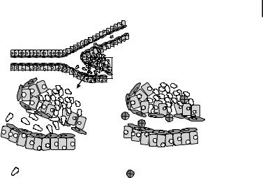
Figure 11.2. A. Normal vaculature with part of cancerous tissue (box). B. Single tumor cells pass through the hypermpermeable vasculature. C. Enhanced permeability and retention effect. Grey circles represent nanoparticles or macromolecules.
junctions, and fenestrae. For comparison, normal vasculature endothelium diameter is less than 2 nm, 6 nm in postcapillary venules, 40–60 nm in kidney glomeruli and 150 nm in sinusoidal epithelium of liver and spleen.
11.4
Drug Delivery to Tumors
The interstitial compartment of a tumor contains a network of collagen and elastic fiber, which is immersed by hyaluronate and proteoglycan-containing fluid. The interstitial pressure within the tumor tissue is elevated due to the lack of a lymphatic drainage system [34, 35]. Increased interstitial pressure and rapid aberrant cell growth are believed to be responsible for the compression and occlusion of blood and lymphatic vessels in solid tumors [25] and for hindering the extravasations and accumulation of the drugs in the tumor tissue.
The transport of a drug into the tumor area is dependent on the interstitial pressure as well on its composition, charge, and the characteristics of the drug (hydrophobicity, size) [36] as the interstitial pressure is higher than the pressure within the blood vessels [37]. In addition the dense packing of tumor cells limits the movement of molecules from the vessel into the interstitial compartment [38]. For example, colloidal particles larger than 50 kDa enter the interstitial compartment through leaky vessels and accumulate in the tumor tissue. This phenomenon is called the enhanced permeability and retention (EPR) effect. The EPR effect is characterized by an accumulation of a drug in the interstitium, exceeding the drug concentration in the plasma [39, 40], which then in turn hinders diffusion of the drug into the interstitial compartment.

294 11 Nanoparticles for Cancer Drug Delivery
The lack of tumor response to a drug (multidrug resistance) can be due to poorly vascularized tumor regions and lack of drug accumulation in the tumor cells at a therapeutically effective concentration. Interaction of macromolecules (drug) with plasma can inactivate the drug by degradation or hydrolysis. The acidic environment of the tumor can inactivate basic molecules by ionization and thereby prevent their diffusion across the cell membrane. Altered drugs can loose their therapeutic efficacy. This cannot be avoided by increasing the dosage of an administered drug to reach the therapeutic effective plasma concentration, because most chemotherapeutics are highly toxic and lead to severe systemic side effects.
It is apparent that the required therapeutic effective drug concentration in the tumor tissue is difficult to achieve and maintain because of vascular and lymphatic characteristics of the tumor tissue. The combination of chemotherapeutics and nanoparticle technology could reduce some of the currently observed problems encountered with systemic treatment regimens, e.g., protecting the drug from degradation, increasing the solubility of lipophilic drugs, increasing the specificity of drugs, and circumventing multidrug resistance.
11.5
Physicochemical Properties of Nanoparticles in Cancer Therapy
Nanoparticles are spherical particles, whether naked or functionalized, measuring less than 100 nm in at least one dimension. It is not surprising that nanoparticles have implications in biological systems in general and as drug delivery systems in particular since most cells are 10,000–20,000 nm in diameter. Nanoparticles can enter cells, even nuclear compartments, and interact with DNA and cellular proteins. They can be fabricated at different sizes and surface modifications, which determines their properties in biological systems. Particles less than 100 nm have longer circulation times, large effective surface areas, low sedimentation rates, and they show enhanced diffusion potential and are easily internalized in tumor cells through the membrane pores. Agglomeration of nanoparticles has to be avoided to prevent thrombosis [41]. Small particles have access to capillaries and are more resistant to the macrophage uptake of the reticuloendothelial system [42–44].
Nanoparticles applicable for cancer treatments need to counter the adverse effects of the current chemotherapeutic approaches described in the previous section. In general, nanoparticles have the potential to improve cancer drug delivery and provide tools to
. |
deliver the pharmacologically required concentration of the drug |
. |
increase drug concentration at the target site through extended or controlled |
|
release |
. |
overcome multidrug resistance |
. |
eradicate side effects to vital organs by reducing systemic exposure |
. |
avoid immune response and hematopoietic toxicity |
. |
destroy malignant cells specifically, sparing normal cells |
. |
kill primary tumors inaccessible to surgery |


296 11 Nanoparticles for Cancer Drug Delivery
. |
Immunogenicity |
. |
Surface properties |
. |
Degradation properties |
. |
Drug loading capacities and release |
. |
Stability of the drug during encapsulation |
. |
Storage and use of the fabricated nanoparticles |
11.5.1
In Vivo Circulation Pathways of Nanoparticles
Nanoparticles injected into biological systems are rapidly coated with plasma proteins such as immunoglobulins and fibronectin and build aggregates. This process is called opsonization. Opsonized particles are recognized by the reticuloendothelial system (RES) or mononuclear phagocytic system (MPS), which is comprised of macrophages related to liver (Kupffer cells), spleen, lymph nodes (perivascular macrophages), nervous system (microglia), and bones (osteoclasts) [45, 46]. The RES is a defense system and comprises highly phagocytotic cells derived from bone marrow. These cells travel in the vascular system as monocytes and reside in their particular tissues.
These macrophages [47] internalize the opsonized nanoparticles through phagocytosis and deliver them to the liver, spleen, kidney, lymph node, and bone marrow (Fig. 11.4). This clearance can occur within 0.5–5 min, [48], thus removing the active nanoparticles from the circulation and prevent their access to the tumor tissue. Coating the nanoparticles and reducing their size to less than 100 nm can mask them that they are no longer recognized by the MPS and remain in circulation for longer [49]. Rather than normal tissue, such nanoparticles preferentially access tumors through their hyperpermeable vasculature [50], which can be regulated by
Figure 11.4. Distribution and routes of nanoparticles after injection.

11.5 Physicochemical Properties of Nanoparticles in Cancer Therapy 297
low dosages of VEGF [33]. Nanoparticles accumulate in the interstitial compartment of the tumors. The retention of nanoparticles is mainly due to the lack of lymphatic clearance in the tumor tissue (the EPR effect) [39, 40, 51]. If nanoparticles are biodegraded in the interstitium, the drug can be released and enter the tumor cells through diffusion.
Cellular uptake mechanisms for nanoparticles and macromolecules are pinocytosis [52], endocytosis [53, 54], and receptor-mediated endocytosis [55] (Fig. 11.5). Macromolecules and DNA, which are susceptible to lysosomal degradation, can be delivered by nanoparticles which escape lysosomal degradation. Polylactide glycolic acid (PLGA) particles are nonspecifically transported into the cells by fluid phase pinocytosis, a process which is mediated by clathrin (Fig. 11.5). At pH 7–7.5 (physiological pH), negatively charged PLGA nanoparticles are transported to primary endosomes, become positively charged, and enter the acidic secondary endosomes and lysosomes, from which the nanoparticles escape into the cytosol. Nanoparticles transported to early or primary endosomes enter the secondary endosomes and become cationic. Local interaction with the membrane releases the particles into the cytoplasm, escaping the lysosomal compartment [56].
Mechanisms of Cellular Uptake
Receptor
mediated |
Endocytosis |
|
|
endocytosis |
Pinocytosis |
|
|
|
Clathrin coated |
|
Pit |
|
Recycling |
|
Clathrin |
|
coated |
|
Vesicle |
|
Endosome |
|
Cytosol |
|
Lysosome |
Drug  Receptor
  Ligand
Figure 11.5. Mechanisms of drug uptake into tumor cells: phagocytosis, clathrin-coated pits, endocytosis, and targeted delivery via receptor mediated endocytosis.
29811 Nanoparticles for Cancer Drug Delivery
11.5.2
Surface Treatment or Coating of Nanoparticles
Through coating with biodegradable matrices, nanoparticles become “invisible” to macrophages. The choice of hydrophilic or hydrophobic matrices for coating determines the fate of the nanoparticles. Hydrophilic coating prevents interaction of nanoparticles with macrophages of the RES, reduces their removal from the circulation, and increases their circulation half-life [57–59]. Hydrophobic coatings are applied to increase opsonization, leading to copious interaction with macrophages, and the nanoparticles are therefore rapidly removed from the circulation. The latter approach is applied for targeted delivery of nanoparticles to the RES of liver and spleen.
Hydrophilic coatings are dextran [60], polyethylene glycol (PEG) [61], polyethylene oxide (PEO), poloxamers and poloxamines [62], and silicones [59, 60, 63]. PEG coating resulted in enhanced circulation time of the particles [64] and reduced opsonization [65, 66]. Ishiwata et al. showed that PEG coating resulted in suppression of macrophage interaction [67].
The concept of particle coating seems promising, although in vivo applications need to be optimized. The choice of the coating polymer can prolong the half-life of the particle in circulation. Polymethylmethacrylate (PMMA) nanospheres coated with polyoxamer 407 or poloxamine 908 showed increased retention and accumulation in B16 and Mtu tumors in mice [68]. However, PMMA is not biodegradable and has no further application in vivo.
Amphiphilic copolymers like polylactic acid, poly e-caprolactone, and polycyanoacrylate were coupled with PEG [69–71], appear to have high circulation profiles, and have not yet been used for tumor targeting.
11.5.3
Polymers for Encapsulation
Nanoparticles as drug carriers can be prepared in two different approaches. One is as a drug reservoir, which consists of an oily core as vehicle, which carries the drug, and a polymeric outer core layer with a coating. Vehicles are vegetable oil, triglycerides, or cotton seed oil. In the other approach, the particles are nanospheres in which the drug is dispersed in a polymeric matrix. Nanoparticles and nanospheres can be prepared from synthetic biodegradable polymers such polyvinylpyrrolidone (PVP) [72], chitosan [73], or polyalkylcyanoacrylates and polylactides [74–77] such as polyisohexylcyanoacrylate (PIHCA), polyethylcyanoacrylate, and polyisobutylcyanoacrylate (PIBCA). PLGA, which has been approved by the Food and Drug Administration for human application, degrades slowly, releasing the drug, and is therefore used for controlled release. Breakdown products are lactic and glycolic acids, which are metabolized through the Krebs cycle. A nonbiodegradable polymer is PMMA, which is not suitable for in vivo injection. Preparations of nanocapsules have been described and characterized by Puisieux and Couvreur et al. [75–77]. Depending on the preparation method, nanocapsules of different sizes have been prepared. An

11.6 Site-Specific Delivery of Chemotherapeutic Agents Using Nanoparticles 299
advantage of synthetic biodegradable polymers lies in the fact that their degradation rate can be predicted and manipulated by the choice of co-polymer composition.
One of the most important characteristics of nanoparticles is their ability to encapsulate drugs. This feature can reduce systemic exposure and deliver pharmaceutically effective concentrations of a drug to the target. Typically, drugs against cancer are administered systemically at high dosages to ensure a therapeutically effective concentration at the tumor site. Commonly, dosages of chemotherapeutics are in the 100-lg range; sometimes they are as high as 1 g per day. In the past 30 years a number of drug delivery devices have been developed, ranging from macrosized (1 mm) to micro- (100–0.1 lm) and nano-sized [78, 79]
Macromolecules or drugs can become altered during plasma exposure and thus lose their potency. Systemic exposure leads to severe side effects as the drugs destroy nonmalignant tissue. Encapsulation of the drug offers a solution for drug protection. Drug encapsulation in a lipophilic environment may even enhance the solubility of a lipophilic drug and reduce severe systemic side effects. Liposomes or polymeric nanoparticles or nanocapsules have been tested in vitro and in vivo for their ability to house drugs or DNA, and their ability to deliver the drugs has been investigated.
11.6
Site-Specific Delivery of Chemotherapeutic Agents Using Nanoparticles
Using nanoparticles to deliver drugs to tumors offers an attractive possibility to avoid obstacles that occur during conventional systemic drug administration. However, new obstacles come into play when nanoparticles are introduced into the system. The route of administration, size of the particles, degradable coatings, biologically acceptable coatings, endocytotic properties, composition of particles, and stability in physiological salinity all determine the fate of nanoparticles. Nanoparticles injected into biological systems should not agglomerate, in order to avoid macrophage uptake and thrombosis. Enhanced accumulation of nanoparticles at the target can be achieved by attaching ligands to the surface of the nanoparticles or, in the case of magnetic nanoparticles, by using an external magnetic field to concentrate them in the area of the tumors.
Nanoparticles can be directed to the tumors by passive or active targeting. Passive targeting includes manipulation of the size and/or hydrophobicity or other physicochemical characteristics and can be applied to target the RES; active targeting involves the direction of magnetic particles by using an external magnetic field or by using ligand-conjugated nanoparticles. The following outline provides examples for the in vivo application of nanoparticles with various coatings and various materials. Polymer-complexed micelles are not discussed in this review.

30011 Nanoparticles for Cancer Drug Delivery
11.6.1
Passive Targeting
Biodegradable nanoparticle systems have been developed to target the lymphatic system; this can be exploited to deliver drugs to the lymph nodes and destroy lymph node metastases and prevent further metastatic progression. Targeting lymphatic systems (to avoid hepatic drug degradation) can be achieved by using different routes of administration: intramuscular, subcutaneous, intraperitoneal, or oral. Coating nanoparticles is advantageous to enhance circulation time and avoid their recognition by the MPS, in turn increasing their uptake and accumulation in tumor. Therefore, long-circulating nanoparticles can reach targets outside the MPS [80].
The importance and effects of different coating materials in passive targeting are indicated in the following examples.
11.6.1.1Targeting Lymph Nodes with Nanoparticles
Hydrophobic coating further enhances lymph node accumulation, because the nanoparticles are incorporated by the MPS and delivered to the lymph nodes. Polyacrylcyanoacrylate particles loaded with insulin have been orally administered to rats and were incorporated into the lymphatic system via the Peyer’s patches in the intestinal lining [81].
The behavior and biodistribution of nanoparticles and egg phosphatidyl-choline (EPC) or phospholipid (PL) emulsions as drug carrier have been compared and revealed that EPC and PL were cleared faster from the injection site: the lymphatic retention time was 17–24 h compared to 131 h in the case of the nanocapsules. Nanocapsules coated with PIBCA were more stable in plasma than the emulsion particles, because the nanoparticles were protected from lipolysis. PIBCA nanoparticles were largely incorporated into lymphocytes, whereas only a small portion of the emulsion particles were detectable in lymphocytes [82]
Nanoparticles of different materials can result in various biodistribution of the drug and therefore alter drug efficacies. Cucurbitacin BE polylactic acid nanoparticles (47–120 nm) were developed for delivery to cervical lymph nodes. The nanoparticles were loaded with 23% of the drug. Since polylactic acid is biodegradable, the release in vivo was slow and the acute toxicity of the cucurbitacin was lowered by 50% [82, 83].
11.6.1.2Increasing Bioavailability of a Compound
Highly hydrophilic compounds are rapidly excreted after systemic injection; their efficacy can be diminished and they may even lose their potency.
Gadolinium neutron capture therapy (GdNCT) is a two-step radiotherapy. Gd-157 emits gamma radiation as a result of its neutron capture reaction from an external neutron source, which inactivates the tumor tissue. Commercially available Gd formulas such as Magnevist – a dimeglumine gadopentetate aqueous solution – are highly hydrophilic, poorly retained in tumors, and rapidly excreted even after intratumoral injection. In order to be effective, Gd has to be delivered and retained at the tumor site at high concentrations. Nanoparticles loaded with gadopentaacetic acid were fabricated from chitosan poly[b-(1–4)-2amino-2-deoxy-D-glucopyranose]. As a

11.6 Site-Specific Delivery of Chemotherapeutic Agents Using Nanoparticles 301
naturally occurring polysaccharide, chitosan is biodegradable, bioadhesive, and biocompatible. The Gd-nanoCP = Gadoliniumpentetic acid in chitosan nanoparticles were fabricated by emulsion droplet coalescence technique, with a particle size of 426 nm, and contained 9.3% Gd. Gd nanoCP particles entered tumor cells through endocytosis and in a comparison to Magnevist significantly enhanced Gd accumulation and retention, by a factor of 200 in B16F10 melanoma cells and SCC-VII squamous cell carcinoma in vitro [84]. Mice bearing subcutaneous B16F10 melanoma were injected intratumorally with the Gd-nanoCP nanoparticles containing 1200 mg natural gadolinium. After thermal neutron irradiation was performed at the tumor site, tumor growth in the nanoparticle-administered group was significantly suppressed compared to that in the gadopentetate solution-administered group. However, a complete treatment was not achieved due to the uneven distribution of Gd in the tumor tissue. This study demonstrated the potential usefulness of Gd-NCT using Gd-loaded nanoparticles [85]. Watanabe et al. tested Gd lipid nanoemulsions (Gd-nanoLE) synthesized from hydrogenated phosphatidyl choline, and gadolinium diethylenetetraaminepentaacetic acid (Gd-DTPA), which were surface-treated with polyoxyethylene to create a hydrophilic moiety. The Gd-nanoLE Gd-nanoLE = Gd lipid nanoemulsion; LE = lipid emulsionparticles (70–90 nm) were injected intravenously and intraperitoneally into melanoma-bearing hamsters (1.5 mg ml–1 Gd). Intravenous injection was advantageous over intraperitoneal injection with respect to bioavailability, tumor retention, and accumulation. When injected intraperitoneally the bioavailability was reduced to 57% compared to intravenous injection. Tumor accumulation was 49.7 mg Gd per gram of tumor at 24 h compared to 21 mg Gd per gram of tumor at 12 h in the intraperitoneally treated groups. However, intravenous administration resulted in higher Gd accumulation in liver, spleen, lung, and kidney compared to intraperitoneally injected groups. Repeated injection with a two-fold enriched formulation led to 100 mg Gd per gram of tissue [86]. The increased efficacy was due to prolonged circulation of the particles, reduced interaction with the RES, reduced excretion of the compound Gd, and increased retention in the tumor tissue.
Polymeric nanoparticles prepared through the polymer–metal complex formation between cisplatinum (CDDP) and poly(ethylene glycol)-poly(glutamic acid) block copolymers were tested for their efficacy in delivering cisplatinum to tumors. The nanoparticles (CDDP/m) had a size of 28 nm and exhibited sustained release in vitro. Lewis lung carcinoma-bearing mice were injected intravenously with free CDDP or CDDP/m 4 mg kg–1. CDDP/m had prolonged blood circulation time, and accumulated in the tumors at a 20-fold higher rate compared to free CDDP, whereas the accumulation in normal tissue was reduced. Complete tumor regression was observed only in mice treated with CDDP/m; no change of body weight occurred during treatment. Free CDDP treatment at the same dosage caused 20% body weight loss and retained tumor survival [87]. These data clearly show the advantages of drug encapsulation in respect of increased bioavailability, increased target accumulation, and reduced accumulation in normal organs.
PIBCA nanospheres containing mitoxantrone coated with poloamine [88] accumulated in tumors of melanoma-bearing mice when injected intravenously. In this study encapsulation of mitoxantrone did not change the biodistribution of the drug

302 11 Nanoparticles for Cancer Drug Delivery
compared to free mitoxantrone. The plasma doxorubicin concentrations in rats injected intravenously with polysorbate-80-coated PIBCA nanoparticles loaded with doxorubicin was 0.1 mg g–1 compared to 6 mg g–1 in the brain 2–4 h after intravenous administration. The nanoparticles were able to pass the blood–brain barrier which is possible through apolipoprotein E adsorption and low-density lipoprotein-receptor- mediated transport [89, 90]. PEG-coated hexadecylcyanoacrylate particles accumulated three-fold compared to uncoated particles in the rat brain [91].
Irinotecan was encapsulated in polylactic nanoparticles from PEG-PPG-PEG block polymers. The drug content of the nanoparticles was 4.5%, the size distribution 80–210 nm. These particles were injected into sarcoma-bearing mice and rats. Irinotecan was antitumorigenic only in the encapsulated form when injected intravenously or subcutaneously. The plasma concentration of irinotecan in nanoparticle injections was increased compared to free CPT 11 = Irinotecan [92]. The small size of particles increased the tumor uptake and therefore improved the chemotherapy.
Taxol is one of the most potent antineoplastic drugs. However, its therapeutic effect has been limited due to poor solubility. Passive targeting by size selection has been tested in vivo on B16F10 melanoma-bearing mice, which were injected intravenously with PVP nanospheres (60 nm) containing paclitaxel (Taxol). Repeated injection resulted in increased survival and reduction of tumor mass compared to treatments with free paclitaxel [72]. This approach is advantageous as paclitaxel shows poor aqueous solubility. Prolonged therapeutic concentration is required to achieve clinical efficacy. Paclitaxel-loaded PLGA nanoparticles with polymer coatings are biodegradable. When intravenously injected the drug was released in a biphasic pattern, with an initial fast release followed by a slower continous release. The fast release may be due to dissolution diffusion of the drug and poor entrapment, whereas the slower release can be attributed to diffusion through the PLGA core. In vitro toxicity was enhanced 10-fold in the PLGA particle approach compared to free paclitaxel in a human small cell lung cancer cell line. Since paclitaxel was more active with cells in the G2/M cell cycle, longer incubation times are more effective. The use of different copolymers in the nanoparticle preparation resulted in prolonged release time and increased the efficacy of the treatment [93].
Paclitaxel has been encapsulated in gelatine nanoparticles of diameter range 600–1000 nm. The large size was chosen to ensure residence of the particles in the urinary bladder, where they were implanted and rapidly released the encapsulated drug. The IC50 was comparable to that of free paclitaxel [94]. This approach prevented systemic drug exposure and therefore prevented the side effects.
A commercially available formulation for i.v. administration is a formulation of paclitaxel in Cremophor EL and alcohol (50:50). The solvent is incompatible with PVC infusion tubing and causes severe side effects such as nephrotoxicity and cardiotoxicity; it could only be administered after steroid and antihistamine premedication [95–97]. The administration was long, requiring up to 3 h infusion time.
In a new approach Cremophor was replaced by biodegradable PLGA nanoparticles containing d-a-tocopherylpolyethyleneglycol 1000 succinate (TPGS) as emulsifier/stabilizer to dissolve paclitaxel for oral administration. The drug encapsulation efficiency was 100% and the release kinetic controllable. The particle size varied from 300 to 1000 nm [98].

11.6 Site-Specific Delivery of Chemotherapeutic Agents Using Nanoparticles 303
Tocosol, a vitamin-E-based paclitaxel emulsion, has been tested in a phase II clinical trial. Taxane-na've patients with ovarian cancer, bladder cancer, colorectal cancer, or non-small cell lung cancer received a 15-min infusion of Tocosol once a week. The treatment was well tolerated and steroid pretreatment was no longer required. A response was seen in 4–26% of patients; half of the patients had stable disease [99].
In a phase I clinical trial using ABI-007, an albumin nanoparticle formulation, encapsulated paclitaxel was administered intravenously to patients. The infusion rate was increased six-fold, no premedication was required, and a higher maximum tolerated dosage (MTD) was achieved during encapsulation compared to the free drug [100]. Hypersensitivity reactions were absent, there was a lower incidence of myelosuppression, and mild hematologic toxicity was observed compared to the free drug. As of 2003, ABI-007 is in phase III clinical trials for metastatic breast cancer.
ABI-007 has also been tested for pulmonary delivery in rats through intratracheal administration. The administered dosage was 5 mg paclitaxel per kilogram body weight. Biodistribution was determined using tritium-labeled paclitaxel/ABI-007. Maximum drug concentration was achieved within 5 min after administration, mean absorption half-life was 0.01 h, and elimination time was 4.7 h. After 10 min, 28% of the drug was discovered in the lungs, less than 1% was detected in other tissue [101].
Oral administration of ABI-007 was tested in rats and resulted in 45% accumulation; this was increased to 100% accumulation in the presence of cyclosporin A. ABI-007/cyclosponrin A/paclitaxel reached maximum concentration six times faster (within 30 min) compared to free paclitaxel. Interestingly, during oral administration saturation was not achieved. Rapid oral uptake and increased bioavailability (100% vs. 38%) suggests a novel mechanism of oral drug uptake [102].
The pharmacologically effective drug concentration in the tumor is a prerequisite for successful treatment, yet difficult to achieve in systemic treatments. Delivery of tamoxifen to estrogen-receptor-positive breast cancer cells was conducted by poly e-capro- lactone nanoparticles. In vitro the intracellular distribution of tamoxifen containing poly e-caprolactone nanoparticles was found in the perinuclear region in MCF-7 cells after 1 h. The intracellular concentration of tamoxifen was saturable [103].
The antiestrogen RU 58668 was encapsulated in biodegradable nanocapsules (110 nm) and nanospheres (250 nm) consisting of PEG-coated polylactic acid nanoparticles. These particles were intravenously injected into MCF-7 xenograft-bearing mice. Coating with PEG prolonged the antiestrogenic potency of RU 58668 compared to noncoated nanoparticles. The antitumoral activity of the encapsulated drug with PEGylated nanocapsules was stronger than from the nanosphere formulation. The particles accumulated in liver, spleen, and tumors [104]
11.6.2
Active Targeting
11.6.2.1 Magnetically Directed Targeting to Tumor Tissue[FettU]
Magnetic nanoparticles were first reported by Gilchrist et al. in 1957. These authors studied the hyperthermic effect of exposure to a magnetic field (1.2 MHz) on tissue, which were immersed in ferrofluids of 20–100 nm (c-Fe2O3) [105]. Magnetic nano-

304 11 Nanoparticles for Cancer Drug Delivery
particles can be composed of iron oxide, magnetite, or nickel, cobalt, or neody- mium–iron boron oxides. All of these particles are magnetic or superparamagnetic, and range in size between 10 and 200 nm. Magnetite (Fe3O4) and maghemite (c-Fe2O3) are preferentially used as they are biocompatible and not toxic to humans. Nickel and cobalt, although highly magnetic, are not suitable for administration to humans due to their toxicity [106].
Iron oxide is easily degradable and therefore useful for in vivo applications. Ferrous nanoparticles are biologically safe. They are metabolized into elemental iron and oxygen by hydrolytic enzymes, the iron joins the normal body stores and is subsequently incorporated into hemoglobin. Iron homeostasis is controlled by absorption, excretion, and storage. Acute toxicity has not been observed; in rats the administered iron was excreted over a period of 4 weeks. In human clinical trials no toxicity was observed [107]. Renal function, hepatic parameters, serum electrolytes, and lactate dehydrogenase remained unchanged from baseline parameters after treatment with ferrofluids [107]. The elevation of serum iron levels persisted for a maximum of 48 h and caused no symptoms. In rats, iron particles (250 mg kg–1) injected intravenously were without any side effects [108]. In patients 2.6 mg iron particles were without any side effects [109].
Numerous studies are in progress to target tumors in vivo for therapeutic applications such as drug delivery and hyperthermic destruction of tumor. Other studies are pursuing the use of nanoparticles for diagnostic applications, such as their use as contrast agents in NMR and MRI. Upon application of an external magnetic field (EMF) the particles can be guided to the tumor tissue, and they should be immobilized when the EMF is then removed. Coating the surface of magnetic nanoparticles increases their circulation time and facilitates the binding of ligands or other molecules on the surface for targeting and receptor-mediated endocytosis.
The efficacy of magnetic therapy is dependent on the applied field strength as well as on the volumetric and magnetic properties of the particles. Magnetic nanoparticles have to meet the following requirements: high magnetization to be controlled by an external magnetic field and exceed linear blood flow rates of 10 cm s–1 in arteries and 0.05 cm s–1 in capillaries, biocompatibility, long circulation time, size of less than 200 nm, and they should not agglomerate. A magnetic field of 0.8 T is sufficient to exceed linear blood flow rates in carriers containing 20% magnetite [110]. For most carriers, flux densities must be 0.2 T with field gradients of 8 T m–1 for femoral arteries. Tissue depth penetration has been observed for the range of 8–12 cm [111].
Increased tissue depth reduces the efficacy of magnetic targeting. Magnetic targeting and delivery are not readily applicable to tumors located deep in the body.
Active targeting with anticancer drugs has been tested in many in vivo studies in animals and humans. Adriamycin has limited application in humans due to its high cardiotoxicity. An approach to avoiding high toxicity in vital organs is to guide mag- netic-particle-bearing drugs to the tumors.
Luebbe et al. fabricated ferrofluids coated with anhydroglucose, reversibly linked to 4-epirubicin. These particles, 100 nm in size, were injected intravenously into tumor-bearing rats and mice. Upon exposure to a magnetic field of 0.2–0.5 T, epiru-


306 11 Nanoparticles for Cancer Drug Delivery
noma-bearing rabbits with exposure to an external magnetic field (1.7 T). Treatment efficacies were compared with respect to magnetic targeting to the tumors. In this model, tumor regression after 12 days was achieved only with intraarterial injection. Intravenous injections were cytostatic, suggesting the intraarterial administration was more successful than intravenous administration. Free mitoxantrone administration (intraarterial) was less effective, causing alopecia, with tumor regression seen after 24 days. Arterial administration resulted in increased drug concentration at the tumor site. Histological examination of the tumor tissue showed that the ferrofluids were concentrated in the intraluminal space and deposited in the endothelium, the tumor interstitium and surrounding tissues. No pathological changes were observed in the liver, kidney, spleen, lung, or brain This study suggests that intravenous injection might be less effective as a route to target tissue through magnetic targeting, because the reticuloendothelial system (RES) removed the injected nanoparticles from the system during the hepatic passage. The macrophages of the RES transport the phagocytosed particles to liver, spleen, bone marrow, and lymph nodes. These organs appear to be a more appropriate target for this approach. Intraarterial injection, however, was successful in transporting the nanoparticles to tumors located in the hind leg of rabbits, by avoiding the liver passage [113].
The application of magnetic force was essential in concentrating the ferrofluids at the tumor site. Linking the mitoxantrone to ferrofluids reduced the circulating mitoxantrone concentration by 50–80% compared to when free mitoxantrone was given [114].
When long-circulating dextran-coated iron oxide particles were injected intravenously into a gliosarcoma rodent model, the particles accumulated in the tumor tissue and were detected by MRI. The particle distribution in the tumor was 19% interstitial, 49% intracellular in glioma cells, and 21% incorporated in tumor-associated macrophages. The nanoparticles crossed the blood–brain barrier [115].
Doxorubicin delivery with magnetic nanoparticles has been conducted in swine [116, 117], rabbits [113], and rats [118, 119]. In all cases the effective doxorubicin concentration could be reduced by a factor of 10. Implanted magnets in the tumor area were used to guide magnetite containing liposome/doxorubicin particles to the tumor tissue. This resulted in an increase of antitumor activity compared to intravenously injected doxorubicin and side effects were eliminated [120, 121].
11.6.2.2Ligand-Directed Active Targeting
The functionalization of nanoparticles with targeting agents such as ligands or antibodies, which bind to receptors on the tumor cell membranes, facilitate specific and increased uptake into the target cells through receptor-mediated endocytosis and thus can increase the in vivo specificity of the drug. In vitro receptor-mediated nanoparticle accumulation has been studied.
Gadolinium hexanedione-containing nanoparticles were fabricated from PEG 400 (<125 nm). A folate ligand was bound to the nanoparticles by linking folic acid to distearoylphosphatidylethanolamine via PEG spacer. Folate-coated nanoparticles specifically targeted the folate receptor expressing human nasopharyngeal epidermal carcinoma cells (KB); MCF-7 cells served as a folate-receptor-negative control.

11.7 Nonviral Gene Therapy with Nanoparticles 307
Folate-coated nanoparticles were incorporated into KB cells by receptor-mediated endocytosis and their concentration was 10-fold higher compared to uncoated particles. Cell death occurred through gadolinium neutron capture therapy [122] in KB cells but not in the receptor-negative MCF-7 cells. Cell death was observed only after gadolinium–folate nanoparticles had accumulated in the KB cells [122]. Similar results were obtained when folate-receptor-positive breast cancer cells and macrophages were incubated with folate-decorated superparamagnetic nanoparticles. Macrophages did not accumulate the folate-decorated particles [123]. These findings show the high specificity of the receptor-mediated process.
Vitamin H receptors are overexpressed in cancer cells compared to normal cells. Pullulan acetate (PA) and vitamin H were self-aggregated to form particles of 80–125 nm diameter and were loaded with doxorubicin (Adriamycin). Using these nanoparticles, tumor targeting, internalization, and controlled drug release were tested on HepG2 cells. The loading efficiency decreased with higher vitamin H content, suggesting a controlled delivery might be feasible. Vitamin-H-decorated nanoparticles showed strong interaction with the HepG2 cells, whereas the PA particles alone showed poor interaction, suggesting receptor-mediated endocytosis. The release of doxorubicin was saturable within 60 min and depended on the loading capacity of the nanoparticles. The release of doxorubicin could be controlled by the vitamin H content of the PA nanoparticles [124].
11.6.2.3Targeted Drug Delivery Using Magnetic Guidance
Due to the large surface area of nanoparticles, drugs or ligands can be covalently bound to the magnetic nanoparticles themselves or to their polymer coating. Exposure to an alternating electric field accumulates the particles in the target tissue. When the forces of the magnetic field exceed the linear blood flow rate (0.05 cm–1 in capillaries), the magnetic particles are retained in the interstitial compartment through the EPR effect. The particles can be internalized into the tumors by ligand-controlled endocytosis or diffusion and can deliver the drug at concentration required for therapy. Recently, silica-coated magnetic holospheres were synthesized [125, 126] with an average size of 150 nm. Carriers have been designed in the form of magnetic particle cores coated with polymers or porous polymer in which magnetic nanoparticles are located inside the pores [127].
11.7
Nonviral Gene Therapy with Nanoparticles
Up to 70% of the European clinical trials use viral gene therapy. Tumor cells frequently have mutations or overexpression of genes which regulate apoptosis and proliferation pathways. These alterations in the genome cause the tumor cells to grow exponentially. Overexpression of certain proteins like Bcl-2 in cancer cells prevents programmed cell death and results in chemoresistance. Other targets for gene therapy are p53 (tumor suppressor) gene transfer, c-myc, and cytokines such as IL-12 to enhance antitumor activity. Antisense oligonucleotides introduced into can-
308 11 Nanoparticles for Cancer Drug Delivery
cer cells can prevent overexpression of certain proteins which prevent the tumor cells from apoptosis, resulting in chemoresistance.
Viral gene therapy encounters problems which can limit the transfection and transcription rate of the gene/DNA. The transfection rate is lowered because the negatively charged tumor cell environment hinders the uptake of negatively charged antisense DNA. Other obstacles are the degradation of viral DNA in circulation and after transfection by endo-/exonucleases, which is caused by lysosomal degradation. Viral therapy can initiate immune response in the patient. Manufacturing viral DNA constructs is costly and requires high safety measures and precautions [128–130].
Many of the problems encountered by viral gene delivery can be avoided by encapsulation of DNA in nanoparticles, which opens a new era in gene therapy [131]. Nonviral gene delivery protects DNA from endonuclear/exonuclear degradation, and avoids viral-initiated immune response. Consequently, nanoparticle delivery of DNA increases the transfection rate and has major advantages for manufacturing and safety reasons [128–130]. To enhance transfection rates, nanoparticles for delivery of DNA into tumor cells have to meet the following requirements: small-size DNA packages (<25 nm) to facilitate DNA delivery to the nucleus; protection from serum endo-/exonucleases; stabilization during the uptake process; and they must bypass the endocytic pathway/lysosomal degradation [132]. Macromolecules which enter the cellular compartment through endocytosis are degraded by lysosomes and hence become inactivated. Escape from endolysosomal degradation has been shown for PLGA nanoparticles. Encapsulated DNA was slowly released, resulting in sustained gene expression [133].
When nondividing cells, growth-arrested neuroblastoma, and hepatoma cells were transfected with DNA liposome mixtures, the transfection increased 7000-fold compared to naked DNA. The size limit of the liposomes for transfection was 25 nm, which is the nuclear pore size. Particles >25 nm showed decreased transfection capability. Gene delivery through viral vectors linked to magnetic coating surface is under development for gene therapy. The advantage of this approach is the extended contact of the viral vector with the target cell, increasing the transfection rate and the expression rate of the introduced gene [134, 135].
Targeted gene delivery has been demonstrated to inhibit angiogenesis in tumor tissue. Cationic polymerized lipid-based nanoparticles (40–50 nm) were covalently coupled to integrin aVb3-ligands, which target angiogenic blood vessels [136]. In vitro, these particles delivered the green fluorescence protein (GFP)-encoding gene specifically to human melanoma cells expressing the integrin receptor. The plasmids were coupled on the surface of the particles via electrostatic attraction. Importantly, nanoparticles without the integrin aVb3-ligands were not incorporated into the cells, hence GFP was not delivered. This approach was tested in vivo on human melanoma xenograft-bearing mice. The mice were injected intravenously with nanoparticles containing the luciferase gene and the aVb3-ligand (aVb3-NP-luciferase) to determine whether aVb3 can deliver genes to angiogenic-tumor-associated blood vessels. Maximal luciferase activity was detected in the tumor area after 24 h but none in vessels of lung, liver, brain, kidney, or skeletal muscle. The tumor-associated

11.8 Hyperthermia 309
blood vessels were specifically targeted as the gene delivery was blocked in the presence of aVb3 inhibitor [136].
Mice bearing subcutaneous M21-L melanoma (aVb3-negative) were injected intravenously with aVb3-NP-Raf(–) to block endothelial signaling and angiogenesis. Treatment resulted in apoptosis of tumor-associated endothelium leading to tumor cell apoptosis regression of primary tumor and metastases after 6 days [136].
Most DNA delivery constructs attach the DNA to the surface of the particles. In vivo systemic delivery of p53 with Transferrin-Lip-p53 complex improved the transfection rate compared to untargeted construct. Complete tumor regression of DU145 human prostate cancer xenografts were observed in mice treated with radiation [137].
DNA delivery through nanotechnological approaches has important future applications in treating diseases of any kind as it facilitates:
. |
Increased transfection and transcription rates |
. |
Sustained transcription through slow release |
. |
Decreased biohazard risk in production and application |
. |
Decreased immune response in the patient |
. |
Protection of DNA and biosystem |
. |
Specific targeting. |
Nonviral gene delivery may be superior to viral gene delivery in the future.
11.8
Hyperthermia
Tumor cells are more sensitive to temperatures above 42 C than normal cells [138, 139] and can be destroyed by increasing the temperature locally to 41–42 C for 30 min [140–143]. Magnetic particles generate heat under an alternating magnetic field (AMF) by hysteresis loss [143, 144].
Heat production occurs during the thermal loss resulting from reorientation of the magnetism of the magnetic material with low electrical conductivity [144]. The heating potential is directly correlated to the size of the magnetic particle, which can be controlled by appropriate synthesis methods. For example, nanoparticles require less AC power than microparticles, and increased dispersion of nanoparticles requires less AC power [145, 146]. Specific absorption power rates (SARs) have been determined in suspensions of magnetite nanoparticles of various size and coating. SARs of dextran ferrites were 180–210 W g–1 Fe (120 nm), sonicated dextran ferrites ranged from 12 to 240 W g–1 Fe, uncoated ferrosuspension from 0 to 45 W g–1 Fe (6–10 nm). Ultrasonification caused dispersion of agglomerated particles and can in part destroy the dextran coating. Carboxymethyldextran magnetite particles (130 nm) had a SAR of 90 W g–1 Fe. SAR data allow an estimate of the amount of particles necessary to heat human tissues [147, 148].
To test the efficacy of hyperthermia treatment, magnetic fluids with a particle size of 3 nm coated with dextran were injected into C3H mammary tumors of mice.

310 11 Nanoparticles for Cancer Drug Delivery
After 30 min the whole bodies of the mice were exposed to an AMF of 6–12 kA m–1 at 520 kHz for 30 min. The intratumoral temperature rose to 46 C and ferrofluid accumulation increased in the tumor tissue from 15% to 45%. Ferrofluid injected alone did not change the tumor histology. Tumor necrosis was observed in mice with ferrofluid and exposure to the AMF. However, tumor growth after treatment was heterogeneous, with a response of 44%, suggesting that the tumor inhomogeneity coincides with ferrofluid distribution [148].
After intravenous injection of 100 mg dextran magnetite (400 mg kg–1) into rats an accumulation of nanoparticles in the mammary tumors was observed. Exposure to an AMF (12 min, 450 kHz) resulted in shrinking and necrosis of the tumor tissue and coagulation of the blood vessels [149]. In mice, iron concentration was increased in liver and spleen after intratumoral injection due to RES uptake. Iron contents in tumors were initially 15% and decreased to 2% after 52 h. Application of AMF resulted in a 2.5-fold increase of iron specifically in the tumor tissue over a time frame of 30 min. Except for the region around the tumor no systemic heating occurred, suggesting that the iron content in the tumors was sufficient to cause hyperthermia. In order to effectively destroy tumors through hyperthermia, the re-
quired amount of iron inside the tumor tissue has been suggested to be 5–10 mg cm–3 [150].
To further increase the accumulation of iron content in the tumors, magnetoliposomes (ML) were conjugated with an antibody which recognizes renal tumor cells. Mice bearing renal implanted tumors were injected with G250F-ML.
The incorporation of iron in the renal tumors was 27-fold higher when G250FML was injected compared to the ML particles alone. Exposure to AMF (5 kW, 118 kHz, 30.6 kA m–1) arrested tumor growth within 2 weeks [151]. Necrosis was observed in tumors but not in livers after AMF exposure, although the liver accumulated ML. Total destruction of tumor tissue was achieved after three AMF exposures, suggesting that repeated exposures are effective. The iron distribution was 50% tumor, 35% liver, 33% blood.
Magnetic nanoparticles injected intratumorally into human breast cancer-bearing mice resulted in partial release of the particles from the tumors and accumulated in liver, spleen, and lung. These organs may be damaged upon body exposure to AMF [145].
A combined hyperthermia therapy approach has been suggested for U251-SP xenograft in nude mice to increase treatment efficacy. TNF-a gene therapy driven by a heat-inducible promoter was combined with hyperthermia using magnetite cationic liposomes. When mice were injected intratumorally with magnetic cationic liposomes and exposed to AMF 118 kHz, 30.6 kA m–1), tumor cell death was induced within 3 min. TNF-a expression increased three-fold during AMF heat induction even in peripheral areas which were not affected by hyperthermia [152].
Specific targeting of cancer cells and incorporation of magnetic nanoparticles conjugated to luteinizing-hormone-releasing hormone (LHRH) showed in vitro accumulation of magnetic nanoparticles through receptor-mediated endocytosis. LHRH-receptor-expressing MCF-7 human breast cancer cells were incubated with LHRH magnetic nanoparticles coated with silica with a final particle size of

11.8 Hyperthermia 311
20–50 nm. These particles accumulated on the surface and inside LHRH-receptor- expressing cells dependent on the LHRH receptor capacities, but not in UCI 107 cells, which do not express LHRH receptor [153]. LHRH-decorated nanoparticles were eight times more potent in destroying MCF-7 cells. The number of lysed cells was linearly dependent on the magnetic field exposure time and the concentration of nanoparticles. UCI 107 cells were not lysed under the same conditions. These results suggest that specific targeting can increase the efficacy of hyperthermic therapy and keep other organs unaffected.
An alternative to AMF-exposure-related thermal damage to tumor tissue has been shown with near-infrared light (NIR). The fabricated gold nanoshells consisted of silica as core particles, which were surrounded by a thin gold shell coated with PEG. Such particles possess a tunable plasmon resonance through light exposure. The relative thickness of the core and shell layers can be controlled and results in various optical absorption from near-UV to mid-IR. NIR light showed minimal absorption in tissue with optimal penetration. The nanoshells were injected into the tumors of xenograft-bearing mice and exposed to NIR light (820 nm, 4 W cm–2). Within 4–6 min a temperature increase of 37 C in the tumor region occurred [154], resulting in necrosis. Gold shell silica nanoparticles show different absorption spectra, dependent on the thickness of the shell: 60-nm core radius particles with a 20-nm shell have an absorption maximum at k=680 nm versus a 5-nm shell which gives k=1000 nm [155]. These fabricated particles are unique. They are tunable because their absorption properties can be designed during the fabrication process, making them highly suitable for imaging (primarily light scattering) or the photothermalbased therapy (primarily absorbing). In vitro exposure of carcinoma cells to NIRabsorbing gold–silica nanoparticles followed by NIR laser irradiation led to photothermal destruction of the cells. In vivo when nanoshells were injected into the tumor (canine TVT cells), irradiation with NIR light for 6 min (820 nm, 4 W cm–2) resulted in tumor cell necrosis within 4–6 min, spanning a zone of thermal damage of 4 mm. The heating occurred in two phases: initial rapid heating and gradual heating. The nanoshells diffused throughout the tumor tissue and adjacent tissue and caused coagulation and cell shrinkage in adjacent areas which were infiltrated by the nanoshell. Further surrounding tissue stayed intact [154].
In yet another approach, nanoscale metallic particles were used to target specific biological structures and tissues through controlled laser-induced breakdown (LIB) process.
So far there have been no reports of successful application of hyperthermia treatment in humans, although the treatment had been very effective in animal studies. In humans it is difficult to acquire adequate quantities of the magnetic nanoparticles in the target tissue without exceeding the tolerable amount, since in humans the applied external magnetic field needs to be stronger than the tolerable field strength. Heating of the tissue is opposed by blood circulation, which creates a cooling effect. The volume of the tumor tissue which needs to be heated is a limiting factor and should not exceed 300 mm3 [150, 156]. The maximum tolerated dose for humans has been reported to be H 0 f = 4.85 6 108 Am-1 s-1 [157]. In addition, the

312 11 Nanoparticles for Cancer Drug Delivery
tissue depth causes loss of field strength. With a particle size of 0.5–5 lm in a swine model, a targeting depth of 8–12 cm was achieved [116].
11.9
Controlled Delivery of Chemotherapeutic Drugs Using Nanoparticles
Controlled release systems continue to draw the attention of several research groups owing to their application in a broad range of areas such as drug delivery, paper, pesticide, printing, cosmetics, and so on [158, 159]. Most of the work so far has focused on achieving controlled release of active ingredients, encapsulated in polymeric matrices, in response to specific stimuli [159–161] or on the use of microfabrication technology [162]. Advances in microelectromechanical systems (MEMS) technology has resulted in further improvements in microchip-based implantable drug delivery systems [136, 158, 163]. In the field of drug delivery, the method of delivery has a tremendous impact on the therapeutic efficacy [159, 164]. More complicated delivery systems are also being designed for targeted delivery of genes using viral vectors, liposomes, and cationic nanoparticles [164].
Of the aforementioned approaches, responsive polymers and microchips appear to be the more versatile methods for controlled release of drugs. Polymer-based drug delivery systems for controlled release are well known and have been in vogue for the delivery of a variety of drugs [165–167]. They have also been designed to respond to specific stimuli such as ultrasound, light, enzymes, temperature, pH, and electric and magnetic fields [164]. Of these stimuli, magnetic stimulus is particularly attractive as it is a soft approach. Reproducible regulation of drug release from polymers was demonstrated in cases where small magnetic spheres or cylindrical magnets were embedded in a polymeric matrix containing drug [168, 169].
The majority of the controlled drug delivery systems being developed are for clinical applications where systemic exposure and release of a drug was desired. Some of these are: delivery of insulin [165, 166], antiarrhythmics [166], gastric acid inhibitors [170], contraception [171, 172], general hormone replacement [173, 174], and immunization [175]. Only recently has there been a growing interest in the development of controlled drug delivery systems for cancer chemotherapy [176, 177]. The approaches can be broadly classified into the following categories:
. |
Sustained release through polymer degradation |
. |
Enzymatically controlled release |
. |
Controlled release through use of thermosensitive polymers |
. |
Photochemically controlled release |
. |
pH-responsive release systems |
. |
Laser-induced breakdown (LIB) |
. |
Ultrasound-mediated release. |
Sustained release using biodegradable polymeric nanoparticles is one of the most popular approaches for controlled release of cancer chemotherapeutic agents. Sustained release depends on the chemical nature of the polymer [178–182] and the

11.10 Nanoparticles to Circumvent MDR 313
method of preparation [98, 183, 184] and surface modification(s) of the polymer– drug nanoparticles [185]. In an interesting study by Yoo and Park, doxorubicin was chemically conjugated to a terminal end group of PLGA by an ester linkage. The doxorubicin–PLGA conjugate and doxorubicin were formulated into nanoparticles and sustained release profiles (over a 1-month period) with minimal initial bursts were observed [186]. In contrast, when unconjugated free doxorubicin was incorporated into nanoparticles, the initial burst was absent and the drug release was complete within 5 days. Conjugation of anticancer drugs to the polymeric nanoparticles resulted in linear drug-release profiles over an extended period [187, 188].
Examples of approaches have been reported where physiological properties of cancer cells have been taken into consideration while designing the controlled release systems for anticancer agents, e.g., angiogenesis, which is a specific physiological property of cancer cells. A controlled release system for an anticancer drug, all-trans-retinoic acid (atRA), was demonstrated based on enzymatic degradation [189]. In this study, PEGylated gelatin nanoparticles carrying atRA released the anticancer drug very sensitively by the action of the enzyme collagenase IV, one of the major metalloproteases involved in angiogenesis. Similarly, pH values in the tumor interstitium are lower than those of normal cells and this difference can be exploited to develop pH-sensitive polymeric nanoparticles for controlled release of anticancer agents. A new class of pH-responsive polymers carrying the anticancer agent doxorubicin (Adriamycin), was prepared using sulfadimethoxine and succinylated pullulan acetate. The nanoparticles were found to be very stable at physiological pH of 7.4 but showed degradation to release doxorubicin at a lower pH close to that of tumor cells [190].
Another approach for controlled release of cancer chemotherapeutics taking advantage of thermal and photochemical properties of polymeric nanoparticles has been investigated. Yoshinobu et al. [191] have reported the design of a novel acrylic composite nanoparticle with a hydrophobic core and a thermosensitively swellable shell demonstrating a thermosensitive mode of drug release. The microcapsules exhibited an exceptionally rapid on–off response of drug release in a thermosensitive manner. Sershen et al. [192] have also demonstrated photothermally modulated drug delivery using gold nanoshells.
11.10
Nanoparticles to Circumvent MDR
Lack of response to chemotherapy is defined as multidrug resistance (MDR) which involves a mechanism to evade chemotherapy. Pgp recognizes drugs when located in the plasma membrane, but not in the cytosol of the lysosomal compartment [193, 194]. Doxorubicin is a Pgp substrate and has been shown to be exported from the cytosol in multidrug-resistant cancer cell lines. Nanoparticles circumventing MDR via the Pgp mechanism have to enter the tumor cells in order to be effective. Two approaches using PIHCA and PIBCA doxorubicin-loaded nanospheres have been reported in an effort to avoid MDR. In the case of PIHCA doxorubicin nanospheres

314 11 Nanoparticles for Cancer Drug Delivery
the intracellular doxorubicin concentration was lower in doxorubicin-resistant glioblastoma cells compared to that seen with free doxorubicin [194]. The doxorubicin nanospheres were only efficient if the MDR was based on Pgp. When a fast-degrad- ing polymer was used for encapsulation of doxorubicin in a doxorubicin-resistant murine leukemia cell line overexpressing Pgp, the intracellular doxorubicin concentration was high and the doxorubicin efflux was comparable to that seen with the free doxorubicin group.
PIBCA nanospheres were degraded before they entered the cellular compartment [195]. Thus doxorubicin at high concentration was associated with the tumor cell membrane being extravasated through the Pgp efflux pump. The coupling of doxorubicin with polymethacrylate resulted in internalization of the particles through endocytosis in U937 doxorubicin-resistant monocyte-like cancer cells. These particles showed sustained slow doxorubicin release, resulting in a significant higher cytotoxicity than free doxorubicin [196].
Several other attempts have been conducted to increase doxorubicin delivery in resistant cancer cells. Doxorubicin incorporated into gelatin nanospheres showed low cytotoxicity in a mouse colon carcinoma xenograft; even increased cardiotoxicity was observed. The different effects were due to slow dissociation of the nanosphere doxorubicin complexes, slow diffusion rate across the cell membrane, and the failure of the complex to enter cells, causing elevated drug release in the cell membrane. MDR can only be avoided when close contact with polycyanoacrylate is achieved [197].
Folate-receptor-mediated uptake of polyethyleneglycol diastearoyl phosphatidylethanolamine liposomes (70–100 nm) loaded with doxorubicin resulted in increased cytosol uptake and release of doxorubicin into the cytoplasm. The drug was released within 2 h in vitro in M109-HiFR multidrug resistant cancer cells. Folate-targeted liposome drug uptake was increased 10-fold compared to free doxorubicin and was more toxic in vivo than free doxorubicin [198].
Doxorubicin delivered by folate-targeted liposomes did not avoid the Pgp efflux system, which was explained by the aggregation form of doxorubicin in the folatetargeted liposome nanoparticle. It has been suggested that encapsulated doxorubicin is dimeric [199]. Doxorubicin loaded into nanospheres (300 nm) consisting of polycyanoacrylate were injected into leukemia cells (P388ADR-) of xenograft-bearing mice intraperitoneally. The treatment prolonged the survival of the mice compared to free doxorubicin administration, which was ineffective. However, no cure was obtained whether nanoparticles or free doxorubicin was administered. IC50 in vitro was 4.3 lM for free dox versus 0.08 lM for doxorubicin-loaded nanospheres. The multidrug-resistant cell lines were 30to 250-fold more sensitive to doxorubicinloaded nanospheres than to free doxorubicin, and even complete reversion of MDR was observed in some cell lines. PIHCA nanospheres loaded with doxorubicin were biodegradable; nanospheres of a size of 200 nm they entered cells by endocytosis and delivered doxorubicin into lysosomes. Doxorubicin-loaded nanospheres were not recognized by Pgp, thus circumventing the MDR.

11.12 Abbreviations 315
11.11
Potential Problems in Using Nanoparticles for Cancer Treatment
Potential problems associated with nanoparticle use in vivo include the risk of thrombosis through agglomerating particles or their breakdown products. Most experiments have been conducted in rodents and are difficult to scale up to humans with respect to distances and volume in circulation, tissue densities, and the amount of particles required. This problem needs to be taken into account if the drug release is uncontrollable with a magnetic field. There is always a potential toxicity of magnetic carrier or coating polymers and their breakdown products, which may occur after long-time exposure or injections. Also, the application of an external magnetic field can lead to cardiac arrhythmia, muscle stimulation, and seizures and needs to be closely monitored. Some patients may not be eligible for these particular applications.
11.12
Future Outlook
Development and research in the field of nanotechnology in applications like biomedicine requires the understanding and interaction of experts from physics, mathematics, the biomedical sciences, and from chemists and medical personnel. The potential for the use of nanoparticles in the task of drug delivery and disease detection is huge and may change the way of currently used drug delivery systems. Nanoparticles offer a broad application range, and represent enough flexibility to customdesign a treatment regimen in the future.
Future emphasis will move on to metastases detection and treatment with a view to eventually eradicating cancer as a disease.
Acknowledgements
The authors would like to thank Janice Keener and Eric Guilbeau of the Pennington Biomedical Research Center for their help in preparing the manuscript and some of the figures.
Abbreviations
AMF – alternating magnetic field
Apo E – apolipoprotein E
CPT – Irinotecan
DNA – desoxyribonucleic acid
EMF – external magnetic field
EPC – Egg Phosphatidylcholine


