

Laser-Tissue Interactions Fundamentals and Applications - Markolf H. Niemz
.pdf198 4. Medical Applications of Lasers
Lasers in Endodontics
Endodontics is concerned with the treatment of infections of the root canal. These arise from either a breakthrough of decay into the pulp or from plaque accumulation beneath the gingiva and subsequent bacterial attacks of the root. In either case, once the pulp or the root canal are infected by bacteria, the only treatment is to sterilize both pulp and root, thereby taking into account the associated death of the tooth. However, even a dead tooth may reside in place for years.
The mechanical removal of bacteria, plaque, infected root cementum, and inflammated soft tissues is regarded as an essential part of systematic periodontal treatment. The excavation of the root itself is a very complicated and time-consuming procedure, since roots are very thin and special tools are required. The procedure can be supported by antimicrobial chemicals to ensure sterility which is a mandatory condition for success of the treatment. Along with the rapid development of medical laser systems, it has been discussed whether lasers could improve conventional techniques of endodontics, especially in removal of plaques and sterilization. First experimental results using CO2 and Nd:YAG lasers in endodontics were published by Weichmann and Johnson (1971) and Weichmann et al. (1972). By means of melting the dentin next to the root, the canal wall appears to be sealed and thus less permeable for bacteria. Indeed, Melcer et al. (1987) and Frentzen and Koort (1990) stated that lasers may have a sterilizing e ect. Sievers et al. (1993) observed very clean surfaces of the root canal after application of an ArF excimer laser. However, both the CO2 laser and the ArF laser will not gain clinical relevance in endodontics, since their radiation cannot be applied through flexible fibers. Even other laser systems will not be applicable exclusively, since suitable fiber diameters of 400μm are still too large for unprepared roots. Thinner fibers are very likely to break inside the root causing severe complications and additional mechanical operation.
Laser Treatment of Filling Materials
In dental practice, not only tooth substance needs to be ablated but also old fillings have to be removed, e.g. when a secondary decay is located underneath. For the removal of metallic fillings, infrared lasers cannot be used, since the reflectivity of these materials is too high in that spectral range. Amalgam should never be ablated with lasers at all. In Figs. 4.40a–b, two samples of amalgam are shown which were exposed to a Nd:YLF laser and an Er:YAG laser, respectively. During irradiation, the amalgam has melted and a significant amount of mercury has been released which is extremely toxic for both patient and dentist. For other filling materials, e.g. composites, little data are available. Hibst and Keller (1991) have shown that the Er:YAG laser removes certain kinds of composites very e ciently. However, it is quite uncertain whether lasers will ever be clinically used for such purposes.
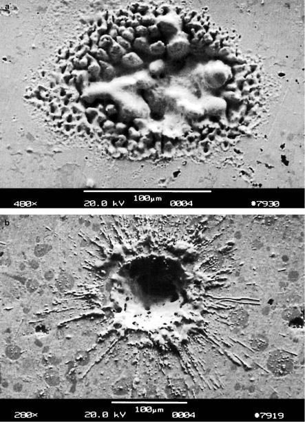
4.2 Lasers in Dentistry |
199 |
Fig. 4.40. (a) Removal of amalgam with a Nd:YLF laser (pulse duration: 30ps, pulse energy: 0.5mJ). (b) Removal of amalgam with an Er:YAG laser (pulse duration: 90μs, pulse energy: 100mJ)

200 4. Medical Applications of Lasers
Another very interesting topic in dental technology is laser-welding of dental bridges and dentures. It can be regarded as an alternative to conventional soldering. During soldering, the parts to be joined are not melted themselves but are attached by melting an additional substance which, in general, is meant to form an alloy between them. Laser-welding, on the other hand, attaches two parts to each other by means of transferring them to a plastic or fluid state. This is achieved with high power densities in the range 102–109 W/cm2. According to van Benthem (1992), CO2 lasers and Nd:YAG lasers are preferably used. Since the reflectivity of metals is very high in the infrared spectrum, it must be assured that either a laser plasma is induced at the surface of the target or that the target is coated with a highly absorbing layer prior to laser exposure. Dobberstein et al. (1991) state that some laser-welded alloys are characterized by a higher tear threshold than soldered samples as shown in Fig. 4.41. However, van Benthem (1992) argues that such behavior cannot be observed in all alloys, but tear thresholds in laser-welded alloys can definitely reach the same values as the original cast. According to his studies, the major advantages of laser-welding are: higher resistence against corrosion, the ability to weld di erent metals, the ability to weld coated alloys, and lower heat load. Moreover, the reproducibility of laser-welded alloys is significantly higher than during soldering. For further results, the interested reader should consult the excellent review given by van Benthem (1992).
Fig. 4.41. Tear thresholds of laser-welded and soldered dental alloys (KCM: cobaltbased alloy, NCA: nickel-based alloy, Sipal: silver-palladium-based alloy). Data according to Dobberstein et al. (1991)

4.3 Lasers in Gynecology |
201 |
4.3 Lasers in Gynecology
Beside ophthalmology, gynecology is one of the most significant disciplines for laser applications. This is mainly due to the high success rate of about 93–97% in treating cervical intraepithelial neoplasia (CIN), i.e. uncommon growth of new cervical tissue, with the CO2 laser. CIN is the most frequent alteration of the cervix and should be treated as soon as possible. Otherwise, cervical cancer is very likely to develop. The cervix represents the connective channel between the vagina and uterus. The locations of the cervix and adjacent organs are illustrated in Fig. 4.42.
Fig. 4.42. Scheme of female reproduction organs
The CO2 laser is the standard laser in gynecology. Beside treating CIN, it is applied in vulvar intraepithelial neoplasia (VIN) and vaginal intraepithelial neoplasia (VAIN). Depending on the type of treatment, CO2 lasers can be operated in three di erent modes – CW radiation, chopped pulse, and superpulse – as shown in Fig. 4.43. Chopped pulses with durations in the millisecond range are obtained from CW lasers when using rotating apertures. Superpulses are achieved by modulation of the high voltage discharge. Thereby, pulse durations less than 1ms can be generated. The peak power is inversely related to the pulse duration. The mean powers of CW radiation and chopped pulses are nearly the same, whereas it decreases in the case of superpulses. As discussed in Sect. 3.2, shorter pulse durations are associated with a reduction of thermal e ects. Hence, by choosing an appropriate mode of the laser, the best surgical result can be obtained.

202 4. Medical Applications of Lasers
Fig. 4.43. CW, chopped pulse, and superpulse modes of a CO2 laser. Dashed lines denote mean powers
Beside selecting the temporal mode, the surgeon has to decide whether he applies a focused or defocused mode as shown in Fig. 4.44. Only in tightly focused mode are deep excisions achieved. In partially focused mode, less depth but a larger surface is vaporized. In defocused mode, the power density decreases below the threshold of vaporization, and tissue is coagulated only.
Fig. 4.44. Coagulation, vaporization, and excision modes of a CO2 laser, depending on a defocused, partially focused, or tightly focused beam
4.3 Lasers in Gynecology |
203 |
In gynecology, there exist several indications for laser treatment:
–vulvar intraepithelial neoplasia (VIN),
–vaginal intraepithelial neoplasia (VAIN),
–cervical intraepithelial neoplasia (CIN),
–endometriosis,
–obstruction of the uterine tube,
–sterilization,
–twin-twin transfusion syndrome.
Vulvar Intraepithelial Neoplasia (VIN). As already mentioned above, neoplasia generally describes uncommon growth of new tissue. In the case of VIN, intraepithelial tissue of the vulva is significantly proliferated. After histologic examination, the altered tissue is usually vaporized with a CW CO2 laser in a partially focused mode. According to Baggish and Dorsey (1981), typical power densities of 100W/cm2 are applied to achieve vaporization depths of about 3–4mm. In the event of bleeding, the laser beam is immediately switched to a defocused mode.
Vaginal Intraepithelial Neoplasia (VAIN). VAIN is a similar diagnosis as VIN, except that it occurs inside the vagina. Initial studies using the CO2 laser were reported by Stafl et al. (1977). Due to the thinner and more sensitive vaginal tissue, slightly lower power densities are applied. The use of a proper surgical microscope is indicated.
Cervical Intraepithelial Neoplasia (CIN). The tissue at risk for the development of cervical cancer is the columnar epithelium which is located in the transformation zone. This type of epithelium can migrate up and down the endocervical channel. Therefore, it is very important to determine its exact location prior to any treatment. This is usually achieved with a colposcope which essentially is an endoscope specially designed for gynecologic purposes. The extent of columnar epithelium is relatively constant. Thus, the more it is exposed at the ektocervix, the less likely is the existence of diseased tissue inside the channel. In order to exclude any potential inflammation, a biopsy specimen is obtained and a second or third control examination is performed after 3–6 months. According to the histologic evaluation of the biopsy, three grades of CIN (I–III) and cervical carcinoma are distinguished, depending on the progress of neoplasia. If the biopsy reveals the presence of cervical carcinoma, a complete resection of the cervix is indicated. At a late stage of cancer, adjacent organs such as the uterus or vagina might have to be removed, as well.
In the case of CIN I, the columnar epithelium is usually located at the ektocervix as shown in Fig. 4.45a. The ablated epithelium is vaporized in a similar fashion as in VIN or VAIN. According to Wright et al. (1983), the procedure should aim at a treated depth of approximately 6mm. Fast movements of the laser beam cause a more homogeneous distribution of heat and thus reduce the probability of carbonization. Scanning mirror devices are

204 4. Medical Applications of Lasers
available to assist the surgeon in steadily moving the beam. In Fig. 4.4b (page 156), a vaporization of cervical tissue is shown as achieved with a CO2 laser at a power of 10W. Escape of smoke is usually inevitable during surgery, but can be managed with specially designed suction tubes.
Fig. 4.45. (a) CO2 laser treatment in the case of CIN I. Diseased epithelium is illustrated by filled ovals. Dashed lines indicate the dome-shaped volume to be vaporized. (b) CO2 laser treatment in the case of CIN II or CIN III. Dashed lines indicate the cylindrical volume to be excised
Fig. 4.46. Alternative CO2 laser treatment in the case of CIN II or CIN III. Diseased epithelium is illustrated by filled ovals. Dashed lines indicate the coneshaped volume to be excised (left). The excised cone is unzipped for histologic examination (right)

4.3 Lasers in Gynecology |
205 |
If CIN II or CIN III is diagnosed, parts of the cervix must be removed to reduce the probability of recurrence. Two di erent treatment techniques were proposed by Dorsey and Diggs (1979) and Wright et al. (1983). Either surgery is performed only after careful evaluation of the obtained biopsy and determination of the cervical length with a micrometer probe. Dorsey and Diggs (1979) suggest excising a cone-shaped volume out of the cervix with a focused CO2 laser as demonstrated in Fig. 4.46. The angle of the cone can be adjusted to individual extents of neoplasia. The procedure itself is called laser conization. The excised cone is then unzipped for histologic examination as shown in Fig. 4.46. By this means, it can be determined whether all altered tissue has been removed.
An alternative method was proposed by Wright et al. (1983) which is illustrated in Fig. 4.45b. Instead of a cone-shaped volume, a cylindrical volume is removed. The vertical cylindrical excisions are achieved with a focused CW CO2 laser, whereas the horizontal excision inside the cervix is performed with either a mechanical scalpel or the same CO2 laser in superpulse mode. After excision of the complete cylinder, the remaining surface is coagulated by defocusing the CW laser beam to achieve local hemostasis. According to Heckmann (1992), cylindrical excisions are better adjusted to individual cases than cone-shaped excisions. In either case, most of the cervical tissue regenerates after being removed. A ten-year review on the treatment of CIN with the CO2 laser was published by Baggish et al. (1989). In Table 4.4, typical cure rates after one laser treatment are summarized.
Table 4.4. Results of CIN treatment with the CO2 laser.
Data according to Baggish et al. (1989)
|
Vaporization |
Conization |
||
|
|
|
|
|
Number of patients |
3070 |
(100%) |
954 |
(100%) |
Cured |
2881 |
(93.8%) |
925 |
(97.0%) |
Persistent |
189 |
(6.2%) |
29 |
(3.0%) |
|
|
|
|
|
According to Wright et al. (1983), laser surgery is an excellent modality for treating CIN when compared to conventional techniques, e.g. cryotherapy. Recently, however, it has been demonstrated by Baggish et al. (1992) that high-frequency electric currents can do the same job as CO2 lasers. When using thin loops of 10–15mm in height, similar thermal damage was observed as with a 40W CO2 laser. Profound studies with low voltage loop diathermy have already been reported by Prendeville et al. (1986) for the purpose of taking cervical biopsies. Since electrically induced excisions are faster and less expensive equipment is required, they probably represent the preferred choice in the near future. This is one of the few applications, where lasers can – and should – be replaced by simpler surgical tools.
206 4. Medical Applications of Lasers
Endometriosis. The cyclic growth of uterine-like mucosa outside the uterus is called endometriosis. It appears as dark burns, deep nodules, or vesicles. Endometriosis can be either coagulated, vaporized, or excised. In the 1980s, Keye and Dixon (1983) reported on the use of an argon ion laser in coagulating endometriosis. Further studies soon followed by Feste (1985) and Lomano (1985) using CO2 and Nd:YAG lasers, respectively. However, treatment of endometriosis with the Nd:YAG laser involves the risk of injuring deeper structures because of the low absorption coe cient at this wavelength. Nevertheless, this laser is being clinically applied in cases of endometriosis which are associated with infertility, and reasonable success rates have been reported, e.g. by Corson et al. (1989) and Shirk (1989). Deeply located endometriosis is usually excised rather than vaporized. A scalpel is still necessary to cut o the distal end.
Obstruction of the Uterine Tube. Tubal obstructions can be caused by either adhesions, proliferated growth of tissue, or tubal pregnancies. In the case of adhesions, salpingolysis is performed, i.e. the recanalization of the salpinx (from Greek: σαλπιγξ = trumpet). By means of CO2 or Nd:YAG laser radiation, the adhesions are vaporized to obtain a free tubal lumen. Care should be taken not to traumatize the endothelium of the tube, since this can cause troublesome bleeding as well as damage to the tube. In the presence of proliferated growth of tissue, additional openings of the tube can be generated in a treatment called salpingostomy. Tubal pregnancies can usually be managed by either salpingostomy or salpingectomy. Salpingectomy denotes the complete removal of one tube. Successful laser treatment of tubal pregnancies was reported by Huber et al. (1989).
Sterilization. A sure way to achieve sterilization is to artifically occlude both uterine tubes. This is performed by either suturing the tube or by coagulating it with a Nd:YAG laser. According to Bailer (1983), safe sterilization is obtained when coagulating both tubes on a length of about 1cm.
Twin-Twin Transfusion Syndrome. This syndrome is caused by a misplaced shunt vessel between the twins. It usually leads to an unbalanced blood supply and is often lethal to both twins. In pilot studies, De Lia et al. (1995) and Ville et al. (1995) have just recently demonstrated that occlusion of this vessel by means of coagulation with a Nd:YAG laser is technically feasible.
Gynecology comprises a wide range of potential laser applications. An excellent review is found in the book by Bastert and Wallwiener (1992). Several minimally invasive techniques have already been described above, but others are yet to be developed. Very promising is the recently established method of laser-induced interstitial thermotherapy (LITT) which can be used to coagulate malformations inside the uterus by means of thin optical fibers. Initial studies have already been reported by Wallwiener et al. (1994). Thus, lasers might turn into irreplaceable gynecologic tools, especially in the presence of pregnancies where conventional surgery is often lethal to the fetus.

4.4 Lasers in Urology |
207 |
4.4 Lasers in Urology
The workhorse lasers of urology are primarily CO2, argon ion, Nd:YAG, and dye lasers. CO2 lasers are best in precise cutting of tissue as already discussed in Sect. 3.2. Argon ion lasers and Nd:YAG lasers are used for the coagulation of highly vascularized tumors or malformations. Among these two lasers, the Nd:YAG laser is preferably applied for the coagulation of large tissue volumes because its radiation deeply penetrates into tissues. Moreover, Q- switched Nd:YAG lasers which interact in the photodisruptive mode have become a standard tool in lithotripsy beside ultrasound fragmentation. Dye lasers have not been investigated until recently in lithotripsy and in photodynamic therapy.
After the development of the first fiberoptic endoscope by Nath et al. (1973), Staehler et al. (1976) performed initial experimental studies with the argon ion laser in urology. Meanwhile, the indications for urologic laser treatments have significantly increased. They extend from the external genital, the lower urinary tract (urethra), the bladder, the upper urinary tract (ureter), all the way up to the kidneys as shown in Fig. 4.47. In addition, very promising results have already been achieved in treating benign hyperplasia of the prostate which embraces the urethra. Various laser therapies for all these different organs require specific strategies and parameters. They shall now be discussed in the above order.
Fig. 4.47. Scheme of male urinary tract