

Recent Progress in Bioconversion of Lignocellulosics
.pdf20 |
D.B. Wilson · D.C. Irwin |
Trichoderma reesei and other microorganisms. Ghent Belgium, Royal Society of Chemistry, Cambridge, UK, p 139
69.Tomme P, Warren AJ, Miller Jr. RC, Kilburn DG, Gilkes NR (1995) Cellulose-binding domains: classification and properties. In: Saddler JN, Penner MH (eds) Enzymatic degradation of insoluble carbohydrates. American Chemical Society, Washington, DC, p142
70.Irwin D, Walker L, Spezio M, Wilson D (1993) Biotech Bioengin 42:1002
71.Zhang S, Lao G, Wilson DB (1995) Biochemistry 34 (10):3386
72.Wilson DB, Irwin D, Sakon J, Karplus PA (1997) Thermomonospora fusca cellulase E4. A processive endocellulase. In: Claeyssens M, Nerinckx W,Piens K (eds) Tricel 97, Carbohydrases from Trichoderma reesei and other microorganisms, Ghent, Belgium, Royal Society of Chemistry, Cambridge, UK, p 133
73.Tomme P, Van Tilbeurgh H, Pettersson G, Van Damme J, Vandekerckhove J, Knowles J, Teeri T, Claeyssens M (1988) Eur J Biochem 170:575
74.Irwin D, Jung E, Wilson DB (1994) Appl Environ Micrbiol 60 (3):763
75.Ferreira LMA, Durrant AJ, Hall J, Hazlewood GP, Gilbert HJ (1990) Biochem J 269:261
76.Johnson PE, Joshi MD, Tomme P, Kilburn DG, McIntosh LP (1996) Biochemistry 35:14381
77.Kraulis PJ, Clore GM, Nilges M, Jones TA, Pettersson G, Knowles J, Gronenborn A (1989) Biochemistry 28:7241
78.Pilz I, Schwarz E, Kilburn DG, Miller RC,Warren RAJ, Gilkes NR (1990) Biochem J 271:277
79.Xu G-Y, Ong E, Gilkes NR, Kilburn dG, Muhandiram DR, Harris-Brandts M, Carver JP, Kay LE, Harvey TS (1995) Biochemistry 34:6993
80.Brun E, Moriaud F, Gans P, Blackledge MJ, Barras F, Marion D (1997) Biochemistry 36 (51):16074
81.Din N, Gilkes NR, Tekant B, Miller RCJ, Warren RAJ, Kilburn DG (1991) Bio/Tech 9:1096
82.Bolam DN, Cirvela A, McQueen-Mason S, Simpson P,Williamson MP, Rixon JE, Boraston A, Hazlewood GP, Gilbert HJ (1998) Biochem J 331 (3):775
83.Jervis EJ, Haynes CA, Kilburn DG (1997) J Biol Chem 272:24016
84.Bothwell MK, Wilson DB, Irwin DC, Walker LP (1997) Micro Technol 20:411
85.Linder M, Teeri TT (1996) Proc Natl Acad Sci USA 93:12251
86.Linder M, Mattinen M-L, Kontteli M, Lindeberg G, Ståhlberg J, Drakenberg T, Reinikainen T, Pettersson G, Annila A (1995) Protein Sci 4:1056
87.Linder M, Salovuori I, Ruohonen L, Teeri TT (1996) J Biol Chem 271:21268
88.Linder M, Lindeberg G, Reinikainen T, Teeri TT, Pettersson G (1995) FEBS Lett 372:96
89.Din N, Forsythe IJ, Burtnick LD, Gilkes NR, Miller RCJ, Warren RAJ, Kilburn DG (1994) Mol Microbiol 11(4):747
90.Béguin P, Alzari PM (1998) Biochem Soc Transact 26:178
91.Doi RH, Park JS, Liu CC, Malburg LM, Tamaru Y, Ichiishi A, Ibrahim A (1998) Extremophiles 2 (2):53
92.Yaron S, Morag E, Bayer EA, Lamed R, Shoham Y (1995) FEBS Lett 360:121
93.Morag E, Yaron S, Lamed R, Kenig R, Shoham Y, Bayer EA (1996) J Biotechnol 51:235
94.Bayer EA, Morag E, Shoham Y, Tormo J, Lamed R (1996) The cellulosome: a cell surface organelle for the adhesion to and degradation of cellulose. In: Fletcher M (ed) Bacterial adhesion: molecular and ecological diversity. Wiley, New York, p 155
95.Shoseyov O, Takagi M, Goldstein MA, Doi RH (1992) Proc Natl Acad Sci USA 89 (8):3483
96.Pagès S, Gal L, Bèlaïch A, Gaudin C, Tardif C, Bèlaïch J-P (1997) J Bact 179 (9):2810
97.Wilson C, Wood T (1992) Appl Microbiol Biotechnol 37:125
98.Hazlewood GP, Gilbert HJ (1997) Structure-function relationships in the cellulasehemicellulase system of anaerobic fungi. In: Claeyssens M, Nerinckx W, Piens K (eds) Tricel 97, Carbohydrases from Trichoderma reesei and other microorganisms. Ghent, Belgium, Royal Society of Chemistry, Cambridge, UK, p 147
99.Johnson EA, Sakajoh M, Halliwell G, Madia A, Demain AL (1982) Appl Environ Microbiol 43 (5):1125
100.Neuman RP, Walker LP (1992) Biotechnol Bioeng 40:226
101.Shen H, Schmuck M, Pilz I, Gilkes NR, Kilburn DG, Miller RC Jr,Warren RAJ (1991) J Biol Chem 266 (17):11335
Genetics and Properties of Cellulases |
21 |
102.Srisodsuk M, Reinikainen T, Penttilä M, Teeri TT (1993) JBC 268 (28):20756
103.Jung ED, Lao G, Irwin D, Barr BK, Benjamin A,Wilson DB (1993) Appl Environ Microbiol 59 (9):3032
104.Wilson D unpublished data
105.Lin SB, Stutzenberger FJ (1995) J Appl Bacteriol 79 (4):447
106.Warren RAJ (1997) Structure and function in b1,4-glycanases. In: Claeyssens M, Nerinckx W, Piens K (eds) Tricel 97, Carbohydrases from Trichoderma reesei and other microorganisms. Structures, biochemistry, genetics and applications. Ghent, Belgium, Royal Society of Chemistry, Cambridge, UK, p 115
107.Mansfield SD, Saake B, Gübitz G, de Jong E, Puls J, Saddler JN (1997) Identificaton, purification and characterisation of the predominant endoglucanases from two brown-rot fungal strains of Gloeophyllum. In: Claeyssens M, Nerinckx W, Piens K (eds) Tricel 97, Carbohydrases from Trichoderma reesei and other microorganisms. Ghent, Belgium, Royal Society of Chemistry, Cambridge, UK, p 227
108.Ware CE, Bauchop T, Gregg K (1989) J Gen Microbiol 135 (4):921
109.Barr BK, Hsieh Y-L, Ganem B, Wilson DB (1996) Biochemistry 35:586
110.Nidetzky B, Steiner W, Claeyssens (1995) Synergistic interaction of cellulases from Trichoderma reesei during cellulose degradation. In: Saddler JN, Penner MH (eds) Enzymatic degradation of insoluble carbohydrate. American Chemical Society, Washington, DC, p 90
111.Walker LP, Wilson DB, Irwin DC, McQuire C, Price M (1992) Biotechnol Bioeng 40:1019
112.Peters LE, Walker LP, Wilson DB, Irwin DC (1991) Bioresour Technol 35:313
113.McCarter JD, Withers SG (1994) Curr Opin Struct Biol 4:885
114.Ruohonen L, Koivula A, Reinikainen T, Valkeajarvi A, Teleman A, Claeyssens M, Szardenings M, Jones TA, Teeri TT (1993) Active site of T. reesei cellobiohydrolase II. In: Suominen P, Reinikainen T (eds) Second Tricel symposium on Trichoderma reesei cellulases and other hydrolases. Espoo, Finland, Foundation for Biotechnical and Industrial Fermentation Research, p 87
115.Wolfgang DW, Wilson DB (1999) Biochemistry, in press
116.Damude H, Withers S, Kilburn D, Miller R, Warren RA (1995) Biochemistry 34 (7):2220
117.Zhang S, Wilson DB (1997) J Biotechnol 57:101
118.Barr BK, Wolfgang DE, Piens K, Claeyssens M, Wilson DB (1998) Biochemistry 37 (26):9220
119.Spiridonov NA, Wilson DB (1999) J Biol Chem 274(19):13127
120.Lin E, Wilson DB (1987) Appl Environ Microbiol 53:1352
121.Lin E, Wilson DB (1988) J Bacteriol 170:3843
122.Walter S, Schrempf H (1996) Mol Gen Genet 251(2):186
123.Lin E, Wilson DB (1988) J Bacteriol 170:3838
124.Gerber L, Neubauer DG, Stutzenberger F (1987) J Bacteriol 169 (5):2267
125.Saloheimo A, Ilmén M, Aro N, Margolles-Clark E, Penttilä M (1997) Regulatory mechanisms involved in expression of extracellular hydrolytic enzymes of Trichoderma reesei. In: Claeyssens M, Nerinckx W, Piens K (eds) Tricel 97, Carbohydrases from Trichoderma reesei and other microorganisms. Ghent, Belgium, Royal Society of Chemistry, Cambridge, UK, p 267
126.Ilmen M, Saloheimo A, Onnela ML, Pentila ME (1997) Appl Environ Microbiol 63 (4): 1298
127.Ilmén M, Onnela ML, Klemsdal S, Keränen S, Penttilä ME (1996) Mol Gen Genet 253:303
128.Ilmen M, Thrane C, Penttila M (1996) Mol Gen Genet 251:451
129.Himmel ME, Baker JO, Overend RP (eds) (1994) Enzymatic conversion of biomass for fuels production. ACS Symposium Series, American Chemical Society, Washington, DC
130.Koivula A, Kinnari T, Harjunpää V, Ruohonen L, Teleman A, Drakenberg T, Rouvinen J, Jones TA, Teeri TT (1998) FEBS Lett 429:341
131.Zhang S unpublished data

Reaction Kinetics, Molecular Action, and Mechanisms
of Cellulolytic Proteins
Nathan S. Mosier1 · Phillip Hall1 · Christine M. Ladisch2 · Michael R. Ladisch1
1Laboratory of Renewable Resources Engineering and Department of Agricultural and Biological Engineering, Purdue University, West Lafayette, IN 47907, USA
(e-mail: ladisch@ecn.purdue.edu)
2Textile Science, Consumer Sciences and Retailing, Purdue University, West Lafayette, IN 47907, USA
Cellulolytic proteins form a complex of enzymes that work together to depolymerize cellulose to the soluble products cellobiose and glucose. Fundamental studies on their molecular mechanisms have been facilitated by advances in molecular biology. These studies have shown homology between cellulases from different microorganisms, and common mechanisms between enzymes whose modes of action have sometimes been viewed as being different, as suggested by the distribution of soluble products. A more complete picture of the cellulolytic action of these proteins has emerged and combines the physical and chemical characteristics of solid cellulose substrates with the specialized structure and function of the cellulases that break it down. This chapter combines the fundamentals of cellulose structure with enzyme function in a manner that relates the cellulose binding and biochemical kinetics at the catalytic site of the proteins to the macroscopic behavior of cellulase enzyme systems.
Keywords. Cellulases, Cellulose, Hydrolysis, Model Mechanism, Structure, Kinetics
1 |
Introduction . . . . . . . . . . . . . . . . . . . . . . . . . . . . . . . |
24 |
2 |
Enzyme Classification . . . . . . . . . . . . . . . . . . . . . . . . . . |
24 |
3 |
Characterization of Cellulosic Materials . . . . . . . . . . . . . . . |
25 |
4 |
Operational Kinetics and Cellulose Binding Models . . . . . . . . . |
27 |
5 |
Development of Kinetic Models . . . . . . . . . . . . . . . . . . . . |
28 |
6 |
Adsorption Modeling . . . . . . . . . . . . . . . . . . . . . . . . . . |
30 |
7 |
Cellulolytic Systems and Synergism . . . . . . . . . . . . . . . . . . |
31 |
8 |
Molecular Architecture . . . . . . . . . . . . . . . . . . . . . . . . . |
32 |
8.1 |
Catalytic Core . . . . . . . . . . . . . . . . . . . . . . . . . . . . . . |
33 |
8.2 |
Cellulose Binding Domain . . . . . . . . . . . . . . . . . . . . . . . |
34 |
8.3 |
Linker Region . . . . . . . . . . . . . . . . . . . . . . . . . . . . . . |
35 |
9 |
Chemical Mechanism . . . . . . . . . . . . . . . . . . . . . . . . . . |
36 |
10 |
Conclusions . . . . . . . . . . . . . . . . . . . . . . . . . . . . . . . |
38 |
|
References . . . . . . . . . . . . . . . . . . . . . . . . . . . . . . . . |
38 |
|
Advances in Biochemical Engineering / |
|
|
Biotechnology, Vol. 65 |
|
|
Managing Editor: Th. Scheper |
|
|
© Springer-Verlag Berlin Heidelberg 1999 |
|
24 |
N.S. Mosier et al. |
1 Introduction
Cellulose, the most abundant biological compound on earth, is highly resistant to degradation. Fungi and bacteria that utilize this polymer as a carbon source have evolved a complex array of enzymes that de-crystallize and hydrolyze cellulose to liberate the individual glucose monomers. These enzymes are traditionally classified into three main groups that act synergistically to form a cellulolytic system. Enzyme kinetics, X-ray diffraction, NMR, and genetic sequencing data gathered over the last several decades have greatly illuminated the chemical mechanisms and the molecular structures that cellulolytic enzymes share and allow more precise classification and identification of these important enzymes.
Cellulolytic enzymes are being used in numerous industrial applications. Their utility in the generation of fermentative sugars is slowly increasing. The largest current industrial use of cellulolytic enzymes is in the textile industry. Cellulases are currently marketed as “biofinishing” agents for cellulose textiles such as denim, rayon, linen, and cotton. Cellulase treatment of these fabrics prevents fuzz and pill formation, increases smoothness and softness, and increases luster and color brightness [1].
Enzymatic action can be defined on three levels: operational kinetics, molecular architecture, and chemical mechanism. Operational kinetic data have given indirect information about cellulolytic enzyme mode of action along with important information useful for modeling cellulose hydrolysis by specific cellulolytic enzyme systems. These data are based on measurement of initial rates of enzyme hydrolysis with respect to purified celluloses and their water soluble derivatives over a range of concentrations of both substrate and products. The resulting kinetic patterns facilitate definition of the enzyme’s mode of action, kinetic equations, and concentration based binding constants. Since these enable the enzymes’ action to be defined with little direct knowledge of its mechanistic basis, the rate equations obtained are referred to as operational kinetics. The rate patterns have enabled mechanisms to be inferred, and these have often coincided with more direct observations of the enzyme’s action on a molecular level [2–4].
Improved protein separation techniques utilizing liquid chromatography and electrophoresis coupled with X-ray diffraction and NMR studies have given insights into the three-dimensional structures of cellulolytic enzymes. This molecular architecture data coupled with DNA sequence information has given clues to the chemical mechanisms of enzymatic hydrolysis and molecular interaction between cellulose and the enzymes.
2
Enzyme Classification
Cellulolytic enzymes are classified into three main groups: cellobiohydrolases, endoglucanases, and b-glucosidases. According to the prevailing hypothesis, cellobiohydrolases (CBHs) attack the chain ends of cellulose polymers to re-
Reaction Kinetics, Molecular Action, and Mechanisms of Cellulolytic Proteins |
25 |
||
Table 1. Glycosyl hydrolase families |
|
|
|
|
|
|
|
Family |
Enzyme |
Source |
|
|
|
|
|
1 |
b-Glucosidase |
Clostridium thermocellum |
|
3 |
b-Glucosidase |
Trichoderma reesi |
|
3 |
b-Glucosidase |
Agrobacterium tumefaciens |
|
5 |
Endoglucanase B |
Clostridium cellulovorans |
|
5 |
Endoglucanase I |
Trichoderma reesi |
|
6 |
Cellobiohydrolase II |
Trichoderma reesi |
|
7 |
Cellobiohydrolase I |
Trichoderma reesi |
|
7 |
Cellobiohydrolase I |
Trichoderma viride |
|
9 |
Endoglucanase C |
Clostridium cellulovorans |
|
11 |
Xylanase |
Trichoderma reesi |
|
11 |
Xylanase |
Trichoderma viride |
|
13 |
a-Amylase |
Homo sapiens (pancreas) |
|
13 |
a-Amylase |
Bacillus subtilis |
|
|
|
|
|
lease cellobiose, the repeat unit of cellulose. Endoglucanases (EGs) decrease the degree of polymerization of cellulose by attacking amorphous regions of cellulose by random scission of the cellulose chains. b-Glucosidase completes the process by hydrolyzing cellobiose to glucose [5]. Cellulolytic systems have enzymes from all three groups. For example, the cellulolytic system from the filamentous fungus Trichoderma reesei contains two cellobiohydrolases, four endoglucanases, and one b-glucosidase [6].
With the advent of technology for identifying and sequencing genes for specific enzymes, another classification system has been proposed as an extension of the International Union of Biochemistry (IUB) Enzyme Nomenclature. The IUB nomenclature is based on the type of chemical reaction catalyzed by the enzyme. The proposed classification scheme adds a fifth number to the IUB classification indicating “families” [7]. These families are based on homology of primary amino acid sequence for all glycosyl hydrolases (EC 3.2.1.x). A difference of three standard deviations between a statistical comparison of two amino acid sequences was considered significantly different [7]. More than 45 families have been established from more than 480 sequenced glycosyl hydrolases with cellulolytic enzymes occupying eleven different families [8]. Table 1 lists several cellulolytic enzymes and a few other glycosyl hydrolases grouped by family. This system can be used as a way of comparing the sequence and thus the structures of different cellulolytic enzymes.
3
Characterization of Cellulosic Materials
Cellulose is a heterogeneous substrate that makes modeling cellulolytic enzyme hydrolysis difficult. Cellulose is composed of chains of glucose connected by b 1–4 glycosidic bonds. One chain end is termed the reducing end because the hemiacetal is able to open to expose the reducing aldehyde. The other chain end is called the non-reducing end because the 1 carbon in the hemiacetal is involv-
26 |
N.S. Mosier et al. |
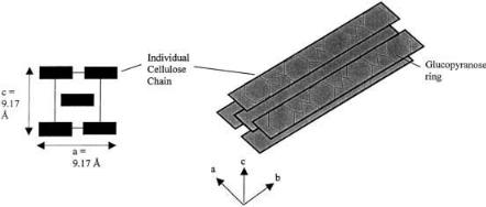
ed in the b 1–4 bond, preventing ring opening. Like starch chains, this gives cellulose chains directionality. Unlike starch, the repeat unit for cellulose is cellobiose, a glucose dimer, not glucose. The b 1–4 bond causes the glucose pyranose structure to align with alternating directionality. X-ray scattering patterns of pure crystalline cellulose from different sources have shown that, in cellulose crystals, chains are arranged in layered sheets. The chains within each sheet tend to align in a parallel fashion (as opposed to anti-parallel) and are linked by hydrogen bonds while they stack using van der Waals forces [9]. Crystalline cellulose can assume different forms. The most common are cellulose I and II. Cellulose I is the native form of cellulose found in cotton, ramie, wood, jute, and flax [10]. The cellulose chains are arranged so that the glucopyranose rings are parallel to the bc plane of the crystal (Fig. 1). This gives distinctly different characteristics between the two crystal faces with exposed glucopyranoside rings and the other faces. The binding of cellobiohydrolase and endoglucanase has been suggested to favor the faces with exposed glucopyranoside rings [11–13]. Highly conserved aromatic amino acid residues that can stack on the exposed rings have been noted between all enzyme binding domains, which are common structural features of carbohydrate binding proteins [14].
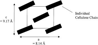
Cellulose II is a “swollen” form of cellulose made by alkali treatment of cellulose I [10]. The cellulose chains are rotated 30° from parallel to the ab crystal face (Fig. 2). Although this 2-chain cell model is correct for most celluloses, some require an 8-chain cell model. However, there is still a great deal of controversy over the nature of crystalline cellulose.
The two most dominating characteristics of cellulose are the specific surface area, SSA, and the crystallinity index, CrI. Specific surface area is defined as the amount of surface area per mass of cellulose. Crystallinty index is the relative amount of crystalline cellulose as opposed to the amount of amorphous cellulose [15]. In biomass, cellulose is closely associated with hemicellulose and lignin. Agricultural residues are composed of 30–40% cellulose, 25–35% hemi-
Fig. 1. Cellulose I crystal. X-ray crystallography in the 1950s solved the basic crystal unit of cellulose I. The axes of the repeat unit (cellobiose) are: a = 8.14 Å, b = 10.3 Å, and c = 9.17 Å. The faces of the glucopyranose rings are parallel to the ab plane of the crystal
Reaction Kinetics, Molecular Action, and Mechanisms of Cellulolytic Proteins |
27 |
Fig. 2. The ac plane of cellulose II crystal. The b plane (not shown) is unchanged (10.3 Å) from cellulose I. However, the orientation of the cellulose chains has shifted from parallel to the ac plane by 30°
cellulose, and 10–15% lignin with the remaining percentage in protein, simple sugars, and minerals [5]. Cellulose is naturally semi-crystalline with regions of high crystallinity averaging approximately 200 glucose residues in length separated by amorphous regions. Due to the large influence of substrate characteristics on cellulose hydrolysis, most research in this area utilizes a more defined form of cellulose as the enzyme substrate. Bacterial micro-crystalline cellulose (BMCC) is the most completely characterized cellulose available. It is recovered as small particles from blue-green algae such as Valonia macrophysa or Acetobacter xylinum. These particles are nearly 100% crystalline cellulose. Due to the cost of bacterial micro-crystalline cellulose, micro-crystalline cellulose derived by acid hydrolysis of wood, such as Avicel (FMC Pharmaceuticals, Philadelphia, Pennsylvania, USA), is more commonly used. Avicel is produced by controlled acid hydrolysis under reflux conditions. Besides micro-crystalline cellulose, Solka Floc (Brown & Co, Berlin, New Hampshire, USA), a mixture of crystalline and amorphous cellulose produced by hammer-milling sulfite wood pulp, is also often used as a substrate for the study of cellulolytic enzyme kinetics. The specific surface area is 3.90 m2/g and the crystallinity index is 77.4 for 270–400 mesh Solka Floc [15].
4
Operational Kinetics and Cellulose Binding Models
Although the Michaelis-Menten model accounts for the kinetic properties of many enzymes, one of the assumptions that this model is built upon does not hold for cellulase systems. Michaelis-Menten modeling assumes a homogeneous system where mass transfer (substrate-to-enzyme and product-from-en- zyme) is not rate limiting, therefore only the catalytic step (enzyme-substrate complex to enzyme-product complex) is the governing rate of the reaction. However, cellulose hydrolysis occurs in a heterogeneous system of two phases. The enzymes are dissolved in an aqueous phase while the cellulose exists in a crystalline solid. To complicate modeling further, cellulose itself is a heteroge-

28 |
N.S. Mosier et al. |
neous polymer that can assume different crystal forms and have varying overall degrees of crystallinity. These physical characteristics all affect hydrolysis rate. Adsorption of the enzyme from the aqueous phase to the solid surface has been shown not to affect significantly the overall rate of hydrolysis [15]. However, adsorption isotherms are important in determining the effective enzyme concentration at the cellulose surface. Therefore, modeling of enzymatic cellulose hydrolysis has been attempted over the years with emphasis on relating hydrolysis kinetics to enzyme adsorption. Although many models have been proposed and used for extrapolating expected conversions from one experimentally measured condition to the next, none are generally applicable for solid substrates. This is due to the heterogeneity of native cellulose, the difficulty in characterizing the important parameters of the substrate, and the complexity of each cellulolytic system in terms of number of different enzymes, synergistic effects, and product inhibition.
5
Development of Kinetic Models
The study of the kinetics of enzymatic hydrolysis of cellulose has been ongoing for nearly 50 years. In 1950, E.T. Reese and his associates suggested that the enzymatic hydrolysis of natural cellulose involves two systems that act consecutively. The original model postulated that the C1 system acts first to make the substrate more accessible to hydrolytic conversion by the Cx system [16]. As protein purification techniques improved and the enzymes composing a cellulolytic system could be isolated, the current hypothesis of three enzyme groups (cellobiohydrolases, endoglucanases, and b-glucosidase) working in concert was proposed. This hypothesis is based on a series of reactions outlined in Table 2.
Because cellobiohydrolases and endoglucanases act on insoluble cellulose, the rate of enzyme adsorption correlates to the rate of enzyme-substrate complex formation. Since b-glucosidase acts upon soluble cellobiose, MichaelisMenten kinetics can be used to model its activity. This is an example of the uti-
Table 2. Cellulolytic enzyme reactions
Enzyme |
Reaction Sequencea |
|
Cellobiohydrolase |
E1+Gx |
´ E1Gx Æ E1G2Gx Æ E1 + G2 + Gx |
Inhibition |
E1+G2 |
´ E1G2 |
Inhibition |
E1+G ´ E1G |
|
Endoglucanase |
Ex+Gx ´ ExGx Æ Ex2Gx Æ Ex+2Gx |
|
Inhibition |
Ex+G ´ ExG |
|
b-Glucosidase |
E+G2 ´ EG2 Æ E2G Æ E+2G |
|
Non-competitive Inhibition |
EG2+G ´ EG2G |
|
Competitive Inhibition |
E+G ´ EG |
|
aE1 = cellobiohydrolase; Ex = endoglucanase; E = b-glucosidase; Gx = cellulose chain; G2 = cellobiose; G = glucose.

Reaction Kinetics, Molecular Action, and Mechanisms of Cellulolytic Proteins |
29 |
Fig. 3. b-Glucosidase inhibition shown by Lineweaver-Burk plot (reproduced from [2]). Lineweaver-Burk plot of kinetic data from “peak 2 cellobiase” (b-glucosidase) at several product inhibitor levels. This is an example of noncompetitive inhibition where the product is not only completing for binding in the active site but also binding to a secondary site on the enzyme that alters the enzyme catalytic ability
lity of operational kinetics. For example (Fig. 3), the initial rate kinetics of b- glucosidase showed initial rate patterns characteristic of non-competitive product inhibition [2]. Another enzyme characteristic, substrate inhibition, was shown in other experiments where enzyme velocity increased with increasing substrate concentration through a maximum.A slow decline in enzyme velocity for the conversion of cellobiose to glucose was noted as the substrate concentration was increased from the maximum velocity value [17]. In addition, cellobiohydrolase and endoglucanase have been shown to be inhibited by their hydrolysis products. Both are inhibited by cellobiose and glucose [5]. One theory is that the inhibition converts the normal enzyme substrate complex to one that is ineffective. Ratios of ineffective bound enzyme to total bound enzyme show an exponential decrease with increased cellobiose concentration and a linear decrease with increased glucose concentration.
One important point to note is that endoglucanases do not directly contribute to the generation of soluble saccharides from insoluble cellulose. Many studies on the kinetics of purified endoglucanases have shown glucose, cellobiose, and cellotriose are produced from small, soluble b 1–4 glucose oligomers. Although endoglucanases hydrolyze b 1–4 glycosidic bonds, they yield insignificant amounts of soluble saccharides from insoluble cellulose. Instead, the primary function of endoglucanases is to increase the number of free insoluble cellulose chain ends. Unfortunately, the number of free chain ends is a
30 |
N.S. Mosier et al. |
parameter that is difficult to measure with any accuracy and must be considered for models based solely on these equations.
Because free cellulose chain ends are so difficult to quantify, other measurable cellulose characteristics have been shown to be factors correlating to hydrolysis susceptibility and rate [15, 18]. These factors affect how well the cellu- lose-binding domain, CBD, adheres to the cellulose and how well the catalytic domain acts on the cellulose. Because of what can and cannot be measured easily, models of enzymatic cellulose hydrolysis combine adsorption behavior with kinetic action.
6
Adsorption Modeling
Many adsorption models have been used for modeling cellulolytic enzyme adsorption, including Langmuir [15], two site Langmuir [19], and combined Langmuir-Freundlich models [20]. One of the earlier models utilized the unpurified T. reesei cellulase system as the basis for a Langmuir adsorption-hy- drolysis model on Solka Floc [15, 18]. By using unpurified cellulase, many problems are inherent in this model. Trichoderma reesei cellulase system consists of seven different enzymes with affinities for adsorbing to cellulose varying from negligible (b-glucosidase) to very strong (cellobiohydrolase I) [21]. Because total protein was measured in these experiments, impurities of other proteins also influence the data.
Endoglucanase activity can increase pores and hydrolysis itself by reducing particle size and changing specific surface area. Changes in crystallinity index due to cellulolytic enzyme activity must also be taken into account. Effect models for these phenomena have not been generated. Another study demonstrated these effects in long-term rate kinetics of hydrolysis again using Solka Floc [18]. It was concluded that the two main factors in the decrease of long-term rates were the change in cellulose structure and the inhibitory effects of products. It was initially found that the specific surface area increased but then leveled off at 24 h (Fig. 4).
Increased specific surface area would thus increase the rate of hydrolysis. The increase in specific surface area is likely due to endoglucanase action of fragmenting the cellulose and opening pores thus increasing the amount of enzymes adsorbed. The crystallinity index, on the other hand, increased greatly in the first 12 h but then slowly decreased but not to its initial level. The crystallinity index increased due to the fast removal of the amorphous cellulose in relation to the crystalline cellulose.
Hydrolysis is the rate-limiting step. The form that the cellulose assumes also greatly affects hydrolysis rate. Initial kinetic rates for cellulolytic enzymes on soluble cellulose (soluble oligosaccharides and carboxymethyl cellulose) are many times greater than on insoluble cellulose [22]. Pretreatment of cellulosic materials is very important for increasing hydrolysis rates as well as overall conversion percentages [23]. Also, the action of the endoglucanase becomes increasingly important by increasing the number of free cellulose chain ends for continued cellobiohydrolase hydrolysis.