

Sodium Channels and Neuronal Hyperexcitability
.pdfNa+ CHANNEL MUTATIONS AND NEUROLOGICAL DISEASE |
73 |
The GAL879-881Q3 mutation in Na + channel SCN2A causes seizures in mice
Muscle and cardiac disease result from mutations that delay the inactivation rate of Na + channels expressed in those tissues, but similar mutations in the neuronal Na + channels have not been described (Bulman 1997). To determine the e¡ect of a delayed inactivation mutation in neurons, we introduced a mutation into the SCN2A cDNA encoding the Na + channel Nav1.2, or brain type 2. We used the neuron-speci¢c enolase promoter to direct expression of the mutant cDNA in transgenic mice. The mutant, GAL879-881QQQ in the cytoplasmic S4^S5 linker of domain 2, has a slightly delayed rate of inactivation and a small increase in persistent current when assayed in Xenopus oocytes (Fig. 1A). The e¡ect on mice expressing the transgene was dramatic (Kearney et al 2001). When the amount of mutant channel was equal to 20% of the wild-type channel, the mice exhibited severe seizures and died by two months of age. When the amount of mutant channel was only 2% of the wild-type channel, as in line Q54, the mice survived for 6^8 months, and had seizures beginning at 3^4 months of age and increasing in frequency and severity. In addition to seizures, the Q54 mice exhibited frozen postures and repetitive behaviours such as grooming or swimming movements (Fig. 2). Tonic/clonic seizures in Q54 mice were accompanied by EEG patterns indicative of hippocampal seizures; these occurred between three and 40 times per day. Na + currents were recorded from hippocampal CA1 neurons by Ted Cummins, who observed persistent currents similar to those generated by the same mutation in the Xenopus oocyte assay (Fig. 1B). A few weeks after the initiation of seizures, neuronal cell loss and gliosis was evident throughout the hippocampus (Kearney et al 2001). The seizure disorder in Q54 mice thus begins with persistent currents in hippocampal neurons, followed typically by initiation
FIG. 1. The GAL879-881QQQ mutation in the S4/S5 linker of domain 2 of SCN2A results in increased persistent current. (A) Kinetics of mutant and wild-type channel in Xenopus oocytes. Normalized current traces during a depolarization to
74 |
MEISLER ET AL |
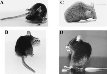
FIG. 2. Appearance of GAL879-881QQQ transgenic mice during seizures. (A) and (B) Focal motor seizures with tonic deviation of the head and body and clonic forelimb movement. (C) This frozen posture was accompanied by spike wave discharges and rhythmic EEG activity. (D) An unusual frozen posture that was maintained for 5 min. Reprinted from Kearney et al (2001) with permission.
of seizures at 2 months, cell loss at 4 months, and death at 6^8 months. Persistent current thus may lead directly to hyperexcitability of the CA1 neurons in Q54 mice.
This mutant line will be a useful model for testing the e¡ectiveness of pharmacological intervention in preventing cell loss, an important goal of epilepsy treatment in human patients. Since other Na + channels are also expressed in hippocampal neurons, it seems likely that delayed inactivation of SCN1A, SCN3A or SCN8A could also result in seizures.
Mutations of SCN1A result in generalized epilepsy in human patients
In 1999, a locus for the human epilepsy syndrome GEFS + (generalized epilepsy with febrile seizures plus) was mapped to the region of chromosome 2q24 that includes the Na + channel genes SCN1A, SCN2A and SCN3A (Baulac et al 1999, Moulard et al 1999). Individuals with GEFS + have a heterogeneous phenotype, with febrile seizures in childhood progressing to several types of generalized seizures in adults. To determine whether the underlying cause of these seizures was similar to that in the Q54 mice, we tested each exon of SCN1A and SCN2A by conformation-sensitive gel electrophoresis (Escayg et al 2000).
Na+ CHANNEL MUTATIONS AND NEUROLOGICAL DISEASE |
75 |
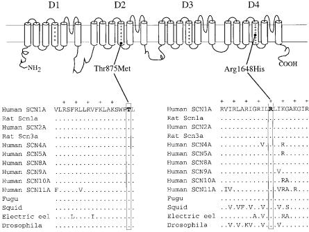
Mutations of SCN1A were detected in both families (Fig. 3). The mutations introduce substitutions for phylogenetically conserved residues in S4 domains of the channel: a threonine to methionine mutation at the cytoplasmic end of D2S4 (T875M) and an arginine to histidine mutation in the R5 residue of D4S4 (R1648H). Al Goldin has investigated the kinetics of the mutant channels in the Xenopus oocyte assay and identi¢ed abnormalities in both mutants (Spampanato et al 2001). These are the ¢rst mutations in a human neuronal Na+ channel a subunit to be associated with neurological disease, and, like the Q54 mice, demonstrate an association between Na + channels and neuronal hyperexcitability.
Mutations of SCN8A cause movement disorders in the mouse
SCN8A is another major Na+ channel that is widely distributed in the CNS and PNS. SCN8A appears to be the only Na + channel located in the nodes of Ranvier in adults, and is also found in cell bodies, dendrites, and presynaptic and postsynaptic membranes (Caldwell et al 2000, Schaller & Caldwell 2000, Krzemien et al 2000). We have been studying four independent spontaneous
FIG. 3. Two SCN1A mutations in patients with familial epilepsy. Thr875 and Arg1648 in D2S4 and D4S4 are conserved in vertebrate and invertebrate channels.
76 |
MEISLER ET AL |
mutations in SCN8A in the mouse, two null alleles, one hypomorph with 2% of the normal level of expression, and one amino acid substitution, A1071T (reviewed in Meisler et al 1997, 2001). Each mutation results in a distinct neurological disorder at the whole animal level and speci¢c changes in Na + currents at the cellular level (Table 1). The phenotypes of the mutant mice have provided insight into the biological role and medical implications of changes in this channel.
Complete loss of SCN8A is lethal
The nodal localization of SCN8A would predict that mice cannot survive without this channel. In fact, null mutants lacking this protein are paralysed and do not survive beyond one month. The timing of death may be explained by an apparent developmental switch in the type of Na + channel located at the node. Between 2 and 3 weeks of age, Na + channels SCN1A (Nav1.1) and SCN2A (Nav1.2) can be detected at the nodes with speci¢c antisera. In the third week of life, during the period of myelination, these channels appear to be replaced by SCN8A (Nav1.6), which is the only channel detected in nodes in adult mice (Caldwell et al 2000). Consistent with this time course, the null mutants lacking SCN8A (med and medtg) can walk at 2 weeks of age but become paralysed by 3 weeks of age. Secondary e¡ects of the null mutations include sprouting of nerve terminals at the neuromuscular junction (Duchen & Stefani 1971) and atrophy of the hind limb muscles.
The SCN8A mutation A1071T in jolting mice results in tremor and ataxia
Transmission at the neuromuscular junction is normal in jolting mice, indicating that this mutation does not impair function at the nodes of Ranvier (Harris & Pollard 1986). Introduction of the jolting mutation into the SCN2A and SCN8A cDNAs by site-directed mutation in Al Goldin’s laboratory resulted in a 10 mV rightward shift in the voltage dependence of activation assayed in Xenopus oocytes (Kohrman et al 1996a, Smith & Goldin 1999). Recordings from cerebellar Purkinje cells isolated from jolting mice by Indira Raman and Bruce Bean (Raman et al 1997) detected major changes in ¢ring patterns. Purkinje cells produce a pattern of complex spikes in response to a single current injection, but fewer spikes were generated in cells with the jolting mutation (Fig. 4). The resurgent current and persistent current which are also characteristic of Purkinje cells were almost completely lost in the mutant (Fig. 4). These data demonstrate that the SCN8A channel is essential for the characteristic ¢ring properties of cerebellar Purkinje cells. Ataxia in jolting mice may be secondary to these cerebellar e¡ects. Spontaneous ¢ring of cartwheel cells of the dorsal cochlear nucleus, which are ontologically related to Purkinje cells, is also reduced in Scn8a mutant mice

TABLE 1 Four independent mutations in the mouse Scn8a gene encoding Nav1.6
|
|
E¡ect on |
Molecular |
Neurological |
Physiological |
Cellular |
Mutant |
Allele |
protein |
mutation |
phenotype |
phenotypes |
phenotypes |
|
|
|
|
|
|
|
med |
Scn8amed |
Null |
Spontaneous |
Paralysis |
Muscle atrophy, slowed |
Sprouting of motor |
|
|
|
line1 insertion |
|
nerve conduction, |
neurons muscle, |
|
|
|
|
|
neurotransmitter release |
atrophy |
|
|
|
|
|
at NMJ function |
|
TgNA4Bs |
Scn8atg |
Null |
Non-targeted |
Paralysis |
Same as above |
Purkinje cells, motor |
|
|
|
transgene |
|
|
neurons, cortical |
|
|
|
insertion |
|
|
pyramidal cells |
medJ |
Scn8amedJ |
Hypomorph |
Splice site |
Weakness, dystonia |
|
P-cells, DCN, |
|
|
(2% level |
deletion |
|
|
motor neurons |
|
|
of transcript) |
|
|
|
|
jolting |
Scn8ajo |
Amino acid |
Nucleotide |
Tremor, ataxia |
|
Purkinje cells but not |
|
|
substitution |
substitution |
|
|
motor neurons |
P-cell, cerebellar Purkinje neurons; DCN, dorsal cochlear nucleus; NMJ, neuromuscular junction.

78 |
MEISLER ET AL |
FIG. 4. Two characteristic currents of cerebellar Purkinje cells are reduced in mice with mutation in Scn8a. Left, resurgent current; right, persistent current. Reprinted from Raman et al (1997) with permission.
(Chen et al 1999). It is interesting that the amino acid substitution A1071T has a major e¡ect on Purkinje neurons but no evident e¡ect on the function of motor neurons.
A modi¢er gene determines survival of mice de¢cient in SCN8A
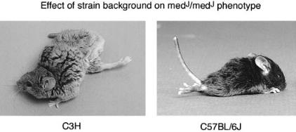
The medJ mutation is a hypomorphic allele of SCN8A. Due to a splice site mutation, the amount of correctly spliced Scn8a transcript in medJ mice is only 2% of the normal level (Kohrman et al 1996b, Sprunger et al 1999). The neurological phenotype of homozygous medJ mice can be changed by crossing the mutation onto di¡erent background strains. On most strains, medJ mice have muscle weakness, persistent tremor and dystonic postures with twisted trunk and limbs that are maintained for several minutes at a time (Fig. 5A). In one strain, C57BL/6, medJ homzygotes have the more severe phenotype of progressive
Na+ CHANNEL MUTATIONS AND NEUROLOGICAL DISEASE |
79 |
FIG. 5. A modi¢er gene alters the clinical consequences of low levels of Scn8a in homozygous medJ mice. The 8 month old homozyogte on strain C3H exhibits muscle weakness and dystonic postures. The 3 week old animal on strain C57BL/6J is paralysed. The di¡erence is caused by the Scnm1 modi¢er locus on chromosome 3.
paralysis and juvenile death, similar to the null mutants (Fig. 5B). This phenomenon is reminiscent of the clinical di¡erences between a¡ected individuals within human pedigrees who share the same mutation but may di¡er at other ‘modi¢er’ genes that in£uence the phenotype. Genetic crosses demonstrated that the di¡erence between C57BL/6J and the other strains is due to allelic di¡erences in one major modi¢er gene, designated SCNM1 (sodium channel modi¢er 1) (Sprunger et al 1999). We are attempting to clone the modi¢er gene, with the goal of identifying a novel protein with a role in Na + channel processing or function.
Conclusions and future prospects
These initial studies of mice with spontaneous or induced mutations in neuronal Na + channel genes suggest that the orthologous human channels are candidate genes for many human disorders. Electrophysiological characterization of neurons isolated from the mutant mice has provided new information about the contributions of these channels in vivo. Future identi¢cation of human disease mutations will be facilitated by the genomic sequence from the Human Genome Project, which permits the design of speci¢c primers used for mutation detection. Mouse models carrying the speci¢c molecular defects found in human patients can be generated using conventional trangenes or targeting in embryonic stem (ES) cells. We anticipate an explosion in the identi¢cation and modelling of neurological disorders resulting from Na + channel mutations during the next few years. The current status of in vivo Na+ channel mutations is summarized in Table 2.
80 |
|
|
|
MEISLER ET AL |
TABLE 2 |
Physiological consequences of Na + channel mutations in vivo |
|||
|
|
|
|
|
Gene |
Protein |
Tissue |
Species |
Major phenotype (mutation) |
|
|
|
|
|
SCN1A |
Nav1.1 |
Neuron |
Human |
GEFS + epilepsy (missense) |
SCN2A |
Nav1.2 |
Neuron |
Mouse |
Brain stem apoptosis (null), |
|
|
|
|
epilepsy (Q54) |
SCN4A |
Nav1.4 |
Muscle |
Human |
Episodic paralysis (missense) |
SCN5A |
Nav1.5 |
Muscle |
Human |
Long QT syndrome (missense) |
SCN7A |
Nax |
Many |
Mouse |
Abnormal salt intake (null) |
SCN8A |
Nav1.6 |
Neuron |
Mouse |
Paralysis (null), ataxia (missense), |
|
|
|
|
dystonia (low expression) |
SCN10A |
Nav1.8 |
Neuron |
Mouse |
Pain sensitivity (null) |
|
|
|
|
|
In addition to monogenic diseases like GEFS + , polymorphic variants of voltage gated Na + channels are likely to contribute to susceptibility to common polygenic disorders including psychiatric diseases. In surveys of human populations, we have detected coding variants in the neuronal Na+ channels, some rare and some common. It will soon be feasible to design a ‘re-sequencing chip’ that would detect all single nucleotide changes in human ion channel genes by hybridization of PCR products against arrayed oligonucleotides. In addition to the medical bene¢ts of identifying disease mutations for diagnosis and development of treatments, biophysical analysis of mutations with impaired in vivo function will expand our understanding of structure/function relationships. Together these two approaches, the detection of human mutations and the generation of mutant mice, will provide a wealth of research material that will lead to better understanding Na + channel function.
Acknowledgements
Our work on the Na + channel genes has been supported by the National Institute of General Medical Sciences (GM24872), the National Institute of Neurological Diseases and Stroke (NS34509), the March of Dimes, and the American Epilepsy Foundation with support from UCB Pharma, Inc.
Note added in proof
During the past year, several mutations responsible for epilepsy have been identi¢ed in SCN1A and SCN2A (reviewed in Meisler 2001).
Na+ CHANNEL MUTATIONS AND NEUROLOGICAL DISEASE |
81 |
References
Baulac S, Gour¢nkel-An I, Picard F et al 1999 A second locus for familial generalized epilepsy with febrile seizures plus maps to chromosome 2q21-q33. Am J Hum Genet 65:1078^1085
Bulman DE 1997 Phenotype variation and newcomers in ion channel disorders. Hum Mol Genet 6:1679^1685
Caldwell JH, Schaller KL, Lasher RS, Peles E, Levinson SR 2000 Sodium channel Na(v)1.6 is localized at nodes of Ranvier, dendrites, and synapses. Proc Natl Acad Sci USA 97: 5616^5620
Chen K, Sprunger LK, Meisler MH, Waller HJ, Godfrey DA 1999 Reduced spontaneous activity in the dorsal cochlear nucleus of Scn8a mutant mice. Brain Res 847:85^89
Duchen LW, Stefani E 1971 Electrophysiological studies of neuromuscular transmission in hereditary ‘motor end-plate disease’ of the mouse. J Physiol 212:535^548
Escayg A, MacDonald BT, Meisler MH et al 2000 Mutations of SCN1A, encoding a neuronal sodium channel, in two families with GEFS + 2. Nat Genet 24:343^345
Harris JB, Pollard SL 1986 Neuromuscular transmission in the murine mutants ‘motor end-plate disease’ and ‘jolting’. J Neurol Sci 76:239^253
Kearney JA, Plummer NW, Smith MR et al 2001 A gain-of-function mutation in the sodium channel gene SCN2A results in seizures and behavioral abnormalities. Neuroscience 102: 307^317
Kohrman DC, Smith MR, Goldin AL, Harris JB, Meisler MH 1996a A missense mutation in the sodium channel gene Scn8a is responsible for cerebellar ataxia in the mouse mutant jolting. J Neurosci 16:5993^5999
Kohrman DC, Harris JB, Meisler MH 1996b Mutation detection in the med and medJ alleles of the sodium channel Scn8a: unusual splicing due to a minor class AT-AC intron. J Biol Chem 271:17576^17581
Krzemien DM, Schaller KL, Levinson SR, Caldwell JH 2000 Immunolocalization of sodium channel isoform NaCh6 in the nervous system. J Comp Neurol 420:70^83
Meisler M 2001 Genetics of epilepsy. Annu Rev Genet 35
Meisler MH, Sprunger LK, Plummer NW, Escayg A, Jones JM 1997 Ion channnel mutations in mouse models of inherited neurological disease. Ann Med 29:569^574
Meisler MH, Kearney J, Escayg A, MacDonald BT, Sprunger LK 2001 Sodium channels and neurological disease: insights from Scn8a mutations in the mouse. Neuroscientist 7:136^145
Moulard B, Guipponi M, Chaigne D, Mouthon D, Buresi C, Malafosse A 1999 Identi¢cation of a new locus for generalized epilepsy with febrile seizures plus (GEFS + ) on chromosome 2q24q33). Am J Hum Genet 65:1396^1400
Raman IM, Sprunger LK, Meisler MH, Bean BP 1997 Altered subthreshold sodium currents and disrupted ¢ring patterns in Purkinje neurons of Scn8a mutant mice. Neuron 19:881^891 Schaller KL, Caldwell JH 2000 Developmental and regional expression of sodium channel
isoform NaCh6 in the rat central nervous system. J Comp Neurol 420:84^97
Smith MR, Goldin AL 1999 A mutation that causes ataxia shifts the voltage-dependence of the Scn8a sodium channel. Neuroreport 10:3027^3031
Spampanato J, Escayg A, Meisler MH, Goldin AL 2001 Functional e¡ects of two voltage-gated sodium channel mutations that cause generalized epilepsy with febrile seizures plus type 2. J Neurosci 21: 7481^7490
Sprunger LK, Escayg A, Tallaksen-Greene S, Albin RL, Meisler MH 1999 Dystonia associated with mutation of the neuronal sodium channel Scn8a and identi¢cation of the modi¢er locus Scnm1 on mouse chromosome 3. Hum Mol Genet 8:471^479
82 |
DISCUSSION |
DISCUSSION |
|
Ptacek: How narrow is the region containing the modi¢er gene? Is it within that 2 Mb?
Meisler: Yes, we are now down to about 1 Mb. I think we can rescue it, because it is a lethal phenotype.
Noebels: Even though it only survives to be 3 weeks old, isn’t the null Nav1.6 mouse living evidence that Nav1.6 can’t be the only nodal Na + channel?
Meisler: We have always wondered why these animals get around much better at 2 weeks of age and then decline by 3 weeks of age. It now seems that between 2 and 3 weeks postnatal in the normal animal there is a replacement of Nav1.2 at the nodes by Nav1.6. So the animals survive until the time that Nav1.6 becomes the major nodal channel at around 3 weeks of age.
Noebels: It would be interesting to expand the expression studies to look at other channels, to see whether there is some attempt at plasticity in these mice that lack the Nav1.6 subunit. The null Nav1.6 mice must still have nerve conduction if they live to be 3 weeks old.
Meisler: I think this is the normal developmental pattern. Other channels like Nav1.2 are present at the nodes in the younger animals, followed by replacement with Nav1.6 between 2 and 3 weeks of age. This doesn’t happen in the null Nav1.6 mice and therefore they die.
Ptacek:In the SCN1A R1648 mutation, it looked like there was a hyperpolarizing shift in the voltage dependence of inactivation.
Goldin: There is an 8 mV negative shift when the a subunit is expressed alone; when it co-expressed with the b subunit, there is no shift.
Ptacek: Then it looked like there was an even smaller rightwards shift of the activation curve.
Goldin: There was no signi¢cant shift. The line is a little bit to the right, but it is not signi¢cantly di¡erent. This was with or without the b1 subunit.
Horn: If there is very fast recovery from inactivation and there was no shift, this means that the entry into the inactivated state must also be very fast.
Goldin: It should mean that, but so far we haven’t looked at this carefully enough. With the two-electrode clamp there were no gross di¡erences in the kinetics of entry into the inactivated state.
Ptacek: Miriam Meisler, early on you said that knockouts won’t have phenotypes. What did you mean by this?
Meisler: What I meant to say was that I don’t think we are going to ¢nd null alleles in human populations, because the e¡ects would be so severe.
Ptacek: From the knockout experiments in mice, it seems the heterozygotes are usually normal.