

Психология
.pdfTo demonstrate the difference between rods and cones in attention to detail, choose a word in this text and focus on it. Do you notice that the words a few inches to the side seem more blurred? This is because the word you are focusing on strikes the detail-oriented cones, while the words surrounding it strike the less-detail-oriented rods, which are located on the periphery.
As you can see in Figure 4.11 "Pathway of Visual Images Through the Thalamus and Into the Visual Cortex", the sensory information received by the retina is relayed through the thalamus to corresponding areas in the visual cortex, which is located in the occipital lobe at the back of the brain. Although the principle of contralateral control might lead you to expect that the left eye would send information to the right brain hemisphere and vice versa, nature is smarter than that. In fact, the left and right eyes each send information to both the left and the right hemisphere, and the visual cortex processes each of the cues separately and in parallel. This is an adaptational advantage to an organism that loses sight in one eye, because even if only one eye is functional, both hemispheres will still receive input from it.
Attributed to Charles Stangor |
Saylor.org |
Saylor URL: http://www.saylor.org/books/ |
181 |

Figure 4.11 Pathway of Visual Images Through the Thalamus and Into the Visual Cortex
The left and right eyes each send information to both the left and the right brain hemisphere.
The visual cortex is made up of specialized neurons that turn the sensations they receive from the optic nerve into meaningful images. Because there are no photoreceptor cells at the place where the optic nerve leaves the retina, a hole or blind spot in our vision is created (see Figure 4.12
Attributed to Charles Stangor |
Saylor.org |
Saylor URL: http://www.saylor.org/books/ |
182 |

"Blind Spot Demonstration"). When both of our eyes are open, we don’t experience a problem because our eyes are constantly moving, and one eye makes up for what the other eye misses. But the visual system is also designed to deal with this problem if only one eye is open—the visual cortex simply fills in the small hole in our vision with similar patterns from the surrounding areas, and we never notice the difference. The ability of the visual system to cope with the blind spot is another example of how sensation and perception work together to create meaningful experience.
Figure 4.12 Blind Spot Demonstration
You can get an idea of the extent of your blind spot (the place where the optic nerve leaves the retina) by trying this demonstration. Close your left eye and stare with your right eye at the cross in the diagram. You should be able to see the elephant image to the right (don’t look at it, just notice that it is there). If you can’t see the elephant, move closer or farther away until you can. Now slowly move so that you are closer to the image while you keep looking at the cross. At one distance (probably a foot or so), the elephant will completely disappear from view because its image has fallen on the blind spot.
Perception is created in part through the simultaneous action of thousands of
feature detector neurons—specialized neurons, located in the visual cortex, that respond to the strength, angles, shapes, edges, and movements of a visual stimulus (Kelsey, 1997; Livingstone & Hubel, 1988). [2] The feature detectors work in parallel, each performing a specialized function. When faced with a red square, for instance, the parallel line feature detectors, the
Attributed to Charles Stangor |
Saylor.org |
Saylor URL: http://www.saylor.org/books/ |
183 |
horizontal line feature detectors, and the red color feature detectors all become activated. This activation is then passed on to other parts of the visual cortex where other neurons compare the information supplied by the feature detectors with images stored in memory. Suddenly, in a flash of recognition, the many neurons fire together, creating the single image of the red square that we experience (Rodriguez et al., 1999). [3]
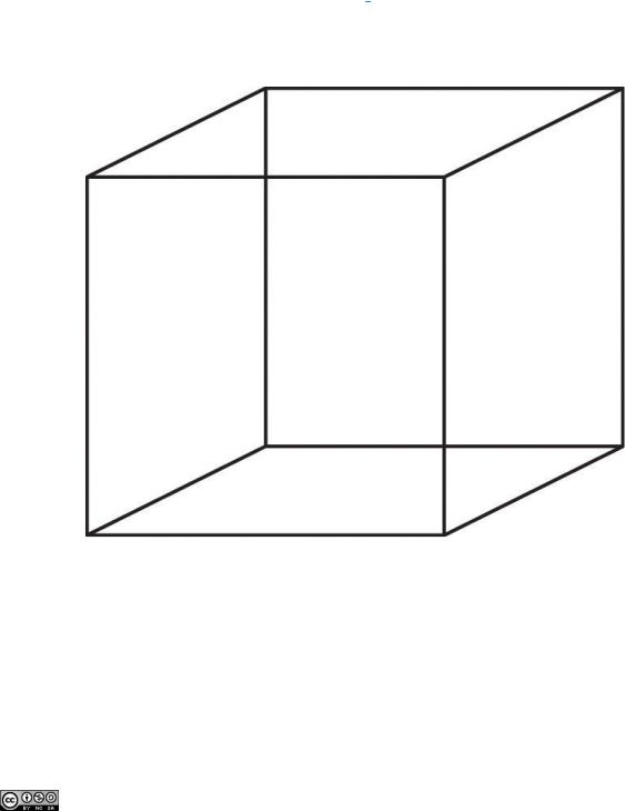
Figure 4.13 The Necker Cube
The Necker cube is an example of how the visual system creates perceptions out of sensations. We do not see a series of lines, but rather a cube. Which cube we see varies depending on the momentary outcome of perceptual processes in the visual cortex.
Some feature detectors are tuned to selectively respond to particularly important objects, for instance, faces, smiles, and other parts of the body (Downing, Jiang, Shuman, & Kanwisher,
Attributed to Charles Stangor |
Saylor.org |
Saylor URL: http://www.saylor.org/books/ |
184 |

2001; Haxby et al., 2001). [4] When researchers disrupted face recognition areas of the cortex using the magnetic pulses of transcranial magnetic stimulation (TMS), people were temporarily unable to recognize faces, and yet they were still able to recognize houses (McKone, Kanwisher, & Duchaine, 2007; Pitcher, Walsh, Yovel, & Duchaine, 2007). [5]
Perceiving Color
It has been estimated that the human visual system can detect and discriminate among 7 million color variations (Geldard, 1972), [6] but these variations are all created by the combinations of the three primary colors: red, green, and blue. The shade of a color, known as hue, is conveyed by the wavelength of the light that enters the eye (we see shorter wavelengths as more blue and longer wavelengths as more red), and we detect brightness from the intensity or height of the wave (bigger or more intense waves are perceived as brighter).
Figure 4.14 Lowand High-Frequency Sine Waves and Lowand High-Intensity Sine Waves and Their Corresponding Colors
Attributed to Charles Stangor |
Saylor.org |
Saylor URL: http://www.saylor.org/books/ |
185 |

Light waves with shorter frequencies are perceived as more blue than red; light waves with higher intensity are
seen as brighter.
In his important research on color vision, Hermann von Helmholtz (1821–1894) theorized that color is perceived because the cones in the retina come in three types. One type of cone reacts primarily to blue light (short wavelengths), another reacts primarily to green light (medium
Attributed to Charles Stangor |
Saylor.org |
Saylor URL: http://www.saylor.org/books/ |
186 |

wavelengths), and a third reacts primarily to red light (long wavelengths). The visual cortex then detects and compares the strength of the signals from each of the three types of cones, creating the experience of color. According to this Young-Helmholtz trichromatic color theory, what color we see depends on the mix of the signals from the three types of cones. If the brain is receiving primarily red and blue signals, for instance, it will perceive purple; if it is receiving primarily red and green signals it will perceive yellow; and if it is receiving messages from all three types of cones it will perceive white.
The different functions of the three types of cones are apparent in people who
experience color blindness—the inability to detect either green and/or red colors. About 1 in 50 people, mostly men, lack functioning in the redor green-sensitive cones, leaving them only able to experience either one or two colors (Figure 4.15).
Figure 4.15
People with normal color vision can see the number 42 in the first image and the number 12 in the second (they are vague but apparent). However, people who are color blind cannot see the numbers at all.
Attributed to Charles Stangor |
Saylor.org |
Saylor URL: http://www.saylor.org/books/ |
187 |

Source: Courtesy
of http://commons.wikimedia.org/wiki/File:Ishihara_11.PNG and http://commons.wikimedia.org/wiki/File:Ishiha
ra_23.PNG.
The trichromatic color theory cannot explain all of human vision, however. For one, although the color purple does appear to us as a mixing of red and blue, yellow does not appear to be a mix of red and green. And people with color blindness, who cannot see either green or red, nevertheless can still see yellow. An alternative approach to the Young-Helmholtz theory, known as the opponent-process color theory, proposes that we analyze sensory information not in terms of three colors but rather in three sets of “opponent colors‖: red-green, yellow-blue, and whiteblack. Evidence for the opponent-process theory comes from the fact that some neurons in the retina and in the visual cortex are excited by one color (e.g., red) but inhibited by another color (e.g., green).
One example of opponent processing occurs in the experience of an afterimage. If you stare at the flag on the left side of Figure 4.16 "U.S. Flag" for about 30 seconds (the longer you look, the better the effect), and then move your eyes to the blank area to the right of it, you will see the afterimage. When we stare at the green stripes, our green receptors habituate and begin to process less strongly, whereas the red receptors remain at full strength. When we switch our gaze, we see primarily the red part of the opponent process. Similar processes create blue after yellow and white after black.
Figure 4.16 U.S. Flag
Attributed to Charles Stangor |
Saylor.org |
Saylor URL: http://www.saylor.org/books/ |
188 |

The presence of an afterimage is best explained by the opponent-process theory of color perception. Stare at the flag for a few seconds, and then move your gaze to the blank space next to it. Do you see the afterimage?
Source: Photo courtesy of Mike Swanson,http://en.wikipedia.org/wiki/File:US_flag(inverted).svg.
The tricolor and the opponent-process mechanisms work together to produce color vision. When light rays enter the eye, the red, blue, and green cones on the retina respond in different degrees, and send different strength signals of red, blue, and green through the optic nerve. The color signals are then processed both by the ganglion cells and by the neurons in the visual cortex (Gegenfurtner & Kiper, 2003). [7]
Perceiving Form
One of the important processes required in vision is the perception of form. German psychologists in the 1930s and 1940s, including Max Wertheimer (1880–1943), Kurt Koffka (1886–1941), and Wolfgang Köhler (1887–1967), argued that we create forms out of their component sensations based on the idea of the gestalt, a meaningfully organized whole. The idea of the gestalt is that the “whole is more than the sum of its parts.‖ Some examples of how gestalt principles lead us to see more than what is actually there are summarized inTable 4.1 "Summary of Gestalt Principles of Form Perception".
Table 4.1 Summary of Gestalt Principles of Form Perception
|
Principle |
Description |
Example |
Image |
|
|
|
|
|
|
|
|
|
We structure |
At right, you may see a vase |
|
|
|
|
input such that |
or you may see two faces, |
|
|
|
|
we always see a |
but in either case, you will |
|
|
|
Figure and |
figure (image) |
organize the image as a |
|
|
|
ground |
against a ground |
figure against a ground. |
Figure 4.1 |
|
|
|
|
|
|
|
|
|
|
|
|
|
Attributed to Charles Stangor |
|
Saylor.org |
|||
Saylor URL: http://www.saylor.org/books/ |
|
189 |
|||

|
Principle |
Description |
Example |
Image |
|
|
|
|
|
|
|
|
|
(background). |
|
|
|
|
|
|
|
|
|
|
|
|
|
Figure 4.1 |
|
|
|
|
You are more likely to see |
|
|
|
|
Stimuli that are |
three similar columns among |
|
|
|
|
similar to each |
the XYXcharacters at right |
|
|
|
|
other tend to be |
than you are to see four |
|
|
|
Similarity |
grouped together. |
rows. |
|
|
|
|
|
|
|
|
|
|
We tend to group |
Do you see four or eight |
|
|
|
Proximity |
nearby figures |
images at right? Principles |
Figure 4.1 |
|
|
|
|
|
|
|
|
|
|
|
|
|
Attributed to Charles Stangor |
|
Saylor.org |
|||
Saylor URL: http://www.saylor.org/books/ |
|
190 |
|||