

Патфиз1 -13 / ACNRMA08_pathology (1)
.pdfNeuropathology Article
Pathology of Intracerebral Haemorrhage
ntracerebral haemorrhage (ICH) is an acute and Ispontaneous extravasation of blood into the brain parenchyma. Bleeding may also extend into the ventricles or subarachnoid space.1 ICH is a subtype of stroke with high morbidity and mortality accounting for about 15% of all deaths from stroke.2 Depending on the underlying cause of bleeding, ICH is classified as either primary or secondary. Primary ICH, which accounts for 78-88% of cases, originates from the spontaneous rupture of small vessels damaged by chronic hypertension or amyloid angiopathy. Secondary ICH occurs in association with trauma, vascular abnormalities, tumours or impaired coagulation.3 The focus of this article is mainly on the aetiology, pathophysiology and pathology of primary ICH with brief
comments on genetics and iatrogenic forms of ICH.
Causes of intracerebral haemorrhage
Table 1 lists the various causes of ICH. Hypertension is still the main cause, being responsible for approximately 55% of cases of ICH.4 Cerebral amyloid angiopathy (CAA) is the other major cause of primary ICH/ lobar haemorrhage in the elderly.5 Post-traumatic haematomas are usually multiple and abut the basal brain surface. Vascular malformations including aneurysms are a common cause of ICH, especially in young normotensive individuals. Coagulopathies may cause multiple or recurrent ICH, sometimes in the presence of systemic bleeding.6
Table 1: Causes of ICH
Primary |
Secondary |
Chronic Hypertension |
Trauma |
Cerebral Amyloid Angiopathy (CAA) |
Ruptured aneurysm |
|
Vascular malformations |
|
Tumours |
|
(primary and metastatic) |
|
Coagulopathies |
|
Drugs or alcohol |
|
Haemorrhagic conversion |
|
of cerebral infarct |
|
Vasculitis |
|
Pregnancy (eclampsia, |
|
venous thrombosis) |
|
Others/ unknown |
|
|
Pathophysiology
ICH was once considered to be a simple, monophasic, rapid bleeding event that stopped quickly as a result of clotting and tamponade.1,3 But ICH has now been shown by serial CT scans to be a dynamic and complex process involving several distinct phases. The two most important new concepts are that firstly, many haemorrhages continue to grow and expand over several hours after the onset of symptoms. Expansion of haematoma: Most haematomas result from rupture of an artery or arteriole. Their expansion is most likely due to continued bleeding from the primary source and to the mechanical disruption of surrounding vessels. Acute hypertension, a local coagulation deficit, or both may be associated with expansion of the haematoma.1,3 Secondly, most of the brain injury and swelling that occurs after ICH is the result of inflammation caused by thrombin and other end products of coagulation.1 Secondary brain injury and oedema: The haematoma initiates oedema and neuronal damage. Oedema typically develops over the first 24-96 hours
and slowly resolves over several weeks. The early oedema is usually secondary to plasma proteins present in the haematoma. Subsequent clotting and complement cascade activation results in disruption of the blood-brain barrier, direct cytotoxicity and more oedema. Lysis of red blood cells with haemoglobin toxicity and formation of free radicals probably accounts for the late onset oedema, which persists for several weeks after the initial haemorrhage.7 Neuronal death in the region around the haematoma is predominantly necrotic, with recent evidence suggesting the presence of programmed cell death (apoptosis).3 Unlike primary tissue injury from the haematoma formation, secondary brain injury and oedema are potential therapeutic targets.5
Pathology
The most common sites of ICH are cerebral hemispheres, basal ganglia, thalamus, brainstem (predominantly the pons), and cerebellum.3 The gross and microscopic changes in the brain depend on the location of ICH, but the general appearance is similar. In the acute stages, the ICH consists of a liquid or semiliquid mass of blood with surrounding oedema. After a few days, the haematoma changes its consistency and adopts a brown colour, while oedema begins to recede. After several months or years, depending on its size, the haematoma becomes a cavity. Small ICH can be reabsorbed almost completely, leaving behind a small linear scar.7
Microscopically, the ICH in its acute stages consists of extravasated well-preserved red blood cells (RBC) without any inflammation. Subsequently, the RBC begins to lyse and neutrophils appear. This is followed by infiltration of macrophages whose main role is to phagocytose blood products and necrotic tissue. The brown discolouration of the slightly older haematomas noted macroscopically is due to the presence of two major haemoglobin-derived pigments, haemosiderin and haematoidin. One of the late events involves proliferation of astrocytes, some containing haemosiderin reflecting their phagocytic activity. The transfer of haemosiderin from macrophages to astrocytes, an event that rarely happens in infants, is common in the adult.7 The pathological evolution of ICH is summarised in Table 2 and a knowledge of this temporal sequence helps in estimating the approximate age of a haematoma in the absence of relevant clinical data.9
Hypertension
Hypertension-related haemorrhages occur typically in deep areas of the brain such as the basal ganglia and thalamus because vessels in these areas are located close to the high pressure of the circle of Willis.9,10 They are less common in the pons, cerebellum or superficial cortex. Pathological studies have shown hyperplasia of the media in artery walls due to proliferation of reactive smooth muscle cells in early hypertension. This has been termed ‘hyperplastic arteriolosclerosis’. Eventually, the smooth muscle cells die and are replaced by collagen fibres which makes the vessel wall brittle and liable for future leakage.10 Miliary or microaneurysms, originally described by Charcot and Bouchard, have been recently questioned as the source of haemorrhage, as complex tortuosities of blood vessels affected by hypertensive changes may be misinterpreted as microaneurysms. Fibrinoid necrosis associated with haemorrhage in acute hypertension appears to be haemostatic, whereas in chronic hypertension it has been suggested that fibrinoid necrosis may be a precursor to haemorrhage.9
Dr Arundhati Chakrabarty, MD, FRCPath, Consultant Neuropathologist in the General Infirmary, Leeds with main interests in the pathology and genetics of CNS tumours.
Dr Aditya Shivane, Specialist Registrar in Neuropathology in General Infirmary, Leeds with interests in CNS tumour and muscle pathology.
Correspondence to:
Dr Aditya Shivane, Dept. of Histopathology, Algernon Firth Building,
The Leeds General Infirmary, Great George Street,
Leeds LS1 3EX, UK.
Tel. +44 (0)113 392 6405 Fax. +44 (0)113 234 7662 Email. Aditya.Shivane@ leedsth.nhs.uk
20 I ACNR • VOLUME 8 NUMBER 1 • MARCH/APRIL 2008
Neuropathology Article
Table 2: Pathological evolution of ICH (Reproduced with kind permission from Nicoll JAR et al. Parenchymal brain haemorrhage. In-Pathology & Genetics-Cerebrovascular Diseases. 2005 ISN, Basel, Switzerland, pp 294-300)
Time |
Macroscopic |
Microscopic |
Seconds |
|
Rupture of blood vessel wall |
Seconds to minutes |
Haematoma formation (bright red acute haemorrhage) |
Extravasation of blood |
Minutes to hours |
Space-occupying effect. Distortion and compression of surrounding tissue, |
Lysis of erythrocytes |
|
(may include: raised intracranial pressure, internal herniations, brain stem compression) |
|
Several hours |
Development of peri-haematoma oedema and ischaemia. Increasing space-occupying effect. |
Oedema, ischaemia, polymorph infiltrate. |
2-3 days to weeks |
Brown coloration of haematoma |
Haemosiderin formation. Phagocytosis by macrophages. |
|
|
Astrocyte hypertrophy and new blood vessel formation |
|
|
at the haematoma margin. |
Weeks to months |
Friable brown clot |
Organisation of the haematoma with phagocytosis of |
|
|
blood and necrotic tissue by macrophages. |
Months to years |
Cavity containing dark blood-stained fluid |
Continued resorption of blood clot |
Months to years |
Cavity containing clear fluid resembling CSF with brown haemosiderin-stained cavity wall |
Cavity lined by hyperplastic and hypertrophic glial cells; |
|
|
residual macrophages and haemosiderin |
|
|
|
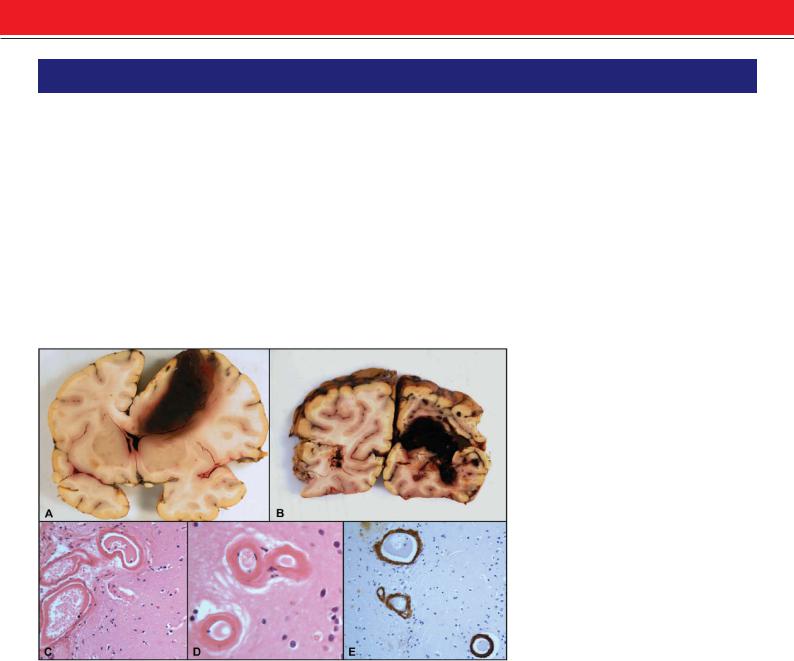
Figure 1: Cerebral amyloid angiopathy. Coronal slice showing a large superficial haemorrhage in the right frontal lobe with extension into the subarachnoid space and ventricle (A). Same case as in (A) showing also a large hematoma in the right occipital lobe (B). Photomicrograph showing thickened hyalinized blood vessels in the leptomeninges (C) and in the brain parenchyma
(D) (H&E x200). Immunohistochemistry for beta-amyloid showing strong positivity in the blood vessel walls (E) (x200).
Cerebral amyloid angiopathy (CAA) |
Genetics |
and amphetamines; and those due to therapeutic manipulations including anticoagulation, fibrinolytic therapy and carotid endarterectomy. Heavy alcohol consumption predisposes to ICH by inducing hypertension (particularly acutely after a binge), by its inhibiting effects on platelet function or by causing liver dysfunction. Cocaine and amphetamines are sympathomimetic agents known to have effects on pulse and blood pressure. Cocaine also induces a cerebral vasculitis, which may be partly responsible for ICH associated with its use. Most patients with drug-related ICH have an associated vascular lesion such as an aneurysm or an arteriovenous malformation.4,7
Conclusions
The prognosis for patients suffering from ICH is still poor. Size and location of the haemorrhage, together with age and presence of hypertension, represent the main prognostic indicators.6 The role of oedema, ischaemia, mass effect, direct cellular toxicity, inflammation, and apoptosis are being evaluated in various experimental studies of ICH.5 The future may see the translation of this basic information into clinical trials and also lead to the development of highly effective treatments.
CAA results from the deposition of the insoluble amyloid-beta (Aβ) peptides (derived from amyloid precursor protein/APP) in the walls of leptomeningeal and cortical arteries, arterioles and capillaries (Figure 1). Replacement of smooth muscle cells in the artery walls by Aβ increases exponentially with age, making the artery less compliant. CAA-associated haemorrhage in some cases may be related to an interaction with other risk factors, notably hypertension. Haemorrhages are superficial, lobar and quite commonly breach the cortical surface resulting in secondary subarachnoid haemorrhage; they can also be multiple or recurrent. Specific vasculopathic complications occur more commonly with CAA-associated haemorrhage than in CAA without haemorrhage. Such complications include concentric splitting of the vessel wall (‘double barrel’ appearance), fibrinoid necrosis, microaneurysms, stenotic lumina, microhaemorrhages and cortical infarction.9,10
Most genetic studies have focussed on the apolipoprotein E alleles ε4 and ε2 that are associated with predisposition to ICH, particularly with CAA and lobar haemorrhages. McCarron and colleagues9 found that patients with ICH who carried the apolipoprotein E ε4 allele had a greater hospital mortality rate than non-carri- ers (40% Vs 25%). The apolipoprotein E ε4 allele is also associated with poor outcome after traumatic brain injury, but there is no such association for ischaemic stroke. Polymorphisms in various other genes such as methylenetetrahydrofolate reductase, angiotensinconverting enzyme, and alpha1-antichy- motrypsin have been found to be associated with ICH in other fairly small studies.2
Iatrogenic forms of ICH
The iatrogenic forms of ICH can be broadly divided into two groups: those due to selfadministration of substances with toxic effects which include mainly alcohol, drugs like cocaine
References
1.Mayer SA, Rincon F. Treatment of intracerebral haemorrhage. Lancet Neurol 2005;4:662-72.
2.Xi G, Keep RF, Hoff JT. Mechanisms of brain injury after intracerebral haemorrhage. Lancet Neurol 2006;5:53-63.
3.Qureshi AI, Tuhrim S, Broderick JP, Batjer HH, Hondo H, Hanley DF. Spontaneous intracerebral hemorrhage. N Engl J Med 2001;344(19):1450-60.
4.MacKenzie JM. Intracerebral haemorrhage. J Clin Pathol 1996;49:360-4.
5.Fewel ME, Thompson BG, Hoff JT. Spontaneous intracerebral hemorrhage: a review. Neurosurg Focus 2003;15:1-16.
6.Sacco S, Marini C, Carolei A. Medical treatment of intracerebral hemorrhage. Neurol Sci 2004;24:S6-9.
7.Kase CS, Caplan LR. Intracerebral hemorrhage. Boston: Butterworth-Heinemann, 1994.
8.Skidmore CT, Andrefsky J. Spontaneous intracerebral hemorrhage: epidemiology, pathophysiology, and medical management. Neurosurg Clin N Am 2002;13:281-8.
9.McCarron MO, Cohen NR, Nicoll JAR. Parenchymal brain hemorrhage. In-Pathology & GeneticsCerebrovascular Diseases. 2005 ISN, Basel, Switzerland, pp 294-300.
10.Sutherland GR, Auer RN. Primary intracerebral hemorrhage. J Clin Neurosci 2006;13:511-17.
ACNR • VOLUME 8 NUMBER 1 • MARCH/APRIL 2008 I 21