

Реваскуляризация 2014, ЕОК
.pdfEuropean Heart Journal Advance Access published September 10, 2014
European Heart Journal |
ESC/EACTS GUIDELINES |
doi:10.1093/eurheartj/ehu278 |
|
|
|
2014 ESC/EACTS Guidelines on myocardial revascularization
The Task Force on Myocardial Revascularization of the European Society of Cardiology (ESC) and the European Association
for Cardio-Thoracic Surgery (EACTS)
Developed with the special contribution of the European Association of
Percutaneous Cardiovascular Interventions (EAPCI)
Authors/Task Force members: Stephan Windecker* (ESC Chairperson) (Switzerland), Philippe Kolh* (EACTS Chairperson) (Belgium), Fernando Alfonso (Spain), Jean-Philippe Collet (France), Jochen Cremer (Germany), Volkmar Falk (Switzerland), Gerasimos Filippatos (Greece), Christian Hamm (Germany), Stuart J. Head
(The Netherlands), Peter Ju¨ni (Switzerland), A. Pieter Kappetein (The Netherlands), Adnan Kastrati (Germany), Juhani Knuuti (Finland), Ulf Landmesser (Switzerland), Gu¨nther Laufer (Austria), Franz-Josef Neumann (Germany), Dimitrios J. Richter (Greece), Patrick Schauerte (Germany), Miguel Sousa Uva (Portugal),
Giulio G. Stefanini (Switzerland), David Paul Taggart (UK), Lucia Torracca (Italy), Marco Valgimigli (Italy), William Wijns (Belgium), and Adam Witkowski (Poland).
* First and corresponding authors: Stephan Windecker, Cardiology, Bern University Hospital, Freiburgstrasse 4, CH-3010 Bern, Switzerland. Tel: +41 31 632 47 70; Fax: +41 31 632 42 99; Email: stephan.windecker@insel.ch
Philippe Kolh, Cardiovascular Surgery Department, University Hospital (CHU, ULg) of Liege, Sart Tilman B 35, 4000 Liege, Belgium. Tel: +32 4 366 7163; Fax: +32 4 366 7164; Email: philippe.kolh@chu.ulg.ac.be
National Cardiac Societies document reviewers: listed in Addenda
The content of these European Society of Cardiology (ESC) Guidelines has been published for personal and educational use only. No commercial use is authorized. No part of the ESC Guidelines may be translated or reproduced in any form without written permission from the ESC. Permission can be obtained upon submission of a written request to Oxford University Press, the publisher of the European Heart Journal and the party authorized to handle such permissions on behalf of the ESC.
‡ Other ESC entities having participated in the development of this document:
Associations: Acute Cardiovascular Care Association (ACCA), European Association for Cardiovascular Prevention & Rehabilitation (EACPR), European Association of Cardiovascular Imaging (EACVI), European Heart Rhythm Association (EHRA), Heart Failure Association of the ESC (HFA).
Working groups: Working Group on Cardiac Cellular Electrophysiology, Working Group on Cardiovascular Magnetic Resonance, Working Group on Cardiovascular Pharmacology and Drug Therapy, Working Group on Cardiovascular Surgery, Working Group on Coronary Pathophysiology and Microcirculation, Working Group on Nuclear Cardiology and Cardiac Computed Tomography, Working Group on Peripheral Circulation, Working Group on Thrombosis, Working Group on Valvular Heart Disease.
Councils: Council for Cardiology Practice, Council on Cardiovascular Primary Care, Council on Cardiovascular Nursing and Allied Professions.
Disclaimer 2014: The ESC Guidelines represent the views of the ESC and were produced after careful consideration of the scientific and medical knowledge and the evidence available at the time of their dating.
The ESC is not responsible in the event of any contradiction, discrepancy and/or ambiguity between the ESC Guidelines and any other official recommendations or guidelines issued by the relevant public health authorities, in particular in relation to good use of healthcare or therapeutic strategies. Health professionals are encouraged to take the ESC Guidelines fully into account when exercising their clinical judgment as well as in the determination and the implementation of preventive, diagnostic or therapeutic medical strategies; however, the ESC Guidelines do not in any way whatsoever override the individual responsibility of health professionals to make appropriate and accurate decisions in consideration of each patient’s health condition and, where appropriate and/or necessary, in consultation with that patient and the patient’s care provider. Nor do the ESC Guidelines exempt health professionals from giving full and careful consideration to the relevant official, updated recommendations or guidelines issued by the competent public health authorities, in order to manage each patient’s case in light of the scientifically accepted data pursuant to their respective ethical and professional obligations. It is also the health professional’s responsibility to verify the applicable rules and regulations relating to drugs and medical devices at the time of prescription.
& The European Society of Cardiology 2014. All rights reserved. For permissions please email: journals.permissions@oup.com.
2014 23, September on guest by org/.oxfordjournals.http://eurheartj from Downloaded

Page 2 of 100 |
ESC/EACTS Guidelines |
|
|
ESC Committee for Practice Guidelines: Jose Luis Zamorano (Chairperson) (Spain), Stephan Achenbach (Germany), Helmut Baumgartner (Germany), Jeroen J. Bax (Netherlands), He´ctor Bueno (Spain), Veronica Dean (France), Christi Deaton (UK), Çetin Erol (Turkey), Robert Fagard (Belgium), Roberto Ferrari (Italy), David Hasdai (Israel), Arno W. Hoes (Netherlands), Paulus Kirchhof (Germany/UK), Juhani Knuuti (Finland), Philippe Kolh (Belgium), Patrizio Lancellotti (Belgium), Ales Linhart (Czech Republic), Petros Nihoyannopoulos (UK), Massimo F. Piepoli (Italy), Piotr Ponikowski (Poland), Per Anton Sirnes (Norway), Juan Luis Tamargo (Spain), Michal Tendera (Poland), Adam Torbicki (Poland), William Wijns (Belgium), and Stephan Windecker (Switzerland).
EACTS Clinical Guidelines Committee: Miguel Sousa Uva (Chairperson) (Portugal).
Document reviewers: Stephan Achenbach (ESC Review Coordinator) (Germany), John Pepper (EACTS Review Coordinator) (UK), Anelechi Anyanwu (USA), Lina Badimon (Spain), Johann Bauersachs (Germany),
Andreas Baumbach (UK), Farzin Beygui (France), Nikolaos Bonaros (Austria), Marco De Carlo (Italy), Christi Deaton (UK), Dobromir Dobrev (Germany), Joel Dunning (UK), Eric Eeckhout (Switzerland), Stephan Gielen (Germany), David Hasdai (Israel), Paulus Kirchhof (UK/Germany), Heyman Luckraz (UK), Heiko Mahrholdt (Germany),
Gilles Montalescot (France), Domenico Paparella (Italy), Ardawan J. Rastan (Germany), Marcelo Sanmartin (Spain), Paul Sergeant (Belgium), Sigmund Silber (Germany), Juan Tamargo (Spain), Jurrien ten Berg (Netherlands), Holger Thiele (Germany), Robert-Jan van Geuns (Netherlands), Hans-Otto Wagner (Germany), Sven Wassmann (Germany), Olaf Wendler (UK), and Jose Luis Zamorano (Spain).
The disclosure forms of the authors and reviewers are available on the ESC website www.escardio.org/guidelines
Online publish-ahead-of-print 29 August 2014
- - - - - - - - - - - - - - - - - - - - - - - - - - - - - - - - - - - - - - - - - - - - - - - - - - - - - - - - - - - - - - - - - - - - - - - - - - -- - - - - - - - - - - - - - - - - - - - - - - - - - - - - - - - - - - - - - - - - - - - - - - - - - - - - - - - - - - - - - - - - - - - - - - - - - -
Keywords |
Acute coronary syndromes † Bare-metal stents † Coronary artery bypass grafting † Coronary artery disease † |
|
Drug-eluting stents † EuroSCORE † Guidelines † Heart Team † Myocardial infarction † Myocardial |
|
ischaemia † Myocardial revascularization † Medical therapy † Percutaneous coronary intervention † |
|
Recommendation † Revascularisation † Risk stratification † Stents † Stable angina † Stable coronary artery |
|
disease † ST-segment elevation myocardial infarction † SYNTAX score |
|
|
Table of Contents
Abbreviations and acronyms . . . . . . . . . . . . . . . . . . . . . . . . |
2544 |
||
1. |
Preamble . . . . . . . . . . . . . . . . . . . . . . . . . . . . . . . . . . . |
2547 |
|
2. |
Introduction . . . . . . . . . . . . . . . . . . . . . . . . . . . . . . . . . |
2548 |
|
3. |
Scores and risk stratification . . . . . . . . . . . . . . . . . . . . . . . |
2549 |
|
4. |
Process for decision-making and patient information . . . . . . . |
2552 |
|
|
4.1 |
Patient information and informed consent . . . . . . . . . . |
2552 |
|
4.2 |
Multidisciplinary decision-making (Heart Team) . . . . . . . |
2553 |
|
4.3 |
Timing of revascularization and ad hoc percutaneous |
|
|
coronary intervention . . . . . . . . . . . . . . . . . . . . . . . . . . |
2553 |
|
5. |
Strategies for diagnosis: functional testing and imaging . . . . . . |
2554 |
|
|
5.1 |
Non-invasive tests . . . . . . . . . . . . . . . . . . . . . . . . . |
2554 |
|
5.2 |
Invasive tests . . . . . . . . . . . . . . . . . . . . . . . . . . . . . |
2554 |
|
5.3 |
Detection of myocardial viability . . . . . . . . . . . . . . . . |
2555 |
6. |
Revascularization for stable coronary artery disease . . . . . . . |
2555 |
|
|
6.1 |
Rationale for revascularization . . . . . . . . . . . . . . . . . . |
2555 |
|
6.2 |
Evidence basis for revascularization . . . . . . . . . . . . . . . |
2555 |
|
6.2.1 Revascularization with the use of percutaneous |
|
|
|
coronary intervention . . . . . . . . . . . . . . . . . . . . . . . . |
2555 |
|
|
6.2.2 Percutaneous coronary intervention with drug-eluting |
|
|
|
stents vs. bare-metal stents . . . . . . . . . . . . . . . . . . . . . |
2557 |
|
|
6.2.3 Revascularization with the use of coronary artery |
|
|
|
bypass grafting . . . . . . . . . . . . . . . . . . . . . . . . . . . . . |
2557 |
|
|
6.3 |
Percutaneous coronary intervention vs. coronary artery |
|
|
bypass grafting . . . . . . . . . . . . . . . . . . . . . . . . . . . . . . . |
2558 |
|
6.3.1 |
Proximal left anterior descending coronary artery |
|
|
disease . . . . . . . . . . . . . . . . . . . . . . . . . . . . . . . . . . |
2558 |
||
6.3.2 |
Left main coronary artery disease . . . . . . . . . . . . . |
2558 |
|
6.3.3 |
Three-vessel coronary artery disease . . . . . . . . . . |
2560 |
|
7. Revascularization in non-ST-segment elevation acute coronary |
|
||
syndromes . |
. . . . . . . . . . . . . . . . . . . . . . . . . . . . . . . . . . . |
2561 |
|
7.1 |
Early invasive vs. conservative strategy . . . . . . . . . . . . . |
2561 |
|
7.2 |
Timing of angiography and intervention . . . . . . . . . . . . |
2562 |
|
7.3 |
Type of revascularization . . . . . . . . . . . . . . . . . . . . . |
2562 |
|
7.3.1 |
Coronary artery bypass surgery . . . . . . . . . . . . . . |
2563 |
|
7.3.2 |
Percutaneous coronary intervention . . . . . . . . . . . |
2563 |
|
8. Revascularization in ST-segment elevation myocardial infarction 2564
8.1 |
Time delays . . . . . . . . . . . . . . . . . . . . . . . . . . . . . . |
2564 |
8.2 |
Selection of reperfusion strategy . . . . . . . . . . . . . . . . |
2564 |
8.3 |
Primary percutaneous coronary intervention . . . . . . . . |
2565 |
8.4 |
Fibrinolysis . . . . . . . . . . . . . . . . . . . . . . . . . . . . . . |
2567 |
8.5 |
Secondary percutaneous coronary intervention . . . . . . . |
2567 |
8.6 |
Coronary artery bypass surgery . . . . . . . . . . . . . . . . . |
2568 |
9. Revascularization in patients with heart failure and cardiogenic |
|
|
shock . |
. . . . . . . . . . . . . . . . . . . . . . . . . . . . . . . . . . . . . . |
2568 |
9.1 |
Chronic heart failure . . . . . . . . . . . . . . . . . . . . . . . . |
2568 |
9.1.1 Revascularization . . . . . . . . . . . . . . . . . . . . . . . |
2568 |
|
9.1.2 Myocardial viability and revascularization . . . . . . . . |
2568 |
|
9.1.3 Ventricular reconstruction . . . . . . . . . . . . . . . . . |
2569 |
|
2014 23, September on guest by org/.oxfordjournals.http://eurheartj from Downloaded

ESC/EACTS Guidelines |
Page 3 of 100 |
|
|
|
9.2 |
Cardiogenic shock . . . . . . . . . . . . . . . . . . . . . . . . . |
2569 |
|
|
9.2.1 |
Revascularization . . . . . . . . . . . . . . . . . . . . . . . |
2569 |
|
|
9.2.2 |
Mechanical circulatory support . . . . . . . . . . . . . . |
2570 |
|
|
9.2.3 |
Right ventricular failure . . . . . . . . . . . . . . . . . . . |
31 |
|
|
9.2.4 |
Mechanical complications . . . . . . . . . . . . . . . . . . |
2571 |
|
10. Revascularization in patients with diabetes . . . . . . . . . . . . . |
2571 |
|||
|
10.1 |
Evidence for myocardial revascularization . . . . . . . . . . |
2571 |
|
|
10.1.1 |
Stable coronary artery disease . . . . . . . . . . . . . . |
2572 |
|
|
10.1.2 Acute coronary syndromes . . . . . . . . . . . . . . . . |
2572 |
||
|
10.2 |
Type of myocardial revascularization . . . . . . . . . . . . . |
2572 |
|
|
10.2.1. Randomized clinical trials . . . . . . . . . . . . . . . . . |
2572 |
||
|
10.2.2 |
Meta-analyses . . . . . . . . . . . . . . . . . . . . . . . . . |
2574 |
|
|
10.3 |
Revascularization with the use of percutaneous coronary |
|
|
|
intervention . . . . . . . . . . . . . . . . . . . . . . . . . . . . . . . . |
2574 |
||
|
10.4 |
Revascularization with the use of coronary artery bypass |
|
|
|
grafting |
. . . . . . . . . . . . . . . . . . . . . . . . . . . . . . . . . . . |
2574 |
|
|
10.5 |
Antithrombotic pharmacotherapy . . . . . . . . . . . . . . |
2574 |
|
|
10.6 |
Anti-diabetic medications . . . . . . . . . . . . . . . . . . . . |
2575 |
|
11. |
Revascularization in patients with chronic kidney disease . . . . |
2575 |
||
|
11.1 |
Evidence-base for revascularization . . . . . . . . . . . . . . |
2575 |
|
|
11.1.1 |
Patients with moderate chronic kidney disease . . . . |
2576 |
|
|
11.1.2 |
Patients with severe chronic kidney disease and end- |
|
|
|
stage renal disease or in haemodialysis . . . . . . . . . . . . . . |
2576 |
||
|
11.2 |
Prevention of contrast-induced nephropathy . . . . . . . . |
2576 |
|
12. |
Revascularization in patients requiring valve interventions . . . |
2577 |
||
|
12.1 |
Primary indication for valve interventions . . . . . . . . . . |
2577 |
|
|
12.2 |
Primary indication for coronary revascularization . . . . . |
2578 |
|
13. |
Associated carotid/peripheral artery disease . . . . . . . . . . . |
2579 |
||
|
13.1 |
Associated coronary and carotid artery disease . . . . . . |
2579 |
|
|
13.1.1 |
Risk factors for stroke associated with myocardial |
|
|
|
revascularization . . . . . . . . . . . . . . . . . . . . . . . . . . . . |
2579 |
||
|
13.1.2 Preventive measures to reduce the risk of stroke after |
|
||
|
coronary artery bypass grafting . . . . . . . . . . . . . . . . . . |
2579 |
||
|
13.1.3 |
Carotid revascularization in patients scheduled |
|
|
|
for myocardial revascularization . . . . . . . . . . . . . . . . . . |
2579 |
||
|
13.1.4 |
Type of revascularization in patients with associated |
|
|
|
carotid and coronary artery disease . . . . . . . . . . . . . . . |
2580 |
||
|
13.2 |
Associated coronary and peripheral arterial disease . . . |
41 |
|
14. |
Repeat revascularization and hybrid procedures . . . . . . . . . |
2581 |
||
|
14.1 |
Early graft failure . . . . . . . . . . . . . . . . . . . . . . . . . . |
2581 |
|
|
14.2 |
Disease progression and late graft failure . . . . . . . . . . |
2582 |
|
|
14.3 |
Acute percutaneous coronary intervention failure . . . .2583 |
||
|
14.4 |
Repeat percutaneous coronary intervention . . . . . . . . |
2583 |
|
|
14.5 |
Hybrid procedures . . . . . . . . . . . . . . . . . . . . . . . . |
2583 |
|
15. Arrhythmias . . . . . . . . . . . . . . . . . . . . . . . . . . . . . . . . |
2585 |
|||
|
15.1 |
Ventricular arrhythmias . . . . . . . . . . . . . . . . . . . . . |
2585 |
|
|
15.1.1 |
Revascularization for prevention of sudden cardiac |
|
|
|
death in patients with stable coronary artery disease and |
|
||
|
reduced left ventricular function . . . . . . . . . . . . . . . . . . |
2585 |
||
15.1.2Revascularization for treatment of electrical storm .2585
15.1.3Revascularization after out-of-hospital cardiac arrest 2585
15.2 Atrial arrhythmias . . . . . . . . . . . . . . . . . . . . . . . . .2585 15.2.1 Atrial fibrillation complicating percutaneous
coronary intervention . . . . . . . . . . . . . . . . . . . . . . . .2585 15.2.2 Atrial fibrillation complicating coronary artery bypass grafting . . . . . . . . . . . . . . . . . . . . . . . . . . . . . . . . . .2585 15.2.3 Post-operative atrial fibrillation and stroke risk . . . .2586
|
15.3 |
Concomitant surgical procedures for atrial fibrillation or |
|
|
|
stroke treatment . . . . . . . . . . . . . . . . . . . . . . . . . . . . . |
2586 |
||
16. Procedural aspects of coronary artery bypass grafting . . . . . . |
2587 |
|||
|
16.1 |
Pre-operative management . . . . . . . . . . . . . . . . . . . |
2587 |
|
|
16.2 |
Blood management . . . . . . . . . . . . . . . . . . . . . . . . |
2587 |
|
|
16.2.1 |
Blood salvage interventions . . . . . . . . . . . . . . . . |
2587 |
|
|
16.2.2 |
Pharmacological strategies . . . . . . . . . . . . . . . . . |
2587 |
|
|
16.2.3 |
Blood transfusion . . . . . . . . . . . . . . . . . . . . . . |
2587 |
|
|
16.3 |
Surgical procedures . . . . . . . . . . . . . . . . . . . . . . . . |
2587 |
|
|
16.3.1 Conduit harvest . . . . . . . . . . . . . . . . . . . . . . . . |
2587 |
||
|
16.3.2 |
Coronary vessel . . . . . . . . . . . . . . . . . . . . . . . |
2587 |
|
|
16.3.3 |
Completeness of revascularization . . . . . . . . . . . |
2587 |
|
|
16.3.4 |
Construction of central anastomosis . . . . . . . . . . |
2587 |
|
|
16.3.5 |
Bypass grafts . . . . . . . . . . . . . . . . . . . . . . . . . |
2588 |
|
|
16.3.6 On-pump and off-pump procedures . . . . . . . . . . |
2589 |
||
|
16.3.7 |
Minimally invasive procedures . . . . . . . . . . . . . . |
2589 |
|
|
16.4 |
Reporting perioperative outcome . . . . . . . . . . . . . . . |
2589 |
|
17. |
Procedural aspects of percutaneous coronary intervention . . |
2589 |
||
|
17.1 |
Percutaneous coronary intervention devices . . . . . . . . |
2589 |
|
|
17.1.1 |
Balloon angioplasty . . . . . . . . . . . . . . . . . . . . . |
2589 |
|
|
17.1.2 |
Coronary stents . . . . . . . . . . . . . . . . . . . . . . . |
2589 |
|
|
17.1.3 |
Bioresorbable stents . . . . . . . . . . . . . . . . . . . . |
2590 |
|
|
17.1.4 |
Drug-coated balloons . . . . . . . . . . . . . . . . . . . . |
2590 |
|
|
17.1.5 |
Other devices . . . . . . . . . . . . . . . . . . . . . . . . |
2590 |
|
|
17.2 |
Adjunctive invasive diagnostic tools . . . . . . . . . . . . . . |
2590 |
|
|
17.2.1 |
Intravascular ultrasound . . . . . . . . . . . . . . . . . . |
2590 |
|
|
17.2.2 Optical coherence tomography . . . . . . . . . . . . . |
2592 |
||
|
17.2.3 |
Pressure-derived fractional flow reserve . . . . . . . . |
2593 |
|
|
17.3 |
Specific lesion subsets . . . . . . . . . . . . . . . . . . . . . . |
2593 |
|
|
17.3.1 |
Bifurcation stenosis . . . . . . . . . . . . . . . . . . . . . |
2593 |
|
|
17.3.2 |
Chronic total coronary occlusion . . . . . . . . . . . . |
2593 |
|
|
17.3.3 |
Ostial lesions . . . . . . . . . . . . . . . . . . . . . . . . . |
2594 |
|
18. |
Antithrombotic treatments . . . . . . . . . . . . . . . . . . . . . . |
2594 |
||
|
18.1 |
Percutaneous coronary intervention in stable coronary |
|
|
|
artery disease . . . . . . . . . . . . . . . . . . . . . . . . . . . . . . . |
2594 |
||
|
18.1.1 |
Oral antiplatelet therapy . . . . . . . . . . . . . . . . . . |
2594 |
|
|
18.1.2 |
Intravenous antiplatelet therapy . . . . . . . . . . . . . |
2595 |
|
|
18.1.3 |
Anticoagulation . . . . . . . . . . . . . . . . . . . . . . . |
2595 |
|
|
18.2 |
Non-ST-segment elevation acute coronary syndrome . . |
2596 |
|
|
18.2.1 |
Oral antiplatelet therapy . . . . . . . . . . . . . . . . . . |
2596 |
|
|
18.2.2 |
Intravenous antiplatelet therapy . . . . . . . . . . . . . |
2597 |
|
|
18.2.3 |
Anticoagulation . . . . . . . . . . . . . . . . . . . . . . . |
2597 |
|
|
18.3 |
ST-segment elevation myocardial infarction . . . . . . . . |
2598 |
|
|
18.3.1 |
Oral antiplatelet therapy . . . . . . . . . . . . . . . . . . |
2598 |
|
|
18.3.2 |
Intravenous antiplatelet therapy . . . . . . . . . . . . . |
2599 |
|
|
18.3.3 |
Anticoagulation . . . . . . . . . . . . . . . . . . . . . . . |
2599 |
|
|
18.4 |
Points of interest and special conditions . . . . . . . . . . . |
2601 |
|
|
18.4.1 |
Pre-treatment with P2Y12 inhibitors . . . . . . . . . . |
2601 |
|
|
18.4.2 |
Intravenous P2Y12 inhibitors . . . . . . . . . . . . . . . |
2601 |
|
|
18.4.3 |
Anticoagulation after percutaneous coronary |
|
|
|
intervention in acute coronary syndrome patients . . . . . . |
2602 |
||
|
18.4.4 |
Anticoagulation during percutaneous coronary |
|
|
|
intervention in patients on oral anticoagulation . . . . . . . . |
2602 |
||
|
18.4.5 |
Antithrombotic therapy after percutaneous |
|
|
|
coronary intervention in patients requiring oral |
|
||
|
anticoagulation . . . . . . . . . . . . . . . . . . . . . . . . . . . . . |
2603 |
||
2014 23, September on guest by org/.oxfordjournals.http://eurheartj from Downloaded

Page 4 of 100 |
ESC/EACTS Guidelines |
|
|
18.4.6 |
Duration of dual antiplatelet therapy after |
|
percutaneous coronary intervention . . . . . . . . . . . |
. . . .2604 |
|
18.4.7 Drug interactions: a clopidogrel-related topic |
. . . .2605 |
|
18.4.8 |
Renal dysfunction . . . . . . . . . . . . . . . . . . . |
. . .2605 |
18.4.9 |
Surgery in patients on dual antiplatelet therapy |
. . .2606 |
18.4.10Antiplatelet therapy monitoring and genetic testing 2608
18.4.11Patients with hypersensitivity to acetylsalicylic acid 2608
18.4.12 Heparin-induced thrombocytopaenia . . . . . . . . .2608 19. Volume–outcome relationship for revascularization
procedures . . . . . . . . . . . . . . . . . . . . . . . . . . . . . . . . . . .2609 19.1 Coronary artery bypass grafting . . . . . . . . . . . . . . . .2609 19.2 Percutaneous coronary intervention . . . . . . . . . . . . .2609
20. Medical therapy, secondary prevention, and strategies for follow-up . . . . . . . . . . . . . . . . . . . . . . . . . . . . . . . . . . . . .2611 21. Addenda . . . . . . . . . . . . . . . . . . . . . . . . . . . . . . . . . .2611 References . . . . . . . . . . . . . . . . . . . . . . . . . . . . . . . . . . . .2612
Abbreviations and acronyms
ACCF/AHA |
American College of Cardiology Founda- |
|||
|
tion/American Heart Association |
|
||
ACCOAST |
A Comparison of Prasugrel at the Time |
|||
|
of Percutaneous |
Coronary Intervention |
||
|
(PCI) Or as Pre-treatment at the Time of |
|||
|
Diagnosis |
in |
Patients |
With |
|
Non-ST-Elevation Myocardial Infarction |
|||
|
(NSTEMI) |
|
|
|
ACE |
angiotensin-converting enzyme |
|
||
ACEF |
age, creatinine, ejection fraction |
|
||
ACS |
acute coronary syndromes |
|
||
ACUITY |
Acute Catheterization and Urgent Interven- |
|||
|
tion Triage strategy |
|
|
|
ADAPT-DES |
Assessment of Dual AntiPlatelet Therapy |
|||
|
with Drug-Eluting Stents |
|
||
AF |
atrial fibrillation |
|
|
|
APPRAISE-2 |
Apixaban for Prevention of Acute Ischemic |
|||
|
and Safety Events |
|
|
|
aPTT |
activated partial thromboplastin time |
|
||
ARCTIC |
Assessment by a double Randomization of |
|||
|
a Conventional antiplatelet strategy vs. a |
|||
|
monitoring-guided strategy for drug-eluting |
|||
|
stent implantation and, of Treatment Interrup- |
|||
|
tion vs. Continuation one year after stenting |
|||
ARMYDA |
Antiplatelet therapy for Reduction of |
|||
|
MYocardial Damage during Angioplasty |
|||
ARTS |
Arterial Revascularization Therapies Study |
|||
ASA |
acetylsalicylic acid |
|
|
|
ASCERT |
American College of Cardiology Founda- |
|||
|
tion–Society of Thoracic Surgeons Data- |
|||
|
base Collaboration |
|
|
|
ATLAS ACS |
Anti-Xa Therapy to Lower cardiovascular |
|||
2–TIMI 51 |
events in Addition to Standard therapy in |
|||
|
subjects with Acute Coronary Syndrome– |
|||
|
Thrombolysis In Myocardial Infarction 51 |
|||
ATOLL |
Acute STEMI Treated with primary PCI and |
|
intravenous enoxaparin Or UFH to Lower is- |
|
chaemic and bleeding events at shortand |
|
Long-term follow-up |
AVR |
aortic valve replacement |
AWESOME |
Angina With Extremely Serious Operative |
|
Mortality Evaluation |
b.i.d. |
bis in diem (twice daily) |
BARI-2D |
Bypass Angioplasty Revascularization Inves- |
|
tigation 2 Diabetes |
BASKET–PROVE |
BASKET–Prospective Validation Examin- |
|
ation |
BMS |
bare-metal stent |
BRAVE |
Bavarian Reperfusion Alternatives Evalu- |
|
ation |
BRIDGE |
Bridging Anticoagulation in Patients who |
|
Require Temporary Interruption of Warfarin |
|
Therapy for an Elective Invasive Procedure |
|
or Surgery |
CABG |
coronary artery bypass grafting |
CAD |
coronary artery disease |
CARDIA |
Coronary Artery Revascularization in |
|
Diabetes |
CAS |
carotid artery stenting |
CASS |
Coronary Artery Surgery Study |
CCS |
Canadian Cardiovascular Society |
CE |
Conformite´ Europe´enne |
CEA |
carotid endarterectomy |
CHA2DS2-VASc |
Congestive heart failure or left ventricular |
|
dysfunction, Hypertension, Age ≥75 |
|
[Doubled], Diabetes, Stroke [Doubled]– |
|
Vascular disease, Age 65–74 and Sex |
|
category [Female] |
CHAMPION |
Cangrelor vs. Standard Therapy to Achieve |
|
Optimal Management of Platelet Inhibition |
CI |
confidence interval |
CIN |
contrast-induced nephropathy |
CKD |
chronic kidney disease |
COMFORTABLE- |
Comparison of Biolimus Eluted From an |
AMI |
Erodible Stent Coating With Bare-Metal |
|
Stents in Acute ST-Elevation Myocardial |
|
Infarction |
COURAGE |
Clinical Outcomes Utilizing Revasculariza- |
|
tion and Aggressive Drug Evaluation |
COX |
cyclo-oxygenase |
CREDO |
Clopidogrel for the Reduction of Events |
|
During Observation |
CRT |
cardiac resynchronization therapy |
CT |
computed tomography |
CTO |
chronic total occlusion |
CURE |
Clopidogrel in Unstable Angina to Prevent |
|
Recurrent Events |
CURRENT-OASIS 7 |
Clopidogrel and Aspirin Optimal Dose |
|
Usage to Reduce Recurrent Events2Seventh |
|
Organization to Assess Strategies in Ischemic |
|
Syndromes 7 |
CYP P450 |
cytochrome P450 |
2014 23, September on guest by org/.oxfordjournals.http://eurheartj from Downloaded

ESC/EACTS Guidelines |
|
|
|
|
|
|
|
|
|
Page 5 of 100 |
|
|
|
|
|||||||
DANAMI |
DANish trial in Acute Myocardial Infarction |
HR |
hazard ratio |
|||||||
DAPT |
dual antiplatelet therapy |
|
|
|
|
iFR |
instantaneous wave-free ratio |
|||
DEB-AMI |
Drug Eluting Balloon in Acute Myocardial |
i.v. |
intravenous |
|||||||
|
Infarction |
|
|
|
|
|
|
IABP |
intra-aortic balloon pump |
|
DELTA |
Drug Eluting stent for LefT main coronary |
IABP-SHOCK |
Intra-aortic Balloon Pump in Cardiogenic |
|||||||
|
Artery disease |
|
|
|
|
|
|
Shock |
||
DES |
drug-eluting stent |
|
|
|
|
|
ICD |
implantable cardioverter defibrillator |
||
DI–DO |
door-in to door-out time |
|
|
|
|
IMA |
internal mammary artery |
|||
DIGAMI |
Diabetes, Insulin Glucose Infusion in Acute |
INR |
international normalized ratio |
|||||||
|
Myocardial Infarction |
|
|
|
|
|
ISAR-CABG |
Is Drug-Eluting-Stenting Associated with |
||
DPP-4 |
dipeptidyl peptidase 4 |
|
|
|
|
|
Improved Results in Coronary Artery |
|||
DTB |
door-to-balloon time |
|
|
|
|
|
|
Bypass Grafts |
||
EACTS |
European Association for Cardio-Thoracic |
ISAR-REACT |
Intracoronary Stenting and Antithrombotic |
|||||||
|
Surgery |
|
|
|
|
|
|
|
Regimen–Rapid Early Action for Coronary |
|
EAPCI |
European |
Association |
of |
Percutaneous |
|
Treatment |
||||
|
Cardiovascular Interventions |
|
|
|
ISAR-SAFE |
Intracoronary Stenting and Antithrombotic |
||||
EARLY-ACS |
Early |
glycoprotein |
IIb/IIIa |
inhibition |
in |
|
Regimen: Safety And eFficacy of a 6-month |
|||
|
non-ST-segment elevation acute coronary |
|
DAT after drug-Eluting stenting |
|||||||
|
syndrome |
|
|
|
|
|
|
IVUS |
intravascular ultrasound imaging |
|
ECG |
electrocardiogram |
|
|
|
|
|
LAA |
left atrial appendage |
||
EF |
ejection fraction |
|
|
|
|
|
LAD |
left anterior descending |
||
EMS |
emergency medical service |
|
|
|
LCx |
left circumflex |
||||
ESC |
European Society of Cardiology |
|
|
LDL-C |
low-density lipoprotein cholesterol |
|||||
EUROMAX |
European |
Ambulance |
Acute |
Coronary |
LM |
left main |
||||
|
Syndrome Angiography |
|
|
|
|
LMWH |
low-molecular-weight heparin |
|||
EXAMINATION |
Everolimus-eluting |
stent |
vs. |
BMS |
in |
LoE |
level of evidence |
|||
|
ST-segment elevation myocardial infarction |
LV |
left ventricle/left ventricular |
|||||||
EXCELLENT |
Efficacy of Xience/Promus vs. Cypher in re- |
LVAD |
left ventricular assist device |
|||||||
|
ducing Late Loss After stenting |
|
|
LVEF |
left ventricular ejection fraction |
|||||
FAME |
Fractional Flow Reserve vs. Angiography for |
LVESVI |
left ventricular end-systolic volume index |
|||||||
|
Multivessel Evaluation |
|
|
|
|
MACCE |
major adverse cardiac and cerebrovascular |
|||
FFR |
fractional flow reserve |
|
|
|
|
|
event |
|||
FINESSE |
Facilitated |
Intervention |
with |
Enhanced |
MACE |
major adverse cardiac event |
||||
|
Reperfusion Speed to Stop Events |
|
|
MADIT II |
Multicentre Automatic Defibrillator Im- |
|||||
FMCTB |
first medical contact to balloon time |
|
|
plantation Trial II |
||||||
FRISC-2 |
Fragmin during Instability in Coronary Artery |
MADIT-CRT |
Multicenter Automatic Defibrillator Im- |
|||||||
|
Disease-2 |
|
|
|
|
|
|
|
plantation Trial – Cardiac Resynchroniza- |
|
FREEDOM |
Future |
Revascularization |
Evaluation |
in |
|
tion Therapy |
||||
|
Patients with Diabetes Mellitus |
|
|
MASS II |
Medical, Angioplasty or Surgery Study II |
|||||
GFR |
glomerular filtration rate |
|
|
|
|
MDCT |
multi-detector computed tomography |
|||
GP IIb/IIIa |
glycoprotein IIb/IIIa |
|
|
|
|
|
MI |
myocardial infarction |
||
GRACE |
Global Registry of Acute Coronary Events |
MIDCAB |
minimally invasive direct coronary artery |
|||||||
GRAVITAS |
Gauging Responsiveness with A VerifyNow |
|
bypass |
|||||||
|
assay: Impact on Thrombosis And Safety |
|
MPS |
myocardial perfusion stress |
||||||
GUSTO |
Global Utilization of Streptokinase and |
MRI |
magnetic resonance imaging |
|||||||
|
Tissue Plasminogen Activator for Occluded |
MT |
medical therapy |
|||||||
|
Coronary Arteries |
|
|
|
|
|
NCDR CathPCI |
National Cardiovascular Database Registry |
||
HAS-BLED |
Hypertension, Abnormal renal/liver func- |
NOAC |
non-vitamin K antagonist oral anticoagulant |
|||||||
|
tion, Stroke, Bleeding history or predispos- |
NSAID |
non-steroidal anti-inflammatory drug |
|||||||
|
ition, Labile INR, Elderly, Drugs/alcohol |
|
NSTE-ACS |
non-ST-segment elevation acute coronary |
||||||
HbA1c |
glycated haemoglobin A1c |
|
|
|
|
syndrome |
||||
HEAT-PCI |
How Effective are Antithrombotic Therapies |
NSTEMI |
non-ST-segment elevation myocardial |
|||||||
|
in PPCI |
|
|
|
|
|
|
|
infarction |
|
HORIZONS-AMI |
Harmonizing Outcomes with Revasculariza- |
NYHA |
New York Heart Association |
|||||||
|
tion and Stents in Acute Myocardial Infarc- |
o.d. |
omni die (every day) |
|||||||
|
tion |
|
|
|
|
|
|
|
OASIS |
Optimal Antiplatelet Strategy for Interventions |
2014 23, September on guest by org/.oxfordjournals.http://eurheartj from Downloaded

Page 6 of 100 |
ESC/EACTS Guidelines |
|
|
OCT |
optical coherence tomography |
On-TIME-2 |
Continuing TIrofiban in Myocardial infarc- |
|
tion Evaluation |
OPTIMIZE |
Optimized Duration of Clopidogrel Therapy |
|
Following Treatment With the Zotarolimus- |
|
Eluting Stent in Real-World Clinical Practice |
OR |
odds ratio |
p.o. |
per os (by mouth) |
PACCOCATH |
Paclitaxel-Coated Balloon Catheter |
PAD |
peripheral artery disease |
PARIS |
Patterns of Non-Adherence to Anti-Platelet |
|
Regimens In Stented Patients |
PCAT |
Primary Coronary Angioplasty vs. Thromb- |
|
olysis |
PCI |
percutaneous coronary intervention |
PEPCAD |
Paclitaxel-Eluting PTCA–Catheter In Cor- |
|
onary Disease |
PES |
paclitaxel-eluting stent |
PET |
positron emission tomography |
PLATO |
Study of Platelet Inhibition and Patient Out- |
|
comes |
PRAMI |
Preventive Angioplasty in Acute Myocardial |
|
Infarction |
PRECOMBAT |
Premier of Randomized Comparison of |
|
Bypass Surgery vs. Angioplasty Using Siroli- |
|
mus-Eluting Stent in Patients with Left Main |
|
Coronary Artery Disease |
PROCAT |
Parisian Region Out of Hospital Cardiac |
|
Arrest |
PRODIGY |
PROlonging Dual Antiplatelet Treatment In |
|
Patients With Coronary Artery Disease |
|
After Graded Stent-induced Intimal Hyper- |
|
plasia studY |
PROTECT AF |
Watchman Left Atrial Appendage System |
|
for Embolic Protection in Patients with |
|
Atrial Fibrillation |
RCT |
randomized clinical trial |
REPLACE |
Randomized Evaluation in PCI Linking Angio- |
|
max to Reduced Clinical Events |
RESET |
Real Safety and Efficacy of a 3-month Dual |
|
Antiplatelet Therapy Following Zotarolimus- |
|
eluting Stents Implantation |
RIVAL |
RadIal Vs. femorAL access for coronary |
|
intervention |
RR |
risk ratio |
RRR |
relative risk reduction |
s.c. |
subcutaneous |
SAVOR-TIMI |
Saxagliptin and Cardiovascular Outcomes in |
|
Patients with Type 2 Diabetes Mellitus |
SCAD |
stable coronary artery disease |
SCAAR |
Swedish Coronary Angiography and Angio- |
|
plasty Registry |
SCD-HEFT |
Sudden Cardiac Death in Heart Failure Trial |
SES |
sirolimus-eluting stent |
SHOCK |
Should We Emergently Revascularize |
|
Occluded Coronaries for Cardiogenic Shock |
SOLVD |
Studies of Left Ventricular Dysfunction |
SPECT |
single photon emission computed tomography |
STE-ACS |
ST-segment elevation acute coronary |
|
syndrome |
STEEPLE |
Safety and Efficacy of Intravenous Enoxaparin |
|
in Elective Percutaneous Coronary Interven- |
|
tion Randomized Evaluation |
STEMI |
ST-segment elevation myocardial infarction |
STICH |
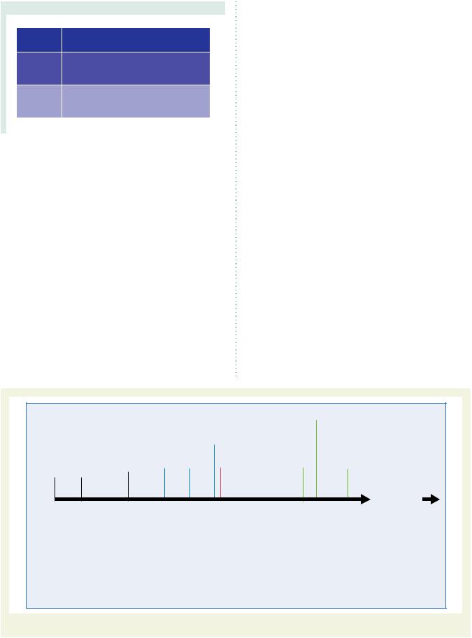
Surgical Treatment for Ischemic Heart |
|
Failure |
STREAM |
STrategic Reperfusion Early After Myocar- |
|
dial infarction |
STS |
Society of Thoracic Surgeons |
SVG |
saphenous vein graft |
SVR |
surgical ventricular reconstruction |
SYNTAX |
Synergy between Percutaneous Coronary |
|
Intervention with TAXUS and Cardiac |
|
Surgery. |
TACTICS-TIMI 18 |
Treat angina with Aggrastat and determine |
|
Cost of Therapy with an Invasive or Conser- |
|
vative Strategy–Thrombolysis in Myocardial |
|
Infarction |
TARGET |
Do Tirofiban and Reo-Pro Give Similar |
|
Efficacy Outcome Trial |
TASTE |
Thrombus Aspiration during PCI in Acute |
|
Myocardial Infarction |
TAVI |
transcatheter aortic valve implantation |
TIA |
transient ischaemic attack |
TIMACS |
Timing of Intervention in Patients with Acute |
|
Coronary Syndromes |
TIME |
Trial of Invasive Medical therapy in the |
|
Elderly |
TIMI |
Thrombolysis in Myocardial Infarction |
TRIGGER-PCI |
Testing Platelet Reactivity In Patients |
|
Undergoing Elective Stent Placement on |
|
Clopidogrel to Guide Alternative Therapy |
|
With Prasugrel |
TRITON TIMI-38 |
TRial to Assess Improvement in Therapeutic |
|
Outcomes by Optimizing Platelet InhibitioN |
|
with Prasugrel–Thrombolysis In Myocardial |
|
Infarction 38 |
TVR |
target vessel revascularization |
UFH |
unfractionated heparin |
VAD |
ventricular assist device |
VF |
ventricular fibrillation |
VKA |
vitamin K antagonist |
VSD |
ventricular septal defect |
VT |
ventricular tachycardia |
WOEST |
What is the Optimal antiplatElet and |
|
anticoagulant therapy in patients with oral |
|
anticoagulation and coronary StenTing |
ZEST-LATE/REAL- |
Zotarolimus-Eluting Stent, Sirolimus-Eluting |
LATE |
Stent, or PacliTaxel-Eluting Stent Implant- |
|
ation for Coronary Lesions - Late Coronary |
|
Arterial Thrombotic Events/REAL-world |
|
Patients Treated with Drug-Eluting Stent |
|
Implantation and Late Coronary Arterial |
|
Thrombotic Events |
2014 23, September on guest by org/.oxfordjournals.http://eurheartj from Downloaded

ESC/EACTS Guidelines |
Page 7 of 100 |
|
|
1. Preamble
Guidelines summarize and evaluate all available evidence, at the time of the writing process, on a particular issue with the aim of assisting health professionals in selecting the best management strategies for an individual patient with a given condition, taking into account the impact on outcome, as well as the risk–benefit ratio of particular diagnostic or therapeutic means. Guidelines and recommendations should help health professionals to make decisions in their daily practice; however, the final decisions concerning an individual patient must be made by the responsible health professional(s), in consultation with the patient and caregiver as appropriate.
A great number of guidelines have been issued in recent years by the European Society of Cardiology (ESC) and the European Association for Cardio-Thoracic Surgery (EACTS), as well as by other societies and organisations. Because of their impact on clinical practice, quality criteria for the development of guidelines have been established in order to make all decisions transparent to the user. The recommendations for formulating and issuing ESC/EACTS Guidelines can be found on the ESC web site (http://www.escardio.org/ guidelines-surveys/esc-guidelines/about/Pages/rules-writing. aspx). These ESC/EACTS guidelines represent the official position of these two societies on this given topic and are regularly updated.
Members of this Task Force were selected by the ESC and EACTS to represent professionals involved with the medical care of patients with this pathology. Selected experts in the field undertook a comprehensive review of the published evidence for management (including diagnosis, treatment, prevention and rehabilitation) of a given condition, according to the ESC Committee for Practice Guidelines (CPG) and EACTS Guidelines Committee policy. A critical evaluation of diagnostic and therapeutic procedures was performed, including assessment of the risk–benefit ratio. Estimates of expected health outcomes for larger populations were included, where data exist. The level of evidence and the strength of recommendation of particular management options were weighed and graded according to pre-defined scales, as outlined in Tables 1 and 2.
The experts of the writing and reviewing panels completed ‘declarations of interest’ forms which might be perceived as real or potential sources of conflicts of interest. These forms were compiled into one file and can be found on the ESC web site (http://www. escardio.org/guidelines). Any changes in declarations of interest that arise during the writing period must be notified to the ESC/ EACTS and updated. The Task Force received its entire financial support from the ESC and EACTS, without any involvement from the healthcare industry.
The ESC CPG supervises and co-ordinates the preparation of new guidelines produced by Task Forces, expert groups or consensus panels. The Committee is also responsible for the endorsement process of these guidelines. The ESC and Joint Guidelines undergo extensive review by the CPG and partner Guidelines Committee and external experts. After appropriate revisions it is approved by all the experts involved in the Task Force. The finalized document is approved by the CPG/EACTS for simultaneous publication in the European Heart Journal and joint partner journal, in this instance the European Journal of Cardio-Thoracic Surgery. It was developed after careful consideration of the scientific and medical knowledge and the evidence available at the time of their dating.
The task of developing ESC/EACTS Guidelines covers not only the integration of the most recent research, but also the creation of educational tools and implementation programmes for the recommendations. To implement the guidelines, condensed pocket versions, summary slides, booklets with essential messages, summary cards for non-specialists, electronic versions for digital applications (smart phones etc.) are produced. These versions are abridged and thus, if needed, one should always refer to the full-text version, which is freely available on the ESC and EACTS web sites. The national societies of the ESC and of the EACTS are encouraged to endorse, translate and implement the ESC Guidelines. Implementation programmes are needed because it has been shown that the outcome of disease may be favourably influenced by the thorough application of clinical recommendations.
Table 1 Classes of recommendations
2014 23, September on guest by org/.oxfordjournals.http://eurheartj from Downloaded
Page 8 of 100 |
ESC/EACTS Guidelines |
|
|
Table 2 Levels of evidence
|
|
|
Level of |
Data derived from multiple randomized |
|
evidence A |
clinical trials or meta-analyses. |
|
Level of |
Data derived from a single randomized |
|
clinical trial or large non-randomized |
|
|
evidence B |
|
|
studies. |
|
|
|
|
|
Level of |
Consensus of opinion of the experts and/ |
|
or small studies, retrospective studies, |
|
|
evidence C |
|
|
registries. |
|
|
|
|
|
|
|
|
|
|
|
Surveys and registries are needed to verify that real-life daily practice is in keeping with what is recommended in the guidelines, thus completing the loop between clinical research, writing of guidelines, disseminating them and implementing them into clinical practice.
Health professionals are encouraged to take the ESC/EACTS Guidelines fully into account when exercising their clinical judgment, as well as in the determination and the implementation of preventive, diagnostic or therapeutic medical strategies; however, the ESC/ EACTS Guidelines do not, in any way whatsoever, override the individual responsibility of health professionals to make appropriate and accurate decisions in consideration of the condition of each patient’s health and in consultation with that patient and, where appropriate and/or necessary, the patient’s caregiver. It is also the health professional’s responsibility to verify the rules and regulations applicable to drugs and devices at the time of prescription.
2. Introduction
Fifty years of myocardial revascularization
In 2014, coronary artery bypass grafting (CABG) celebrates the 50th anniversary of the first procedures performed in 1964.1 Thirteen
years later, the first percutaneous coronary intervention (PCI) was performed.2 Since then both revascularization techniques have undergone continued advances, in particular the systematic use of arterial conduits in the case of CABG, and the advent of stents. In the meantime, PCI has become one of the most frequently performed therapeutic interventions in medicine,3 and progress has resulted in a steady decline of periprocedural adverse events, resulting in excellent outcomes with both revascularization techniques. Notwithstanding, the differences between the two revascularization strategies should be recognized. In CABG, bypass grafts are placed to the mid-coronary vessel beyond the culprit lesion(s), providing extra sources of bloodflow to the myocardium and offering protection against the consequences of further proximal obstructive disease. In contrast, coronary stents aim at restoring normal bloodflow of the native coronary vasculature by local treatment of obstructive lesions without offering protection against new disease proximal to the stent.
Myocardial revascularization has been subject to more randomized clinical trials (RCTs) than almost any other intervention (Figure 1). In order to inform the current Guidelines, this Task Force performed a systematic review of all RCTs performed since 1980, comparing head-to-head the different revascularization strategies—including CABG, balloon angioplasty, and PCI with bare-metal stents (BMS) or with various US Food and Drug Administration-approved drug-eluting stents (DES)—against medical treatment as well as different revascularization strategies, and retrieved 100 RCTs involving 93 553 patients with 262 090 patient-years of follow-up.4
Formulation of the best possible revascularization approach, also taking into consideration the social and cultural context, will often require interaction between cardiologists and cardiac surgeons, referring physicians, or other specialists as appropriate. Patients need help with taking informed decisions about their treatment and the most valuable advice will probably be provided to them by the
|
|
|
1994 |
|
|
|
2001 |
2010 |
|
2012 |
||||
|
|
|
|
GABI |
|
|
|
ERACI II |
|
CARDia |
|
VA CARDS |
||
|
|
|
|
n=359 |
|
|
|
n=450 |
|
n=510 |
|
n=198 |
||
|
|
|
|
|
|
|
|
|
|
|
|
|
||
|
|
|
|
|
|
|
|
|
|
|
|
|
||
|
|
|
1993 |
|
1995 |
2000 |
2001 |
2009 |
2011 |
|
||||
|
|
|
ERACI |
|
MASS |
SIMA |
ARTS |
LE MANS |
PRECOMBAS |
|
||||
|
|
|
n=127 |
|
n=214 |
n=123 |
n=1205 |
n=105 |
n=600 |
|
||||
|
|
|
|
|
|
|
|
|
|
|
|
|
|
|
|
|
1986 |
|
1994 |
|
1996 |
|
2001 |
2009 |
|
2012 |
|||
|
|
|
EAST |
|
BARI |
|
|
AWESOME |
SYNTAX |
|
FREEDOM |
|||
1964 |
1977 |
CORONARY |
|
n=392 |
|
n=1829 |
|
|
n=454 |
n=1800 |
|
n=1900 |
||
STENTS |
|
|
|
|
|
|
|
|
|
|
|
|
||
FIRST CABG |
CORONARY |
|
|
|
|
|
|
|
|
|
|
|
|
|
|
|
|
|
|
|
|
|
|
|
|
|
|
||
PROCEDURES |
ANGIOPLASTY |
|
|
|
|
|
|
|
|
|
|
|
|
|
|
|
|
1993 |
|
1995 |
1997 |
2002 |
2007 |
|
2011 |
|
|||
|
|
|
RITA |
|
CABRI |
FMS |
|
SOS |
MASS II |
LEIPZIG LM |
EXCEL |
|||
|
|
|
n=1011 |
|
n=1054 |
n=152 |
|
n=988 |
n=611 |
n=201 |
n=2600 |
|||
1964 |
|
|
|
|
|
|
|
|
|
|
|
|
|
|
|
|
|
|
|
|
|
|
|
|
2014 |
|
|
|
|
|
|
|
|
|
|
|
|
|
|
|
|
|
|
|
|
|
|
|
|
|
|
|
|
|
|
|
|
|
|||
|
|
|
|
|
|
|
|
|
|
|
|
|
|
|
|
|
|
|
|
|
|
|
|
|
|
|
||||
|
|
|
|
|
|
|
|
|
|
|
|
|
|
|
|
|
|
|
|
|
|
|
|
|
|
|
|
|
|
|
1980 |
|
1984 |
|
|
|
|
|
|
1999 |
|
2003 |
|
2007 |
|
2009 |
|
2012 |
|
|
ISCHEMIA |
||||||||||
|
ECSS |
|
VA |
|
|
|
|
|
|
|
|
|
|
|
||||||||||||||||
|
|
|
|
|
|
|
AVERT |
|
ALKK |
|
SWISS-II |
|
BARI-2D |
|
|
FAME-2 |
|
|
n=8000 |
|||||||||||
|
n=768 |
n=686 |
|
|
|
|
|
|
|
|
|
|
|
|
||||||||||||||||
|
|
|
|
|
|
n=341 |
|
n=300 |
|
n=201 |
|
n=384 |
|
|
n=888 |
|
|
|
|
|
||||||||||
|
|
|
|
|
|
|
|
|
|
|
|
|
|
|
|
|
|
|
|
|
||||||||||
|
|
|
|
|
|
|
|
|
|
|
|
|
|
|
|
|
|
|
|
|
|
|
|
|
|
|
|
|
|
|
|
|
|
|
|
|
1997 |
2001 |
|
|
2006 |
|
2008 |
|
|
|
|
|
|
|
|
|
|||||||||
|
|
|
1984 |
|
|
|
RITA-2 |
|
|
TIME |
|
|
OAT |
|
JSAP |
|
|
|
|
|
|
|
|
|||||||
|
|
|
|
|
|
n=1018 |
|
|
n=305 |
|
|
|
|
|
|
|
|
|
|
|
||||||||||
|
|
|
CASS |
|
|
|
|
|
|
n=2166 |
|
n=384 |
|
|
|
|
|
|
|
|
||||||||||
|
|
|
|
|
|
|
|
|
|
|
|
|
|
|
|
|
|
|
|
|
|
|
||||||||
|
|
|
n=780 |
|
|
|
|
|
|
|
|
|
|
|
|
|
|
|
|
|
|
|
|
|
|
|
|
|
||
|
|
|
|
|
|
|
|
|
|
|
|
|
|
|
|
|
|
|
|
|
|
|
|
|
|
|
|
|
|
|
|
|
|
|
|
|
|
|
|
|
|
|
|
|
|
|
|
2007 |
|
|
|
2011 |
|
|
|
|
|
|
|||
|
|
|
|
|
|
|
|
|
|
|
|
|
|
|
|
|
|
COURAGE |
|
|
|
STICH |
|
|
|
|
|
|||
|
|
|
|
|
|
|
|
|
|
|
|
|
|
|
|
|
|
n=2287 |
|
|
|
n=1212 |
|
|
|
|
|
|||
|
Revascularization vs. MT |
|
|
|
Balloon angioplasty vs. CABG |
|
|
|
BMS vs. CABG |
|
DES vs. CABG |
|
|
|
|
|
|
|
|
|||||||||||
|
|
|
|
|
|
|
|
|
|
|
|
|
|
|
|
|||||||||||||||
BMS = bare-metal stent; CABG = coronary artery bypass grafting; DES = drug-eluting stent.
2014 23, September on guest by org/.oxfordjournals.http://eurheartj from Downloaded
Figure 1 Randomized trials in myocardial revascularization therapy over the past five decades.

ESC/EACTS Guidelines |
Page 9 of 100 |
|
|
‘Heart Team’.5 Recognizing the importance of the interaction between cardiologists and cardiac surgeons, the leadership of both the ESC and the EACTS has given this Joint Task Force, along with their respective Guideline Committees, and the reviewers of this document the mission to draft balanced, patient-centred, evidencedriven practice guidelines on myocardial revascularization. The respective Chairpersons of these two associations and CPG Chairperson were also given the task to adapt to the declaration of interest policy and to ensure that their Task Force members followed it throughout the development process of the Guidelines. In the event that any of the Task Force members had a potential conflict of interest to declare, he/she did not participate in the final decision of the Task Force on the given subject.
3. Scores and risk stratification
Myocardial revascularization in the elective setting is appropriate when the expected benefits, in terms of survival or health outcomes (symptoms, functional status, and/or quality of life), exceed the expected negative consequences of the procedure. Whether medical therapy, PCI, or CABG is preferred should depend on the risk–benefit ratios of these treatment strategies, weighting the risks of periprocedural death, myocardial infarction and stroke against improvements in health-related quality of life, as well as longterm freedom from death, myocardial infarction or repeat revascularization. The Heart Team should take into consideration the coronary anatomy, disease, age and comorbidities, patient preference, and hospital/operator experience.
Numerous models have been developed for risk stratification, focussing on anatomical complexity or clinical risk, and have demonstrated their value during decision-making.6 Those models most frequently used in a clinical setting are summarized in the Tables of recommendation [risk models to assess short-term (in-hospital or 30-day) and medium-to-long-term (≥1 year) outcomes].
(1)The EuroSCORE predicts surgical mortality.7,8 It is based on an old data set and has been shown to overestimate the risk of mortality, and should therefore no longer be used.9,10
(2)The EuroSCORE II is an update of the logistic EuroSCORE model and is derived from a more contemporary data set better reflecting current cardiac surgical practice.11 Its value has been demonstrated in specific cohorts of patients undergoing CABG.12 Compared with its original version, the EuroSCORE II may have a better ability to predict mortality.12 – 14
(3)The Society of Thoracic Surgeons (STS) score is a risk-prediction model, validated in patients undergoing cardiac surgery, with a
specific model for CABG surgery and combined CABG and valve surgery.15,16 It can be used to predict in-hospital or 30-day mortality (whichever occurs last) and in-hospital morbidity.
(4)The SYNTAX score (Table 3) was developed to grade the anatomical complexity of coronary lesions in patients with left main or three-vessel disease, and was found to be an independent predictor of long-term major adverse cardiac and cerebrovascular event (MACCE) in patients treated with PCI but not CABG.17,18 It facilitates the selection of optimal treatment by
identifying patients at highest risk of adverse events following PCI. The interobserver variability of the Synergy between Percutaneous Coronary Intervention with TAXUS and Cardiac Surgery (SYNTAX) score is significant,19 although development of non-invasive assessments may simplify calculation of the SYNTAX score.20
(5)The National Cardiovascular Database Registry (NCDR
CathPCI) risk score has been developed to predict risk in PCI patients and should only be used in this context.21
(6)The age, creatinine, ejection fraction (ACEF) model is a simple
score as it contains only three variables, and was developed using data from a cohort of surgical patients.22 ACEF has also been validated to predict mortality in patients undergoing PCI.23
(7)The clinical SYNTAX score is a combination of the ACEF and SYNTAX scores. Originally established as an additive model, the subsequent development of a logistic model has provided more tailored risk assessment.24
(8)The SYNTAX II score is a combination of anatomical and clinical factors [age, creatinine clearance, left ventricular (LV) function, gender, chronic obstructive pulmonary disease, and peripheral vascular disease] and predicts long-term mortality in patients with complex three-vessel or left main (LM) coronary artery disease (CAD).25 It was found to be superior to the conventional SYNTAX score in guiding decision-making between CABG and PCI in the SYNTAX trial, and subsequently validated in the drug-eluting stent for left main coronary artery disease DELTA registry.
(9)For the American College of Cardiology Foundation–Society of Thoracic Surgeons Database Collaboration (ASCERT) study,26
two large datasets from the National Cardiovascular Data Registry (NCDR) and STS were used to develop several models to
predict mortality at different time points following CABG and
PCI.27,28
Comparative analyses of these models are limited because available studies have largely evaluated individual risk models in different patient populations, with different outcome measures being reported at various time points, and most models are restricted to one type of revascularization. In addition, several important variables, such as frailty, physical independence and porcelain aorta, are not incorporated in current risk scores. An ideal risk–benefit model enables comparison of the short-term benefits of PCI to the longterm benefits of CABG; however, even though risk models may provide useful information for predicting mortality and major adverse events, prediction of which patients will receive benefit in terms of quality of life is so far unavailable.
These limitations restrict the ability to recommend one specific risk model. It is also important to acknowledge that no risk score can accurately predict events in an individual patient. Moreover, limitations exist in all databases used to build risk models, and differences in definitions and variable content can affect the performance of risk scores when they are applied across differing populations. Ultimately, risk stratification should be used as a guide, while clinical judgement and multidisciplinary dialogue (The Heart Team) remain essential.25
2014 23, September on guest by org/.oxfordjournals.http://eurheartj from Downloaded
Page 10 of 100 |
|
ESC/EACTS Guidelines |
||||
|
|
|
|
|
||
|
|
|
|
|
|
|
|
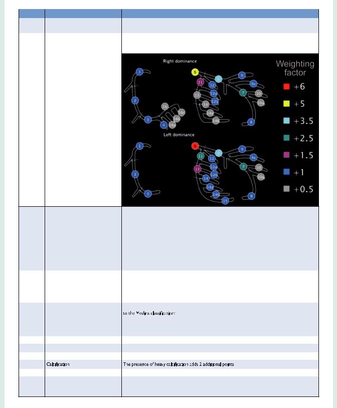
Table 3 Guide to calculate the SYNTAX score |
|
||||
|
|
|
|
|
|
|
|
|
|
|
|
|
|
|
|
Steps |
Variable assessed |
Description |
||
|
|
|
|
|
|
|
|
|
Step 1 |
Dominance |
The weight of individual coronary segments varies according to coronary artery dominance (right or |
||
|
|
|
|
left). Co-dominance does not exist as an option in the SYNTAX score. |
|
|
|
|
Step 2 |
Coronary segment |
The diseased coronary segment directly affects the score as each coronary segment is assigned a |
||
|
|
|
|
weight, depending on its location, ranging from 0.5 (i.e. posterolateral branch) to 6 (i.e. left main in case |
||
|
|
|
|
of left dominance). |
||
|
Step 3 |
Diameter stenosis |
The score of each diseased coronary segment is multiplied by 2 in case of a stenosis 50–99% and by 5 |
|||
|
|
|
in case of total occlusion. |
|
|
|
|
|
|
In case of total occlusion, additional points will be added as follows: |
|||
|
|
|
- Age >3 months or unknown |
+1 |
|
|
|
|
|
- Blunt stump |
|
+1 |
|
|
|
|
- Bridging |
|
+1 |
|
|
|
|
- First segment visible distally |
+1 per non visible segment |
||
|
|
|
- Side branch at the occlusion |
+1 if <1.5mm diameter |
||
|
|
|
|
|
+1 if both <1.5 and ≥1.5mm diameter |
|
|
|
|
|
|
+0 if ≥1.5mm diameter (i.e. bifurcation lesion) |
|
|
Step 4 |
Trifurcation lesion |
The presence of a trifurcation lesion adds additional points based on the number of diseased segments: |
|||
|
|
|
- 1 segment |
+3 |
|
|
|
|
|
- 2 segments |
+4 |
|
|
|
|
|
- 3 segments |
+5 |
|
|
|
|
|
- 4 segments |
+6 |
|
|
|
|
|
|
|
||
|
Step 5 |
Bifurcation lesion |
The presence of a bifurcation lesion adds additional points based on the type of bifurcation according |
|||
|
|
|
|
29 |
|
|
|
|
|
- Medina 1,0,0 or 0,1,0 or 1,1,0: add 1 additional point |
|||
|
|
|
- Medina 1,1,1 or 0,0,1 or 1,0,1 or 0,1,1: add 2 additional point |
|||
|
|
|
Additionally, the presence of a bifurcation angle <70° adds 1 additional point. |
|
||
|
Step 6 |
Aorto-ostial lesion |
The presence of aorto-ostial lesion segments adds 1 additional point |
|
||
|
Step 7 |
Severe tortuosity |
The presence of severe tortuosity proximal of the diseased segment adds 2 additional points |
|||
|
|
|
|
|
||
|
Step 8 |
Lesion length |
Lesion length >20 mm adds 1 additional point |
|||
|
|
|
|
|
|
|
|
Step 9 |
|
|
|
|
|
|
|
|
|
|
||
|
Step 10 |
Thrombus |
The presence of thrombus adds 1 additional point |
|||
|
|
|
|
|
||
|
Step 11 |
Diffuse disease/small vessels |
The presence of diffusely diseased and narrowed segments distal to the lesion (i.e. when at least 75% of |
|||
|
|
|
the length of the segment distal to the lesion has a vessel diameter of <2mm) adds 1 point per segment |
|||
|
|
|
number |
|
|
|
|
|
|
|
|
|
|
|
|
|
|
|
|
|
2014 23, September on guest by org/.oxfordjournals.http://eurheartj from Downloaded