

Учебники / Pediatric Sinusitis and Sinus Surgery Younis 2006
.pdfxx |
|
|
|
Contents |
Coding Surgical Sinus Procedures |
. . . . 246 |
|
||
Charge Entry and Account Follow-Up . . . . |
249 |
|||
Claim Submission . . . . |
249 |
|
|
|
Watching Reimbursement |
. . . . |
250 |
|
|
Informed Consent/Medicolegal Issues . . . . |
251 |
|||
Credentialing . . . . |
253 |
|
|
|
Occupational Safety and Health Administration . . . . 254 |
||||
Ancillary Services . . . . |
255 |
|
|
|
Office Administration . . . . 256 |
|
|
||
Conclusion . . . . |
257 |
|
|
|
Index . . . . 259
Contributors
Frank C. Astor Departments of ENT and Otolaryngology, University of Miami, Miami, Florida, U.S.A.
Fuad M. Baroody Section of Otolaryngology—Head and Neck Surgery, Pritzker School of Medicine, University of Chicago, Chicago, Illinois, U.S.A.
Michael S. Benninger Department of Otolaryngology—Head and Neck Surgery, Henry Ford Hospital, Detroit, Michigan, U.S.A.
Roy R. Casiano Department of Otolaryngology, University of Miami School of Medicine, Miami, Florida, U.S.A.
Raphael Chan Bossier City, Louisiana, U.S.A.
Sam J. Daniel Department of Otolaryngology, McGill University, Montreal, Quebec, Canada
Craig S. Derkay Departments of Otolaryngology—Head and Neck Surgery and Pediatrics, Eastern Virginia Medical School, The Children’s Hospital of the King’s Daughters, Norfolk, Virginia, U.S.A.
Tina P. Elkins University of Texas, Cypress, Texas, U.S.A.
xxi
xxii |
Contributors |
Joshua A. Gottschall Department of Otolaryngology—Head and Neck Surgery, Henry Ford Hospital, Detroit, Michigan, U.S.A.
Edward Hepworth Division of Otolaryngology Head and Neck Surgery, Department of Surgery, University of New Mexico, Albuquerque,
New Mexico, U.S.A.
Melissa A. M. Hertler Division of Otolaryngology Head and Neck Surgery, Department of Surgery, University of New Mexico, Albuquerque, New Mexico, U.S.A.
Sarita Kaza New York, New York, U.S.A.
Gary Kleiner Department of Pediatrics and Division of Immunology and Infectious diseases, University of Miami School of Medicine, Miami, Florida, U.S.A.
Rande H. Lazar Pediatric Otolaryngology Fellowship Program, Le Bonheur Children’s Medical Center, Memphis, Tennessee, U.S.A.
Mary LeGrand KarenZupko & Associates Inc., Chicago, Illinois, U.S.A.
Ron B. Mitchell Department of Otolaryngology, Virginia Commonwealth University, Richmond, Virginia, U.S.A.
Samantha M. Mucha SectionofOtolaryngology—HeadandNeck
Surgery,PritzkerSchoolofMedicine,UniversityofChicago,Chicago,
Illinois,U.S.A.
Maria T. Pen˜a Department of Otolaryngology, Children’s Research Institute, Children’s National Medical Center, Washington, D.C., U.S.A.
Kevin D. Pereira Medical Center of Otolaryngology Department, University of Texas, Houston, Texas, U.S.A.
Hassan H. Ramadan Department of Otolaryngology, Head and Neck Surgery, West Virginia University, Morgantown, West Virginia, U.S.A.
Scott A. Schraff Department of Otolaryngology—Head and Neck Surgery, Eastern Virginia Medical School, Norfolk, Virginia, U.S.A.
Gavin Setzen Department of Surgery and Albany ENT and Allergy Services, Albany Medical College, Albany, Newy York, U.S.A.
Contributors |
xxiii |
Michael Setzen Department of Otolaryngology, North Shore University Hospital, Manhasset, New York, U.S.A.
Ramzi T. Younis Department of Pediatrics, University of Miami, Miami, Florida, U.S.A.
George H. Zalzal Department of Otolaryngology, Children’s Research Institute, Children’s National Medical Center, Washington, D.C., U.S.A.
This book is dedicated to
My dad—God bless his soul—for the great education he has provided, my mom for the love and care she planted in me,
my wife, Dina, and my children, Karen, Jessica, and Tamer for their support encouragement, understanding, and everlasting love

1
Embryology and Anatomy of the Nose
and Paranasal Sinuses
Raphael Chan
Bossier City, Louisiana, U.S.A.
Frank C. Astor
Departments of ENT and Otolaryngology, University of Miami, Miami, Florida, U.S.A.
Ramzi T. Younis
Department of Pediatrics, University of Miami, Miami, Florida, U.S.A.
EMBRYOLOGY OF THE NOSE
At the fourth week of gestation, the stomodeum is formed superiorly by the midline frontonasal process. The maxillary processes of the first branchial arch form the lateral borders.
By the end of the fourth week, nasal placodes, which are bilateral ovalshaped ectodermal thickenings, form on each side of the frontonasal process. The proliferation of mesenchyme around the placode creates the medial and lateral processes and the placodes eventually come to lie in depressions called nasal pits.
The maxillary processes grow toward each other and to the medial nasal processes. The lateral nasal process is separated from the maxillary process by a cleft or furrow, called the nasolacrimal groove.
The medial nasal processes merge with each other and with the maxillary processes during the sixth and seventh weeks. This merging results in the formation of the philtrum, the premaxillary process, and the primitive septum.
1

2 |
Chan et al. |
Figure 1 Undersurface of the head of a human embryo about 29 days old. Source: From Ref. 1.
Figure 2 Model of the chondrocranium of a human embryo, 8-cm long. Source: From Ref. 1.
Anatomy of Nose and Paranasal Sinuses |
3 |
The frontonasal process develops into the forehead and the bridge and tip of the nose, while the sides of the nose are derived from the lateral nasal processes.
The primary palate develops from the premaxillary process at the end of the fifth week while the secondary palate develops from the horizontal mesodermal projections from the inner maxillary processes. These are the palatine processes and grow medially, fusing with each other and with the primary palate and nasal septum.
The primitive nasal capsule forms the framework for the development of the bony and cartilaginous structures of the upper face. The cartilaginous nasal capsule is formed from the condensation of mesenchyme during the third fetal month. Later, in-growth of connective tissue divides the structure into the lower lateral and upper lateral and septal cartilage. Most of the posterior capsule ossifies into the portions of the sphenoid bones, ethmoid bone including the ethmoid turbinates, sinus walls, and perpendicular plate. The nasal bone and maxilla form upon its lateral surface. Ossification of the cartilaginous capsule is not complete as cartilaginous segments remain in the anterior nose (Figs. 1, 2).
THE DEVELOPMENTAL ANATOMY OF THE LATERAL
NASAL WALL
The progenitor of the inferior turbinate first appears as a swelling just above the palatal shelf in the anterior aspect of the lateral nasal wall at 38–40 days of gestation. This is the maxilloturbinal swelling. Another swelling, the ethmoturbinal, arises at 40–43 days at the junction of the nasal septum and nasal roof. The space between the maxilloturbinal and the ethmoturbinal develops into the future middle meatus. The ethmoturbinal swelling forms the middle, superior, and supreme turbinates. The supreme turbinate is seen by 95–105 days. The supreme turbinate differentiates further after the seventh month, but by puberty, they are usually absorbed, attaining the adult configuration by 12 years of age. The nasoturbinal swelling can be seen at approximately 60 days. This embryologic anlage is representative of the future agger nasi.
A thickened area develops at the junction of the ascending and descending parts of the middle meatus at 40–60 days. This sinks into the lateral nasal wall forming a furrow that is the future infundibulum, and the ridge that develops anterior to it on the nasoturbinal swelling becomes the uncinate process.
Further deepening of the floor of the infundibulum as a pouch forms the precursor of the maxillary sinus by 60–75 days. The frontal recess cells develop medial to the uncinate and between it and the anterior attachment of the middle turbinate at approximately 105 days. The ethmoid bulla forms posterior to the infundibular furrow.
4 |
Chan et al. |
The superior meatus differentiates in a truncated manner; at 110 days, the anterior end develops a superior and inferior arm (like the infundibulum) and a crista (like the bulla). The bony lamellae of the lateral nasal wall consist of the middle, superior, and the supreme turbinates, the ethmoid bulla, and the uncinate. These lamellae are attached to the lamina papyracea and remain constant throughout development, although with differentiation, they may be distorted by pneumatization. The lamellae partition the lateral wall into compartments in which cellular development is organized. The basal lamella of
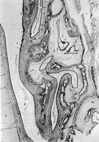
Figure 3 Horizontal section of the left ethmoid bone and the ethmoidal sinuses of a 7-month fetus. 1. ethmoidal sinuses, 2. middle turbinate, 3. superior turbinate, 4. middle meatus, 5. superior meatus, 6. nasal chamber, 7. nasal septum. Source: From Ref. 2.