

Аорта
.pdfe294 |
Circulation |
April 6, 2010 |
Figure 16. Takayasu arteritis with involvement of the thoracoabdominal aorta and great vessels as shown on contrast-enhanced CT and MR examinations. Note narrowing of the arterial lumen and circumferential soft tissue thickening of the walls of the great vessels and thoracic and abdominal aorta. Panel A, Image through the great vessels with narrowing of the left common carotid and left subclavian arteries. Panel B, Mid descending thoracic aorta (arrowheads). Panel C, Aorta just above the diaphragm (arrowheads). Panel D, Infrarenal aorta. Panel E, Volume-rendered image from CT demonstrates the extent of involvement. Panel F, Oblique sagittal MR of the thoracic aorta. Panel G, Coronal MR of the abdominal aorta. CT indicates computed tomographic imaging; and MR, magnetic resonance imaging.
Hiratzka et al |
2010 Guidelines on Thoracic Aortic Disease |
e295 |
nearly 32% of the patients had aortic aneurysm formation.170 Most commonly, aneurysm formation developed in the descending aorta, followed by the abdominal, then ascending aortic segments. In the National Institutes of Health series of 60 patients with Takayasu arteritis, 23% had aortic aneurysm formation.158 Aneurysms most commonly formed in the aortic arch or root, abdomen, and then other thoracic segments. Stenosis of the aorta is more common than is aneurysm formation, occurring in 53% of patients in the National Institutes of Health series. Any segment of the aorta may be involved, but the abdominal aortic segment is affected more than 70% of the time if stenosis is found.
Treatment of Takayasu arteritis begins with inflammation reduction with corticosteroids. Steroids are typically started at high dose, 40 to 60 mg daily at initiation to lower the erythrocyte sedimentation rate or C-reactive protein to normal, and are required for 1 to 2 years to ensure proper disease treatment.169 Despite the prolonged regimen, nearly half of the patients will relapse during tapering, requiring additional immunosuppression. Second-line agents that have been used include methotrexate, azathioprine, and antitumor necrosis factor-alpha agents.158,169 Unfortunately, markers of inflammation are imperfect barometers of disease activity. Disease progression has been shown to occur in the setting of normal marker levels.158
Revascularization for aortic stenosis or aneurysm occurs for the same indications as in noninflammatory disorders: secondary organ vascular insufficiency or risk of rupture. There are no randomized trials of percutaneous or surgical intervention in this disease.158,171–173 Nonrandomized reports have shown that revascularization of either variety may be appropriate, with one caveat. The risk of graft failure is higher in patients with active local inflammation.162 Moreover, the presence of aneurysmal disease itself may be problematic. One report documented a 12% incidence of anastomotic aneurysms over 2 decades of follow-up related to the presence of aneurysms at surgery.174
7.3. Giant Cell Arteritis
GCA, also known as temporal arteritis, is an elastic vessel vasculitis involving the aorta and its secondary and tertiary branches. Distinguishing it from Takayasu arteritis, GCA affects patients above the age of 50 years, with an incidence peaking in the eighth decade of life.175 The disease affects women in a 3:2 ratio to men and has a predilection for those of northern European ancestry.29 In the United States, epidemiologic investigation reports a prevalence of 20 cases per 100 000 persons. The incidence is higher in Scandinavian nations but lower in southern Europe, suggesting a genetic predisposition in certain populations.176
The clinical presentation of GCA is varied, requiring a heightened suspicion by clinicians for early diagnosis. Half of the patients report constitutional symptoms, such as weight loss, night sweats, malaise, and fever.177 Because of the predilection for secondary and tertiary thoracic branches of the aorta, cranial symptoms are common. Scalp tenderness and headache are present in two thirds of patients and in up to 90% of patients with biopsy-proved disease.177 Jaw claudication is common and affects half of the patients, 20%
develop visual changes, and other neurologic symptoms such as stroke or neuropathy occur in nearly one third.29 Visual changes are particularly important to notice, because early treatment may prevent permanent blindness. Patients may report diplopia, amaurosis fugax, or blurriness prior to blindness. Polymyalgia rheumatica characterized by a generalized inflammatory state with proximal muscle involvement is found in nearly half of patients with GCA.29 Patients with polymyalgia rheumatica report muscular pain and stiffness, particularly on initiation of movement.
Extracranial vascular involvement is less common in GCA than in Takayasu arteritis, occurring in 25% of patients. In a 50-year study of Olmsted County, Minn, that included 168 patients with GCA, aortic aneurysm/dissection was found in 18% of the subjects, whereas large-artery stenosis was noted in 13% of patients.178 No patient had stenosis of the aorta. Aortic aneurysm formation represents an important marker. Although aneurysm formation per se does not reduce survival compared with the GCA cohort as a whole, AoD in the setting of an aneurysm reduces survival to an average of 1.1 years.178 Similarly, aortic aneurysm rupture or dissection caused two thirds of deaths in a series of patients with GCA in California.179
The American College of Rheumatology diagnostic criteria for GCA include 1) age older than 50 years, 2) recent-onset localized headache, 3) temporal artery pulse attenuation or tenderness, 4) erythrocyte sedimentation rate greater than 50 mm/h, and 5) an arterial biopsy demonstrating necrotizing vasculitis.164 Three or more criteria confer a sensitivity and specificity above 90% for the disease. With intracranial disease, temporal artery biopsies are diagnostic in up to 80% of cases.180 The rate of positivity declines with initiation of glucocorticoid therapy, but this should not delay treatment to avoid GCA complications. Biopsies performed within 7 days of steroid initiation retain a high diagnostic yield.181
The pathophysiology of GCA shares important features with Takayasu arteritis.28 GCA is marked by a T-cell clonal expansion suggesting a specific antigenic response, which currently remains unelucidated. The inflammatory response, which begins in the adventitial layer, is marked by augmented cytokine and MMP production causing granuloma formation. Granuloma formation both shields the vessel from the inciting antigen and causes vessel destruction. The inflammatory environment within the vessel wall with the possible formation of aneurysms or vessel stenosis is histologically identical to that of Takayasu arteritis. Because of the multiyear cyclical nature of disease incidence, some have posited an infectious etiology.29
Corticosteroids represent the standard in therapy for patients with GCA.182 The typical treatment regimen includes starting prednisone dose of 40 to 60 mg daily, although recent evidence suggests a similar efficacy with 30 to 40 mg daily.183 Therapy is typically required for 1 to 2 years to avoid recurrence, although the dose may be tapered beginning 2 to 3 months after initiation. Patients commonly report feeling much better rapidly but, as with Takayasu arteritis, new vascular involvement may occur in up to half of patients treated with steroids.184 In contrast to Takayasu arteritis, additional immunomodulatory agents do not seem to modulate the disease’s progress. Methotrexate studied in a double-
e296 |
Circulation |
April 6, 2010 |
blind, placebo-controlled study as an adjunct to prednisone did not reduce morbidity, erythrocyte sedimentation rate level, or cumulative prednisone dose.184a Revascularization recommendations follow the same pattern as in Takayasu arteritis.
7.4. Behc¸et Disease
In 1937, Hulusi Behc¸et described his eponymous syndrome based on a set of 3 symptoms: uveitis, aphthous stomatitis, and genital ulcers. Most common in Turkey, with a prevalence of 80 to 370 cases per 100 000 persons,185,186 the disease is much less common in the United States, with an estimated prevalence of 1 to 3 cases per million persons.187 The diagnostic criteria were established by the International Group for Behc¸et’s disease and require oral ulceration and 2 of these 3 lesions: recurrent genital ulceration, uveitis or retinal vasculitis, or skin lesions, such as erythema nodosum, pseudofolliculitis, or pathergy.188 In addition to these cardinal manifestations, vascular involvement may occur in one third of patients. A small vessel vasculitis commonly associated with the human leukocyte antigen (HLA) B51 allele,187 Behc¸et disease is 1 of 2 vasculitides that may also involve veins. Venous involvement is most commonly superficial thrombophlebitis, but deep vein thrombosis in the vena cava, varices, and cerebral sinuses has been reported.189 The small vessel involvement of Behc¸et disease may result in nonvascular complaints, such as erythema nodosum, arthritis, and gastrointestinal involvement with diarrhea, gastrointestinal bleeding, or perforation.187 Treatment of Behc¸et disease varies based on the manifestation of disease. Systemic corticosteroids are the typical therapy for those with vascular involvement.
Specifically with regard to vascular manifestations of Behc¸et disease, any artery or vein, large or small, systemic or pulmonary, may be involved by the vasculitic process. Aortic histopathology shows lymphocytic infiltration mixed with histiocytes and eosinophils with giant cells around vasa vasorum of media and adventitia. Destruction of media leads to aneurysm formation and may proceed to pseudoaneurysm formation and rupture. Aneurysm formation may occur in multiple sites and in different sites over a period of follow-up. Aneurysm, stenotic lesions, and occlusion of brachiocephalic arteries may occur with or without aortic involvement. Although aortic involvement is unusual for patients with Behc¸et vasculitis, aneurysm rupture can be unpredictable and fatal.190,191 With regard to surgical repair, anastomotic pseudoaneurysms often occur (12.9% within 18 months in 1 series) and may be related to ongoing inflammatory changes in the area of anastomotic suture lines.192 Endovascular repair with stent grafts has also been described.193
7.5. Ankylosing Spondylitis (Spondyloarthropathies)
The group of diseases labeled spondyloarthropathies are linked by the strong association of major histocompatibility complex HLA B-27 and the absence of rheumatoid factor.194,195 Several features are common to the spondyloarthropathies, including sacroilitis, inflammatory arthritis or enthesitis (inflammation of tendon insertions), associations with inflammatory bowel disease or psoriasis, and aortitis and heart block.195
Ankylosing spondylits is the most common variant and often begins with back pain and stiffness in the second or third decade of life. It affects men 2 to 3 times as often as women, worsens with inactivity, and commonly takes years for the diagnosis to be made.196 The diagnosis requires 4 of the 5 criteria: onset of pain at younger than 40 years, back pain for longer than 3 months, morning stiffness, subtle symptom onset, and improvement with exercise.197 Patients may also report constitutional symptoms, such as malaise or fever. Acute anterior uveitis is reported in up to 40% of patients. Aortic root and aortic valve involvement are reported in up to 80% of patients.198 When involved, the aortic valve may have a nodular appearance, and aortic valvular regurgitation is present in nearly half of the patients.198 Treatment of aortic root expansion and aortic valvular abnormalities is the same as for other conditions.
7.6. Infective Thoracic Aortic Aneurysms
Infection (due to bacterial, fungal, viral, spirochetal, or tubercular organisms) is a rare cause of thoracic aortic aneurysms. Originally named mycotic endarteritis by Osler,199 the terms infected aneurysm or infectious aortitis are now used more commonly, because the majority of etiologic agents are nonfungal. Saccular aneurysms are most common, but infected aneurysms can be fusiform and often even pseudoaneurysms. The ascending thoracic aorta, aortic arch, and descending thoracic aorta can all be affected, as can prosthetic aortic grafts and aortic homografts. Typically, the sites of infected aneurysms are opposite the great vessels in the aortic arch or opposite the visceral ateries in the abdomen. There are several mechanisms by which the aortic infection may arise. First, there may be contiguous spread from adjacent thoracic structures, such a mediastinitis, abscess, infected lymph nodes, infectious pericarditis, empyema, or paravertebral abscess. Second, there may be septic emboli from underlying bacterial endocarditis. Third, there may be hematogenous dissemination of bacteria in the setting of sepsis or intravenous drug abuse. Infection most often arises in a diseased aorta, either in a preexisting aneurysm, at the site of an atherosclerotic plaque, or at the site of some accidental or iatrogenic aortic trauma. Indeed, infected thoracic aortic aneurysms may arise as a late complication of cardiac surgery, often associated with postoperative mediastinitis, typically at the sites of aortic cannulation or anastomotic suture lines.200,201
Various organisms can infect the aorta, with most infections being bacterial. Staphylococcus aureus and Salmonella are the organisms most commonly identified.202–204 Pneumococcus and Escherichia coli are relatively common grampositive and gram-negative pathogens, respectively.
Treponema pallidum, the gram-negative spirochete bacterium that causes syphilis, as well as other Treponema species, can cause infected aortitis, with the ascending thoracic aorta most often involved. However, in syphilitic aortitis, thoracic aortic aneurysm does not appear for 10 to 25 years after the initial spirochetal infection. Fungal infections of the aorta, with either Candida or Aspergillus, occur less often205 and typically occur in the setting of impaired immunity, such as
Hiratzka et al |
2010 Guidelines on Thoracic Aortic Disease |
e297 |
patients with systemic illness, human immunodeficiency virus, or prior organ or bone marrow transplant.
Indeed, patients with impaired immunity are also at increased risk of tuberculous aortitis attributable to mycobacterium tuberculosis. Tuberculous aortitis has until now been exceedingly rare, but the incidence may rise as the prevalence of tuberculosis rises worldwide. Tuberculous aortitis typically affects the distal aortic arch and descending thoracic aorta, likely because the aorta is thought to become infected via direct extension from continuous infected lymph nodes, empyema, or pericarditis.206
Finally, there appears to be an independent association between human immunodeficiency virus and ascending thoracic aortic dilatation, although its mechanisms are poorly understood.207,208 Moreover, the incidence of frank aneurysms remains extremely low.
8. Acute Aortic Syndromes
Acute aortic syndromes consist of 3 interrelated conditions with similar clinical characteristics and include AoD, IMH, and PAU.209
8.1. Aortic Dissection
8.1.1. Aortic Dissection Definition
AoD is defined as disruption of the media layer of the aorta with bleeding within and along the wall of the aorta resulting in separation of the layers of the aorta. In the majority of patients (90%), an intimal disruption is present that results in tracking of the blood in a dissection plane within the media. This may rupture through the adventitia or back through the intima into the aortic lumen (Figure 17). This classic dissection results in a septum, or “flap,” between the 2 lumens (Figure 18). The false lumen may thrombose over time (Figure 19). While on noninvasive imaging, 15% of patients with aortic dissection syndromes have an apparent IMH without evidence of an intimal tear, autopsy studies show only 4% have no visible intimal tear; indeed, at the time of surgery a tear is found in most patients.210,211 Occasionally, AoD originates from a small atheromatous ulcer that is difficult to identify. On the other hand, extensive atheromatous disease of the aorta may lead to PAU or a localized IMH.
The true incidence of acute AoD is difficult to define for 2 principal reasons: 1) acute AoD can be rapidly fatal, and when patients die prior to hospitalization, death may be erroneously attributed to another cause and 2) acute AoD is frequently missed on initial presentation, and early mortality among this group may be misclassified as non– dissection related. Population-based studies suggest that the incidence of acute AoD ranges from 2 to 3.5 cases per 100 000 personyears, which correlates with 6000 to 10 000 cases annually in the United States.75,212,215–217 A review of 464 patients from IRAD reported a mean age at presentation of 63 years, with significant male predominance (65%).47 The prevalence of AoD appears to be increasing, independent of the aging population, as noted by Olsson and colleagues,218 who found the incidence of AoD among Swedish men has increased to 16 per 100 000 men yearly. It may be that 2 to 3 times as many patients die from AoD than from ruptured AAA; approximately 75% of patients with AAA will reach an emer-
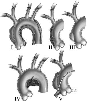
Figure 17. Classes of intimal tears. I. Classic dissection with intimal tear and double lumen separated by septum. Communication between lumens is typically in descending aorta at sheared-off intercostal arteries or distal reentry site. II. IMH. No intimal tear or septum is imaged but is usually found at surgery or autopsy. DeBakey Types II and IIIa are common extent of this lesion. III. Intimal tear without medial hematoma (limited dissection) and eccentric aortic wall bulge. Rare and difficult to detect by TEE or CT. Patients with Marfan syndrome prone to this type. May result in aortic rupture or extravasation. IV. PAU usually to the adventitia with localized hematoma or saccular aneurysm. May propagate to Class I dissection, particularly when involving ascending aorta or aortic arch. V. Iatrogenic (catheter angiography or intervention)/traumatic (deceleration) dissection. CT indicates computed tomographic imaging; IMH, intramural hematoma; PAU, penetrating atherosclerotic ulcer; and TEE, transesophageal echocardiography. Figure reprinted with permission from the Cleveland Clinic Foundation. Legend adapted from Svensson et al,212 Chirillo et al,213 and Murray et al.214
gency department alive, whereas for AoD, the prognosis appears to be worse, with 40% dying immediately, 1% per hour dying thereafter, and between 5% and 20% dying during or shortly after surgery.219 –221 Furthermore, only 50% to 70% will be alive 5 years after surgery depending on age and underlying etiology.222 Because AoD tends to occur in areas of aneurysmal dilatation, treatment of aneurysms before dissection occurs is important to long-term survival3 (see Section 8.1).
Regarding time from onset of initial symptoms to time of presentation, acute dissection is defined as occurring within 2 weeks of onset of pain; subacute, between 2 and 6 weeks from onset of pain; and chronic, more than 6 weeks from onset of pain.
8.1.2. Anatomic Classification of Aortic Dissection
Anatomically, acute thoracic AoD can be classified according to either the origin of the intimal tear or whether the dissection involves the ascending aorta (regardless of the site of origin). Accurate classification is important as it drives decisions regarding surgical versus nonsurgical management. The 2 most

e298 |
Circulation |
April 6, 2010 |
Figure 18. Type A aortic dissection and extent of involvement depicted on axial CT images from the cranial to caudal direction. Although the flap appears to disappear in the infrarenal, it is actually compressed against the anterior wall of the aorta in Panel G (arrowheads) and it is clearly present caudally in the common iliac arteries in Panel H. Hemopericardium (asterisk) is visible in Panel D. Bowel wall thickening (arrowheads) indicates ischemia in Panel I. Panel A, Aortic arch. Panel B, Mid thorax. Panel C, Aortic root. Panel D, Just above the diaphgram. Panel E, At the level of the celiac axis. Panel F, Mid kidneys. Panel G, Infrarenal aorta. Panel H, Proximal common iliac arteries. Panel I, Image through the mid abdomen at narrow window/level settings demonstrates small bowel wall thickening due to bowel ischemia caused by apposition of the flap against the origins of the celiac axis and superior and inferior mesenteric arteries. CT indicates computed tomographic imaging; F, false lumen; and T, true lumen.
commonly used classification schemes are the DeBakey and the Stanford systems (Figure 20). For purposes of classification, the ascending aorta refers to the aorta proximal to the brachiocephalic artery, and the descending aorta refers to the aorta distal to the left subclavian artery.
The DeBakey classification system categorizes dissections based on the origin of the intimal tear and the extent of the dissection:
●Type I: Dissection originates in the ascending aorta and propagates distally to include at least the aortic arch and typically the descending aorta (surgery usually recommended).
●Type II: Dissection originates in and is confined to the ascending aorta (surgery usually recommended).
●Type III: Dissection originates in the descending aorta and propagates most often distally (nonsurgical treatment usually recommended).
–Type IIIa: Limited to the descending thoracic aorta.
–Type IIIb: Extending below the diaphragm.
The Stanford classification system divides dissections into 2 categories, those that involve the ascending aorta and those that do not.

Hiratzka et al |
2010 Guidelines on Thoracic Aortic Disease |
e299 |
Figure 19. Type A Aortic dissection with thrombosed false lumen and left renal artery involvement depicted on axial CT images. Demonstrates marked narrowing of the true lumen, patent right renal artery arising from the true lumen (bottom left, arrow), and narrow left renal artery compressed by thrombus in the false lumen, with secondary decreased enhancement of the left kidney compared with the right kidney. Top left, At the level of the left main coronary artery. Top right, At the celiac axis. Bottom left, At the right renal artery (arrow). Bottom right, At the left renal artery (arrow). *Thrombus in false lumen. CT indicates computed tomographic imaging; L, left kidney; and R, right kidney.
●Type A: All dissections involving the ascending aorta regardless of the site of origin (surgery usually recommended) (Figures 18 and 20).
●Type B: All dissections that do not involve the ascending aorta (nonsurgical treatment usually recommended). Note involvement of the aortic arch without involvement of the ascending aorta in the Stanford classification is labeled as Type B (Figure 21).
At this time, there is no unanimity regarding which classification system is the ideal one to use. Some of the writing committee members believe that a more pragmatic approach is to refer to the dissection involving the aorta as either proximal or distal to the left subclavian artery. Others of the writing committee do not use this approach. Thus, if a patient has an arch dissection even without ascending aortic involvement, then immediate surgery would be recommended by some, if feasible and the patient is viable. Others on the writing committee would select medical management if the patient has only an arch dissection without proximal extension, malperfusion, or bleeding, as long as repeat imaging demonstrates stability. If there is evidence of malperfusion or bleeding in such a patient, then the writing committee would usually select a surgical approach.
The intimal tear and AoD can also be categorized into classes that may have a bearing on treatment212,213 (Figure 17).
8.1.3. Risk Factors for Aortic Dissection
Risk factors for AoD include conditions that result in aortic medial degeneration or place extreme stress on the aortic wall
(Table 9). Two thirds to three quarters of patients have hypertension, which is often uncontrolled. Genetic predisposition (see Section 5) to AoD can occur in the context of a syndrome, such as Marfan syndrome or Loeys-Dietz syndrome, or can be inherited in families in the absence of syndromic features.3 IRAD data showed that of patients under 40 years of age with AoD, 50% had a history of Marfan syndrome.223 Other congenital or genetically based diseases as well as inflammatory conditions associated with a higher risk of AoD are noted in Sections 6.3, 6.4, and 7.
First and foremost, a family history of thoracic aortic aneurysm is an important risk factor. In 2 separate clinical studies, 13% to 19% of patients without an identified genetic syndrome with thoracic aortic aneurysms had first-degree relatives with thoracic aortic aneurysms or AoD.127,130 The term “familial thoracic aortic aneurysm and dissection syndrome” is often applied (see Section 5). In taking a history for thoracic aortic disease, one should be careful to distinguish a history of an abdominal aortic aneurysm from a thoracic aortic aneurysm. Many people, even healthcare providers, mistakenly use the terms AAA or triple A for any aortic aneurysm, regardless of location. Clarifying that the aneurysm was thoracic rather than abdominal affects one’s consideration of risk. Also, one must consider the potential underlying diagnosis when a patient reports a family history of “sudden death” or “heart attack” when there was no confirmatory autopsy. If the patient’s father, at the age of 45, had sudden onset of chest pain and then died moments later,

e300 |
Circulation |
April 6, 2010 |
Figure 20. Aortic dissection classification:
DeBakey and Stanford Classifications.
Reprinted with permission from the
Cleveland Clinic Foundation.
there is a chance that the death may have been from an acute AoD rather than an acute MI.
The history may reveal syndromic causes of thoracic aortic aneurysm and dissections, especially Marfan, Loeys-Dietz, and vascular Ehlers-Danlos syndromes. In some cases, patients have only some of the features of Marfan or LoeysDietz syndrome, rather than the full-blown clinical syndrome, so a history of any phenotypic features, such as mitral valve prolapse or pectus excavatum, should prompt consideration of thoracic aortic aneurysms or dissections.224,225 Bicuspid aortic valve is a strong risk factor for ascending thoracic aortic aneurysms, as well as coarctation of the aorta. In addition, a history of extreme exertion or emotional distress may preceed the onset of pain.226
8.1.4. Clinical Presentation of Acute Thoracic Aortic Dissection
The clinical presentation of acute AoD spans a spectrum from the overt with classic pain and physical examination findings to the enigmatic as a painless process with few physical manifestations of the disease (Table 10). Given its exceed-
ingly high mortality, clinicians must maintain a high index of suspicion for acute AoD, as noted in Section 8.6 (Figure 22).
8.1.4.1. Symptoms of Acute Thoracic Aortic Dissection
Patients with acute aortic syndromes often present in a similar fashion, regardless of whether the underlying condition is AoD, IMH, PAU, or contained aortic rupture. Pain is the most commonly reported presenting symptom of acute AoD regardless of patient age, sex, or other associated clinical complaint.228 –235 Pooled data from over 1000 patients in 8 studies found that the pain of acute dissection is perceived as abrupt in onset in 84% of cases (95% CI 80% to 89%) and of severe intensity in 90% of cases (95% CI 88% to 92%).236 Although classically described as having a tearing or ripping quality, registry data suggest patients are more likely to describe the pain of acute dissection as sharp or stabbing (51% to 64%, respectively) and that report of a migrating quality to pain is highly variable (12% to 55%).228,236 Pain may subsequently ease or abate, leading to a false reassurance on the part of the patients and physicians.
Figure 21. Type B aortic dissection with mediastinal hematoma and pleural blood. Ruptured Type B aortic dissection with mediastinal hematoma (*) and pleural blood. Left, Flap arises in the proximal descending thoracic aorta, with faint contrast-enhanced blood adjacent to the site of rupture outside the confines of the aortic wall (arrow). Right, At the level of the aortopulmonary window. FL indicates false lumen; PL, pleural blood; and TL, true lumen.

Hiratzka et al
Table 9. Risk Factors for Development of Thoracic Aortic Dissection
Conditions associated with increased aortic wall stress
Hypertension, particularly if uncontrolled
Pheochromocytoma
Cocaine or other stimulant use
Weight lifting or other Valsalva maneuver
Trauma
Deceleration or torsional injury (eg, motor vehicle crash, fall) Coarctation of the aorta
Conditions associated with aortic media abnormalities
Genetic
Marfan syndrome
Ehlers-Danlos syndrome, vascular form
Bicuspid aortic valve (including prior aortic valve replacement) Turner syndrome
Loeys-Dietz syndrome
Familial thoracic aortic aneurysm and dissection syndrome Inflammatory vasculitides
Takayasu arteritis
Giant cell arteritis
Behçet arteritis
Other
Pregnancy
Polycystic kidney disease
Chronic corticosteroid or immunosuppression agent administration
Infections involving the aortic wall either from bacteremia or extension of adjacent infection
Pain location and other associated symptoms reflect the site of initial intimal disruption and may change as the dissection extends along the aorta or involves other arteries or organ systems.236 Data from 464 patients enrolled in IRAD found that patients with Type A dissections most frequently present with chest pain (80%), more commonly described as anterior (71%) than as posterior (32%).228 Although less common, patients with Type A dissection report back pain (47%) and abdominal pain (21%), presumably as a result of antegrade dissection into the descending aorta.228 In contrast, patients with Type B dissections are most likely to present with back pain (64%) followed by chest and abdominal pain (63% and 43%, respectively).228 Some patients present with abdominal pain in the absence of chest pain or with only painful or numb lower extremities related to end-organ ischemia. In 1 retrospective study of 44 patients ultimately diagnosed with acute thoracic AoD, the location of the patient’s pain was highly predictive of the clinician’s suspicion for acute AoD; dissection was suspected in 86% of patients who presented with chest and back pain, 45% of those who presented with chest pain alone, and only 8% of those primarily abdominal pain.237
Although uncommon, acute AoD may present without pain.238 –240 In a separate analysis of 977 IRAD patients, 63
2010 Guidelines on Thoracic Aortic Disease |
e301 |
Table 10. International Registry of Acute Aortic Dissection (IRAD) Physical Findings of 591 Patients With Type A Aortic Dissection
Presenting Hemodynamics and Clinical Findings |
Frequency/Finding |
Hypertensive |
32% |
Normotensive |
45% |
Hypotensive |
14% |
Shock |
13% |
Cardiac tamponade |
5% |
Murmur of aortic insufficiency |
45% |
Pulse deficits |
26% |
Pericardial friction rub |
2% |
Cerebrovascular accident |
8% |
Ischemic peripheral neuropathy |
3% |
Ischemic spinal cord damage |
2% |
Ischemic lower extremity |
10% |
Coma/altered consciousness |
12% |
Congestive heart failure |
5% |
First blood pressure systolic, mean |
130 mm Hg |
First blood pressure diastolic, mean |
75 mm Hg |
|
|
Adapted from Pape et al.227 |
|
patients (6.4%) presented without pain.241,242 This group of patients was noted to be older and more likely to present with syncope, stroke, or congestive heart failure than were patients with painful dissection.241 Patients on steroids and patients with Marfan syndrome may be more prone to present without pain.243
8.1.4.2. Perfusion Deficits and End-Organ Ischemia
Perfusion deficits as a result of dissection-related obstruction of aortic branch vessels have long been recognized as a common clinical manifestation, resulting in organ complications at initial presentation (Table 11). End-organ involvement in acute thoracic AoD can occur via several mechanisms. Most occlusions are caused by obstruction by the dissection flap, which can either prolapse across a vessel origin without entering it (dynamic obstruction) or directly extend into a vessel (static obstruction)244 (Figure 18). Other causes include postobstructive arterial thrombosis, embolism to branches of either the true or false lumen, direct compression of an aortic branch artery or adjacent structures by an expanding false lumen,245 rupture or leakage of the false lumen into contiguous structures, and occlusion or dissection of coronary arteries and/or aortic valve distortion leading to heart failure.
Physical examination is insensitive to renal and mesenteric ischemia early in the course of acute AoD. Elevated serum creatinine or refractory hypertension may be due to renal ischemia but may represent the clinical baseline in a patient with poorly documented or inadequately treated prior medical conditions. Serologic markers of mesenteric ischemia may not be present until hours after onset.
Combined data from over 1500 patients in 16 studies found that pulse deficits were present in 31% of cases (95% CI 24%

e302 |
Circulation |
April 6, 2010 |
Figure 22. Acute surgical management pathway for AoD. *Addition of ‘if appropriate’ based on Patel et al.226a AoD indicates aortic dissection; CABG, coronary artery bypass graft surgery; CAD, coronary artery disease; TAD, thoracic aortic disease; and TEE, transesophageal echocardiogram.
to 39%) and, when present, were strongly suggestive of AoD (positive likelihood ratio 5.7; 95% CI 1.4 to 23)37 and predict increased risk. Of 513 cases of Type A dissection, patients with perfusion deficits were more likely to present with hypotension, shock, neurologic deficits, and tamponade and were more likely to have higher rates of hospital complications and mortality (41% versus 25%, P 0.0002).246 Furthermore, overall mortality rates correlated with the number of
pulse deficits present, likely as a reflection of the extent of vascular compromise and associated end-organ ischemia.246 Similarly, of 118 patients with Type A acute dissection, limb ischemia (defined as loss of pulse with associated pain and neurologic symptoms) was present in 38 cases (32%).247 The presence of limb ischemia was associated with an increased likelihood of other end-organ ischemia (ie, cerebral, visceral, or coronary) and a significant increase in overall mortality.247

Hiratzka et al |
2010 Guidelines on Thoracic Aortic Disease |
e303 |
Table 11. End-Organ Complications of Acute Aortic Dissection
Type |
End-Organ Complication |
Cardiovascular |
Aortic insufficiency |
|
Syncope |
|
Pericardial tamponade |
|
Myocardial ischemia or infarction |
|
Congestive heart failure |
Neurologic |
Ischemic stroke or transient ischemic attack |
|
Peripheral neuropathy |
|
Paraplegia/paraparesis |
|
Spinal ischemia |
Pulmonary |
Pleural effusion |
|
Aortopulmonary fistula with hemorrhage |
Gastrointestinal |
Mesenteric ischemia or infarction |
|
Aortoenteric fistula with hemorrhage |
Renal |
Renal failure |
|
Renal ischemia or infarction |
Extremities |
Limb ischemia |
|
|
Among the 38 patients with limb ischemia, in-hospital mortality was 45% compared with 15% among the 61 patients without organ malperfusion.247
These studies underscore the clinical importance of an adequate vascular examination to help both identify the disease and stratify risk once the diagnosis is established. Every patient being evaluated for possible acute AoD should have pulses checked in all extremities to identify the presence of perfusion deficits. In patients with acute limb ischemia versus those without, renal and mesenteric malperfusion were nearly 2-fold more frequent and mortality was twice as high, further highlighting the importance of this finding.248
8.1.5. Cardiac Complications
The heart is the most frequently involved end organ in acute AoD involving the ascending aorta. In distinction to other end-organ pathology, most cardiac complications are a direct result of dissection-related disruption of normal anatomic relationships.215,245
8.1.5.1. Acute Aortic Regurgitation
Acute aortic regurgitation is the most commonly recognized cardiac complication of Type A dissection,228 –234 occurring in 41% to 76% of cases.228 –232 Three distinct dissection-related mechanisms for acute aortic valve incompetence have been identified, and they can occur in combination: 1) acute dilatation of the aortic root by an expanding false lumen, resulting in incomplete aortic valve closure; 2) a dissection extending into the aortic root and disrupting aortic valve commissural attachments, resulting in valve leaflet prolapse; and 3) a portion of dissection flap prolaping through the aortic valve in diastole, preventing adequate leaflet closure.235 Clinical manifestations of dissection-related aortic regurgitation span the spectrum from only a hemodynamically insignificant diastolic murmur to congestive heart failure and cardiogenic shock.236,249
8.1.5.2. Myocardial Ischemia or Infarction
Myocardial ischemia or infarction is an infrequent but serious complication of acute AoD. Registry and review data suggest that ECG evidence of myocardial ischemia was present in up to 19% of patients with acute AoD, whereas pooled data from 988 patients in 9 different studies found that acute MI was present in 7% of cases (95% CI 4% to 14%).37,47,250 Coronary artery flow can be compromised by an expanding false lumen compressing the proximal coronary or by extension of the dissection flap into the coronary artery ostium.251
Clinically, a dissection-related cardiac malperfusion syndrome may present with ECG changes that are indistinguishable from those of primary myocardial ischemia or infarction, increasing the likelihood of misdiagnosis and inappropriate therapeutic intervention.252
8.1.5.3. Heart Failure and Shock
Heart failure is a relatively uncommon complication of AoD, found to occur in approximately 6% of cases.236 In this setting, heart failure may result from acute aortic insufficiency, acute myocardial ischemia or infarction, or cardiac tamponade. Registry data suggest that patients with acute AoD complicated by heart failure are often atypical in their presentation, frequently leading to a delay in diagnosis.236 The largest study to evaluate heart failure in acute AoD included 1069 patients from the IRAD database and found that patients with AoD and concomitant heart failure were more likely to present in shock but were less likely to complain of chest pain and that, when chest pain was present, the pain was more often mild and less often abrupt in onset.236
8.1.5.4. Pericardial Effusion and Tamponade
Pericardial pathology is a frequent complication of acute Type A AoD and can occur via 2 distinct mechanisms.37,253–256 Most commonly, transudation of fluid across the thin wall of an adjacent false lumen into the pericardial space leads to a hemodynamically insignificant pericardial effusion,256 which is present in about one third of patients.257 Less often, the dissected aorta ruptures directly into the pericardium, leading rapidly to tamponade physiology and hemodynamic compromise.245,258,259 Cardiac tamponade is diagnosed in 8% to 10% of patients presenting with acute Type A AoD and is an ominous clinical predictor of poor outcomes,260 as well as the leading cause of mortality in this group.47,215,231 Consequently, the presence of cardiac tamponade should prompt truly urgent aortic repair.260
8.1.6. Syncope
Syncope is a well-recognized dissection-related complaint occurring in approximately 13% of cases242,261 with multiple potential etiologies, including: 1) cardiac (eg, severe aortic regurgitation, ventricular outflow obstruction, cardiac tamponade), 2) vascular (eg, impaired cerebral blood flow and aortic baroreceptor activation); 3) neurologic (eg, vasovagal in response to pain), and 4) volume-related (eg, false lumen rupture into the pleural space) causes.240,261–267 Regardless of its etiology, syncope in the setting of AoD increases the risk of near-term adverse events. In a review of 728 cases of acute AoD, patients with a history of syncope were significantly more likely to die than were those without syncope (34%