

Аорта
.pdfe314 |
Circulation |
April 6, 2010 |
ers.3,321,357,358 Thus, in selected patients at high risk for other injuries and bleeding, delayed repair of traumatic contained rupture of the aorta may be an option.
The open surgical repair of TRA has evolved over time. In a meta-analysis of 596 TRA patients by Svensson et al,359 the highest mortality rate was noted with cardiopulmonary bypass (16.7%), the rate was less with shunts (11.4%), and the rate was lowest with a simple “clamp and sew” approach (5.8%, P 0.01). There was no difference in the risk of postoperative paralysis. Subsequently, von Oppell and colleagues360 reviewed 1742 patients and found the risk of death was 18.2% with cardiopulmonary bypass, 11.9% for distal perfusion with atriofemoral bypass, 12.3% for shunts, and 16% for the “clamp and sew” method. The respective paralysis rates were 2.4%, 1.7%, 11.1%, and 19.2%, respectively. The key factor in open repairs has been to keep the total aorta cross-clamp time to as short a period as possible, especially less than 45 minutes.3,359,361–364
The latest evolution in managing TRA is the use of endovascular deployed graft covered stents. Although endovascular stent grafting has not been prospectively studied for this clinical scenario, US Food and Drug Administration– approved devices are being used “off label,” with considerable success reported based on retrospective studies. In a collected series of 284 patients reported in the literature, Lettinga-van de Poll and colleagues reported the procedurerelated mortality was 1.5%, 6.7% had endoleaks, and 14.4% had procedure-related complications.358,365–370 In a multicenter study of 30 patients with 100% implantation success, 6% to 7% of patients died, 1 had a stroke (3.3%), and 1 had partial stent collapse (3.3%).370 Reporting bias of favorable results may be an issue regarding interpretation of the safety and efficacy of this approach.
The problems with endovascular grafting for TRA have included the need to cover the left subclavian artery; the acute sharp angle of the distal aortic arch, particularly in young patients; and the lack of sufficiently small prostheses for use in young patients. The size and angle problem can result in the “bird beak” deformity, where the proximal edge of the stent is not in contact with the aortic wall and can result in lifting or collapse of the stent366 (Figure 27). Similarly, when the stent graft is larger than the aortic diameter, enfolding of the stent and collapse can occur. On cross-sectional views, the stent graft has the appearance of a diagrammatic heart.
Over time, it is hoped newer iterations of endografts will be developed that are better able to accommodate the angulation of the distal arch and are smaller. It is unlikely that a prospective randomized study will be performed for this because of the small number of patients who make it to any surgical center and because initial results with endografting of TRA, with exception of the problems listed earlier, have been reasonable. The Expert Opinion Committee of the Society of Thoracic Surgeons and the American Association of Thoracic Surgeons suggested that both acute and chronic ruptures be considered for treatment with endografts. Some measures of caution must be taken because these are young patients who may be subjected to cumulative radiation
exposure with multiple CT scans and because the longterm durability of endovascular stent grafts is not known.371
9. Thoracic Aortic Aneurysms
Most thoracic aortic aneurysms are caused by degenerative disease resulting in dilatation of the aorta (Figure 28). The incidence of thoracic aortic aneurysms is estimated to be increasing and there are around 10.4 cases per 100 000 person-years.372
Risk factors for development of thoracic aortic aneurysms include hypertension, smoking, and chronic obstructive pulmonary disease. In addition, several genetic syndromes with a predisposition for thoracic aortic aneurysms have been identified and are listed in Section 5. Thoracic aortic aneurysms are also associated with bicuspid aortic valve (see Section 6.1) and other congenital cardiovascular anomalies (see Section 6) and inflammatory diseases (see Section 7). Some thoracic aortic aneurysms are due to an inheritance of a predisposition for the disease, termed familial thoracic aortic aneurysm syndrome (see Section 5.1.6), and still others are idiopathic.
Many patients with a thoracic aortic aneurysm are asymptomatic and diagnosed by chest x-ray or CT scan obtained for other reasons. An aneurysm may cause compressive symptoms on adjacent structures including hoarseness, from left recurrent laryngeal nerve stretching; stridor, from tracheal or bronchial compression; dyspnea, from lung compression; dysphagia, from esophageal compression; and plethora and edema, from superior vena cava compression. Aortic valve regurgitation may develop due to aortic root or ascending aortic dilatation and result in heart failure. Neck and jaw pain may occur with aortic arch aneurysms, whereas back, interscapular, and/or left shoulder pain may occur with descending thoracic aortic aneurysms. Embolization of atherosclerotic debris with endorgan symptoms may occur. Finally, acute syndromes including dissection or rupture without dissection may occur with potentially catastrophic outcomes as described in Section 8.5.
Thoracic aortic aneurysms may involve different segments of the aorta. The ascending thoracic aorta and/or root is most commonly involved, with the descending aorta involved less often. Involvement of the aortic arch occurs in only 10%. The etiology, natural history, and treatments differ somewhat for aneurysms in each location. In Marfan syndrome, aneurysms typically arise in the aortic root, a process often referred to as annuloaortic ectasia. Because the leaflets of the aortic valve are suspended within the root, successful repair of the aortic root may require performance of a valve-sparing root repair or, in some cases, a composite aortic graft.
The average rate of expansion of thoracic aortic aneurysms is estimated to be 0.10 to 0.42 cm/y.373–375 Medical and surgical treatment considerations and selection criteria are noted in Section 9.2.
A leaking or ruptured aneurysm (see Section 9.1.2.1) may also present as chest pain with hypotension due to

Hiratzka et al |
2010 Guidelines on Thoracic Aortic Disease |
e315 |
Figure 27. Beaking of thoracic endoprosthesis. Top left, Baseline thoracic aortography in left anterior oblique projection shows a traumatic pseudoaneurysm several cm distal to the left subclavian artery. Aortic diameter proximal to the left subclavian artery measured 23 mm, and distal to the pseudoaneurysm measured 21 mm. Top middle, Thoracic aortography following deployment of a 23-mm- diameter cuff distally and a 26 mm 10 cm thoracic endograft proximally. Considerable beaking of the leading edge of the endograft is present (arrow), with lack of conformity of the proximal endograft with the tight inner curve of the aortic arch. The left subclavian artery was covered intentionally to exclude the pseudoaneurysm, which does not opacify. Top right, CT examination of the chest 2 days following implantation of the endografts shows collapse of the leading edge of the endograft (arrow). Bottom left, On a CT slice several centimeters more caudally, the endograft remains collapsed posterolaterally, resulting in revascularization of the pseudoaneurysm (arrow). Bottom middle, Thoracic aortography confirms posterior collapse of the endograft and reopening of the pseudoaneurysm. Bottom right, After placement of a self-expanding z-stent in the proximal endograft and reballooning of the endoprostheses, the endograft has been reexpanded and the pseudoaneurysm was once again excluded. Although beaking persists, the endograft has remained fully expanded and the pseudoaneurysm has remained excluded for 3 years of follow-up. CT indicates computed tomographic imaging.
hemorrhage into the left or right pleural space or pericardium376; an aortoesophageal fistula may manifest as gastrointestinal hemorrhage.377 An unusual manifestation reported is a hemoptysis from a ruptured ascending aortic aneurysm eroding into the left lung bronchus.378
Further anatomic classifications refer to segments of the descending thoracic aorta and thoracoabdominal aorta divided into subsections according to the extent of the disease that is replaced at the time of surgery. These extents have an important influence on the risk of death and complications after surgery or stenting (Figure 29) (see Section 9.2.2.3).
9.1. General Approach to the Patient
9.1.1. Recommendation for History and Physical Examination for Thoracic Aortic Disease
Class I
1.For patients presenting with a history of acute cardiac and noncardiac symptoms associated with a significant likelihood of thoracic aortic disease, the clinician should perform a focused physical examination, including a careful and complete search for arterial perfusion differentials in both upper and lower extremities, evidence of visceral ischemia, focal neurologic defi-
e316 |
Circulation |
April 6, 2010 |
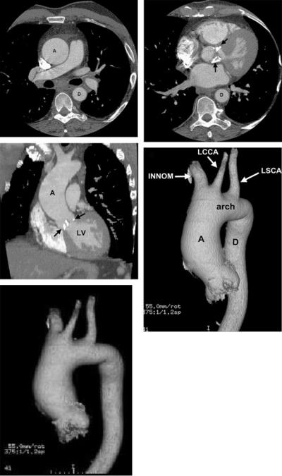
Figure 28. Ascending thoracic aortic aneurysm in a patient with calcific aortic stenosis. Top left, Axial CT image demonstrates an enlarged ascending thoracic aorta (A) and normal caliber descending thoracic aorta (D). Top right, Axial CT image demonstrates extensive aortic valve leaflet calcification (arrows). Middle left, Coronal CT image also demonstrates the dilated ascending aorta (A) and aortic valve leaflet calcification (arrows). Middle right, Volume rendered CT image demonstrates the dilated ascending thoracic aorta (A), normal caliber aortic arch and descending thoracic aorta (D) and great vessels with a bovine arch configuration (INN, LCCA, LSCA). Bottom, Volume rendered rotating image of the thoracic aorta can be used to depict the anatomy, particularly the relationship of an aortic abnormality to the great vessels, for surgical planning. The full cine video for the bottom panel is available in the online-only Data Supplement at http://circ.ahajournals.org/cgi/content/full/CIR. 0b013e3181d4739e/DC1. CT indicates computed tomographic imaging; INN, innominate artery; LCCA, left common carotid artery; LSCA, left subclavian artery; and LV, left ventricle.
cits, a murmur of aortic regurgitation, bruits, and findings compatible with possible cardiac tamponade.339–341
(Level of Evidence: C)
The physical findings of thoracic aortic diseases may be subtle indirect manifestations of uncommon underlying genetically predisposing conditions. In evaluating the evidence base for physical examination of patients with thoracic aortic disease, there are no controlled or blinded experimental research studies that have stratified patients into different treatment categories based on physical findings.
For all patients with thoracic aortic disease, the first and foremost issue is to identify those who are acutely at risk
for catastrophic harm as early as possible. Establishing a set of “triggers” or “red flags” may serve as alerts to either exclude or identify life-threatening thoracic aortic disease.
Given the growing awareness of an extensive variety of diseases associated with nonemergent thoracic aortic disease, it is important to be aware of the many different physical findings associated with extracardiovascular etiologies particularly those of genetic origin (see Section 5).
9.1.1.1. Coronary Artery Disease
The frequency of coexisting CAD varies widely among patient subgroups with thoracic aortic disease as does the

Hiratzka et al |
2010 Guidelines on Thoracic Aortic Disease |
e317 |
etiology of the coronary artery abnormalities, if present. Patients with atherosclerotic aneurysms of the descending aorta are at elevated risk for coronary atherosclerosis, particularly if they have multiple atherosclerotic risk factors. While patients with Type A dissection or annuloaortic ectasia may be protected from atherosclerosis,383 patients with Takayasu arteritis may occasionally have inflammatory coronary involvement with coronary aneurysms (less than 10%).384,385 Similarly, an occasional patient with GCA may have coronary artery involvement.386,387 If ascending aortic surgery is being considered, with or without aortic valve surgery, then identification of the coronary anatomy and any underlying CAD is important for planning the best operation.
9.1.1.2. Emboli
Embolization of material thrombus, atheromatous debris, or cholesterol crystals may affect any distal arterial bed (see Section 11.3). Embolization may occur in patients with thoracic aortic aneurysms or atheromas and in those who have undergone angiography, major vessel surgery, or thrombolytic therapy.388 –397 Clinical consequences of such embolization vary considerably, from being completely asymptomatic to presenting with acute multiorgan failure, including progressive renal failure or cutaneous involvement, with a mortality rate as high as 70% to 90%.398
9.1.1.3. Associated Renal Ischemia
Renal complications of thoracic aortic disease may be acute, subacute, and chronic.399 Patients may present with severe abdominal or flank pain, hematuria, fever, nausea, or a combination of these signs and symptoms.400
9.1.1.4. Associated Mesenteric Ischemia
Patients with acute intestinal ischemia have severe abdominal pain that is initially out of proportion to physical findings.401 Hours to days later, peritonits and sepsis correlate with
Figure 29. Descending aneurysm classification. Descending aneurysms are classified as involving thirds of the descending thoracic aorta and various combinations. A involves the proximal third, B the middle third, and C as the distal third. Thus, an aneurysm involving the proximal two thirds is an AB extent aneurysm. Practically, these groupings can be combined into proximal or distal aneurysm, because these extents influence the risk of paralysis after either open or endovascular repairs. Thoracoabdominal aneurysms are classified according to the Crawford classification: Type I extends from proximal to the sixth rib and extends down to the renal arteries. Type II extends from proximal to the sixth rib and extends to below the renal arteries. Type III extends from distal to the sixth rib but from above the diaphragm into the abdominal aorta. Type IV extends from below the diaphragm and involves the entire visceral aortic segment and most of the abdominal aorta. Juxtarenal and supraenal aneurysms are excluded.379 –382 Image reprinted with permission from the Cleveland Clinic Foundation.
intestinal perforation. Findings suggestive of intestinal ischemia as well as specific arterial or venous obstruction require further surgical or vascular specialist evaluation.402– 406
9.1.1.5. Associated Peripheral Ischemia
Acute limb ischemia results in pain, pallor, paraesthesias, and paralysis.4 Noninvasive vascular diagnostic testing (eg, ankleand toe-brachial indices, segmental pressure measurements, pulse volume recordings, duplex ultrasound imaging, and Doppler waveform analysis) may document ischemia with additional use of angiographic imaging when necessary.407
9.1.2. Differential Diagnosis
9.1.2.1. Symptoms
Symptoms are most commonly related to pain or discomfort. Particularly large thoracic aneurysms may be associated with chest discomfort. Rarely, dysphagia (dysphagia lusoria) or dyspnea is present, usually related to congenital distal arch lesions, such as aberrant right subclavian artery and Kommerell diverticulum or Felson and Palayew Type I or II right-sided aortic arch lesions.408
History of fevers may be related to inflammatory disease or mycotic aneurysms. Occasionally, with chronic dissection and leaking aneurysms, the reabsorption of blood may be associated with fever or jaundice.
9.1.2.2. Physical Findings
Most physical findings are not specific for thoracic aortic disease. Other findings may be related to genetic syndromes and connective tissue disorders (see Section 5) or inflammatory diseases (see Section 7). Findings associated with coarctation of the aorta include brachial-femoral pulse delay and murmurs.

e318 |
Circulation |
April 6, 2010 |
|
|
Table 13. Studies of Medical Treatment of Thoracic Aortic Aneurysm |
||||
|
|
|
|
|
Treatment |
|
|
Studies |
Results |
|
|
|
||
Beta blockers |
Genoni M, Paul M, Jenni R, et al410 |
Retrospective, case-record review of 78 patients with chronic Type B dissection who received |
||
|
|
|
|
medical treatment. 51 of 71 received beta-blocker treatment, 20 of 71 were treated with |
other antihypertensive drugs. 10 of 51 (20%) of the beta-blocker–treated patients and 9 of 20 (45%) from the other treatment group needed dissection-related surgery (P 0.002). The incidence of increasing aortic diameter was 12% (6 of 51) in the beta-blocker group and 40% (8 of 20) in the other treatment group (P 0.002).
Shores J, Berger KR, Murphy EA,
et al88
Open-label, randomized, control study of propranolol in 70 patients with Marfan syndrome. The treated group received a mean daily propranolol dose of 212 68 mg/d. Propranolol therapy slowed aortic root dilation (0.023 vs 0.084 per year, P 0.001).
|
Ladouceur M, Fermanian C, |
Retrospective evaluation of aortic dilation in children with Marfan syndrome. Aortic dilatation |
|
Lupoglazoff JM, et al411 |
was slowed by 0.2 mm/y in children treated with beta blockers. |
Angiotensin-converting |
Ahimastos AA, Aggarwal A, D’Orsa |
Randomized, double-blind, placebo-controlled trial of 17 patients with Marfan syndrome |
enzyme inhibitors |
KM, et al412 |
taking beta-blocker therapy to perindopril or placebo. After 24 weeks of therapy, the |
|
|
perindopril-treated subjects compared with placebo-treated subjects had smaller growth in |
|
|
the ascending aortic diameter during systole (1.2 vs 0.3 mm/m2, P 0.01) and a significant |
|
|
reduction in ascending aortic diameter during diastole (0.4 vs 1.2 mm/m2, P 0.001), |
|
|
respectively. |
Angiotensin receptor |
Mochizuki S, Dahlof B, Shimizu M, |
blockers |
et al413 |
3081 Japanese patients with hypertension, coronary heart disease, heart failure, or a combination were randomly assigned either to open-label valsartan (40 to 160 mg/d) or to other treatment without angiotension receptor blockers. Patients randomized to valsartan had reduction in composite cardiovascular outcome (OR 0.61, 95% CI 0.47 to 0.79) and reduction in aortic dissection (OR 0.18, 95% CI 0.04 to 0.88). Open-label, randomized.
Brooke BS, Habashi JP, Judge DP,
et al89
The clinical response to angiotension receptor blockers (losartan in 17 patients and irbesartan in 1 patient) were evaluated in pediatric patients with Marfan syndrome with severe aortic root enlargement. The mean ( SD) rate of change in aortic root diameter decreased significantly from 3.54 2.87 mm/y during previous medical therapy to 0.46 0.62 mm/y during angiotension receptor blocker therapy (P 0.001). The deviation of aortic root enlargement from normal, as expressed by the rate of change in z scores, was reduced by a mean difference of 1.47 z scores/y (95% CI 0.70 to 2.24, P 0.001) after the initiation of angiotension receptor blocker therapy. The sinotubular junction showed a reduced rate of change in diameter during angiotension receptor blocker therapy (P 0.05), whereas the distal ascending aorta was not affected by angiotension receptor blocker therapy.
Statins |
Diehm N, Decker G, Katzen B, |
|
et al414 |
A nonrandomized propensity-score–adjusted study of statin use effect on long-term mortality of patients after endovascular repair of AAA (731 patients) or TAA (59 patients) was done. Statin use was associated with decreased long-term mortality in patients with AAA (adjusted HR 0.613, 95% CI 0.379 to 0.993, P 0.047), but not for patients with TAA (adjusted HR 1.795, 95% CI 0.147 to 21.942; P 0.647).
AAA indicates abdominal aortic aneurysm; CI, confidence interval; SD, standard deviation; and TAA, thoracic aortic aneurysm.
9.1.3. Considerations for Imaging
Because most cases of chronic thoracic aortic disease are asymptomatic and difficult to detect on physical examination, the clinician must have a low threshold for screening for thoracic aortic disease. CT or MR is required to adequately visualize the affected aorta. There has been no cost– benefit analysis of screening in these populations (see Section 18.1).
9.2. General Medical Treatment and Risk Factor Management for Patients With Thoracic
Aortic Disease
9.2.1. Recommendation for Medical Treatment of Patients With Thoracic Aortic Diseases
Class I
1.Stringent control of hypertension, lipid profile optimization, smoking cessation, and other atherosclerosis risk-reduction measures should be instituted for patients with small aneurysms not requiring surgery, as well as for patients who are not considered
to be surgical or stent graft candidates. (Level of Evidence: C)
The patient’s general status should be optimized where possible. Respiratory illness, a common comorbid problem, may be improved by stopping smoking, clearing bronchitis, and exercising regularly with walking. Because other atherosclerotic disease is usually present in patients with thoracic aortic aneurysms or atheroma, risk-reduction measures as outlined in other guidelines are appropriate.409 Additional medical management rationales are noted in Table 13.
Patients who are not candidates for operative intervention include those whose aneurysms or other aortic pathology do not meet the criteria for surgical intervention and those in whom the criteria are met but who are considered inoperable, most commonly because of coexisting disease. Patients with large aneurysms who are considered inoperable may benefit from stringent control of risk factors (see Section 3.2) to potentially slow the rate of growth and reduce the risk of rupture or dissection. Recommendations for periodic imaging are noted in Section 14.
Hiratzka et al |
2010 Guidelines on Thoracic Aortic Disease |
e319 |
9.2.1.1. Recommendations for Blood Pressure Control
Class I
1.Antihypertensive therapy should be administered to hypertensive patients with thoracic aortic diseases to achieve a goal of less than 140/90 mm Hg (patients without diabetes) or less than 130/80 mm Hg (patients with diabetes or chronic renal disease) to reduce the risk of stroke, myocardial infarction, heart failure, and cardiovascular death.415– 419 (Level of Evidence: B)
2.Beta adrenergic– blocking drugs should be administered to all patients with Marfan syndrome and aortic aneurysm to reduce the rate of aortic dilatation unless contraindicated.88 (Level of Evidence: B)
Class IIa
1.For patients with thoracic aortic aneurysm, it is reasonable to reduce blood pressure with beta blockers and angiotensin-converting enzyme inhibitors412 or angiotensin receptor blockers89,413 to the lowest point patients can tolerate without adverse effects.88,410,411 (Level of Evidence: B)
2.An angiotensin receptor blocker (losartan) is reasonable for patients with Marfan syndrome, to reduce the rate of aortic dilatation unless contraindicated.89,90 (Level of Evidence: B)
Treatment of hypertension to reduce end points of MI, stroke, and death is well established with many randomized clinical trials.420 In the Jikei Heart Study, Japanese patients who received valsartan along with other antihypertensive therapy had a significantly lower rate of cardiovascular morbidity and mortality compared with patients treated without valsartan. Reductions noted in particular included lower incidence of stroke, transient ischemic attack (TIA), angina pectoris, and heart failure. Moreover, pertinent to this guideline, there was a significant reduction in the incidence of AoD in the valsartan-treated patients, which contributed to the reduction in overall cardiovascular morbidity and mortality.413
Currently, beta adrenergic blockade serves as the foundation of the medical regimen because of demonstrated inhibition of aneurysm expansion in patients with Marfan syndrome. Shores and colleagues88 randomized 70 patients with Marfan syndrome to propranolol or placebo in a open-label study demonstrating an attenuated rate of expansion over the 10-year follow-up. Dietz and colleagues91 demonstrated that angiotensin receptor blocker therapy reduces aneurysm expansion in animal models of Marfan syndrome. This group has also recently demonstrated that angiotension receptor blocker therapy slowed the rate of progression of progressive aortic root dilatation in a preliminary study of 18 pediatric patients with Marfan syndrome.89 Both beta blockade and angiotensin II receptor blockade therapy are being further investigated in a randomized trial for patients with Marfan syndrome.90
Lifestyle modifications of diet, weight reduction for overweight or obese patients, moderation of alcohol consumption,
and aerobic exercise are standard approaches to treat hypertension,421 but pharmacological therapy is usually required for patients with thoracic aortic diseases.
9.2.1.2. Recommendation for Dyslipidemia
Class IIa
1.Treatment with a statin to achieve a target LDL cholesterol of less than 70 mg/dL is reasonable for patients with a coronary heart disease risk equivalent such as noncoronary atherosclerotic disease, atherosclerotic aortic aneurysm, and coexistent coronary heart disease at high risk for coronary ischemic events.422– 425 (Level of Evidence: A)
The National Cholesterol Education Program ATP III recommends that patients with noncoronary atherosclerosis be treated like patients with established coronary heart disease.426 Atherosclerosis in the aorta, like atherosclerosis in any noncoronary vascular bed, markedly increases the risk of MI and stroke. As a result of this high-risk status (greater than 20% event rate in 10 years), the goal for hypolipidemic therapy is an LDL level less than 100 mg/dL. Initial therapy in these patients should be a statin. After the National Cholesterol Education Program ATP III guidelines were released in 2001, the Heart Protection Study reported in 2002 that patients with atherosclerosis and a total cholesterol level greater than 135 mg/dL benefited from the addition of simvastatin 40 mg/d.427 The RR reductions remained even when LDL started at less than 100 mg/dL. In concert with data from patients with acute coronary syndromes, the more recent ACC/AHA Guidelines for the Management of Patients With Peripheral Arterial Disease also gave a Class IIa recommendation suggesting the use of a statin to achieve a target LDL of less than 70 mg/dL for patients at very high risk of ischemic events is reasonable.4
There are experimental data demonstrating a delayed development of atherosclerosis and prevention of aneurysm development by statins.428 – 430 However, there are no clinical outcomes data that justify their use acutely or suggest that statins prevent expansion after thoracic aortic aneurysms have developed.
9.2.1.3. Recommendation for Smoking Cessation
Class I
1.Smoking cessation and avoidance of exposure to environmental tobacco smoke at work and home are recommended. Follow-up, referral to special programs, and/or pharmacotherapy (including nicotine replacement, buproprion, or varenicline) is useful, as is adopting a stepwise strategy aimed at smoking cessation (the 5 A’s are Ask, Advise, Assess, Assist, and Arrange).431– 432b (Level of Evidence: B)
There are no randomized or prospective trials that have investigated the effect of smoking cessation on thoracic aortic disease. Patients with thoracic aortic aneurysm who smoke have double the rate of aneurysm expansion.433 Aneurysm expansion and rupture after Type B dissection are not

e320 |
Circulation |
April 6, 2010 |
affected by cigarette smoking.434 Smoking cessation reduces the rate of MI and death in patients with noncoronary atherosclerosis.435 Patients who smoke require close follow-up in conjunction with medical and other support to achieve complete smoking cessation.
9.2.2. Surgical and Endovascular Treatment by Location of Disease
9.2.2.1. Ascending Aorta and Aortic Sinuses
9.2.2.1.1. Recommendations for Asymptomatic Patients With Ascending Aortic Aneurysm
Class I
1.Asymptomatic patients with degenerative thoracic aneurysm, chronic aortic dissection, intramural hematoma, penetrating atherosclerotic ulcer, mycotic aneurysm, or pseudoaneurysm, who are otherwise suitable candidates and for whom the ascending aorta or aortic sinus diameter is 5.5 cm or greater, should be evaluated for surgical repair.371 (Level of Evidence: C)
2.Patients with Marfan syndrome or other genetically mediated disorders (vascular Ehlers-Danlos syndrome, Turner syndrome, bicuspid aortic valve, or familial thoracic aortic aneurysm and dissection) should undergo elective operation at smaller diameters (4.0 to 5.0 cm depending on the condition; see Section 5) to avoid acute dissection or rup-
ture.81,114,143,371,436 – 439 (Level of Evidence: C)
3.Patients with a growth rate of more than 0.5 cm/y in an aorta that is less than 5.5 cm in diameter should be considered for operation. (Level of Evidence: C)
4.Patients undergoing aortic valve repair or replacement and who have an ascending aorta or aortic root of greater than 4.5 cm should be considered for concomitant repair of the aortic root or replacement of the ascending aorta. (Level of Evidence: C)
Class IIa
1.Elective aortic replacement is reasonable for patients with Marfan syndrome, other genetic diseases, or
bicuspid aortic valves, when the ratio of maximal ascending or aortic root area ( r2) in cm2 divided by the patient’s height in meters exceeds 10.16,143 (Level of Evidence: C)
2.It is reasonable for patients with Loeys-Dietz syndrome or a confirmed TGFBR1 or TGFBR2 mutation to undergo aortic repair when the aortic diameter reaches 4.2 cm or greater by transesophageal echocardiogram (internal diameter) or 4.4 to 4.6 cm or greater by computed tomographic imaging and/or magnetic resonance imaging (external diameter).78
(Level of Evidence: C)
Aortic diameter is a major criterion for recommending elective operation in asymptomatic patients with aneurysm of the thoracic and thoracoabdominal aorta. This assumes that the risk of operation is low (less than 5%). Currently, aortic diameter perpendicular to the axis of flow as measured by CT is the dimension most often used to determine the size of the enlarged
Figure 30. Effect of aortic aneurysms diameter on risk of complication. For thoracic aortic aneurysms of all etiologies. Adapted from Elefteriades et al.437
aorta. This recommendation is based on the observation that the risk of an adverse event (rupture, dissection, death) exceeds the risk of elective operation when the maximum diameter exceeds 5.5 to 6.0 cm374,436,437,440 (Figure 30). Formulas that incorporate height and aortic cross-sectional area have been developed to establish thresholds for operation in shorter patients but are less widely used.16,143
The morphology and histopathology of thoracic aortic enlargements affect the natural history of aortic diseases, including the risk of rupture or dissection, and thus can influence the decision to intervene (Figures 31 and 32). Fusiform aneurysms are most common and behave in a relatively predictable manner. Aortic dimension can thus be used as an indication for operation. Saccular aneurysms occur less frequently and may be associated with a greater risk of rupture. Many of these are actually pseudoaneurysms, which can develop after previous trauma or aortic surgery, or with PAUs, and which result in focal disruption or weakening of the layers of the aorta.
Patients with substantial dilatation of the aortic sinuses may develop asymptomatic aortic regurgitation as a result of loss of coaptation of the otherwise normal aortic valve cusps. Patients with associated bicuspid aortic valve disease may have asymptomatic stenosis or regurgitation of the valve. In these patients, the valvular disease may be an indication for operative intervention.5
9.2.2.1.2. Recommendation for Symptomatic Patients With Thoracic Aortic Aneurysm
Class I
1.Patients with symptoms suggestive of expansion of a thoracic aneurysm should be evaluated for prompt surgical intervention unless life expectancy from comorbid conditions is limited or quality of life is substantially impaired. (Level of Evidence: C)
Symptoms associated with thoracic aneurysms usually develop later in the course of enlargement of the aorta and most commonly result from impingement of the aneurysm on adjacent structures. Aneurysms of the ascending aorta and aortic sinuses may result in symptoms related to the

Hiratzka et al |
2010 Guidelines on Thoracic Aortic Disease |
e321 |
Figure 31. Ascending aortic aneurysm of degenerative etiology. CABG indicates coronary artery bypass graft surgery; CAD, coronary artery disease; CT, computed tomographic imaging; and MR, magnetic resonance imaging.
aortic regurgitation that develops as a result of the progressive aortic enlargement. Chest or back pain in the presence of an enlarged thoracic aorta is a predictor of aortic rupture.336,441 Patients who develop an acute Type A AoD commonly present with severe chest or back pain.
9.2.2.1.3. Endovascular Grafting for Ascending Aortic Aneurysm. At the time of this writing, endovascular stent grafts have not been approved by the US Food and Drug Administration for treatment of aneurysms or other conditions of the ascending aorta.
9.2.2.1.4. Recommendations for Open Surgery for Ascending Aortic Aneurysm
Class I
1.Separate valve and ascending aortic replacement are recommended in patients without significant aortic root dilatation, in elderly patients, or in young patients with minimal dilatation who have aortic valve disease. (Level of Evidence: C)
2.Patients with Marfan, Loeys-Dietz, and EhlersDanlos syndromes and other patients with dilatation

e322 |
Circulation |
April 6, 2010 |
Figure 32. Ascending aortic aneurysms associated with genetic disorder. *Depends on specific genetic condition. †See Recommendations for Asymptomatic Patients With Ascending Aortic Aneurysm (Section 9.2.2.1.1) and Recommendations for Bicuspid Aortic Valve and Associated Congenital Variants in Adults (Section 6.1). CABG indicates coronary artery bypass graft surgery; CAD, coronary artery disease; CT, computed tomographic imaging; and MR, magnetic resonance imaging.
of the aortic root and sinuses of Valsalva should undergo excision of the sinuses in combination with a modified David reimplantation operation if technically feasible or, if not, root replacement with valved graft conduit.134,164,442–444 (Level of Evidence: B)
Ascending Aortic Aneurysms: The extent of aortic resection and the need for ancillary procedures are determined by preoperative testing and intraoperative findings. Ancillary procedures that may be performed concurrently include coronary artery bypass graft surgery, valve replacement or
repair, repair of cardiac septal defects, closure of vascular fistulas, and ablative therapy for arrhythmias.
For patients with isolated aneurysms confined to the ascending aorta, resection and graft replacement is the most commonly performed and recommended procedure. Alternatively, reduction aortoplasty with or without external reinforcement has only been performed in very limited circumstances.445,446
For patients with aortic valve stenosis who require valve replacement, the choice of valve substitute is determined by age of the patient, presence of comorbid disease, risk of
Hiratzka et al |
2010 Guidelines on Thoracic Aortic Disease |
e323 |
complications related to anticoagulation and reoperation, and life expectancy.139
For patients with aortic regurgitation associated with a bicuspid aortic valve, repair of the aortic valve with or without root remodeling or tailoring of the sinotubular junction is preferable if the valve is not severely fibrotic or calcified.99,140 For patients with a dilated aortic root, particularly those with stenotic bicuspid valves, composite valve grafts containing either mechanical or biological valves are implanted.
Ascending aneurysms larger than 4.5 to 5.0 cm require repair or tube graft replacement when aortic valve repair or replacement is the primary indication for operation.5 In elderly patients, ascending aortic aortoplasty when the aortic diameter does not exceed 5.0 cm may be an acceptable alternative.
Aortic Valve and Root: In patients with aortic valve regurgitation and root dilatation, aortic valve repair and rootsparing procedure may be the primary procedure. In patients with Marfan syndrome or with tricuspid aortic valve regurgitation, a modification of the David reimplantation operation may be considered. 94,95,97–99,447 Composite valve grafts with either biological or mechanical valves are an alternative option, particularly for valvular stenosis.99,140,441,448
9.2.2.2. Recommendations for Aortic Arch Aneurysms
Class IIa
1.For thoracic aortic aneurysms also involving the proximal aortic arch, partial arch replacement together with ascending aorta repair using right subclavian/axillary artery inflow and hypothermic circulatory arrest is reasonable.222,449,450 (Level of Evidence: B)
2.Replacement of the entire aortic arch is reasonable for acute dissection when the arch is aneurysmal or there is extensive aortic arch destruction and leakage.222,450 (Level of Evidence: B)
3.Replacement of the entire aortic arch is reasonable for aneurysms of the entire arch, for chronic dissection when the arch is enlarged, and for distal arch aneurysms that also involve the proximal descending thoracic aorta, usually with the elephant trunk procedure.451– 453 (Level of Evidence: B)
4.For patients with low operative risk in whom an isolated degenerative or atherosclerotic aneurysm of the aortic arch is present, operative treatment is reasonable for asymptomatic patients when the diameter of the arch exceeds 5.5 cm.374 (Level of Evidence: B)
5.For patients with isolated aortic arch aneurysms less than 4.0 cm in diameter, it is reasonable to reimage using computed tomographic imaging or magnetic resonance imaging, at 12-month intervals, to detect enlargement of the aneurysm. (Level of Evidence: C)
6.For patients with isolated aortic arch aneurysms 4.0 cm or greater in diameter, it is reasonable to reimage using computed tomographic imaging or magnetic resonance imaging, at 6-month intervals,
to detect enlargement of the aneurysm. (Level of Evidence: C)
Aneurysms of the aortic arch are commonly associated with aneurysmal disease or dissection of the ascending aorta or the adjacent descending thoracic aorta, and the indications for operative intervention in these patients are those for the adjacent aortic segment. This relates to the need for hypothermic cardiopulmonary bypass and an interval of hypothermic circulatory arrest, and to higher operative mortality and stroke rates than those observed following operation for isolated aneurysms of the ascending or descending thoracic aorta.451– 459 As with ascending aneurysms, a growth rate of more than 0.5 cm/y in the absence of symptoms could be considered an indication for operation.
Symptoms associated with aortic arch aneurysms such as hoarseness resulting from stretching of the left recurrent laryngeal nerve, dysphagia, dyspnea, and chest or back pain are indications for operative intervention for patients with arch aneurysms unless life expectancy is quite limited. Suitability for operative intervention involves similar risk assessment to that for aneurysm or other disorders of the ascending aorta and aortic root.
The innominate, left carotid, and left subclavian arteries may require separate grafting. For short periods of circulatory arrest, the use of retrograde or antegrade brain perfusion has not conclusively been shown to add further brain protection; however, use of the subclavian or axillary artery bypass with a side graft reduces the risk of stroke.449
9.2.2.2.1. Open Surgery. At present, endovascular stent grafts have not been approved by the US Food and Drug Administration for treatment of aneurysms or other conditions of the aortic arch. For patients with large aneurysms who are not candidates for conventional open operation, experience is accumulating with operative procedures that involve translocation of the brachiocephalic arteries from the aortic arch using branch grafts from the proximal ascending aorta, and placement of an endovascular graft into the distal ascending aorta, the entire aortic arch, and a segment of the adjacent descending thoracic aorta.371,460,461
Cardiopulmonary bypass with some degree of hypothermia is required for operations that require replacement of the aortic arch. Brain protection can be achieved by profound hypothermia alone, direct antegrade perfusion of 1 or more of the brachiocephalic arteries, or retrograde perfusion using cold oxygenated blood that is infused into the superior vena cava during the arrest period211,449,462– 467 (see Section 14.5.1). The aortic arch is replaced with a synthetic graft. The brachiocephalic arteries are attached to the graft using a patch of the aorta which contains the origins of the 3 vessels or separately using a graft that contains 3 branches. The proximal and distal ends of the aortic graft are attached to normal segments of ascending and descending thoracic aorta.
An “elephant trunk” procedure has been used to reconstruct the arch and then provide a Dacron graft landing zone for endovascular stent graft treatment of descending thoracic aortic aneurysms (Figure 33).