

Аорта
.pdfe304 |
Circulation |
April 6, 2010 |
versus 23%, respectively; P 0.01).242 Additionally, patients who presented with syncope more frequently had associated cardiac tamponade, stroke, decreased consciousness, and evidence of spinal cord ischemia.242
8.1.7. Neurologic Complications
Acute AoD frequently presents with dissection-related neurologic complications. Pooled data from more than 1300 patients in 13 studies that included both Type A and B dissections found that neurologic symptoms were reported in 17%.37 Neurologic complications may result from hypotension, malperfusion, distal thromboembolism, or nerve compression.251,253,254 In a recent study of 102 patients with Type A AoD, 29% had neurologic symptoms on initial presentation253; of those with neurologic symptoms, 53% had ischemic stroke (predominantly right hemispheric) and 37% had ischemic neuropathy (described as limb pain with sensory or motor deficit).253
Last, although uncommon, acute paraplegia as a result of spinal cord malperfusion has been described as a primary manifestation of thoracic AoD, occurring in 1% to 3% of
patients.215,251,253,254,268,269
Of clinical note, up to 50% of dissection-related neurologic symptoms may be transient and as many as one-third of patients with neurologic symptoms present without complaints of chest pain, complicating appropriate diagnosis and
treatment.253,255,256,262,269 –271
8.1.8. Pulmonary Complications
Pleural effusion, the most common pulmonary complication of acute AoD, is noted in 16% of cases at presentation37; whereas large effusions may result from leaking of blood from the aorta into the pleural space, small effusions are typically a nonhemorrhagic exudate believed to be inflammatory in origin.37,272–275
Other pulmonary complications of acute AoD include dissection-related compression of the pulmonary artery and development of an aortopulmonary fistula, either of which may present with dyspnea as a prominent symptom.276 –278 Hemoptysis, noted in 1 study in 3% of patients presenting with thoracic AoD, may result from compression of lung parenchyma by an expanding false lumen or via direct aneurysmal rupture into the lung, leading to massive hemoptysis and death.215,279,280
8.1.9. Gastrointestinal Complications
Mesenteric ischemia is the most common gastrointestinal complication of acute AoD228 and can result from malperfusion or systemic hypotension. It is the most common cause of death among those with Type B AoD. Mesenteric ischemia is associated with abdominal pain, but pain may be nonspecific and out of proportion to the physical examination of the abdomen, so the cause of pain often goes unrecognized early on. Unfortunately, by the time serum markers of bowel ischemia or infarction turn positive, it is often too late to salvage the bowel or the patient. Therefore, it is essential to be viligant for mesenteric ischemia in every patient with acute AoD and associated abdominal pain.
Gastrointestinal hemorrhage is a rare but potentially catastrophic complication of acute AoD.281–283 Dissection-related
gastrointestinal bleeding may present with limited bleeding as a result of mesenteric infarction or as massive hemorrhage secondary to an aortoesophageal fistula or false lumen rupture into proximal small bowel.281–283 Although rare, dissection-related gastrointestinal hemorrhage should be in the differential of all patients presenting with bleeding and complaints of thoracic or abdominal pain.
8.1.10. Blood Pressure and Heart Rate Considerations
Blood pressure abnormalities are common in patients presenting with acute thoracic AoD. About half of patients are hypertensive at presentation, with 71% of Type B patients having a systolic blood pressure greater than 150 mm Hg versus only 36% of Type A patients.284 –287 Conversely, nearly 20% present with either hypotension or shock.37 Hypotension and shock can result from cardiac tamponade, aortic hemorrhage, severe aortic insufficiency, myocardial ischemia or infarction, true lumen compression by distended false lumen, or an intra-abdominal catastrophe. Of more than 1000 patients with acute AoD, those with hypotension on admission were found more likely to have neurologic complications; myocardial, mesenteric, or limb ischemia; and death.249
Accurate systemic blood pressure measurement may be complicated by dissection-related occlusion of aortic branch arteries, resulting in erroneously low blood pressure readings in the affected limb. Accordingly, blood pressures may need to be measured in both arms and, at times, both legs to determine the highest central blood pressure.
8.1.11. Age and Sex Considerations
Acute AoD presentation varies with patient age and sex. In 951 IRAD patients, 7% were younger than 40 years. Compared with patients 40 years of age and older, this group was less likely to have a history of hypertension and significantly more likely to have Marfan syndrome, bicuspid aortic valve, or a history of prior aortic surgery.223 Clinically, young patients in this study were more likely to describe pain as abrupt in onset but less likely to be hypertensive at presentation (25% versus 45%).223 In contrast, a separate study of IRAD data evaluating 550 patients with Type A dissection found that among patients more than 70 years of age (32% of total), typical symptoms (abrupt onset of pain) and signs (murmur of aortic regurgitation or pulse deficits) were significantly less common, suggesting that extra vigilance may be required to identify acute AoD in young and elderly patients.288
Sex appears to affect the presentation of acute AoD as well. In a study of 1078 patients enrolled in IRAD, 32% were women. Women were older; were less likely to present within 6 hours of symptom onset, to complain of abrupt onset of pain, and to have a pulse deficit; and were more likely to present with either altered mental status or congestive heart failure. Consequently, women were less likely to be diagnosed within 24 hours of symptom onset and had significantly higher in-hospital mortality (30% versus 21%, P 0.001) than men.289
8.2. Intramural Hematoma
Among acute aortic syndromes, acute dissection is the most common, but approximately 10% to 20% of patients290 –292

Hiratzka et al |
2010 Guidelines on Thoracic Aortic Disease |
e305 |
Figure 23. Intramural hematoma demonstrated as a low-attenuation band of hematoma (arrows) in the aortic wall on CT images. Top left, Axial image at the level of the aortic arch. Top middle, Through the mid thorax. Top right, At the level of the superior mesenteric artery with narrowing of the aortic lumen. Bottom left, Oblique sagittal reformatted image through the thorax (note band artifact evident without the use of ECG gating). Bottom right, Coronal reformatted image through the abdomen demonstrate the length of the hematoma, and an incidental infarenal aortic aneurysm. CT indicates computed tomographic imaging; and ECG, electrocardiogram.
with a clinical picture of dissection exhibit an IMH via imaging without identification of blood flow in a false lumen or an intimal lesion. Some believe IMH arises from hemorrhage of the vasa vasorum located within the medial layer of the aorta,293,294 whereas others argue that the hematoma arises from microscopic tears in the aortic intima. The resulting hematoma may then propagate in an antegrade or a retrograde manner, producing symptoms that may be impossible to differentiate clinically from those of a classic AoD.61 IMH has a variable radiologic appearance (Figure 23) according to the area of the aorta involved. In some cases, IMH may be associated with a PAU (see Section 8.2 or 8.3).
Clinically, IMH most commonly occurs in the descending aorta and in older patients. Pain is characteristic of IMH, whereas malperfusion and pulse deficit are much less likely than with classic AoD.295,296
Imaging criteria of IMH are based on the appearance of fresh thrombus in the aortic wall. These include crescentic or circular thickening of the aortic wall with maximal thickness greater than or equal to 7 mm on TEE without intimal flap or tear or longitudinal flow in the false lumen. The thickened wall has a higher tissue density than unenhanced blood on CT and is without enhancement after contrast on the CT/ MR.290,296 –298 When the term IMH is used strictly, no intimal defect such as a tear or an ulcer is present. But in practice, the term is used loosely to mean a thrombosed false lumen regardless of a small intimal defect. The distinction is further blurred by the facts that the intimal defect may be subtle and difficult to exclude and that some patients with IMH begin with a CT scan that shows a thrombosed false lumen with no apparent intimal defect and then over the course of 1 or 2
months develop 1 or more distinct ulcerlike communications.299,300 Because of this overlap in imaging findings, it is difficult and perhaps somewhat arbitrary to base treatment on the appearance of the CT snapshot of the aorta in its disease progression.
The natural history of IMH is variable. The hematoma may entirely resolve (10%),301 it may convert to a classic dissection, or the aorta may enlarge and potentially rupture. The clinical behavior of IMH varies according to the location and mimics that of classic AoD. IMH involving the ascending aorta has a high, early risk of complication and death with medical treatment alone, and surgery is usually indicated. IMH involving the descending aorta may be treated with blood pressure control, and the use of beta blockers has been shown to improve the long-term survival rate.296 Conversion of the IMH to a more classic picture of dissection occurs in 3% to 14% of cases involving the descending aorta297,298,302,303 and in 11% to 88% of cases involving the ascending aorta,302–305 with that figure increasing with increased length of follow-up. Progressive increase in aortic diameter has been demonstrated by serial imaging studies.298,306 In 1 study, the mortality after 2 years of patients with acute proximal Type A IMH versus that of patients with classic dissection was not significantly different.307 Another group found improved actuarial survival rates at 1, 2, and 5 years in patients with IMH versus classic dissection: 90%, 90%, and 90% versus 67%, 66%, and 62%, respectively, for Type A,304 and 100%, 97%, and 97% versus 83%, 79%, and 79%, respectively, for Type B.297 Song et al also described increased risk for complications or mortality for patients with IMH involving the ascending aorta when ascending aortic

e306 |
Circulation |
April 6, 2010 |
Figure 24. Penetrating atherosclerotic ulcer of the proximal descending thoracic aorta. Axial CT images at the level of the aortopulmonary window (left) and at the level of the left pulmonary artery (right) demonstrate a small penetrating ulcer (long arrow, U) that extends beyond the expected confines of the aortic lumen with adjacent IMH both at the level of the ulcer itself and that extends a few centimeters caudally in the wall of the descending thoracic aorta (short arrows). CT indicates computed tomographic imaging; IMH, intramural hematoma; and U, penetrating ulcer.
diameter is greater than 4.8 cm or IMH thickness is greater than 11 mm.308
8.3. Penetrating Atherosclerotic Ulcer
PAU refers to an atherosclerotic lesion with ulceration that penetrates the internal elastic lamina and allows hematoma formation within the media of the aortic wall.309 This lesion sets the stage for development of IMH, AoD, or frank vessel rupture309 (Figure 24). Anatomically PAUs develop in aortic segments where atherosclerotic changes are most common and therefore are localized to the descending thoracic aorta in over 90% of cases.310 When viewed tangentially, the classic appearance of the lesion is a mushroom-like outpouching of the aortic lumen with overhanging edges, resembling a gastric ulcer, as depicted on a barium study (Figure 24). The typical patient is elderly (usually over 65 years of age) and has hypertension and diffuse atherosclerosis, who presented with chest or back pain but without signs of aortic regurgitation or malperfusion. Less commonly, patients presented only with signs of distal embolization.311 Asymptomatic patients may also be found with aortic lesions that are indistinguishable, by imaging criteria, from PAUs.310,311
Two entities can mimic PAUs. A branch artery pseudoaneurysm represents a small collection of flowing blood within an otherwise thrombosed aortic false lumen, which is created by an injury to a small branch artery during the propagation of the IMH.312 These are usually incidental findings, are distinguished from ulcers by the apparent absence of a communication with the aortic lumen by CT, and do not usually require specific treatment. A dissection entry or reentry tear may develop in an area of IMH as detected by several imaging studies over a period of several
months.297,299,300,306
8.4. Pseudoaneurysms of the Thoracic Aorta
Pseudoaneurysms of the thoracic aorta are frequently related to deceleration or torsional trauma to the aorta from motor vehicle accidents, falls, and sports injuries.3,313–319 Aortic pseudoaneurysms are relatively rare, with posttraumatic pseudoaneurysms having an incidence of 3% to 4% after blunt trauma.51 Other pseudoaneurysms may occur following aortic surgery, catheter-based interventions, or penetrating trauma. Pseudoaneurysms often have a slim “neck” that leads to the “aneurysm” that corresponds to points of penetration and containment.320 Aortic infections (mycotic aneurysms)
and penetrating ulcers may also result in pseudoaneurysms (see Section 9.2.2). Penetrating injuries are usually repaired immediately whenever recognized and feasible.317,318,321
8.5. Traumatic Rupture of the Thoracic Aorta
A UK survey of all motor vehicle accident fatalities found that approximately 20% of patients had an autopsy finding of a ruptured aorta, emphasizing the importance of traumatic rupture of the aorta (TRA). In the United States, there are around 40 000 motor vehicle deaths annually, and it is likely that around 8000 of the victims had TRA. It is estimated that only 9% to 14% of patients with TRA reach a hospital alive and only 2% ultimately survive. In this survey, 29% were involved with frontal impact crashes and 44% were involved with side impact crashes.318,319
Parmley and colleagues318 noted the correlation of high risk of early death and the sites of TRA on the basis of autopsies in 275 deaths from unrelated aortic rupture. In 45%, the tear was at the aortic isthmus; 23%, in the ascending aorta; 13%, in the descending aorta; 8%, in the transverse aorta; 5%, in the abdominal aorta; and 6%, multiple sites.
Examination of the patient usually reveals signs similar to those of coarctation of the aorta with arm blood pressure higher than leg blood pressure, delay between radial versus femoral artery pulsation, and a harsh interscapular murmur.3 Evidence of polytrauma is, however, common.
The best method for detection of a TRA is debated. A chest x-ray with a nasogastric tube in position has 80% sensitivity for suggesting TRA by showing displacement of the nasogastric tube by the hematoma. However, signs of hemomediastinum are more often false positive than true positive.40 Even when present, mediastinal blood is less likely to be due to arterial/aortic injury than to less-consequential venous bleeding. A biplane contrast aortogram may fail to detect the tear until the development of a pseudoaneurysm. TEE may be used, but if dilatation has not occurred, the diagnosis may still be in doubt. CT is used but is not absolutely certain to establish the diagnosis. In questionable cases, intravascular ultrasound can also be used3,322,323 (Figure 8). Realistically, the imaging sequence often depends on the stability of the patient and the need for the diagnosis of concomitant injuries. Sometimes, this may even fail to detect the tear, and the study may have to be repeated at a later date to detect the tear.
Hiratzka et al |
2010 Guidelines on Thoracic Aortic Disease |
e307 |
8.6. Evaluation and Management of Acute
Thoracic Aortic Disease
8.6.1. Initial Evaluation and Management
8.6.1.1. Recommendations for Estimation of Pretest Risk of Thoracic Aortic Dissection
Class I
1.Providers should routinely evaluate any patient presenting with complaints that may represent acute thoracic aortic dissection to establish a pretest risk of disease that can then be used to guide diagnostic decisions. This process should include specific questions about medical history, family history, and pain features as well as a focused examination to identify findings that are associated with aortic dissection, including:
a.High-risk conditions and historical features47,127,223,261
(Level of Evidence: B):
●Marfan syndrome, Loeys-Dietz syndrome, vascular Ehlers-Danlos syndrome, Turner syndrome, or other connective tissue disease.
●Patients with mutations in genes known to predispose to thoracic aortic aneurysms and dissection, such as FBN1, TGFBR1, TGFBR2, ACTA2, and MYH11.
●Family history of aortic dissection or thoracic aortic aneurysm.
●Known aortic valve disease.
●Recent aortic manipulation (surgical or catheter-based).
●Known thoracic aortic aneurysm.
b.High-risk chest, back, or abdominal pain fea-
tures37,47,215,223,261,264,288 (Level of Evidence: B):
●Pain that is abrupt or instantaneous in onset.
●Pain that is severe in intensity.
●Pain that has a ripping, tearing, stabbing, or sharp quality.
c.High-risk examination features37,47,253,257,261,324
(Level of Evidence: B):
●Pulse deficit.
●Systolic blood pressure limb differential greater than 20 mm Hg.
●Focal neurologic deficit.
●Murmur of aortic regurgitation (new).
2.Patients presenting with sudden onset of severe chest, back, and/or abdominal pain, particularly those less than 40 years of age, should be questioned about a history and examined for physical features of Marfan syndrome, Loeys-Dietz syndrome, vascular Ehlers-Danlos syndrome, Turner syndrome, or other connective tissue disorder associated with thoracic aortic disease.223 (Level of Evidence: B)
3.Patients presenting with sudden onset of severe chest, back, and/or abdominal pain should be questioned about a history of aortic pathology in immediate family members as there is a strong familial component to acute thoracic aortic disease.223 (Level of Evidence: B)
4.Patients presenting with sudden onset of severe chest, back, and/or abdominal pain should be questioned about recent aortic manipulation (surgical or catheter-based) or a known history of aortic valvular disease, as these factors predispose to acute aortic dissection. (Level of Evidence: C)
5.In patients with suspected or confirmed aortic dissection who have experienced a syncopal episode, a focused examination should be performed to identify associated neurologic injury or the presence of pericardial tamponade (see Section 8.1.6). (Level of Evidence: C)
6.All patients presenting with acute neurologic complaints should be questioned about the presence of chest, back, and/or abdominal pain and checked for peripheral pulse deficits as patients with dissectionrelated neurologic pathology are less likely to report thoracic pain than the typical aortic dissection patient253 (see Section 8.1.7). (Level of Evidence: C)
8.6.1.2. Laboratory Testing
Several plasma markers have been investigated for their utility in the evaluation of acute AoD. Plasma smooth muscle myosin heavy chain protein, D-dimer, and high-sensitivity C-reactive protein have shown diagnostic promise, although a lack of large prospective studies precludes a recommendation regarding their use.325–328
Elevation of D-dimer levels occurs with intravascular activation of the coagulation cascade and secondary fibrinolysis and in conditions such as venous thromboembolism, sepsis, disseminated intravascular coagulation, malignancies, recent trauma or surgery, and acute MI and following fibrinolytic therapy. The ACEP has published guidelines regarding the use of certain D-dimer assays to rule out pulmonary embolism in low-risk patients.329
Regarding the potential role of plasma D-dimer levels to screen for AoD, significant elevations of D-dimer were seen in all 24 patients with documented acute AoD involving either the ascending or descending thoracic aorta regardless of time from presentation, ranging from 1 to 120 hours.325 A meta-analysis of 11 studies330 noted that the pooled sensitivity of D-dimer in 349 patients with documented acute AoD was 94% (95% CI 91% to 96%) with specificity ranging from 40% to 100%. Two patients had limited ascending aortic IMH without intimal flap and had negative D-dimer assays.331
Some authors325,332 recommend that D-dimer assays be performed in all patients where clinical suspicion exists, to help identify those who do not require definitive imaging studies. However, the efficacy and safety of this strategy have not been tested in a large clinical trial, and several caveats should apply. The negative likelihood ratio provided by the most sensitive D-dimer assay is not of sufficient magnitude to provide useful information in high-risk individuals and therefore cannot be used to “rule out” the disease in this group. Clinical scoring systems to identify the true pretest probability for AoD in individual patients have not been developed or validated, thus limiting an accurate determination of the true “posttest” probability associated with a negative D-dimer result. Finally, there are reports of negative D-dimer assays
e308 |
Circulation |
April 6, 2010 |
associated with ascending aortic IMH or a thrombosed false lumen, such that further studies are needed regarding the sensitivity of D-dimer levels to detect the presence of IMH or PAU.331 Given these issues, the writing committee cannot recommend serum D-dimer screening for all patients being evaluated for AoD.
Where a high level of suspicion for acute AoD exists, laboratory testing aimed at presurgical screening (blood count, serum chemistries, coagulation profiles, and blood type and screen) may reduce preoperative delays.
8.6.1.3. Recommendations for Screening Tests
Class I
1.An electrocardiogram should be obtained on all patients who present with symptoms that may represent acute thoracic aortic dissection.
a.Given the relative infrequency of dissection-related coronary artery occlusion, the presence of STsegment elevation suggestive of myocardial infarction should be treated as a primary cardiac event without delay for definitive aortic imaging unless the patient is at high risk for aortic dissection.37,47,333
(Level of Evidence: B)
2.The role of chest x-ray in the evaluation of possible thoracic aortic disease should be directed by the patient’s pretest risk of disease as follows:
a.Intermediate risk: Chest x-ray should be performed on all intermediate-risk patients, as it may establish a clear alternate diagnosis that will obviate the need for definitive aortic imaging. (Level of Evidence: C)
b.Low risk: Chest x-ray should be performed on all low-risk patients, as it may either establish an alternative diagnosis or demonstrate findings that are suggestive of thoracic aortic disease, indicating the need for urgent definitive aortic imaging. (Level of Evidence: C)
3.Urgent and definitive imaging of the aorta using transesophageal echocardiogram, computed tomographic imaging, or magnetic resonance imaging is recommended to identify or exclude thoracic aortic dissection in patients at high risk for the disease by initial screening.42–46,67,73 (Level of Evidence: B)
Class III
1.A negative chest x-ray should not delay definitive aortic imaging in patients determined to be high risk for aortic dissection by initial screening. (Level of Evidence: C)
8.6.1.4. Recommendations for Diagnostic Imaging Studies
Class I
1.Selection of a specific imaging modality to identify or exclude aortic dissection should be based on patient variables and institutional capabilities, including immediate availability. (Level of Evidence: C)
2.If a high clinical suspicion exists for acute aortic dissection but initial aortic imaging is negative, a second imaging study should be obtained.212 (Level of Evidence: C)
8.6.1.5. Recommendations for Initial Management
Class I
1.Initial management of thoracic aortic dissection should be directed at decreasing aortic wall stress by controlling heart rate and blood pressure as follows:
a.In the absence of contraindications, intravenous beta blockade should be initiated and titrated to a target heart rate of 60 beats per minute or less. (Level of Evidence: C)
b.In patients with clear contraindications to beta blockade, nondihydropyridine calcium channelblocking agents should be used as an alternative for rate control. (Level of Evidence: C)
c.If systolic blood pressures remain greater than 120 mm Hg after adequate heart rate control has been obtained, then angiotensin-converting enzyme inhibitors and/or other vasodilators should be administered intravenously to further reduce blood pressure that maintains adequate end-organ perfusion. (Level of Evidence: C)
d.Beta blockers should be used cautiously in the setting of acute aortic regurgitation because they will block the compensatory tachycardia.5 (Level of Evidence: C)
Class III
1.Vasodilator therapy should not be initiated prior to rate control so as to avoid associated reflex tachycardia that may increase aortic wall stress, leading to propagation or expansion of a thoracic aortic dissection. (Level of Evidence: C)
8.6.1.6. Recommendations for Definitive Management
Class I
1.Urgent surgical consultation should be obtained for all patients diagnosed with thoracic aortic dissection regardless of the anatomic location (ascending versus descending) as soon as the diagnosis is made or highly suspected. (Level of Evidence: C)
2.Acute thoracic aortic dissection involving the ascending aorta should be urgently evaluated for emergent surgical repair because of the high risk of associated life-threatening complications such as rupture.47 (Level of Evidence: B)
3.Acute thoracic aortic dissection involving the descending aorta should be managed medically unless life-threatening complications develop (eg, malperfusion syndrome, progression of dissection, enlarging aneurysm, inability to control blood pressure or symptoms).285,288,334 –337 (Level of Evidence: B)
Early identification of acute thoracic dissection is challenging. During the initial evaluation, the correct diagnosis of
Hiratzka et al |
2010 Guidelines on Thoracic Aortic Disease |
e309 |
AoD has been made in only 15% to 43% of patients initially thought to have the disease.215,237,252 Factors that may impede accurate diagnosis of AoD include the following:
1.Acute AoD is often believed to be a rare disease (2.9 to 3.5 cases per 100 000 person-years),215,217 whereas the incidence of acute MI is several orders of magnitude greater (more than 200 cases per 100 000 person-years).338 Data previously cited in this guideline from UHC suggest that acute AoD is not “rare.” Some common explanations that clinicians give for this underlying belief include the following:
a.It is difficult for clinicians to effectively separate patients with AoD from the multitude of other patients who present to emergency departments and primary care physicians with common complaints that are much more often not due to acute AoD.
b.Front-line medical providers may have little direct experience with acute AoD and are unlikely to be aware of the subtleties of its presenting signs and symptoms.
c.Institutional multidisciplinary pathways developed for other emergencies such as ST-elevation myocardial infarction (STEMI) or acute stroke have not generally existed for acute AoD.
2.In contrast to other more common cardiovascular emergencies, acute AoD may occur in younger patients. Accurate identification of acute AoD; however, requires that clinicians recognize on a routine basis that the disease may present in younger patients. Case reports exist of children as young as 3 years of age presenting with acute AoD.105
3.Acute AoD may present with a wide range of unusual manifestations that do not conform with classic “textbook findings.”339–341
4.There are no well-studied, rapidly available, and effective screening tests for acute AoD.
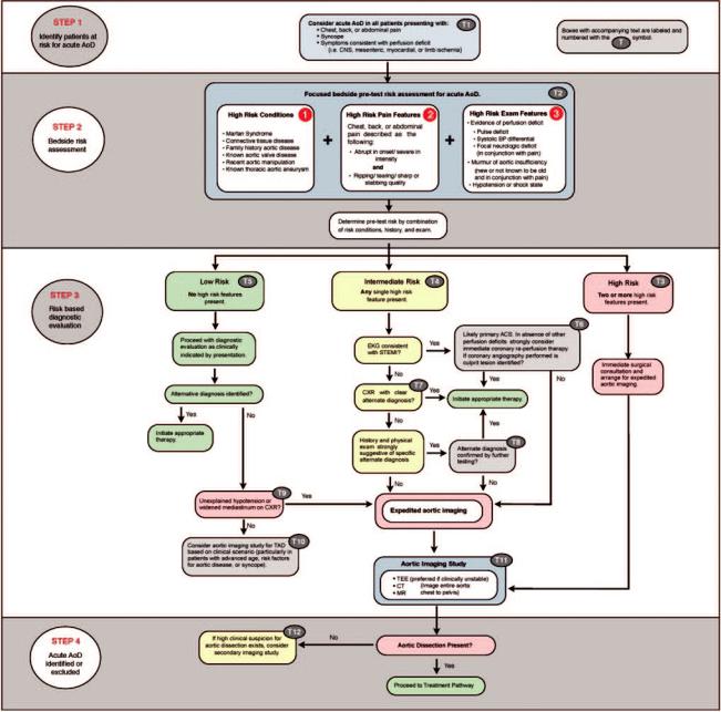
8.6.2. Evaluation and Management Algorithms
The provided algorithms guide the initial evaluation of patients whose presentations are concerning for AoD and the management of patients in whom the diagnosis of acute thoracic AoD is confirmed. Although clinicians may ultimately choose to deviate from the pathway for patient-specific reasons, the algorithms provide a framework with which to quickly diagnose (Figure 25) and provide early management (Figure 26) of AoD. This decision model is supported by several large studies that indicate a targeted history and physical examination are likely to identify the vast majority of patients who present with acute AoD, suggesting that adequate screening need not be time intensive or technology dependent.47,215,264 Using a target history and physical examination, patients can be placed into 1 of 3 categories: 1) those with immediately apparent acute AoD requiring emergent surgical evaluation and expedited aortic imaging, 2) those whose presentation is concerning for acute AoD and in the absence of a clear alternative diagnosis require expedited aortic imaging, and 3) those whose clinical presentation is not initially suggestive of acute AoD but may benefit from aortic imaging in the absence of a likely alternative diagnosis at the completion of the initial evaluation.
Several high-risk conditions (Figure 25, T2–1) greatly increase the likelihood that presenting complaints that could be a result of acute AoD.
Pain (Figure 25, T2–2) is the most commonly reported presenting symptom of acute AoD regardless of patient age, sex, or other associated clinical complaint.37,47,215,223,242,264,289 Pain described as abrupt or instantaneous in onset, that is severe in intensity, or that has a ripping or tearing quality establishes a high pretest probability for AoD.47,261
The combination of 2 or more high-risk features (Figure 25, T3) is strongly suggestive of acute AoD.
The presence of a single high-risk feature (ie, high-risk condition, pain, or physical examination) may trigger immediate concern for acute AoD; however, other diagnostic considerations may exist. Pain with high-risk features, although suggestive of acute AoD, may occur as a result of an alternate disease process (Table 12). For patients who are determined to have an intermediate probability of acute AoD on the basis of initial bedside assessment, Figure 25, T4, provides a pathway for further evaluation.
Figure 25, T5, provides a pathway for further evaluation of patients without any high risk features. Delay in diagnosis and increased mortality is common in this group.241,288,289 For patients presenting with new ST-segment elevations on the initial ECG and without high-risk AoD features, immediate coronary angiography and reperfusion therapy (ie, thrombolysis or percutaneous coronary intervention) are indicated.333 However, if coronary angiography is performed and no culprit coronary lesion is identified, then Figure 25, T6, provides a pathway to dedicated aortic imaging. As approximately 40% of chest films in acute AoD lack a widened mediastinum, and as many as 16% are normal, the absence of radiographic abnormalities does not exclude the
diagnosis of AoD37,47 (Figure 25, T7).
Missed or delayed diagnosis of acute AoD is most commonly ascribed to an incorrect working diagnosis of acute coronary syndrome, a condition that may require a prolonged time interval to correctly identify (ie, serial cardiac biomarkers) and whose management with antiplatlet and antithrombin agents may cause harm to the patient with AoD. For patients with an intermediate-risk profile for acute AoD and who do not have diagnostic STEMI but who are being evaluated for a possible acute coronary syndrome, aortic imaging may detect AoD prior to the administration of antiplatelet and antithrombin agents (Figure 25, T8).
Unexplained hypotension is present in approximately 20% of patients with acute AoD.37,47 Similarly, a widened mediastinum on chest x-ray strongly suggests the need for additional definitive diagnostic aortic imaging, particularly in patients without a clear alternative explanation for their presenting complaint342 (Figure 25, T9).
Some patients with acute AoD present without any highrisk features, making early diagnosis difficult. If a clear alternative diagnosis is not established after the initial evaluation, then obtaining a diagnostic aortic imaging study, particularly in patients with advanced age (older than 70 years), syncope, focal neurologic deficit, or recent aortic manipulation (surgery or catheter based), should be considered47,242,253,288,343 (Figure 25, T10).
Multidetector CT, TEE, and MR all provide acceptable diagnostic accuracy for the diagnosis of acute AoD. In patients with hemodynamic instability, requiring close mon-
e310 |
Circulation |
April 6, 2010 |
Figure 25. AoD evaluation pathway. ACS indicates acute coronary syndrome; AoD, aortic dissection; BP, blood pressure; CNS, central nervous system; CT, computed tomographic imaging; CXR, chest x-ray; EKG, electrocardiogram; MR, magnetic resonance imaging; STEMI, ST-elevation myocardial infarction; TAD; thoracic aortic disease; and TEE, transesophageal echocardiogram.
itoring, bedside TEE is preferred to avoid moving the patient out of the acute care environment (Figure 25, T11).
The most recent comparative study with nonhelical CT, 0.5 Tesla MR and TEE showed 100% sensitivity for all modalities, with better specificity of CT (100%) than for TEE and MR.44 A recent meta-analysis that evaluated the diagnostic accuracy of TEE, helical CT, and MR for suspected AoD found that all 3 imaging techniques provided equally reliable diagnostic values.46 Accordingly, selection of an imaging modality is influenced by individual patient variables and institutional capabilities.
The diagnosis of acute AoD cannot be excluded definitively based on the results of a single imaging study.
Although TEE, CT, and MR are all highly accurate for the evaluation of acute AoD; false-negative studies can and do occur47 (Figures 9 and 15). If a high clinical suspicion exists for acute AoD but initial aortic imaging is negative, strongly consider obtaining a second imaging study (Figure 25, T12).
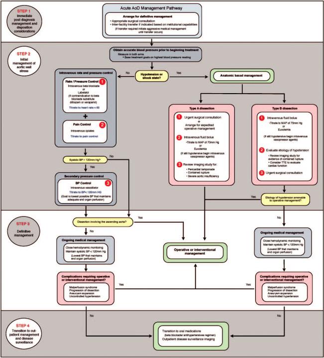
8.6.3. Initial Management
Once the diagnosis of AoD or one of its anatomic variants (IMH or PAU) is obtained, initial management is directed at limiting propagation of the false lumen by controlling aortic shear stress while simultaneously determining which patients will benefit from surgical or endovascular repair (Figure 26).
Hiratzka et al |
2010 Guidelines on Thoracic Aortic Disease |
e311 |
Figure 26. Acute AoD management pathway. AoD indicates aortic dissection; BP, blood pressure; MAP, mean arterial pressure; and TTE, transthoracic echocardiogram.
8.6.3.1. Blood Pressure and Rate Control Therapy
Aortic wall stress is affected by the velocity of ventricular contraction (dP/dt), the rate of ventricular contraction, and blood pressure. Initial medical stabilization using beta blockers controls these 3 parameters by reducing heart rate and blood pressure to the lowest amounts that will still maintain adequate end-organ perfusion.61 Reasonable initial targets are a heart rate less than 60 bpm and a systolic blood pressure between 100 and 120 mm Hg.61
Intravenous propranolol, metoprolol, labetalol, or esmolol is an excellent choice for initial treatment. In patients who have a potential contraindication to beta blockade (eg, those with asthma, congestive heart failure, or chronic obstructive pulmonary disease), esmolol may be a viable option given its extremely short half-life. Use of labetalol, which is both an alphaand beta-receptor antagonist, offers the advantage of potent heart rate and blood pressure control from a single agent, potentially eliminating the need for a secondary

e312 |
Circulation |
April 6, 2010 |
Table 12. Differential Diagnosis for High-Risk Pain or Examination Features
Chest pain
●Acute myocardial infarction
●Pulmonary embolism
●Spontaneous pneumothorax
●Esophageal rupture Abdominal pain
●Renal/biliary colic
●Bowel obstruction/perforation
●Non– dissection-related mesenteric ischemia Back pain
●Renal colic
●Musculoskeletal pain
●Intervertebral disk herniation
Pulse deficit
●Non– dissection-related embolic phenomena
●Non– dissection-related arterial occlusion Focal neurologic deficit
●Primary ischemic cerebrovascular accident
●Cauda equina syndrome
vasodilator. In patients who are unable to tolerate beta blockade, nondihydropyridine calcium channel antagonists (verapamil, diltiazem) offer an acceptable, although lessestablished, alternative.61 Beta blockers, verapamil, or diltiazem for rate control in patients with significant aortic regurgitation may be problematic because of deleterious affects on reflex tachycardia.
8.6.3.2. Additional Antihypertensive Therapy
It is frequently difficult to reduce blood pressure to optimum levels.285–287,344 –346 In 1 study, patients required a median of 4 different antihypertensive drugs.347 In addition to beta blockade, vasodilators may be required to control blood pressure. Intravenous sodium nitroprusside is the most established agent and offers the advantage of being rapidly titratable.61 Nicardipine,348 nitroglycerin, fenoldopam, and various other intravenous antihypertensive agents are appropriate for this situation. Vasodilator therapy without prior beta blockade may cause reflex tachycardia and increased force of ventricular contraction leading to greater aortic wall stress and potentially causing false lumen propagation.42 Following initial stabilization with intravenous antihypertensives, most patients will require long-term antihypertensive treatment including the use of a beta blocker plus additional classes of agents. Angiotensin-converting enzyme inhibitors or angiotension receptor blockers may retard aortic dilatation and their use may be indicated as outlined in Section 9.2.1.1.
8.6.3.3. Pain Control
Adequate pain control is essential in the setting of acute AoD to decrease sympathetic mediated increases in heart rate and blood pressure. Appropriate use of intravenous opiate anal-
gesia will help augment the effects of rate control and vasodilator agents.
8.6.3.4. Hypotension
Medical management options for all forms of dissectionrelated hypotension are limited. Volume administration titrated to improvement of blood pressure is a reasonable first approach. Vasopressors can be added, if needed, to maintain adequate perfusion but have the potential to cause further false lumen propagation. Inotropic agents are likely to increase the force and rate of ventricular contraction and therefore increase sheer stress on the aortic wall.
Pericardiocentesis for dissection-related hemopericardium has been associated with recurrent pericardial bleeding and associated mortality.307,349,350 Several articles from the Asian literature suggest that pericardiocentesis may be safe in the setting of acute Type A IMH.351,352 Other cardiac complications that may result in hypotension include severe dissection-related aortic regurgitation, true lumen obstruction by a compressing false lumen, and acute MI. All require definitive operative management. Hypotension or shock in the setting of AoD may also result from contained rupture of the false lumen into adjacent structures (ie, pleural space or mediastinum), a scenario that also mandates immediate operative intervention.
Ultimately, hypotension or shock in the acute AoD patient suggests the need for immediate operative management. For patients with hemopericardium and cardiac tamponade who cannot survive until surgery, pericardiocentesis can be performed by withdrawing just enough fluid to restore perfusion.
8.6.3.5. Determining Definitive Management
In the clinically stable patient, the decision for surgical versus medical management of patients with acute AoD is based primarily on the location of the dissection as described by the Stanford and DeBakey classification systems61,353 (see Section 8.1.2). A prompt cardiac surgical consultation provides the best management resource, regardless of location of the AoD, as it is impossible to predict which complications may develop or when they may occur.
8.6.4. Recommendation for Surgical Intervention for Acute Thoracic Aortic Dissection
Class I
1.For patients with ascending thoracic aortic dissection, all of the aneurysmal aorta and the proximal extent of the dissection should be resected. A partially dissected aortic root may be repaired with aortic valve resuspension. Extensive dissection of the aortic root should be treated with aortic root replacement with a composite graft or with a valve sparing root replacement. If a DeBakey Type II dissection is present, the entire dissected aorta should be replaced. (Level of Evidence: C)
When a Type A AoD involves the aortic root, resuspension of the valve with preservation of the aortic sinuses and excision of the sinuses and resuspension of the valve within a polyester graft are suitable options. If the aortic root is dilated, or if there is extensive dissection and disruption of the
Hiratzka et al |
2010 Guidelines on Thoracic Aortic Disease |
e313 |
aortic sinuses, replacement with a composite graft is necessary.
8.6.5. Endovascular Interventions
Endovascular stent grafts are not approved for AoD involving the ascending aorta or aortic arch. Endovascular stent grafts used for descending thoracic aortic dissection is discussed in Section 9.2.2.3.1. Indications for either surgical or endovascular interventions are discussed in Section 9.
8.6.6. Principles of Treatment for Intramural Hematoma and Penetrating Atherosclerotic Ulcer
The goals of treatment are to prevent aortic rupture or progression to classic AoD, allow patient stabilization before urgent surgery, and reduce complexity of unavoidable aortic surgery. Aggressive medical treatment usually includes, particularly in symptomatic patients, beta blockers and other antihypertensive therapy. Indications for open or endograft treatment are based on the anatomic features of the lesion, clinical presentation and course, patient comorbidities, and anatomic constraints related to endograft technology. Treatment by endografts or open aortic reconstruction can be discussed in the context of 3 overlapping aortic lesions: intimal defect without IMH, intimal defect with IMH, and IMH without an intimal defect.
8.6.6.1. Intimal Defect Without Intramural Hematoma
These are localized lesions and may involve a limited segment of the aorta. They are often an incidental finding. By imaging criteria, they include uncomplicated aortic ulcers, blebs, and eccentric or saccular aneurysms of the aorta. They are treated as saccular aneurysms based on their maximum diameter and clinical feature.212,306,354 These lesions can be treated with open reconstruction and are the most suitable of the 3 groups for treatment by endografts, if in the descending thoracic aorta. They involve a limited segment, which can easily be excluded from the circulation, as long as there is an adequate distance from a critical branch artery. When these limited dissections involve the ascending aorta, emergency surgery is indicated as for other types of AoD because rupture or cardiac tamponade can occur.212,306,354
8.6.6.2. Intimal Defect With Intramural Hematoma
The intimal defect again presents a target lesion for endovascular treatment in the descending thoracic aorta, but the associated IMH involves a longer segment of aorta than the first category. If the patient becomes asymptomatic in response to aggressive medical treatment, it may be possible to delay endovascular or open reconstruction until the IMH has reabsorbed and organized. (Of note, some writing committee members have observed healing of IMH such that immediate reconstruction was not required but have continued to follow that small number of patients closely.) Two considerations affect the length of aorta bordering the intimal defect, which is to be included in the segment targeted for treatment. Evidence of adjacent atheromatous wall should favor more extensive treatment of the aorta with longer endografts, because radiographic imaging underestimates shallow ulcerated atheromas, and the ulcer typically arises in a bed of atheromatous intima. Treatment with longer endografts provides a safety margin against undertreating the intimal defect.
The second consideration is the extent of associated IMH. The self-expanding endograft may tear through the intimal surface into underlying thrombosed false lumen. When treatment of this lesion in the acute stage is clinically necessary (eg, persisting pain, evidence for expansion or rupture, compromise of critical branches), it is preferable to anchor the endograft in the noninvolved wall above and below the intimal defect.
8.6.6.3. Recommendation for Intramural Hematoma Without Intimal Defect
Class IIa
1.It is reasonable to treat intramural hematoma similar to aortic dissection in the corresponding segment of the aorta. (Level of Evidence: C)
As noted earlier, some authors suggest treating IMH as an AoD in the corresponding aortic territory. Others recommend invasive treatment regardless of location or aortic diameter.354 However, small patient series, incomplete anatomic description of case material, and lack of explicit anatomic or clinical guidelines indicating open or endovascular aortic repair make it difficult to generalize from the literature.
The absence of an intimal defect, which can serve as a target lesion, presents a diagnostic as well as treatment challenge. Intimal tears can be extremely subtle, depending on the size of the intimal tear and the amount of intramedial thrombus, which can sometimes fill the cavity flush with the aortic lumen. The intimal tear may be remote in the aorta despite leaking into the chest. IMH in a normal caliber aorta without an apparent intimal tear precludes limited treatment of a target lesion. There are no data supporting prophylactic implantation of endografts covering the entire descending aorta, yet in unusual circumstances one may be forced to propose such treatment. IMH in an aneurysmal aorta presents a particularly urgent problem, because this complication may be a precursor to aneurysm rupture. Although the literature gives no compelling guidelines for treatment, the writing committee believes that treatment of IMH corresponding to treatment of AoD in the corresponding segment of the aorta is reasonable.
8.7. Treatment for the Management of Traumatic Aortic Rupture
The management of blunt TRA is evolving.3,318,319,355,356 On the basis of the report by Parmley and colleagues,318 most surgeons have recommended immediate surgical repair. However, when other serious traumatic injuries are present including head injuries and long bone or pelvic fractures, immediate surgery may not be feasible or may be dangerous. Multiple studies appear to show that if careful blood pressure control is used, many patients can be treated initially conservatively and then undergo operation once their other injuries have been stabilized.3,321,357–359 In a review by Svensson et al359 of 44 patients initially treated with careful blood pressure control who subsequently had delayed open surgery, there were no operative deaths. This approach has also been reported as being safe by Pate and colleagues and oth-