


Burn Management |
67 |
|
|
Sushma Sagar, Kamal Kataria, and Maneesh Singhal |
|
A 50-year-old male patient with history of alcohol abuse was admitted to the emergency department with burns while sleeping in a closed room. On arrival, he was conscious and oriented with cold, clammy extremities and feeble pulse. Blood pressure was 80/50 mmHg. He had 60% burns involving face, torso, and extremities. Over the right lower limb, there were circumferential burns and swelling with absent pulsations. There was hoarseness of voice with production of sooty sputum. Chest radiograph was normal.
The quality of life and outcome of the burn patient has improved due to improvement in the care over the past few decades. The removal of deep wounds and biological closure helps to attenuate the development of wound sepsis. The care of the burn patient requires very advanced critical care, preferably in the burn unit.
First Day
Step 1: Initial assessment and resuscitation
All burn patients should be approached as a polytrauma patient. Ideally, these patients should be managed in a dedicated burn unit.
Airway
•The airway is assessed first by asking the name of the patient and listening to hoarseness, which signifies upper airway burn.
•One hundred percent oxygen is administered, and oxygen saturation is monitored using pulse oximetry. Beware of falsely high saturation due to high
S. Sagar, M.S. (*) • K. Kataria, M.S. • M. Singhal, M.S., M.Ch. Department of Trauma Surgery, J.P.N. Apex Trauma Centre, AIIMS, New Delhi, India
e-mail: sagar.sushma@gmail.com
R. Chawla and S. Todi (eds.), ICU Protocols: A stepwise approach, |
535 |
DOI 10.1007/978-81-322-0535-7_67, © Springer India 2012 |
|
536 |
S. Sagar et al. |
|
|
carboxyhemoglobin levels in cases of carbon monoxide intoxication due to inhalation injury. Confirm oxygenation by blood gas analysis in a CO-oximeter.
•Wheezing, tachypnea, stridor, and hoarseness indicate impending airway obstruction due to an inhalation injury or edema, and immediate treatment is required.
•If the patient is not breathing or has labored respiration or signs of obstruction, clear the airway by oral/nasal suction followed by orotracheal intubation with inline stabilization of the neck if an injury to the cervical spine is a consideration.
•Risk of upper airway obstruction increases with the following:
–Inhalation burns—carbonaceous sputum, singed nasal hairs
–All patients with deep burns of more than 35–40% TBSA (total burn surface area)
–Burns involving face, neck, and upper torso
•Intubate early if progressive airway edema is suspected in cases of extensive burns or if the patient has signs of airway obstruction.
•Early intubation is also performed if the patient requires prolonged transport. Properly securing airway is of utmost importance in the patient.
•Awake fiberoptic intubation should be performed in difficult cases.
Breathing
•Breathing problem may be due to smoke inhalation injury, deep circumferential chest burn, or associated chest injury.
•Carbon monoxide (CO) is a by-product of incomplete combustion. Its intoxication is diagnosed by carboxyhemoglobin levels:
–Less than 10% is normal.
–More than 40% is severe.
•Treatment for carbon monoxide intoxication is to remove source and give 100% oxygen. Hyperbaric oxygen is also used to treat this condition. Patients with smoke inhalation injury often present with hoarseness, wheezing, carbonaceous sputum, facial burns, and singed nasal vibrissae.
•Diagnosis is often established by the use of bronchoscopy, which reveals early inflammatory changes such as erythema, edema, ulceration, sloughing of mucosa, and prominent vasculature in addition to infraglottic soot. Management of inhalation injury is directed at maintaining open airways and maximizing gas exchange.
•A patient who is able to cough with a patent airway can clear secretions very effectively, and efforts should be made to treat the patient without mechanical ventilation.
•If respiratory failure is imminent, intubation is instituted early, and frequent chest physiotherapy and suctioning are performed to maintain pulmonary hygiene.
•Frequent bronchoscopies may be needed to clear inspissated secretions.
•In addition to the preceding measures, adequate humidification and appropriate treatment for bronchospasm is indicated.
•These patients should be ventilated as per ARDSnet protocol with low tidal volume (6 mL/kg ideal body weight). Try to keep the plateau pressure below 30 cm
H2O. Deep circumferential chest burn may limit chest wall motility, so a higher plateau pressure up to 40 cm H2O may be tolerated.
•Frequent escharotomies may improve breathing and airway pressures.

67 Burn Management |
|
537 |
|
|
|
|
|
Table 67.1 |
Resuscitation formulas |
|
|
|
|
|
5% dextrose or plain water |
Formula |
Crystalloid volume |
Colloid volume |
by the nasogastric tube |
Parkland |
4 mL/kg/percent TBSA burn |
None |
None |
Brooke |
1.5 mL/kg/percent TBSA |
0.5 mL/kg/percent |
2.0 L |
|
burn |
TBSA burn |
|
Galveston |
5,000 mL/m2 |
None |
None |
(pediatric) |
burned + 1,500 mL/m2 total |
|
|
TBSA total body surface area
Circulation
•Obtain IV access anywhere possible and start giving fluids:
–Unburned areas are preferred.
–Burned areas are acceptable.
–Central access is obtained if expertise available.
–Cutdowns.
•Perform resuscitation in burn shock (first 24 h):
–Massive capillary leak occurs after major burns.
–Fluids shift from intravascular space to interstitial space.
–Fluid requirement increases with greater severity of burn (larger percent total body surface area(TBSA), increased depth, inhalation injury, associated injuries)
•IV fluid rate depends on physiologic response and goals:
–Sensorium—comfortable, arousable.
–Base deficit—less than 2.
–Goal for adults—urine output of 0.5 mL/kg/h.
–Goal for children—urine output of 1 mL/kg/h; if urine output is below these levels, increase fluid rate.
–Preferred fluid—lactated Ringer’s solution as it is isotonic, cheap, and easily stored.
–Resuscitation formulas—resuscitation formulas are just a guide for initiating resuscitation (Table 67.1).
Parkland formula is most commonly used for fluid calculation:
•Give half of the calculated volume in the first 8 h (from the time of injury).
•Give the other half in the next 16 h.
•Warning: Despite the formula suggesting decrease the fluid rate to half at 8 h, the fluid rate should be gradually reduced throughout the resuscitation to maintain the targeted urine output.
Resuscitation endpoint
When maintenance rate is reached (approximately 24 h), change fluids to D5/NS with 20 mEq KCl at the maintenance level:
• Maintenance fluid rate = basal requirements + evaporative losses

67 Burn Management |
539 |
|
|
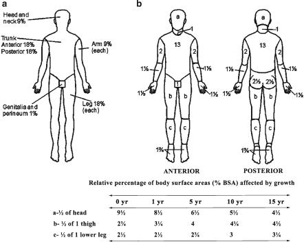
Fig. 67.1 (a) Rule of “nines” and (b) Lund–Browder diagram for estimating extent of burns (Adapted from Artz CP, Moncrief JA. The treatment of burns. 2nd ed. Philadelphia: WB Saunders Company; 1969)
•In children, adjust percents because they have proportionally larger heads (up to 20%) and smaller legs (13% in infants) than adults.
•Lund–Browder diagrams improve the accuracy of the percent TBSA for children.
•Palmar surface of the hand is approximately 1% TBSA helps in estimating percent total body surface area in children affected by burns.
Depth of burn injury
A. Superficial burns (first-degree and superficial second-degree burns):
•First-degree burns
–Damage above the basal layer of epidermis.
–Dry, red, painful (“sunburn”).
•Second-degree burns
–Damage into dermis.
–Skin adnexa (hair follicles, oil glands, etc.) remain intact.
–Heal by reepithelialization from skin adnexa.
–The deeper the second-degree burn, the slower the healing (fewer adnexa for reepithelialization).
–Moist, red, blanching, blisters, extremely painful.
540 |
S. Sagar et al. |
|
|
–Superficial burns heal by reepithelialization and usually do not scar if healed within 2 weeks.
B.Deep burns (deep second-degree to fourth-degree burns):
•Deep second-degree burns (deep partial thickness)
–Damage to deeper dermis
–Less moist, less blanching, less pain
–Heal by scar deposition, contraction, and limited reepithelialization
•Third-degree burns (full thickness)
–Entire thickness of skin destroyed (into fat)
–Any color (white, black, red, brown), dry, less painful (dermal plexus of nerves destroyed)
–Heal by contraction and scar deposition (no epithelium left in middle of wound)
•Fourth-degree burns
–Burn into muscle, tendon, and bone.
–Need specialized care.
–Deep burns usually need skin grafts to optimize results and lead to hypertrophic (raised) scars if not grafted.
Age
•Mortality for any given burn size increases with age.
•Children/young adults can survive massive burns.
•Children require more fluid per TBSA burns.
•The elderly may die from small (<15% TBSA) burns.
Associated injuries
•Other trauma increases severity of injury.
Use of alcohol or drugs
•It makes assessment of the patient more difficult.
Role of antibiotics
•It can be started later on when signs/symptoms of infection are present.
Step 5: Burn wound care and control of infection
Burn wound care: Current therapy for burn wounds can be divided into the following three stages: assessment, management, and rehabilitation:
•Once the extent and depth of the wounds have been assessed and the wounds are thoroughly cleaned and debrided, each wound should be dressed with an appropriate covering that serves three functions. First, it protects the damaged epithelium. Second, the dressing should be occlusive to reduce evaporative heat loss. Third, the dressing should provide comfort over the painful wound.
•The choice of dressing should be individualized based on the characteristics of the treated wound:
•First-degree wounds are minor with minimal loss of barrier function. These wounds require no dressing and are treated with topical agents to decrease pain and keep the skin moist.
67 Burn Management |
541 |
|
|
•Second-degree wounds can be treated by daily dressing changes with an antibiotic ointment such as silver sulfadiazine covered with several layers of gauze under elastic wraps. Alternatively, the wounds can be covered with a temporary biologic or synthetic covering to close the wound. These coverings eventually slough as the wound reepithelializes underneath.
•Deep secondand third-degree burns will not heal in a timely fashion without autografting. These burned tissues serve as a nidus for inflammation and infection that can lead to death of the patient. Early excision and grafting of these wounds is preferred in terms of survival, less blood loss, and decreased length of hospitalization.
•Early excision should be reserved for third-degree wounds typically caused by flame. A deep second-degree burn can appear clinically to be a third-degree wound at 24–48 h after injury, particularly if it has been treated with topical antimicrobials that combine with wound drainage to form a pseudoeschar.
•Escharotomy: With circumferential deep secondand third-degree burns to an extremity, peripheral circulation to the limb can be compromised. The entire constricting eschar must be incised to relieve the obstruction to blood flow. Increased pressures in the underlying musculofascial compartments are treated with standard fasciotomies to avoid compartment syndrome.
•Control of infection: Decreasing invasive infections in the burn wound is due to early excision and closure and the timely and effective use of antimicrobials. The antimicrobials that are used can be divided into those given topically and those given systemically. Topical antibiotic includes 11% mafenide acetate, 1% silver sulfadiazine, polymyxin B, neomycin, bacitracin, and mupirocin:
–Mafenide acetate has a broad spectrum of activity, particularly for Pseudomonas and Enterococcus species. Mafenide sulfate is typically reserved for small full-thickness injuries and ear burns to prevent chondritis.
–Silver sulfadiazine, the most frequently used topical agent, has a broad spectrum of activity from its silver and sulfa moieties that cover Gram-positive organisms, most Gram-negative organisms, and some fungi. It is painless upon application, has a high patient acceptance, and is easy to use.
–Petroleum-based antimicrobial ointments with polymyxin B, neomycin, and bacitracin are clear on application, are painless, and allow for observation of the wound. These agents are commonly used for the treatment of facial burns, graft sites, healing donor sites, and small partial-thickness burns.
–Mupirocin has improved activity against Gram-positive bacteria, particularly methicillin-resistant Staphylococcus aureus, and selected Gram-negative bacteria.
–Nystatin in the powder form can be applied to wounds to control fungal growth, and nystatin powder can be combined with topical agents such as polymyxin B to decrease colonization of both bacteria and fungi.
–Available agents for application as a soak include 0.5% silver nitrate solution, 0.5% sodium hypochlorite, 5% acetic acid, and 5% mafenide acetate solution.

542 |
S. Sagar et al. |
|
|
–The use of perioperative systemic antimicrobials also has a role in decreasing sepsis in the burn wound until it is healed. Common organisms that must be considered when choosing a broad-spectrum perioperative regimen include S. aureus and Pseudomonas species, which are prevalent in wounds. After massive burns, gut flora are often found in the wounds mandating coverage of these species as well.
Step 6: Fluid of choice on the second day
• Dextrose 5% in water is fluid of choice.
Step 7: Supportive treatment—nutrition
•Nutritional support is best accomplished by early enteral nutrition that can abate the hypermetabolic response to a burn. Therefore, duodenal or jejunal tube feeding should be commenced as early as within the first 6 h after burn.
•The caloric requirements are needed to gain weight and achieve nitrogen balance and have been calculated from linear regression analysis of weight change versus predicted dietary intakes in adults at 25 kcal/kg plus 40 kcal/% TBSA burn for 24 h (Table 67.3). Protein needs are approximately 2.5 g/kg. Estimation of 24-h urinary urea nitrogen for calculating nitrogen balance should be obtained (see Chap. 43 on nutrition).
The pediatric formulas have been derived from retrospective analyses of dietary intake, which is associated with maintenance of average body weight over hospital stay (Table 67.4).
Ulcer prophylaxis and deep venous thrombosis prophylaxis should be started along with the rest of the management unless there are contraindications.
Table 67.3 Curreri formula for estimating caloric requirements for adult burn patients
Age |
Formulas |
6–60 years |
25 kcal/kg/day + 40 kcal/percent burn/day |
>60 years |
25 kcal/kg/day + 65 kcal/percent burn/day |
Table 67.4 Formulas for estimating caloric requirements for pediatric burn patients
Age |
Formulas |
|
|
0–1 years |
2,100 kcal/m2 |
TBSA/day + 1,000 kcal/m2 |
TBSA burn/day |
1–11 years |
1,800 kcal/m2 |
TBSA/day + 1,300 kcal/m2 |
TBSA burn/day |
12–18 years |
1,500 kcal/m2 |
TBSA/day + 1,500 kcal/m2 |
TBSA burn/day |
Shriners Hospitals for Children at Galveston, Texas
Step 8: Manage complications
Successful management of burns involves management of predicted complications.