

Cognitive function_fMRI
.pdfManuscripts Author Funders PMC Europe
Manuscripts Author Funders PMC Europe
Tomassini et al. |
Page 21 |
187.Bosnell R, et al. Reproducibility of fMRI in the clinical setting: implications for trial designs. Neuroimage. 2008; 42:603–610. [PubMed: 18579411]
188.Bassett DS, Bullmore ET. Human brain networks in health and disease. Curr Opin Neurol. 2009; 22:340–347. [PubMed: 19494774]
189.Tomassini V, et al. Structural and functional bases for individual differences in motor learning. Hum Brain Mapp. 2011; 32:494–508. [PubMed: 20533562]
190.Stinear CM, et al. Functional potential in chronic stroke patients depends on corticospinal tract integrity. Brain. 2007; 130:170–180. [PubMed: 17148468]
191.Dijkhuizen RM, et al. Functional MRI and Diffusion Tensor Imaging of Brain Reorganization After Experimental Stroke. Transl Stroke Res. 2012; 3:36–43. [PubMed: 22408692]
192.Biswal B, Yetkin FZ, Haughton VM, Hyde JS. Functional connectivity in the motor cortex of resting human brain using echo-planar MRI. Magn Reson Med. 1995; 34:537–541. [PubMed: 8524021]
Nat Rev Neurol. Author manuscript; available in PMC 2014 March 18.

Manuscripts Author Funders PMC Europe
Manuscripts Author Funders PMC Europe
Tomassini et al. |
Page 22 |
Box 1
Criteria for the definition of behaviourally relevant brain functional reorganization after brain damage. Recovery studies in the fields of stroke and MS suggest that behaviourally meaningful functional reorganization - that is, adaptive functional reorganization - can be defined if:
1.There is a relationship between extent of changes of functional patterns and associated pathology
2.Altered patterns of functional activation accompanied by preserved or completely recovered behaviour
3.Learning or rehabilitation induce similar changes in functional activation
4.Facilitation of reorganization increases the rate of, or the potential for recovery
5.Interruption of reorganization processes and/or maladaptation results in functional impairment
Nat Rev Neurol. Author manuscript; available in PMC 2014 March 18.

Manuscripts Author Funders PMC Europe
Manuscripts Author Funders PMC Europe
Tomassini et al. |
Page 23 |
Box 2
The neurophysiological basis of functional MRI. Functional MRI (fMRI) has millimetrescale spatial resolution, providing a large-scale average of neural activity. The parameter measured by this imaging technique is the blood oxygenation level-dependent (BOLD) signal, which is principally affected by changes in the local balance between neuronal excitation and inhibition. Increased neural activity results in increased cerebral metabolic rate of oxygen (CMRO2) and local vasodilatation. As a consequence, cerebral blood flow increases. The fractional increase in blood flow is greater than the fractional increase in CMRO2. This difference reduces the quantity of (paramagnetic) deoxy-haemoglobin in the veins and is equivalent to increased oxygenation, which increases local magnetic field homogeneity around capillaries and veins and, thereby, increases net signal intensity in that area. This change in BOLD signal varies depending on the magnetic field strength, the brain region and the underlying physiology or pathology. Within a scan, periods of active stimulation (‘ON’ condition) are contrasted with rest periods (‘OFF’ condition). The choice of baseline is crucial in the interpretation of fMRI data.
The most powerful experimental designs use equal-duration alternating ON and OFF periods (block design), each lasting 10-60 s. Brief stimuli can be used in event-related designs where the functions under investigation dictate. Investigations of resting-state activity, in which the volunteer does not perform a particular task, have grown in popularity in recent years, especially in patient populations, removing the potential complication of disease-associated impaired task performance. These investigations seek to identify temporally co-varying BOLD signals, which are thought to reflect dynamically co-varying levels of neural activity, from different brain regions. Such signals are thought to represent functional connectivity between the brain regions in question. This approach can identify networks of brain regions that could underlie a specific function, such as motor output or vision192, and can be used to detect their potential modulation in disease.
Nat Rev Neurol. Author manuscript; available in PMC 2014 March 18.

Manuscripts Author Funders PMC Europe
Manuscripts Author Funders PMC Europe
Tomassini et al. |
Page 24 |
Box 3
Considerations for future studies to promote functional recovery in MS.
Type of study
Hypothesis-driven studies are preferable when targets of intervention are known. Translational studies, from bench to bedside, should be encouraged. Exploratory methodologies can be used for identification of potential new therapies or targets.
Design
Optimized trial designs (for example, sequential, adaptive or enrichment methodologies) should be prioritized over traditional trial designs. Appropriate control groups are essential. A post-intervention study phase is desirable to confirm the effect of interventions and to test for sustained effects.
Groups
Cohorts with disabilities should be prioritized when investigating the potential benefits of new interventions. Nondisabled cohorts can be studied to define biological mechanisms of successful recovery. They may be also considered for studies of interventions that have potential to increase the capacity for recovery by delaying accrual of disability Eligibility criteria for study group should be based on “rehabilitation criteria” (performance over disease characteristics), when effects of interventions are tested, or on “standard clinical criteria” (disease characteristics over performance), when the influence of specific disease characteristics on effects of interventions is to be tested.
Sample size
Fixed sample size or adaptive sample size re-estimation can be considered, depending on the type of study design.
End point
The study end point should be clinically relevant. Both clinical and paraclinical measures such as imaging should be collected to define mechanisms of therapeutic benefit. Efforts should be accelerated to better validate imaging or other paraclinical measures of brain recovery. Patient-related outcome measures provide an important complementary perspective.
Interventions
Behavioural, pharmacological or electrophysiological interventions should all be considered. Interventions and key aspects of imaging or other paraclinical measures should be standardized as much as possible to allow comparisons between studies. To facilitate the development of such standardised methods, the scientific community should work towards sharing of methodology and data.
Analysis methods
Hypothesis testing and exploratory studies should be clearly identified as such and appropriate statistical approaches used for each. Confidence intervals should be regularly
Nat Rev Neurol. Author manuscript; available in PMC 2014 March 18.

Tomassini et al. |
Page 25 |
reported and consideration should be given to the potential effects of heterogeneity in patient populations. When imaging is combined with behavioural studies, multimodal approaches are desirable.
Manuscripts Author Funders PMC Europe
Manuscripts Author Funders PMC Europe
Nat Rev Neurol. Author manuscript; available in PMC 2014 March 18.

Manuscripts Author Funders PMC Europe
Manuscripts Author Funders PMC Europe
Tomassini et al. |
Page 26 |
Search strategy and selection criteria
References for this Review were identified through searches of PubMed using the search terms “functional reorganization”, “brain plasticity”, “recovery”, “rehabilitation”, “pharmacological modulation” and “MRI” from January 1949 until April 2012. Articles were also identified through searches of the authors’ own files. Only papers published in English were reviewed. The final reference list was generated on the basis of originality and relevance to this Review.
Nat Rev Neurol. Author manuscript; available in PMC 2014 March 18.

Manuscripts Author Funders PMC Europe
Manuscripts Author Funders PMC Europe
Tomassini et al. |
Page 27 |
Key points
•Evidence across brain systems supports a behaviourally relevant role for neuroplasticity in multiple sclerosis (MS) across ages, stages and phases of the disease, which is preserved despite widespread pathology.
•Together with adaptive plasticity, maladaptive plasticity can occur owing to disuse with impairment and may contribute to disability.
•Interventions that drive neuroplasticity can promote functional restoration by inducing adaptive changes or by predisposing functional systems to adaptive plasticity.
•Individual and disease-related factors influence both spontaneous and intervention-driven adaptive functional reorganization, as well as its assessment using imaging.
•Improving the interpretability of functional MRI measures is important for characterization and quantification of the effects of recovery interventions and, thereby for development of recovery-oriented strategies.
Nat Rev Neurol. Author manuscript; available in PMC 2014 March 18.
Manuscripts Author Funders PMC Europe
Manuscripts Author Funders PMC Europe
Tomassini et al. |
Page 28 |
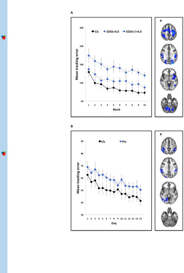
Figure 1.
Non-pharmacological modulation of brain plasticity in MS14,94. Patients with MS and healthy volunteers performed a visuomotor task in which they tracked a continuously moving bar on a computer screen by altering pressure applied to a handle held in the right
Nat Rev Neurol. Author manuscript; available in PMC 2014 March 18.

Manuscripts Author Funders PMC Europe
Manuscripts Author Funders PMC Europe
Tomassini et al. |
Page 29 |
hand. The task was performed in blocks of 38 sec. Participants practised the task in shortterm (10 blocks for a total of ~25 min) and in the longer-term (daily for 15 days consecutively) settings. Performance was measured as the mean tracking error across each block (short-term) or day (longer-term) of practice. During the first and last session, participants underwent fMRI scanning. As depicted in the graphs, short-term (a) and longerterm (b) task practice significantly improved visuomotor performance in both healthy controls and patients with MS, across levels of disability according to EDSS scores. As shown in the fMRI scans, these performance improvements were associated with a reduction in blood oxygenation level-dependent signal in brain regions involved in visuomotor integration. Abbreviations: EDSS, Expanded Disability Status Scale; fMRI, functional MRI; MS, multiple sclerosis; R, right hemisphere. Permission obtained from SAGE Publications © Tomassini, V. et al. Mult. Scler. 17, 103–115 (2011), and Tomassini, V. et al. Neurorehabil. Neural Repair 26, 581–593 (2012).
Nat Rev Neurol. Author manuscript; available in PMC 2014 March 18.

Manuscripts Author Funders PMC Europe
Manuscripts Author Funders PMC Europe
Tomassini et al. |
Page 30 |
Figure 2.
Pharmacological modulation of brain plasticity in MS16. Patients with MS and healthy volunteers underwent a counting Stroop task during fMRI scanning. Patients had comparable cognitive performance to controls, but a significantly greater BOLD signal change in the left prefrontal cortex - a difference that reflects functional reorganization. BOLD signal changes in these regions correlated with cognitive performance and brain volume. A functional score, the activation ratio (AR), representing the ratio between the magnitude of prefrontal cortex activation on the left (found in MS patients) relative to right hemisphere, was calculated to test the effect of pharmacological modulation of brain adaptive plasticity with rivastigmine, a cholinesterase inhibitor. Before rivastigmine administration or following administration of placebo, mean AR in patients was greater than in controls. After rivastigmine administration, mean AR in patients was reduced to within the range of controls. Abbreviations: BOLD, blood oxygenation level-dependent; fMRI, functional MRI; MS, multiple sclerosis; R, right hemisphere. Permission obtained from Oxford University Press © Parry, A. M. et al. Brain 126,2750–2760 (2003).
Nat Rev Neurol. Author manuscript; available in PMC 2014 March 18.