

Sensory Organ Replacement and Repair - Gerald E. Miller
.pdf6 SENSORY ORGAN REPLACEMENT AND REPAIR
Decibel |
Sound |
|
|||
|
|
|
One hand moving in air |
||
10−19 |
|||||
Softest sound |
|||||
20−29 |
Softest whisper |
||||
40−49 |
Quiet office (no typing) |
||||
50−59 |
Average home with A/C |
||||
60−69 |
Normal conversations |
||||
70−79 |
Living room music, radio, busy traffic |
||||
80−89 |
Food blender, noisy restaurant |
||||
90−99 |
Motorcycle at 25 ft, subway |
||||
100−109 |
Airplanes 1000 ft away |
||||
110−119 |
Rock concerts |
||||
>120 |
Jets, rockets |
||||
FIGURE 7: Decibel scale representing common sounds at various amplitudes.
Hearing is a very powerful sense—far more powerful than most people realize. Hearing allows individuals to determine where a sound is coming from, even if it is behind them or they cannot see the source of the sound. In addition, in a noisy environment, individuals can selectively filter out “extraneous” sound and concentrate on one voice or sound source. Try this
FIGURE 8: Frequency range of human hearing compared to other species.

HEARING AIDS 7
experiment: In a crowded room, close your eyes. Then try to concentrate on one person’s voice and determine where that person is merely from the sounds reaching your ear. As such, hearing is truly three dimensional and, of more importance, can be selective among several blended sounds. This is important when one has a hearing impairment that requires augmentation with a hearing aid or cochlear implant. One may regain sound amplitude and frequency processing, but loses the spatial resolution feature that is such a powerful aspect of normal hearing.
1.2Hearing Loss
When the problem is in the inner ear, a sensorineural hearing loss occurs. Sensorineural hearing loss is the most common type of hearing loss. More than 90% of all hearing aid wearers have sensorineural hearing loss. The most common causes of sensorineural hearing loss are agerelated changes, noise exposure, inner ear blood circulation, inner ear fluid disturbances, and problems with the hearing nerve. Conductive hearing loss occurs when sound is not conducted efficiently through the ear canal, eardrum or the tiny bones of the middle ear, resulting in a reduction of loudness of sound. Conductive loss may result from earwax blocking the ear canal, fluid in the middle ear, middle ear infection, obstructions in the ear canal, perforations (hole) in the eardrum, or disease of any of the three middle ear bones. People with conductive hearing loss may notice that their ears seem to be full or plugged. They may speak softly because they hear their own voice loudly. Crunchy foods, such as celery or carrots, seem very loud to the person with a conductive hearing loss and this person may have to stop chewing to hear what is being said. All conductive hearing losses should be evaluated by an audiologist and a physician to explore medical and surgical options.
Audiometry is the testing of a person’s ability to hear various sound frequencies. The test is performed with the use of electronic equipment called an audiometer. This testing is usually administered by a trained technician called an audiologist. Audiometry testing is used to identify and diagnose hearing loss. The equipment is used in health screening programs, for example, in grade schools, to detect hearing problems in children. It is also used in the doctor’s office or in hospital’s audiology department to diagnose hearing problems in children, adults, and the elderly. With correct diagnosis of a person’s specific pattern of hearing impairment, the right type of therapy, which might include hearing aids, corrective surgery, or speech therapy, can be prescribed.
The person being tested wears a set of headphones that blocks out other distracting sounds and delivers a test tone to one ear at a time with the amplitude slowly increasing. When the patient hears the sound of a tone, he holds up a hand or finger to indicate that the sound is detected. The audiologist lowers the volume and repeats the sound until the patient can no longer detect it. This process is repeated over a wide range of tones or frequencies from very

8 SENSORY ORGAN REPLACEMENT AND REPAIR
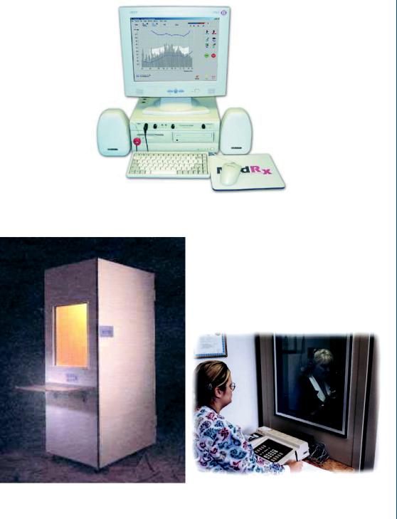
FIGURE 9: Small desktop audiometer.
deep, low sounds, like the lowest note played on a tuba, to very high sounds, like the pinging of a triangle. Each ear is tested separately. It is not unusual for levels of sensitivity to sound to differ from one ear to the other. The results of the audiometry test may be recorded on a grid or graph called an audiogram. This graph is generally set up with low frequencies or tones at one end and high ones at the other end, much like a piano keyboard. Low notes are graphed on the left and high notes on the right. The graph also charts the volume of the tones used, from soft, quiet sounds at the top of the chart to loud sounds at the bottom. Hearing is measured in a unit called decibels. Most of the sounds associated with normal speech patterns are generally spoken in the range of 20–50 dB. An adult with normal hearing can detect tones between 0 and 20 dB.
A typical desktop audiometer is shown in Figure 9 with a computer-driven system shown in Figure 10.
Some hospitals and schools utilize an acoustic chamber type audiometer that further isolates interfering sound. This chamber is sometimes called an anechoic chamber and is shown in Figure 11.
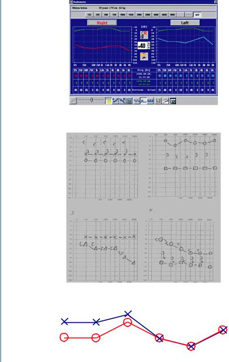
The audiograms noted above can be computer generated as seen in Figure 12 or hand drawn as with a desktop system as seen in Figure 13.
As can be seen from the audiograms given in Figures 12 and 13, there are data points at various frequencies for both ears. Normal hearing is designated by a fairly parallel set of points for each ear as is shown in Figure 14.
HEARING AIDS 9
FIGURE 10: Computer-controlled audiometer.
FIGURE 11: Acoustic chamber type audiometer.
10 SENSORY ORGAN REPLACEMENT AND REPAIR
FIGURE 12: Computer-generated audiogram.
FIGURE 13: Hand-drawn audiogram from a desktop audiometer.
FIGURE 14: Normal bilateral hearing with fairly parallel data points at all frequencies.
HEARING AIDS 11
FIGURE 15: Upper frequency hearing loss in both ears.
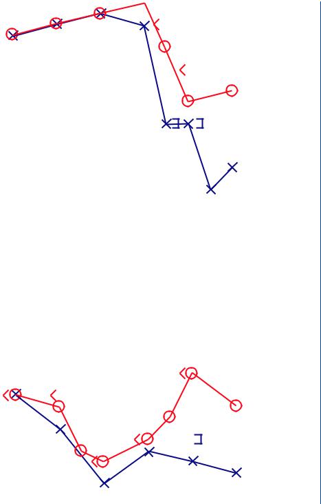
Elderly individuals or those who have been repeatedly exposed to loud sounds often lose hearing at the upper frequencies as shown in Figure 15. Still other patients may have hearing loss in mid-frequency ranges due to childhood illnesses or recurring tympanic membrane problems, as is seen in Figure 16.
There are sometimes hearing losses in only one ear, typically due to recurring infection in one ear or damage to the outer or middle ear in one ear. When both ears are affected, the patient may utilize hearing aids in both ears, although that is not required. Obviously, when only one ear is affected, a single hearing aid may be needed. Figure 17 depicts one-sided hearing loss with a patient who has a recurring right ear infection.
FIGURE 16: Mid-range hearing loss in both ears.

12 SENSORY ORGAN REPLACEMENT AND REPAIR
FIGURE 17: One-sided hearing loss at most frequencies due to a recurring right ear infection.
1.3Hearing Aid Technologies
Hearing aids are actually miniature sound systems with a microphone, speaker, audio amplifier, PA and associated electronics. As with many sound systems, the amplitude of the amplified sound can be adjusted as can the frequency range. In addition, as with many stereo systems, issues such as total harmonic distortion, signal to noise ratio, overall gain, and other signal processing factors are also elements in hearing aids. Since any individual patient has an individualized hearing loss, which can be frequency and amplitude dependent, there are settings for gain at varying frequencies that are preset for each patient, and then are fixed. The patient can have control over the overall gain and whether the unit is turned on or off, but cannot normally set the gain at individual frequencies, as these are internally set within the casing of the hearing aid within the electronics. A small dime-sized battery, which is normally on a pivot holder to allow for ease of replacement, powers the device. A typical hearing aid is shown in Figure 18 that depicts an ear mold (not used in all types of hearing aids), volume control, and on–off switch.
FIGURE 18: Typical hearing aid configuration with volume control and on–off switch.

HEARING AIDS 13
FIGURE 19: BTE hearing aid with clear tube fitting into the ear canal.
There are four styles or configurations of hearing aids. These include the “behind-the- ear” (BTE) style, the “in-the-canal” (ITC) style, and the “completely-in-the-canal” (CIC) style. The BTE hearing instruments are extremely flexible for all types of hearing loss.
The hearing device is housed within a curved shell that sits behind each ear and delivers sound through a clear tube, as is seen in Figure 19. The clear tube fits into a mold that has been customized to comfortably fit inside each ear.
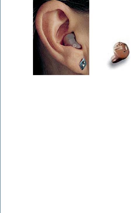
The in-the-ear (ITE) hearing instruments are very easy to operate, even if the user has poor dexterity. The hearing device is housed within a custom-made shell that fits comfortably inside each ear and delivers sound directly to the ear. This style is shown in Figure 20 and is
FIGURE 20: ITE style of hearing aid with the device easily accessible and removable.
14 SENSORY ORGAN REPLACEMENT AND REPAIR
FIGURE 21: ITC style hearing aid with the device positioned further into the canal.
one of the most common styles of hearing aids, particularly for the elderly or for individuals wearing glasses who cannot easily utilize the BTE style.
The ITC hearing instruments can barely be seen and are very easy to operate, even if the user has poor dexterity. The hearing device is housed within a custom-made shell that fits comfortably inside each ear canal and delivers sound directly to the ear. However, this style is not as accessible as the ITE style. This version is seen in Figure 21 and is often utilized by younger patients as opposed to elderly patients.
The CIC hearing instruments are virtually invisible to others. The hearing device is housed in a tiny shell that fits comfortably and completely into each ear canal. The device is removed from the ear canal by pulling a tiny cord. Where these miniature instruments are both powerful and cosmetically appealing, some features such as manual volume control are not available simply because the devices are so small. These types of devices are worn almost exclusively by younger patients who wish to avoid the appearance of a hearing aid. This style is shown in Figure 22.
Hearing aid batteries are small dime-sized devices housed within a pivoting holder within the shell of the device. As is the case with many electronic devices, there are many types of hearing aids. However, in order to avoid confusion for a health care device, hearing aid batteries are color coded, so that it is easy to purchase the correct replacement battery, as is seen in Figure 23.
Typically, batteries last 7–14 days based on 16 h per day use cycle. Batteries are inexpensive, costing less than a dollar each. Generally, the smaller the battery size, the shorter the battery

HEARING AIDS 15
FIGURE 22: CIC style with a protruding cord to remove the device.
life. The sizes of hearing aid batteries are listed below along with their standard numbers and color codes:
Size 5: Red
Size 10 (or 230): Yellow
Size 13: Orange
Size 312: Brown
Size 675: Blue
FIGURE 23: Color-coded hearing aid batteries with easy punch out sections and easily seen plus/minus poles.