

Sensory Organ Replacement and Repair - Gerald E. Miller
.pdf46 SENSORY ORGAN REPLACEMENT AND REPAIR
FIGURE 53: IOL injection style holders with push button or screw release.
a few days. In addition, the patient is given a plastic disk that must be taped over the affected eye at night. This is to protect the eye from accidental rubbing during the night. Normally, the patient is seen by the ophthalmic surgeon the day after surgery, in order to check the incision, the healing process, and to determine if there is any swelling or infection. Approximately 1 week after surgery, the patient returns to have the outer sutures removed. The ophthalmic surgeon carefully snips the ends of the sutures and uses a small tweezers to pull the sutures out. The patient can feel the light pulling of the sutures, but rarely feels any pain.
On some occasions, the lens sac/capsule may become cloudy at some point following the surgery as is shown in Figure 54. This is normally remedied by means of an out-patient visit to the surgeon’s office. An excimer laser is used to burn small holes in the sac at various points. The patient is placed in a head mount device and the affected eye is held open. The patient sees red flashes and hears “pops,” but does not feel anything during the process. The holes in the sac supply sufficient light such that the clear vision is restored. The sac is otherwise still intact.
At times, a patient may experience a “floater.” Floaters, or muscae volitantes (Latin—“flying flies”), are characterized by shadowlike shapes that appear singly or together with several others in one’s field of vision. They can take the form of spots, threads, or fragments of cobweblike
FIGURE 54: Lens sac becomes cloudy some time after surgery.

INTRAOCULAR LENS 47
shapes that float slowly before one’s eyes. Floaters are suspended in the vitreous humor, the thick fluid or gel that fills the eye. Thus, they generally follow the rapid motions of the eye while drifting slowly within the fluid. When they are first noticed, the natural reaction is to attempt to look directly at them. However, attempts to shift the gaze toward them are frustrating, because the floaters follow the motion of the eye, and remain to the side of the direction of gaze. Although the blood vessels of the eye also obstruct light, they are invisible under normal circumstances (and thus not annoying) because they are fixed in the location relative to the retina, and the brain “tunes out” stabilized images. This does not occur with floaters and they remain visible, and, in some cases, when large and numerous, are very irritating. Despite the name “floaters,” many of these specks have a tendency to sink toward the bottom of the eyeball, in whichever way the eyeball is oriented. Floaters are not uncommon, although they rarely cause problems for those who have them. Floaters can be a nuisance and a distraction to those who suffer from severe cases, as the spots seem to drift through the field of vision. Normally, there is no treatment indicated, particularly for mild cases, as the floater will eventually sink toward the bottom of the vitreous body and out of the field of vision. Surgical or laser treatments have been employed for severe cases with mixed results.
4.4History of the IOL and Current Research
Nicholas Harold Lloyd Ridley, most commonly known as Harold Ridley, was a British ophthalmologist who pioneered IOL surgery for cataract patients. He was famous as a Surgeon at Moorfields Eye Hospital and St. Thomas’ Hospital in London, specializing in ophthalmology. While working with Royal Air Force casualties during World War II, he noticed that when splinters of perspex from aircraft cockpit canopies became lodged in the eyes of wounded pilots, they did not trigger rejection, leading him to propose the use of artificial lenses in the eye to correct cases of cataracts. He had a lens manufactured using an identical plastic, and on November 29, 1949, at St. Thomas’ Hospital, he achieved the first implant of an IOL, although it was not until 1950 that he implanted an artificial lens permanently in an eye. He went on to develop widespread programs for cataract surgery with intraocular implants and pioneered this treatment in the face of initially strong opposition from the medical community. He continually refined the technique; until by the late 1960s, with his pupil Peter Choyce, he eventually achieved widespread support for the technique. The IOL was finally approved for use in the United States by the FDA in 1981. IOL implantation and cataract surgery is now a common type of eye surgery.
Although the most common form of an IOL is for the treatment of a cataract, there is now another form of IOL used for the treatment of myopia. The alternative IOL is placed in the anterior chamber (with respect to the lens capsule) and is primarily used to treat nearsightedness, as opposed to a cataract. In this fashion, it is similar to that of

48 SENSORY ORGAN REPLACEMENT AND REPAIR
glasses or contact lenses, but is permanently placed within the eye, as is done for more standard IOLs. This new IOL is the latest type to receive FDA approval as noted at http://www.fda.gov/bbs/topics/ANSWERS/2004/ANS01313.html. As befits a new style of IOL for a different purpose that was originally intended to treat cataracts, there have been numerous studies of late including those by Alio et al. (2005), Benedetti et al. (2005), Leccisotti and Fields (2005), Lifshitz et al. (2004), Lovisolo and Reinstein (2005), and Sridhar et al. (2005).
For traditional IOLs to treat cataracts, recent studies have focused on the materials used for an IOL as well as surgical and follow-up complications of various IOL designs. Examples of studies involving materials used for an IOL include a study of the use of PMMA by Frazer et al. (2005) as well as a comparison of PMMA, silicone and acrylics by Smith et al. (2005). Examples of studies involving surgical and postsurgical complications of an IOL include those by Cakmak et al. (2005), Collins et al. (2006), Martin and Sanders (2005), Oshika et al. (2005), Parsons et al. (2005), Tassignon et al. (2005), Tognetto et al. (2005), and Werner et al. (2006).
5 ARTIFICIAL AND REPLACEMENT CORNEA
The cornea is the outer coating of the human eye as can be visualized in Figures 40 and 41 and provides most of an eye’s optical power. Together with the lens, the cornea refracts light and consequently helps the eye to focus. The cornea gives a larger contribution to the total refraction than the lens, but whereas the curvature of the lens can be adjusted to alter the focus based on accommodation, the curvature of the cornea is fixed. The cornea has sensitive nerve endings such that a touch of the cornea causes an involuntary reflex that closes the eyelid. As transparency is of prime importance, the cornea does not have blood vessels, but rather receives nutrients via diffusion from tears on the outside surface and the aqueous humor on the inside surface. The adult cornea has a diameter of approximately 12 mm (about one half inch) and a thickness of 0.5–0.7 mm in the center and 1.0–1.2 mm at the periphery. In humans, the refractive power of the cornea is approximately 45 D, which is approximately three-fourths of the eye’s total refractive power. The cornea consists of five layers. From the outside to the inside they are as follows:
•Corneal epithelium: a thin epithelial layer of fast-growing and easily regenerated cells. Tears keep this layer moist.
•Bowman’s layer: a tough layer that protects the corneal stroma. It consists of irregularly arranged collagen fibers.
•Corneal stroma: a thick, transparent middle layer responsible for most of the focusing power of the cornea. It consists of regularly arranged collagen fibers along with a few fibroblasts. If the stroma is damaged, for example, by injury or infection, it can lose its

ARTIFICIAL AND REPLACEMENT CORNEA 49
transparency, causing vision problems. The corneal stroma consists of approximately 200 layers of type I collagen fibrils. The ordering of the fibrils is responsible for the transparency of the tissue.
•Posterior limiting membrane (also called Descemet’s membrane): a thin acellular layer that serves as the modified basement membrane of the corneal endothelium.
•Corneal endothelium: a simple scaly style epithelium, an inner lining acting as a barrier to prevent water inside the eyeball from moving into and hydrating the cornea, which would lead to blurred vision.
The cornea is composed mostly of dense connective tissue, similar to the surrounding sclera. However, the collagen fibers are arranged in a parallel pattern, allowing light waves to constructively interfere, letting the light pass through relatively uninhibited. The cornea is innervated by the long posterior ciliary nerves.
Various refractive eye surgery techniques, such as LASIK surgery, can change the shape of the cornea in order to reduce the need for glasses or otherwise improve the refractive state of the eye. In the techniques used today, parts of the cornea are removed with lasers. If the corneal stroma has developed opaque patches known as leukomas, a cornea of a deceased donor can be transplanted. Because there are few blood vessels in the cornea, there are also few problems with rejection of the new cornea. Replacement corneas are normally obtained from eye banks, which store donor corneas. There are also synthetic corneas in development. Most are merely plastic inserts, but there are also some made of plastics that encourage the eye tissue to grow into the synthetic cornea, making it a full replacement. In addition tissue-based artificial corneas are now in development.
5.1Corneal Transplant
A corneal transplant is the replacement of a damaged cornea with an undamaged one. Corneal transplant is also called keratoplasty. A corneal transplant is not the same thing as laser-assisted in situ keratomileusis; LASIK involves reshaping, not removal, of the cornea, in order to correct focusing problems. Although the cornea is tough, due to its vulnerable location, it is quite susceptible to damage from accidents, bacteria, and particles in the air. The well-organized arrangement of the collagen is required in order for the cornea to remain transparent. Corneal transplantation is undertaken for blurry vision resulting from a cornea that no longer enables light to pass through uninterrupted, due to cloudiness or scarring. Reasons for opaqueness of the cornea may be
•degenerative disease;
•dystrophies (inherited diseases in which impurities gradually accumulate in the cornea);
50SENSORY ORGAN REPLACEMENT AND REPAIR
•infection (infection of the cornea can also occur as a complication of other infections); and
•trauma.
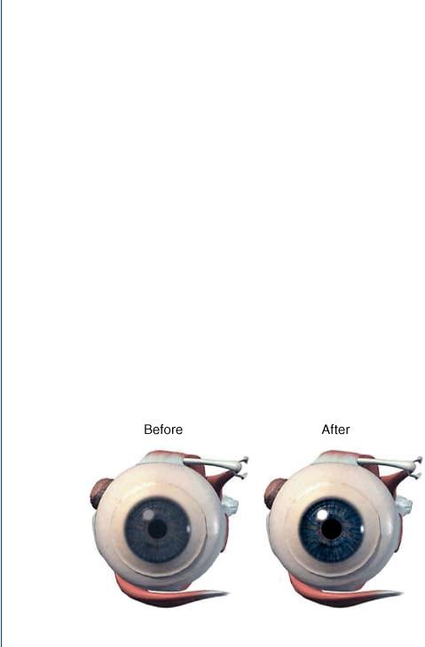
In the procedure, the central region of the cornea is cut out with a trephine (an instrument resembling a cookie cutter). The replacement cornea is then inserted and sutured into place using extremely fine thread (narrower than a human hair). The patient is awake during the operation, which is conducted under local anesthesia. The eye is held open by special device. Surgery requires 30–90 min. After the surgery, eye drops are used to aid healing. Nevertheless, recovery from keratoplasty occurs extremely slowly; complete healing takes months to years, and so the sutures remain in place for at least several months. Complications include the possibility of rejection of the foreign tissue graft or bleeding or infection risks of surgery. The use of large amount of steroids following the operation improves the success rate. Nevertheless, about one in five patients rejects the new cornea. Over 40 000 corneal transplants are performed in the United States every year. Medicare reimbursement for a corneal transplant in one eye was about $1200 in 1997. Figure 55 depicts a cornea before and after corneal transplant surgery.
Figure 56 depicts the sequence of the surgical procedure for corneal transplantation. There are numerous regional and local eye banks dedicated to receiving donor corneal
tissue with proper storage until needed by a patient with a cloudy cornea. One can contact local eye banks by looking in the local yellow pages under “eye bank” or by searching yahoo.com under “eye bank, state name (or city name).” There will be links on how to become a donor including downloading a donor card. Physicians involved with corneal transplantation usually know how to access the local eye bank for donor tissue. Although blood-typing is not a necessity (as the
FIGURE 55: The cornea before and after transplant surgery. The cloudy cornea on the left is replaced with a clear transplant cornea on the right.

ARTIFICIAL AND REPLACEMENT CORNEA 51
(a) |
(b) |
(c) |
(d) |
FIGURE 56: Corneal transplant surgery: (a) damaged cornea; (b) cornea removal; (c) replacement with donor cornea from eye bank or cadaver; and (d) suture replacement cornea in place.
corneal is avascular), it is often prudent to match blood type between donor and patient. The United Network for Organ Sharing, which is responsible for organ donation and transplantation for a variety of organs, does not handle eye tissue. This is done via eye banks at the local and regional level.
Replacement corneas that are either plastic or tissue based have been developed in recent years. An artificial (nontissue) cornea has been developed by AlphaCor produced by Argus Biomedical Corp. AlphaCor is a biocompatible, flexible, one-piece artificial cornea (keratoprosthesis) designed to replace a scarred or diseased native cornea. It is technically listed as a hydrogel. AlphaCor is designed for use in patients who have had multiple failed corneal transplants or in those patients in whom a donor graft is likely to fail. AlphaCor is available in two versions, to suit those with a natural lens (phakic) or artificial lens (pseudophakic) and for those without a lens (aphakic). AlphaCor is a one-piece convex disk consisting of a central transparent optic and an outer skirt that is entirely manufactured from poly(2-hydroxyethyl methacrylate) or PHEMA. The outer skirt is an opaque, high water content, PHEMA sponge. The porosity of the sponge encourages biointegration with host tissue and thus promotes retention of the implanted device. The central optic core is a transparent PHEMA gel providing a refractive power similar to that of the human cornea. The junctional zone between the skirt and the central optic is the interpenetrating polymer network (IPN). This is a permanent bond formed at the molecular level and is designed to prevent the down growth of cells around the optic, which can lead to the formation of retroprosthetic membranes, one of the major complications historically associated with artificial corneas. The AlphaCor device is shown in Figure 57.
The implantation of AlphaCor is a unique two-stage procedure. The first stage of the surgery involves the surgeon creating a pocket within the existing scarred cornea and inserting the device (after removing a portion of the scarred cornea first). This is usually performed under general anesthetic. Approximately 3 months after this initial surgery, a second, smaller procedure is performed. At this stage, the scarred tissue that is covering the AlphaCor is removed to allow the light to enter.

52 SENSORY ORGAN REPLACEMENT AND REPAIR
4.5mm optic
0.6mm
IPN |
Skirt |
7mm diameter
FIGURE 57: AlphaCor artificial cornea. Specifics of the outer skirt and the IPN are explained in the text.
Another type of artificial (nontissue) cornea is the Boston keratoprosthesis, manufactured by the Massachusetts Eye and Ear Hospital, which is affiliated to the Harvard Medical School. This device is a multipiece system as shown in Figure 58.
The Boston system appears to be similar to a bolt, washer and screw assembly, albeit biocompatible and sterilizable versions. The Boston keratoprosthesis has been under development since the 1960s and has gradually been perfected. It received FDA approval in 1992. About 600 implantations have been performed. It is one of the most commonly used keratoprosthesis in the United States. The keratoprosthesis is made of clear plastic with excellent tissue tolerance and optical properties. It consists of two parts but when fully assembled, it has the shape of a collar button (see Figure 58). The device is inserted into a corneal graft, which is then sutured into the patient’s cloudy cornea. If the natural lens is in place, it is also removed. Finally, your
FIGURE 58: Boston keratoprosthesis consisting of three sections.

ARTIFICIAL AND REPLACEMENT CORNEA 53
FIGURE 59: Boston keratoprosthesis 3 months after implantation.
physician may recommend that a soft contact lens may be applied to the surface. The Boston keratoprosthesis is shown implanted in a patient 3 months postsurgery in Figure 59.
Postoperative medication might be fluoroquinolone and vancomycin drops (14 mg/mL) once or twice daily. Prednisolone acetate 1% (a steroid) may also be used if needed. If pressure problems appear, steroids may be eliminated but the antibiotics should be used for life, on a daily basis. Soft contact lens should be worn around the clock on a long-term basis. The lens is very protective of the corneal tissue hydration.
Tissue-based artificial corneas consist of the following versions: 1) an artificial scaffold in which cells and nerves grow to form a natural corneal replacement, or 2) a tissue-engineered replacement that can replace a damaged cornea with a true tissue analog that will again grow into a finalized complete cornea. Scientists from the University of Ottawa created an artificial cornea that is grown around a “scaffold” of plastic and protein implanted into the eye. It regenerates the cells necessary to make a fully functioning cornea within a matter of weeks. So far the scaffold has been successfully tested only in pigs with corneal damage. Pigs given traditional corneal transplants showed no nerve regeneration in the weeks following transplantation. They reported, in the journal Proceedings of the National Academy of Sciences, that other corneal substitutes have been produced and tested, but that their implantable matrix performs as a physiologically functional tissue substitute and not simply as a prosthetic device (Griffith et al. 2002).
Another alternative is the true tissue-engineered replacement cornea. Human eye tissue has been grown in a laboratory and then successfully transplanted into patients. All the 14 patients in the University of California study had badly affected eyesight, but in 10 cases following treatment, vision was either restored or significantly improved. The scientists used the latest “bioengineering” techniques to grow corneal cells in the laboratory from a very small number supplied either by the patient, if there was one good eye, or by a related donor. Similar techniques are frequently used to “grow” new skin to lay over severe burns. The eye cells, called corneal stem cells, are naturally found just underneath the outside of cornea itself, which protects the delicate eye from damage. The stem cells mature into adult corneal cells that are needed to

54 SENSORY ORGAN REPLACEMENT AND REPAIR
replace ageing corneal cells or repair injuries. Certain corneal injuries, such as burns—from fire, radiation, or chemicals—and some rare diseases and tumors can destroy the patient’s stem cells. This means that the eye is no longer able to repair itself, and an accumulation of slight injuries destroys the eyesight. In these cases, a traditional corneal transplant, in which the outermost layer is taken from an organ donor, is simply not enough, as not enough stem cells are carried to replace those lost. The technique, perfected at the University of California Davis School of Medicine and Medical Center, harvests just a few stem cells under local anesthetic. These are converted into films just one cell thick in laboratory dishes, and placed on to a sterile membrane that gives them a framework on which to grow. A thicker layer is eventually produced, which is tough enough to be transplanted. The stem cells then mature into adult corneal cells so that sight can be restored. This work was initially reported by Schwab and Isserhoff (2000) followed by Han et al. (2002), and Schwab et al. (2000). Obviously, as was the case for the University of Ottowa study, the field of tissue and/or cellular based artificial corneas is still in its infancy. This is, however, the holy grail of corneal transplant, as is the case for organ transplant for other purposes—the ability to replace a failing organ with a replacement that looks, acts, and feels like the original.
REFERENCES
Aarts NL, Caffee CS. Manufacturer predicted and measured REAR values in adult hearing aid fitting: accuracy and clinical usefulness. Int J Audiol 2005 May;44(5):293–301.
Alio JL, Mulet ME, Gutierrez R, Galal A. Artisan toric phakic intraocular lens for correction of astigmatism. J Refract Surg 2005 Jul–Aug;21(4):324–31.
Beadle EA, McKinley DJ, Nikolopoulos TP, Brough J, O’Donoghue GM, Archbold SM. Long-term functional outcomes and academic-occupational status in implanted children after 10 to 14 years of cochlear implant use. Otol Neurotol 2005 Nov;26(6):1152–60.
Benedetti S, Casamenti V, Marcaccio L, Brogioni C, Assetto V. Correction of myopia of 7 to 24 diopters with the Artisan phakic intraocular lens: two-year follow-up. J Refract Surg 2005 Mar–Apr;21(2):116–26.
Bentler RA. Effectiveness of directional microphones and noise reduction schemes in hearing aids: a systematic review of the evidence. J Am Acad Audiol 2005 Jul–Aug;16(7): 473–84.
Berg AL, Herb A, Hurst M. Cochlear implants in children: ethics, informed consent, and parental decision making. J Clin Ethics 2005 Fall;16(3):239–50.
Biernath KR, Reefhuis J, Whitney CG, Mann EA, Costa P, Eichwald J, Boyle C. Bacterial meningitis among children with cochlear implants beyond 24 months after implantation.
Pediatrics 2006 Jan 3.

REFERENCES 55
Cakmak SS, Caca I, Unlu MK, Cakmak A, Olmez G, Sakalar YB. Surgical technique and postoperative complications in congenital cataract surgery. Med Sci Monit 2005 Dec 19;12(1):CR31–35.
Clark G, Shepherd R, Patrick J, Black R, Tong Y. Design and fabrication of the banded electrode array. Ann NY Acad Sci 1983;405:191–201.
Cohen-Mansfield J, Infeld DL. Hearing aids for nursing home residents: current policy and future needs. Health Policy 2005 Dec 30.
Collins JF, Gaster RN, Krol WF, VA Cooperative Cataract Study Group. Outcomes in patients having vitreous presentation during cataract surgery who lack capsular support for a nonsutured PC IOL. Am J Ophthalmol 2006 Jan;141(1):71–78.
Fabry D. Creating the evidence: lessons from cochlear implants. J Am Acad Audiol 2005 Jul– Aug;16(7):515–22.
Ferlito A, Arnold W, Rinaldo A, Niedermeyer HP, Bozorg Grayeli A, Devaney KO, McKenna MJ, Anniko M, Pulec JL, McCabe BF, van den Broek P, Shea JJ Jr. Viruses and otosclerosis: chance association or true causal link? Acta Otolaryngol 2003 Aug;123(6):741–6.
Flipsen P Jr, Colvard LG. Intelligibility of conversational speech produced by children with cochlear implants. J Commun Disord 2005 Dec 20.
Frazer RQ, Byron RT, Osborne PB, West KP. PMMA: an essential material in medicine and dentistry. J Long Term Eff Med Implants 2005;15(6):629–39.
Fu QJ, Chinchilla S, Nogaki G, Galvin JJ 3rd. Voice gender identification by cochlear implant users: the role of spectral and temporal resolution. J Acoust Soc Am 2005 Sep;118(3 Pt 1):1711–8.
Gordon KA, Tanaka S, Papsin BC. Atypical cortical responses underlie poor speech perception in children using cochlear implants. Neuroreport 2005 Dec 19;16(18):2041–5.
Griffith M, Hakim M, Shimmura S, Watsky MA, Li F, Carlsson D, Doillon CJ, Nakamura M, Suuronen E, Shinozaki N, Nakata K, Sheardown H. Artificial human corneas: scaffolds for transplantation and host regeneration. Cornea 2002 Oct;21(7 Suppl):S54– 61.
Gustav Mueller H. Fitting hearing aids to adults using prescriptive methods: an evidence-based review of effectiveness. J Am Acad Audiol 2005 Jul–Aug;16(7):448–60.
Gustav Mueller H, Bentler RA. Fitting hearing aids using clinical measures of loudness discomfort levels: an evidence-based review of effectiveness. J Am Acad Audiol 2005 Jul– Aug;16(7):461–72.
Hachmeister JE. An abbreviated history of the ear: from Renaissance to present. Yale J Biol Med 2003 Mar 1;76(2):81–6.
Haensel J, Ilgner J, Chen YS, Thuermer C, Westhofen M. Speech perception in elderly patients following cochlear implantation. Acta Otolaryngol 2005 Dec;125(12):1272–6.