

Sensory Organ Replacement and Repair - Gerald E. Miller
.pdf16 SENSORY ORGAN REPLACEMENT AND REPAIR
Today’s hearing aid batteries are “zinc-air.” Because the batteries are air activated, a factory-sealed sticker keeps them “inactive” until you remove the sticker or push the battery from within its sealed package. Once the sticker is removed from the back of the battery, oxygen in the air contacts the zinc within the battery, and the battery is activated. Zinc-air batteries have a “shelf life” of up to 3 years when stored in a cool, dry environment. Storing zinc-air hearing aids in the refrigerator has no beneficial effect on their shelf life. In fact, quite the opposite may happen. The cold air may actually form little water particles under the sticker.
Hearing aid electronics may be analog or digital. The analog versions may be programmable, but the digital versions are all programmable. Most people recognize that digital hearing aids provide better quality to the user; however, some people still use analog hearing aids for the simple fact that digital models are considerably more expensive. Analog hearing aids usually cost anywhere from $200 to $1000 and digital hearing aids usually cost between $800 and $2000. This means that digital hearing aid models can potentially cost more than twice as much as their analog counterpart. In general, digital hearing aids provide a higher quality, more “realistic” sound with the ability to filter out ambient, nonspeech sounds. In addition, they are more easily programmable in terms of frequency ranges to be augmented and/or depressed and are also smaller in size than their analog counterparts.
There are various auxiliary devices used by individuals with hearing impairments who also use hearing aids. These include modified telephones, alarm clocks, television audio controls and interfaces, and radio/stereo interfaces. One vital component to hearing aids is the use of a telecoil (T-coil), which provides an interface to a telephone while avoiding feedback to the hearing aid. In some cases, a telephone earpad is placed over the earpiece such that the incidence of feedback to the hearing aid is also reduced, as is shown in Figure 24.
In most cases, an amplified telephone is used by individuals with hearing aids to allow for selectively increased amplification of the spoken voice over the phone. The telephone bandwidth is 300–3000 Hz. Amplified telephones are T-coil compatible, often have tone controls, may have audio jacks for headsets or attachment to cochlear implants (to be discussed below), and they amplify the speech up to 40 dB, which is considerable. The ringer is also adjustable up to 95 dB, which is the sound level equivalent to a motorcycle or subway. In addition, there is typically a large flashing visual display for the ringer to further alert a hearing impaired individual to an incoming call. There are third-party add-ons to standard telephones that allow for adjustable volume control and a visual display as shown in Figure 25.
In order to avoid outside interfering sounds and also to provide adjustable volume control, those with impaired hearing or who wear hearing aids sometimes employ an infrared wireless system that can attach to a standard television as shown in Figure 26.

HEARING AIDS 17
FIGURE 24: Telephone earpad to allow the hearing aid to be more distant to the telephone speaker and reduce interference and feedback.
As was noted above, special alarm clocks can be purchased that allow for adjustable volume increase up to 40 dB and have a large (sometimes projecting) visual display when the alarm sounds.
The Federal Trade Commission (FTC) is responsible for monitoring the business practices of hearing aid dispensers and vendors. The FTC can take action against companies that mislead or deceive consumers. Such companies may use misleading sales and advertising
FIGURE 25: Adjustable volume control as an attachment to a standard telephone.
18 SENSORY ORGAN REPLACEMENT AND REPAIR
FIGURE 26: Infrared wireless headset for television listening and interface.
practices, giving inaccurate information about hearing loss, hearing aid performance, refund policies, or warranty coverage. The law further requires companies offering warranties to fully disclose all terms and conditions of their warranties.
The Food and Drug Administration (FDA) enforces regulations that deal specifically with the manufacture and sale of hearing aids. According to the FDA, the following conditions must be met by all dispensers before selling a hearing aid:
1.Dispensers must obtain a written statement from the patient, signed by a licensed physician. The statement must be dated within the previous 6 months, state that the patient’s ears have been medically evaluated, and that the patient is cleared for fitting with a hearing aid.
2.A patient of age 18 years or older can sign a waiver for a medical examination, but dispensers must avoid encouraging the patient to waive the medical evaluation requirement. Dispensers also must advise the patient that waiving the examination is not in his best health interest.
3.Dispensers must advise patients who appear to have a hearing problem to consult promptly with a physician.
4.The FDA regulations also require that an instruction brochure be provided with the hearing aid that illustrates and describes its operation, use, and care. The brochure must list sources for repair and maintenance, and include a statement that the use of a hearing aid may be only a part of a rehabilitative program.
The FDA Web site that notes standards for hearing aids is at http://www.accessdata. fda.gov/scripts/cdrh/cfdocs/cfStandards/Detail.CFM?STANDARD IDENTIFICATION

MIDDLE EAR REPLACEMENT 19
NO=14730. Hearing aids are regulated by the FDA within the Center for Devices and Radiological Health, which can be accessed at http://www.fda.gov/cdrh/.
Numerous studies have been performed regarding hearing aid use, design, and evaluation. Studies involving the use of hearing aids by the elderly have been published by Cohen-Mansfield and Infeld (2005), and van Hooren et al. (2005), among others. Studies regarding the evaluation of hearing aid electronic design include those by Bentler (2005), Lewis et al. (2005), Moore et al. (2005), and Ricketts and Hornsby (2005), among others. Still other studies have examined the methods of fitting hearing aids in patients or evaluation of first-time users, which include Aarts and Caffee (2005), Gustav Mueller (2005), Gustav Mueller and Bentler (2005), Killion and Gudmundsen (2005), Reber and Kompis (2005), Reese and Hnath-Chisolm (2005), and Uriarte et al. (2005), among others. General reviews regarding the use of hearing aids include those by Vuorialho et al. (2005).
2 MIDDLE EAR REPLACEMENT
2.1Introduction
Otosclerosis is a condition that affects hearing as a result of hardening of a bone or bones in the middle ear. The hearing loss associated with this disease is called a conductive hearing loss because the extra bone growth around the middle ear bones prevents sound from being conducted into the inner ear in a normal way. Otosclerosis is inherited and it tends to run in families although you may not know who in your family passed it on to you. About 80% of people with otosclerosis will have the disease in both ears. If you have otosclerosis, it is estimated that there is less than a one in four chance of passing it on to your children.
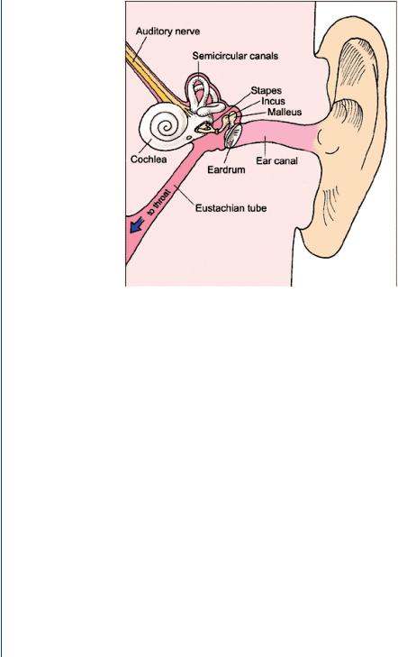
Sound vibrations that reach the eardrum are usually relayed to the inner ear by way of three small bones in the middle ear. These tiny bones, called the hammer (malleus), anvil (incus), and stirrup (stapes), act as a kind of transformer to change sound waves into liquid waves in the inner ear. The stapes bone is the final link in the hearing chain of bones and is the bone most often affected by otosclerosis. When the stapes hardens, a conductive hearing loss occurs. As the hardening continues over time, your hearing worsens. When the hardening spreads to the inner ear, a sensorineural hearing loss occurs. A sensorineural hearing loss is a nerve hearing loss that is usually permanent and can only be helped with a hearing aid.
Hearing loss is the most common symptom of otosclerosis. Some people also have tinnitus, a ringing noise in the head or ear, and almost half of all people with this disease have dizziness. A woman with otosclerosis that becomes pregnant might find that her hearing loss becomes worse.
Unfortunately, there is no medicine that will help or stabilize the hearing loss in people who have otosclerosis. For many people, however, surgery can help or even overcome the hearing
20 SENSORY ORGAN REPLACEMENT AND REPAIR
FIGURE 27: Bones of the middle ear.
loss they have. There are several surgery techniques used to correct the hearing loss associated with otosclerosis. A stapedectomy or stapedotomy is usually recommended. These operations are usually done in a hospital or surgery center with a local anesthetic. After your ear has been numbed, it is cleaned and then a cut is made down inside the ear canal and the eardrum is lifted up to uncover the middle ear. The diseased stapes bone is removed, the inner ear is sealed with tissue, and a new stapes is inserted. Sometimes a laser is used to open the bottom of the stapes and a pistonlike stapes replacement is used. It is also possible that a replacement malleus and/or incus is required. Figure 27 depicts the middle ear among the various anatomical features of the human ear.
2.2Technology and Replacement Components
When the incus is eroded, broken, or absent, the ossicular chain is reconstructed with an incus replacement prosthesis. The one depicted in Figure 28 is a cylinder with a notch that fits under the handle of the malleus and a circular groove that sits on the head of the stapes.
When both the incus and the malleus are eroded or absent, the ossicular chain is reconstructed with a partial ossicular replacement prosthesis (PORP). The one shown in the Figure 29 is a cylinder with a circular flange that fits under the drum. A piece of cartilage is

MIDDLE EAR REPLACEMENT 21
FIGURE 28: Incus replacement with artificial incus.
removed from the tragus and inserted between the prosthesis and the inner surface of the drum to minimize rejection.
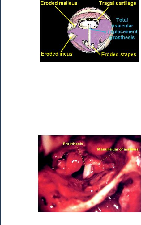
When the incus and arch of the stapes are eroded, or when the malleus, incus, and arch of the stapes are absent, the ossicular chain is reconstructed with a total ossicular replacement prosthesis (TORP). The one depicted in Figure 30 has a circular flange that fits under the drum and a slender shaft that is placed over the footplate of the stapes. Here again, a piece of cartilage is inserted between the drum and the flange.
Surgical replacement of the malleus is shown in Figure 31 and surgical replacement of the incus in Figure 32.
The actual size of an artificial stapes, malleus, or incus is shown in Figure 33 as compared to a common dime.
FIGURE 29: PORP replacement of both the incus and the malleus.
22 SENSORY ORGAN REPLACEMENT AND REPAIR
FIGURE 30: TORP replacement of both the stapes and the incus.
2.3History of Ossicle Surgery and Replacement
Otosclerosis surgery has developed through three eras. The mobilization era began in the late 1800s when Kessel attempted stapes mobilization without ossicular chain reconstruction in cases where it was noted to be fixed (Heermann, 1969). Later, Jack removed the stapes, leaving the oval window open (Schuknecht, 1968). Both techniques allowed increased transmission of sound through the oval window but did not use middle ear amplification structures. Furthermore, fatal cases of meningitis from intraoperative exposure of perilymph to bacteria occurred, and
FIGURE 31: Surgical implantation of artificial malleus. Image is significantly magnified with the actual prosthesis of the size of a letter “i” in an 800 × 600 display.

MIDDLE EAR REPLACEMENT 23
FIGURE 32: Surgical implantation of an artificial incus. Image is significantly magnified.
any gains in hearing frequently were temporary because any remaining stapes footplate often refixed.
The fenestration era began in 1923, when Holmgren created a fistula in the horizontal semicircular canal and sealed it immediately with periosteum (Shea, 1998). This procedure allowed sound conduction preferentially through the fistula, rather than the ossicular chain. Sourdille popularized the procedure when his three-stage technique was widely published during the 1930s (Shea, 1998). Lempert developed a one-stage technique for horizontal semicircular
FIGURE 33: Actual size of artificial ossicle (middle bone) components.

24 SENSORY ORGAN REPLACEMENT AND REPAIR
fenestration, which went on to gain worldwide acceptance after it proved to enhance hearing (Lempert et al., 1956). Results, however, were short-lived because the fenestra often resealed with bone.
The stapedectomy era began before the fenestration era closed. Rosen revisited stapes mobilization in 1952 (Shea, 1998). Later, Shea removed the stapes, sealed the oval window with an autograft vein wall, and then reconstructed the sound-conducting mechanism with an artificial prosthesis (House et al., 1960; Shea, 1976).
This technique gained wide acceptance and has been improved since inception. In the 1970s, Myers conducted stapedotomy using a piston prosthesis (Myers et al., 1970; Myers and Myers, 1968). In the early 1980s, Perkins began using the laser for stapedotomy in a procedure in which a small hole is made in the footplate, as opposed to complete or subtotal removal (Perkins, 1980). Several techniques and approaches are commonly used today, with largely excellent results. A few challenges remain, such as those patients enduring sensorineural hearing loss and unsteadiness, but many think that surgical treatment for otosclerosis has reached perfection.
Evidence has recently mounted that the measles virus plays an important role in gene activation of otosclerosis (Ferlito et al., 2003; Karosi et al., 2004; Karosi et al., 2005; Niedermeyer et al., 2001). This hypothesis is supported by a declining incidence of otosclerosis since measles vaccinations became widespread.
3 COCHLEAR IMPLANT
3.1Introduction
Individuals who have extreme sensorineural hearing loss, are born completely deaf, acquire deafness through illness or injury, or who cannot be adequately treated with the use of a hearing aid or artificial ossicle are candidates for a cochlear implant. The cochlear implant is a prosthetic replacement for the inner ear (cochlea) and is appropriate only for people who receive minimal or no benefit from a conventional hearing aid. The cochlear implant bypasses damaged parts of the inner ear and electronically stimulates the hair cells and adjacent nerves within the cochlea. Part of the device is surgically implanted in the skull behind the ear and tiny wires are inserted into the cochlea at set intervals depending on the number of channels or number of frequency bands to excite. The other part of the device is external and has a microphone, a speech processor (that converts sound into electrical impulses), and connecting cables. It is battery powered, adjustable, and expensive.
A surgical procedure places the implant on the outside of the skull and under the skin just above and behind the ear. An electrode array attached to the implant is inserted into the cochlea. Within a few weeks of the surgery, the user is fitted with external devices—a microphone, processor, and transmitter coil. Sound waves are received by the microphone that
COCHLEAR IMPLANT 25
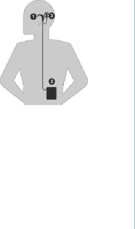
FIGURE 34: Schematic view of a cochlear implant with (1) BTE speech processor or (2) on-the-body speech processor. Either is connected to (3) the connector to the electrode array.
is located either above the ear (Figure 34, point 1) or on the transmitter coil (Figure 34, point 3). The signal from the microphone is sent to the speech processor, which comes in two designs. It may be either a BTE model, which looks like a hearing aid (Figure 34, point 1), or a body-worn device (BWD) that rests on the belt (Figure 34, point 2). In either case, the speech processor deciphers the sound and decides how it will be presented to the ear by means of which electrodes (representing frequencies/regions of the cochlea) will be excited. There are many ways that a processor can translate sound into an electronic code, each of which is called a speech strategy. New speech strategies are being constantly developed to help cochlear implant recipients hear more accurately. A schematic of a cochlear implant depicting both of the potential sites for the speech processor is shown in Figure 34.
3.2Cochlear Implant Components and Surgery
As can be seen in Figure 35, the electronic code is sent to the transmitter coil (Figure 35, point 3) that is held above the implant (Figure 35, point 4) by a magnet. The transmitter coil uses an FM radio signal to transmit the signal through the intact skin into the implant. The implant package decodes the signal and sends a pattern of very rapid, small electrical pulses to