

Physics of biomolecules and cells
.pdfLecture 3: Going After Epigenetics |
391 |
to form a disordered region. Thus, for DNA mapping applications using the posts one can use electrophoretic high velocities and simply work backwards from the taut end and still maintain high spatial resolution.
5 Conclusions
The real use of this technology will be realized when the channels which the molecule traverses are made on the same scale as or smaller than the persistence length of the polymer, so that the molecule must enter the channel not as a coil but as an extended strand. Under those conditions truly high spatial resolution of the length of the molecule will occur allowing this technique to do high resolution dynamic mapping of single dsDNA molecules. Given the high degree of discrimination between polarized light oriented parallel and perpendicular to the slit (Fig. 2), it also seems likely that this device can be used to measure fluorescence polarization [13] of single molecules as they pass successive slits. This can be achieved by placing the polarized light source on the solution side of the device, and the detector below the slit. In this configuration the relaxation time of a polarized signal [14] could also be followed as a function of slit number.
References
[1]J. Lin, R. Qi, C. Aston, J. Jing, T.S. Anantharaman, B. Mishra, O. White, J.C. Venter and D.C. Schwartz, Science 285 (1999) 1558-1562.
[2]X. Michalet, R. Ekong, F. Fougerousse, S. Rousseaux, C. Schurra, N. Hornigold, M. van Slegtenhorst, J. Wolfe, S. Povey, J.S. Beckmann and A. Bensimon, Science 277 (1997) 1518-1523.
[3]Optics at the Nanometer Scale, E. Nieto-Vesperinas and N. Garcia (eds.) (Kluwer Acad. Publ., 1996).
[4]M. Miles, Science 277 (1997) 1845-1847.
[5]M. Hogan, R.H. Austin and J. LeGrange, Nature 304 (1983) 752-754.
[6]H.A. Bethe, Phys. Rev. 66 (1944) 163-182.
[7]E. Betzig, A. Harootunian, A. Lewis and M. Isaacson, Appl. Opt. 25 (1986) 18901900.
[8]Classical Electrodynamics, J.D. Jackson, 3rd edition (John Wiley & Sons, N.Y., 1998).
[9]T.W. Ebbessen, H.J. Lezec, H.F. Ghaemi, T. Thio and P.A. Wol , Nature 391 (1998) 667-669.
[10]M. Born and E. Wolf, Principles of Optics, 6th edition (Pergamon Press, Oxford, 1984).
[11]P.K. Wei, R.L. Chang, J.H. Hsu, S.H. Lin, W.S. Fann and B.R. Hsieh, Opt. Lett. 21 (1996) 1876.
[12]O.B. Bakajin, T.A.J. Duke, C.F. Chou, S.S. Chan, R.H. Austin and E.C. Cox, Phys. Rev. Lett. 80 (1998) 2737-2740.
[13]F. Perrin, J. Phys. Radium 7 (1926) 390-401
[14]X. Chen, L. Levine and P.-Y. Kwok, Genome Res. 9 (1999) 492-498.

392 |
Physics of Bio-Molecules and Cells |
Abstract
I discuss in this lecture a magnetic separation idea which utilizes several ideas from microfabrication and nanomagnetics. The basic idea comes from our earlier work using asymmetry in obstacles and brownian motion to e ect separation of objects [10] by moving them in streams whose angle to the hydrodynamic average velocity is a function of the di usion coe cient of the object.
1 Introduction
Cellular and molecular biologists have developed a variety of ways to sort living cells according to certain characteristics. It is frequently desirable to sort cells according to their chemical content, enzyme activity, surface antigens, or size. Such steps allow physicians to purify a patient’s blood sample for further analysis and allow scientists to isolate rare cell types to study biological processes. The possibility of isolating rare cells, such as hematopoietic stem cells and metastatic cancer cells, carries important medical applications as well. Density centrifugation, fluorescence-activated cell separation (FACS), and magnetic-activated cell separation (MACS) have proven to be very e ective cell sorting methods to this point. However, the great importance of such methods requires constant innovation and refinement of the technique.
The device I propose here is not technically a brownian ratchet device but uses the idea of force which acts at angle to the hydrodynamic flow. In our case, the force is generated by a magnetic field gradient which comes from an array of magnetized wires which lie at an angle θ to a hydrodynamic field flow. The sum of the hydrodynamic force and the magnetic force create a new vector which as in the case of the brownian ratchet moves the cell out of the main stream direction. Figure 1 shows how the two ideas are correlated.
2 Blood specifics
Blood carries nourishment and oxygen to, and waste products away from, all parts of the body through the arteries, veins, and capillaries. Blood also mediates the immune system, recognizing foreign macromolecules and mounting an attack against them. Humans contain approximately five liters of blood, which accounts for 7% of our body weight. There are three main types of blood cells: erythrocytes (red blood cells), leukocytes (white blood cells), and platelets. However, all three arise from precursor cells in the bone marrow, called hematopoietic stem cells.
Erythrocytes are the most common type of blood cell, existing at concentrations around 5×10¹² cells per liter [2]. They constitute approximately

Lecture 4: Fractionating Cells |
393 |
Fig. 1. a) The original thermal ratchet concept. As molecules are moved down in a flow field, the odds of moving to the left or right are not equal. b) The magnetic force separation idea. High magnetic field gradients provide forces at an angle to the flow of cells.
45% of the total volume of blood. Erythrocytes are very small (7.8 µm) and are normally shaped as biconcave disks. Aside from rare exceptions, mature erythrocytes have no nuclei or internal membranes. Their purpose is the first listed above: they are primary components of the circulatory system. Leukocytes exist at nearly one-thousandth the concentration of erythrocytes, and they serve an entirely di erent purpose. Leukocytes protect the body from infection, in cooperation with the organs of the immune system. They are typically classified into three types: granulocytes, monocytes, and lymphocytes. Granulocytes, so named because of granules in their cytoplasm, make up the majority of leukocytes at 5 × 10⁹ cells per liter. They range from twelve to fifteen microns in diameter. Monocytes exist at only 4×10⁸ cells per liter, but they range from fifteen to eighteen microns across. Lymphocytes, at 3 × 10⁹ cells per liter, are only slightly larger than erythrocytes, though each has a very large nucleus which occupies most of the cell.
All adaptive immune responses are mediated by B-lymphocytes (B cells) and T-lymphocytes (T cells). All lymphocytes bear variable cell-surface receptors to detect antigens, or foreign macromolecules and cells. Of special interest to cellular and molecular biologists are the blood-borne proteins called antibodies, for they can be used as highly-specific probes to identify and distinguish between di erent cell populations. Antibodies are Y-shaped proteins of the immunoglobulin (Ig) family. The body produces antibodies as a defense against extracellular materials. For instance, an antibody may bind to a virus or toxin to prevent it from infecting a cell, or it may coat a foreign bacterial cell and mark it for destruction. However, a particular antibody can only bind to select molecules that fit into its antigen-binding site. Although the amino acid sequence among all antibodies is mostly constant, the end of each “arm” of the Y-shaped molecule sports a variable

394 |
Physics of Bio-Molecules and Cells |
region. These arms form the binding site of an antibody, and it is their variability that accounts for the specificity in what they can bind to. Today, antibodies can be obtained which distinguish between two polypeptides that di er by only a single amino acid [3]. Once families of antibodies have been produced, they can be conjugated to small fluorescent molecules, such as fluorescein or rhodamine, by which they can be detected under a fluorescent microscope.
Cell biologists have isolated families of antibodies that selectively recognize di erent subpopulations of leukocytes by specific proteins contained in the cells’ outer membranes. Though they are natural components of the leukocytes, these cell-surface molecules are called “antigens” because antibodies can be raised against them. A more fitting name for them is “markers” because they are characteristic of specific cell populations. Markers can be grouped into multiple categories; some are specific for cells of a particular lineage, while the presence of others may vary according to the stage of di erentiation of cells of the same type [4]. Any cell surface marker that identifies a particular lineage or di erentiation stage and is recognized by a group of monoclonal antibodies is part of a “cluster of di erentiation”. All leukocyte surface markers whose clusters are defined are designated with a CD, followed by a number.
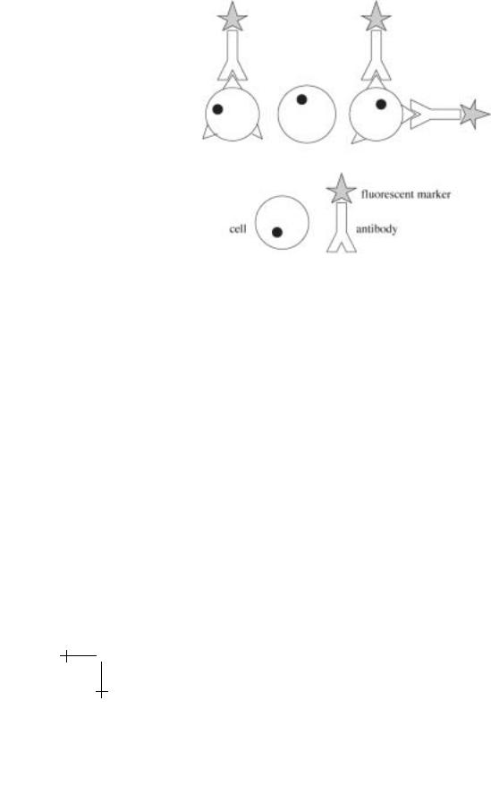
The CD system has allowed immunologists to identify cells participating in various immune responses. For instance, most helper T cells are CD3+ CD4+ CD8−, while cytotoxic T cells are CD3+ CD4− CD8+ [4]. Fluorescent molecules can be conjugated to the antibody clusters that recognize their specific markers, and then incubated with the cells in a sample. However, the CD system also enables the fractionation of blood cells according to their specific surface antigens. Clusters of antibodies can be used to selectively bind fluorescent molecules or magnetic beads to certain leukocytes, which are then isolated by flow cytometry techniques. This system is crucial to our method of separating cells as well as to the existing technologies. Figure 2 gives a picture of how antibodies can be labeled and attached to cells.
Fluorescence-activated cell separation provides scientists with one way to isolate cells of a uniform type from a tissue or cell suspension. Specific cells in a sample are labeled with antibodies of an appropriate cluster, coupled to a fluorescent dye. The cells are sent single file in a fine stream through the path of a laser beam. As each cell passes the beam, it is monitored for fluorescence. A nozzle then forms droplets containing single cells and gives each a positive or negative charge, depending on whether the cell it contains is fluorescent. Finally each droplet is deflected by a strong electric field into a collection chamber [2]. This process results in two collection tubes, one containing labeled cells and one containing unlabeled cells. Consequently, the specific cells in the sample that were labeled are isolated from the rest
Lecture 4: Fractionating Cells |
395 |
Fig. 2. Labeled antibodies and their attachment to a cell membrane.
of the sample. A schematic view of a FACS machine and a magnetic sorting device is shown in Figure 3.
The beads themselves are composed of iron (II) oxide nanocrystals approximately 50 nm in diameter, coated with a polysaccharide which provides functional groups for the attachment of antibodies [6]. Thus, each bead is smaller than the average leukocyte by a volume factor of more than 106. Their extremely small size makes them very gentle on the cells to which they attach, and they bear apparently e ect on the cells’ function or viability. However, although only a few dozen beads are needed to separate a cell, several thousand may be bound to a particular cell after incubation [7].
An interesting physics result of the nanosize of the beads is that they are superparamagnetic, which means that they are single domain but too small to form a stable magnetic moment (ferromagnetic materials have stable magnetic moments in the absence of an external magnetic field H). Ferromagnetism comes from the Fermi exchange interaction between two atoms, each of which has a net electronic unpaired spin S, and the net odd symmetry that the total wavefunction must have. The sign of the exchange interaction is such that the spins have a parallel alignment energy on the order of 400 K, and if the system is big enough can be ferromagnetic at room temperature. However, for small volumes thermal fluctuations will be su cient to overcome the anisotropy energy and cause the spontaneous loss

396 |
Physics of Bio-Molecules and Cells |
Fig. 3. Two conventional ways to sort labeled cells from whole blood.
of a permanent magnetic moment. Under such conditions the material is classified as superparamagnetic. In zero external field, the net magnetic moment of the superparamagnetic beads is zero. In the presence of an external magnetic field the beads can be highly magnetized.
The high specificity and e ciency of magnetic methods have made them quite useful in obtaining rare cell types. Hematopoietic stem cells, residual tumor cells, and antigen-specific B and T cells can be isolated and used in a variety of functional assays. Hematopoietic stem cells can be isolated by their expression of the CD34 antigen [8]. Stem cell purification techniques are of great value for both science and medicine. Pure populations of stem cells will make possible scientific studies of blood cell formation and differentiation. Additionally, they are necessary for successful transplantation procedures. Stem cells from the bone marrow and peripheral blood are transplanted in combination with chemotherapy for the treatment of certain malignant and genetic disorders. The success of a transplant procedure depends on the e ectiveness of techniques that are used to isolate the cells for the transplant. Any additional cells lingering in the preparation may pose a risk to the recipient.
Cancer patients benefit from cell separation techniques by their capacity to remove residual tumor cells from the bone marrow. Since conventional cancer therapy is toxic to bone marrow stem cells, a fraction of bone marrow must be removed from a patient before high dose therapy can be given. The

Lecture 4: Fractionating Cells |
397 |
bone marrow can subsequently be reinfused into the patient. But before this occurs, it is desirable to eliminate all tumor cells from it. Tumor cells can be selected by specific antibodies to surface markers and then removed from the sample. Scientists have achieved removal of 99.9% of the malignant cell population using magnetic separation techniques [9].
We describe an alternative way to isolate biological cells from a larger sample. Our method is similar to MACS in that specific cells are attached to antibody-coated magnetic beads in a high magnetic field gradient. However, our device is entirely confined to one single microchip. Additionally, it confers the advantages of continuous input and two-dimensional separation. It is believed that our device could potentially yield a higher e ciency and a greater degree of purification than existing cell separation techniques.
3 Magnetic separation
In our device, cell fractionation is made possible by an array of very thin magnetized “wires” which are aligned at an angle to a net hydrodynamic flow direction. There are two advantages to these microfabricated wires: the extremely thin ferromagnetic layer forces the spin system to be single domain, or at least “few domain” with resultant very high magnetic fields. Secondly, because of the small length scale of the wires such small structures have large magnetic field gradients at their edges. Since magnetic force depends on field gradients, the path of a paramagnetic object exposed to this array of wires will be altered. Thus, our device can separate paramagnetic objects from diamagnetic ones. Paramagnetic beads attached to cells are attracted to the wires and are deflected away from unlabeled cells.
Since there are no magnetic monopoles, forces result from field gradients acting on magnetic dipole moments. If an object is ferromagnetic with a permanent magnetic moment, µ, it will feel a force in the presence of a magnetic field gradient given by:
Fm = (µ · )B. |
(3.1) |
|
|
|
|
The magnetic moment of a paramagnetic object is induced by an external field. A paramagnetic object with magnetic susceptibility χ will feel a force given by:
Fm = (χB · )B. |
(3.2) |
|
|
|
|
In our apparatus, beads attached to the cells have an induced magnetic
moment ( ) aligned parallel to the field produced by the wires. The pre-
χB
ferred magnetization direction of the magnetic stripes is to place mag-
B netized perpendicular to the plane of the wafer, so the field points in the

398 |
Physics of Bio-Molecules and Cells |
+y-direction. Since beads travel through our device at a fixed height yo above the wires, the force that they encounter is:
F = µ∂By /∂x. |
(3.3) |
Our beads are single domain, 50 nm particles of ferrous iron oxide (FeO). They are superparamagnetic, which means that they are too small to sustain a stable dipole moment but that they will exhibit a net moment in an external applied field.
4 Microfabrication
Microfabricated devices are capable of accessing the small length scales of biological cells and providing magnetic field gradients high enough for cell fractionation. Microfabrication was performed at the Princeton Center for Photonics and Optoelectronic Materials (POEM). The facility o ers a full range of sophisticated processes and equipment for scientific research, which enables the fabrication of devices that could not have been made several years ago.
The fabrication of our device required a series of steps to make it suitable for magnetic cell separation. First, the outer design was exposed onto a silicon wafer by photolithography. This included the channels, inlets, outlets, and central chamber, constituting the framework of the apparatus. Then, this design was etched 16 µm into the wafer and the remnant photoresist was stripped o . A separate pattern for the diagonal magnetic wires was exposed onto the central chamber, again by photolithography. Grooves for these wires were then etched an additional 0.2 µm so that the wires would be countersunk and not impede the flow of cells. A ferromagnetic metal alloy was deposited onto the wafer in a uniform coat. The unexposed photoresist was then removed from the wafer, lifting o the overlapping metal with it. This left behind only the thin wires on the wafer. Finally, a protective layer of SiO2 was deposited on top of all structures.
Masks for positive imaging were made commercially by Adtek Photomask of Montreal, Quebec. Our device required two masks: one for the outer channels and outlets, and one for the magnetic wires. Each mask was a square quartz plate, 5 × 5 × 0.09 . The masks were designed on L-Edit, a computer graphics program, and they were fabricated by electron beam lithography. A pair of alignment marks at opposite ends of each mask ensured that the patterns overlapped each other precisely.
To form magnetic wires, a cobalt-chrome-tantalum (Co-Cr-Ta) alloy was sputtered onto silicon wafers that had been exposed to the diagonal wire pattern. Co-Cr-Ta was used because of its high remanent magnetization. Co-Cr-Ta was chosen as a suitable alloy for our magnetic structures because

Lecture 4: Fractionating Cells |
399 |
it has an exceptionally high remanent magnetization of 6000 Gauss [16]. This should be compared to the saturation magnetization of its main component, cobalt (1400 Gauss) [15]. As an alloy, Co-Cr-Ta has the ability to acquire and retain a greater magnetic moment than each of its three components. Co-Cr-Ta occupies a hexagonal close packed (hcp) lattice structure, and, like most sputtered films, it is isotropic [17]. Additionally, as the wires in our device are very thin films, it is expected that they are single domain. Evidently the wires are capable of acquiring a large, uniform magnetization in an applied field and retaining it when removed from the field. As they are isotropic, they may be magnetized either in the plane of the wafer or perpendicular to it.
Wires were magnetized perpendicular to the plane of the wafer, in a uniform external field. The magnetic field was provided by a large electromagnet and measured at 5 kG. It is believed that wires achieved their saturation magnetization in this field and retained a nearly uniform internal field of 6000 Gauss as the electromagnet was disabled.
The strength of the magnetic remanence of the wires was tested using paramagnetic and latex beads. First, fluorescent paramagnetic beads were suspended above the wires. Beads were attracted by the wires in a striking fashion, accumulating at the edges where the field gradients were the highest. As a control, nonmagnetic polymer beads were then suspended. They disseminated uniformly, showing no preference for the magnetic wires.
5 Magnetic field gradients
Magnetic fields are produced by Co-Cr-Ta wires positioned at a 45o angle to the input stream of cells. Each wire is 10 µm wide and 0.2 µm thick, and neighboring wires are separated by 25 µm. The following calculations presume uniform magnetization of the wires, with a remanent field of 6000 Gauss.
Wires may be magnetized in either of two directions: in the plane of the wafer or perpendicular to the wafer. The two orientations are shown in Figure 4, with a consistent coordinate system. For neither method is there a force directed along the length of the wire; ∂B/∂z = 0 in each case. However, for both orientations, there are very large gradients in the magnetic fields near their edges. These gradients attract paramagnetic objects to the wires and redirect their flow through the chamber. The orientation that can provide higher field gradients should be chosen for optimal results.
Field gradients were also calculated analytically. Since cells travel at a fixed height yo above the wires, only ∂B/∂x is significant. Although gradients for both in-plane and perpendicularly magnetized wires are comparable at large yo, gradients at lower heights are significantly greater for the case

400 |
Physics of Bio-Molecules and Cells |
Fig. 4. Two possible directions for magnetization.
of perpendicular magnetization. Thus, in order to maximize the magnetic force for separation, wires were magnetized perpendicular to the plane of the wafer.
A bead that is not attached to a cell will roll along the floor of the chamber. The bead is separated from the wires by only a 0.2 µm layer of
SiO2. Since its magnetic moment aligns with the field and Fm = (µ · )B, |
|
|
|
the bead will experience horizontal forces of up to 3 × 10⁻¹¹ Newtons, or 3 ×10⁻⁶ dynes. A bead attached to a cell will feel a smaller force because it is elevated above the floor of the chamber. Lymphocytes are approximately 8 µm in diameter. Beads will therefore be, on average, 4 µm above the wires in the chamber. At this height, fields are 0.0045 Tesla and gradients are 1300 T/m. Consequently, beads will experience forces up to 1.5 × 10⁻¹³ N, or 1.5 × 10⁻⁸ dynes. For comparison, a constant force of 1.5 × 10⁻⁸ dynes will cause a leukocyte to reach a terminal velocity in water of 1 µm/s, a modest speed for a cell in a microchip. In reality, though, the forces on the cells will be considerably higher. Because the beads are so small, several thousand may bind to each cell. Also, many of these beads will be closer to the wires than 4 µm, where forces are significantly greater. Cells will experience the net e ect of the magnetic forces on all beads bound to them.
It is important to note that magnetic forces point in the x-direction, perpendicular to the wires. The magnetic force is greatest at the edges, and it is always attractive. Since the cells flow at a 45o angle to the wires, there is an x-component to the hydrodynamic force which should exactly cancel the magnetic force. All that is left is the z-component to the hydrodynamic force (and viscous drag). As a result, the cells flow in the z-direction, along the lengths of the wires. Although they continue to be propelled by the input jet stream, they are constrained to follow the wires by the high field gradients at their edges.