


Dev Genes Evol (2006) 216:743–754 |
745 |
|
|
The budding sequence of each species was inferred through static observations of the ontogenetic sequence of developing zooids. The organization of zooids along a mature portion of the stem was first established. The regularity of siphonophore organization made it possible to then determine the identity of immature zooids further to the anterior based on their relative position.
Results
A note on terminology
Haddock et al. (2005) consolidated and standardized the usage of terms for axes and orientation in siphonophores, and the present article adheres to this clarified scheme. The nomenclature used to describe the functionally specialized zooids of siphonophores is similar to that used for the Hydrozoa in general, but also includes several terms that are specific to the group (Dunn 2005). The term gonodendron is used here to describe any compound reproductive structure consisting of multiple zooids that are attached to the stem via a common peduncle. The structure and zooid composition of gonodendra vary widely across taxa; it is not at all clear if they are homologous across siphonophores or have independently arisen multiple times.
Material examined
Specimen data for the examined material are listed in Table 1. Specimens of Apolemia sp. and the cystonects are from Philip R. Pugh’s collection. Most Agalma elegans material was collected in the bay at Villefranche-Sur-Mer, France, in the months of March–May in 2003 and 2004; the remaining A. elegans specimens were collected by bluewater diving from the RV Oceanus in the summers of 2000–2002 along the east coast of the USA. Nanomia bijuga and Forskalia formosa were both collected in Monterey Bay, CA, and the bay at Villefranche-Sur-Mer, France. These newly acquired specimens have been deposited in the Yale Peabody Museum (catalog numbers can be found in Table 1). The Lychnagalma utricularia specimen quickly deteriorated after being caught and was not preserved.
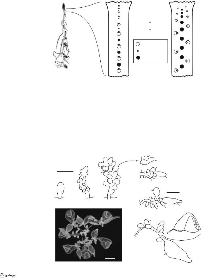
Bathyphysa sibogae
The gastrozooids and gonodendra of Bathyphysa sibogae were found to be in a uniserial sequence (Fig. 2b). The gastrozooids first appear as isolated buds at the anterior end of the siphosome and grow a tentacle on their anterior side as they mature and are carried to the posterior by the elongating stem. The first several gastrozooids at the
Table 1 Specimens examined
Species |
Specimen |
Collection |
|
|
|
Bathyphysa sibogae |
BWP796-20-7 |
PRP |
Rhizophysa filiformis |
BWP1077-3 |
PRP |
|
BWP567-8 |
PRP |
|
BWP566-17 |
PRP |
|
BWP566-16 |
PRP |
|
BWP588-21 |
PRP |
|
BWP442 |
PRP |
Rhizophysa eysenhardti |
BWP539-13 |
PRP |
|
BWP1611-12 |
PRP |
|
BWP634-6 |
PRP |
|
BWP741-3 |
PRP |
|
BWP778-12 |
PRP |
|
BWP465 |
PRP |
|
BWP465 |
PRP |
|
BWP802-1 |
PRP |
|
BWP1086-6 |
PRP |
|
BWP814-12 |
PRP |
Agalma elegans |
YPM 36365-36390 |
YPM |
Nanomia bijuga |
YPM 36427-36444 |
YPM |
Apolemia sp. |
JSLII 1450-SS3 |
PRP |
Forskalia formosa |
YPM 36445-36448 |
YPM |
Lychnagalma utricularia |
Tiburon 676-D6 |
(specimen |
|
|
destroyed) |
|
|
|
The numbers given in YPM rows are museum catalog numbers. The specimens in PRP’s collection are identified by their mode
of collection and a unique identifier.
PRP Personal collection of Philip R. Pugh (National Oceanographic Centre, UK); YPM the Yale Peabody Museum (New Haven,
CT); BWP blue-water plankton series; Tiburon the remotely operated vehicle Tiburon (Monterey Bay Aquarium Research Institute);
JSL II the manned submersible Johnson Sealink II
(Harbor Branch Oceanographic Institute)
anterior end of the sequence (i.e., the youngest gastrozooids) have no sign of gonodendra between them. Gonodendra arise further to the posterior as isolated buds between and in line with the maturing gastrozooids.
The young gastrozooids of B. sibogae have pronounced lateral ridges known as ptera (Leloup 1936), which give the gastrozooids a bract-like appearance. The striking similarity of these Bathyphysa gastrozooids to “physonect” bracts may indicate that physonect bracts are in fact modified polyps. Each young gastrozooid also has a lamella extending part way up its posterior side and further anchoring it to the stem, just as physonect bracts do. This holds the young gastrozooid so that its distal end faces to the posterior. Although conspicuous in young gastrozooids, the ptera and lamellae are absent by the 30th gastrozooid in the examined specimen.
Leloup (1936) figured and described the early stages of development of Bathyphysa gonodendra. He did not, however, trace the fate of the young buds or describe their
746 |
Dev Genes Evol (2006) 216:743–754 |
|
|
Fig. 2 Schematic illustrations of the budding sequences of long-stemmed cystonects (the Rhizophysidae). (b, c ventral view) a Rhizophysa eysenhardti
(adapted from Kawamura 1910). b Uniserial budding, in which the gastrozooids arise in a single line, and the gonodendra are later intercalated between them. The tentacles are borne on the anterior side of the gastrozooids. c Triserial budding, in which the gastrozooids arise in two outer rows, and the gonodendra are found in a row between them. The tentacles are borne on the side of the gastrozooid facing the ventral midline
a |
b Uniserial |
c Triserial |
A
R

 L
L
P
Gastrozooid
Tentacle
Gonodendron
 Gastrozooid
 Gonodendron
Bathyphysa sibogae |
Rhizophysa eysenhardti |
Rhizophysa filiformis |
|
later stages of development. The material examined here was in good condition, and the full ontogenetic sequence of gonodendron development was described (Fig. 3). The youngest gonodendron buds are at first simple, smooth evaginations that protrude from the stem (Fig. 3a). These then elongate and become warty in appearance (Fig. 3b,c). Each wart-like protuberance then elongates and develops into a gonodendron branch (Fig. 3d–g). The branches do not ramify further, and are all attached directly to the
central style (i.e., the main axis of the gonodendron). A single bud first arises on the side of the branch (Fig. 3d). This bud takes on a distinctive shape and matures into a nectophore. The portion of the branch distal to the nectophore matures into the palpon that terminates the branch. Gonophores then form from evaginations that arise along the branch (Fig. 3e), some of which arise distal to the nectophore. Each branch bears on the order of seven to nine gonophores at maturity. The gonodendra of this species,
Fig. 3 Developmental sequence |
|
|
d |
N |
|
|
of Bathyphysa sibogae gono- |
|
|
|
|
||
|
|
|
P |
|
||
dendron. a–c Young gonoden- |
|
|
|
|
|
|
dra showing the origin of the |
|
0.5 mm |
|
|
N |
|
side branches as evaginations of |
|
e |
|
|
|
|
the main gonodendron axis. |
|
|
|
|
|
|
|
|
|
|
|
|
|
d–g Close-ups of isolated side |
a |
b |
c |
|
P |
|
branches following the develop- |
|
|
|
Go |
|
|
|
|
|
|
|
||
mental stage shown in (c). |
|
|
|
|
|
|
|
|
|
|
|
|
|
h Photograph of preserved |
|
|
|
N |
1 mm |
|
gonodendron. Scale bar |
|
|
f |
|
|
|
(0.5 mm) applies to a–e, Scale |
|
|
|
|
|
|
bar (1 mm) applies to f, g; scale |
|
|
|
|
|
|
bar (2 mm) applies to h. Go, |
|
|
|
|
Go |
P |
gonophore; N nectophore; |
|
|
|
|
||
|
h |
|
|
|
||
P gonopalpon |
|
|
|
|
|
|
|
|
|
g |
Go |
N |
|
2 mm |
P |
|

Dev Genes Evol (2006) 216:743–754 |
747 |
|
|
like those of the other Cystonectae, are thought to break away from the colony at maturity before releasing their gametes (Totton 1965, P.R. Pugh, personal communication).
Rhizophysa filiformis
All examined specimens of Rhizophysa filiformis had the same uniserial development of gastrozooids and gonodendra as B. sibogae (Fig. 2b). There were from one to six gonodendra found between the mature gastrozooids. The gonodendra develop in the same manner as those of B. sibogae (Fig. 3) and have the same general structure at maturity. The gastrozooids of R. filiformis do not have pronounced ptera at any stage.
Rhizophysa eysenhardti
The origin of the siphosomal elements of Rhizophysa eysenhardti is triserial (Fig. 2c). The buds of the two outer rows give rise to gastrozooids. The gastrozooids are staggered with respect to each other. The single tentacle of each gastrozooid forms on the side facing the midline. When the gastrozooids are mature, it is not entirely obvious that they arose in two separate rows, but the alternate attachment of the tentacle to the left and right sides of the gastrozooids is clearly discernable.
The median row of siphosomal elements, found between the left and right rows of gastrozooids, consists solely of gonodendra. Gonodendron buds are found just as far to the anterior as the gastrozooid buds are. The gonodendron buds are not exactly in phase with the gastrozooid buds, and there are a variable number of gonodendra between gastrozooids at maturity (but always at least one). The gonodendra of R. eysenhardti develop in the same manner as those of B. sibogae (Fig. 3).
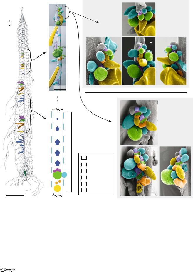
Agalma elegans
Our observations of the zooid organization of A. elegans (Fig. 4b,c) are consistent with Totton’s (1954, frontispiece— reproduced here as Fig. 4a). Each cormidium consists, from posterior to anterior, of a large palpon (marked as “B” in Totton’s (1954) frontispiece so it will be referred here as the B-palpon), a female gonodendron with an associated palpon, a gastrozooid with multiple gastric palpons attached to its peduncle, and several clusters of male elements. Each cluster of male elements has a palpon, several male gonophores, and bracts in various stages of development. Bracts also are spread along the full length of the stem. Each cormidium can span up to 4 cm or more of the stem, and there are few developing cormidia within the growth zone. This makes the inference of the budding sequence quite difficult in this species because there are
few intermediate stages of cormidial development in each colony. It also implies that zooids may be added to the colony at a very slow rate.
There is not a pronounced horn (sensu, Dunn 2005) in the siphosomal growth zone of mature specimens of A. elegans, although the tip of the siphosomal growth zone does form a slight overhang (Fig. 4d–j, marked “T” in each pane). Totton (1956) noted a well-developed horn (which he called a nectostyle) in the larvae of this species, so it seems to recede as the colony matures. The anteriormostdifferentiated cormidium consists of six buds on a common peduncle (as seen in cormidium 1 of the specimens figured in Fig. 4d–g and h–j). The largest of these is a bud that was inferred to be the gastrozooid based on its rounded shape and consistent position relative to the other buds. There are two buds on the anterior side of the gastrozooid base. The proximal bud, which was identified at various stages of development by its anteriormost position within the cormidia, differentiates into the primary male reproductive elements. The distal bud on the anterior side of the gastrozooid peduncle differentiates into one, or perhaps several, gastric palpons. There is also a bud on the left side of the gastrozooid base at the same level as the bud of the male reproductive elements. This lateral bud was inferred to be a bract based on the differentiation of buds in the same position in more posterior cormidia. Finally, there are two buds to the posterior of each young gastrozooid. The one furthest to the posterior had a characteristic shape even early in development that indicated that it was a palpon, and its precocious development and location indicate that it is the B-palpon. Between the B-palpon and the gastrozooid is a bud that in posterior cormidia differentiates into the female reproductive elements. The B-palpon grows larger than all the other zooids of a cormidium by the point where the cormidium is second in the sequence. Totton (1954) considered the gastrozooid to be the posterior element of each cormidium. In light of the developmental sequence, we differ with him on this point and consider the B-palpon to be the posteriormost structure.
In addition to the bract that arises on the left of the base of each gastrozooid, there is a lateral row of bracts on both the left and right sides of the stem. These bracts are further from the ventral midline than any other zooids and are more developed than adjacent bracts arising from the base of the gastrozooids. No buds that could give rise to these lateral bracts were ever observed within the derivatives of the probud, and the lamellae of these bracts sometimes extended along the stem slightly to the anterior of the tip of the siphosomal growth zone. These findings suggest that they arise directly on the side of the stem.
The male bud comes to be attached to the stem independently from the gastrozooid, gastric palpon, and gastrozooid-associated bract, and the female bud comes to
748 |
Dev Genes Evol (2006) 216:743–754 |
|
|
a
A
V

 D
D
P
1 cm
b
Cormidium
1 mm
A
R

 L c P
L c P
Cormidium
d
|
|
T |
2 |
Specimen 1 |
|||
|
|
1 |
50 µm |
||||
|
|
|
|||||
|
|
3 |
|
||||
|
|
2 |
3 |
|
|
|
|
|
|
|
|
|
|
|
|
e |
|
(Anterior) |
|
g |
|
|
|
|
f |
R L |
T |
||||
|
|
|
A |
|
|
|
|
T |
|
T |
P |
1 |
|
|
|
|
|
1 |
|
|
|
||
|
1 |
|
|
|
|
|
|
|
1 |
2 |
1 |
2 |
|
||
1 |
2 |
|
|||||
|
|
2 |
|
|
|
||
|
|
2 |
|
|
|
|
|
2 |
|
|
|
|
|
||
|
|
3 |
|
|
|
||
|
|
|
|
|
|
|
|
3 |
|
3 |
|
3 |
|
|
|
3 |
|
3 |
|
|
|
||
(Right) |
|
(Ventral) |
|
(Left) |
|
|
|
h
|
|
T |
1 |
|
|
|
|
|
Specimen 2 |
|||
|
|
|
|
|
|
|
|
|||||
|
|
|
1 |
2 |
|
|
|
|
50 µm |
|||
|
|
|
|
|
|
|
|
|||||
|
|
|
|
|
|
|
|
|
||||
|
|
|
3 |
|
2 |
|
|
|
|
|
|
|
|
|
|
|
|
|
|
|
|
|
|
|
|
|
(Anterior) |
|
|
|
|
|
|
|
|
|
||
|
i |
T 1 |
|
R L |
j |
|
|
|||||
|
|
|
|
|
|
A |
|
|
|
|
||
|
|
|
|
|
|
|
|
|
|
|
T |
|
|
Male |
|
1 |
2 |
|
P |
1 |
|||||
|
|
|
|
|
||||||||
|
|
|
|
|
|
|
|
|||||
|
Bract |
|
|
|
|
|
2 |
|
||||
|
|
|
|
|
|
|
|
|||||
|
|
|
|
|
|
|
|
|
|
|
||
|
|
|
|
|
|
|
|
|
|
|
|
|
|
Gastric Palpon |
|
3 |
|
2 |
|
|
|
3 |
|
2 |
|
|
|
|
|
|||||||||
|
Gastrozooid |
|
|
|
|
|
|
|
|
|
||
|
|
|
|
|
|
|
|
|
|
|
||
|
|
|
|
|
|
|
|
|
|
|
|
|
|
Female |
|
3 |
|
|
|
|
|
|
|
|
|
|
|
|
|
|
|
|
|
|
|
|
||
|
B Palpon |
|
|
|
|
|
|
(Left) |
|
|
||
|
(Ventral) |
|
|
|
|
|
|
|
||||
|
|
|
|
|
|
|
|
|
||||
Fig. 4 Agalma elegans—false coloring indicates zooid identity. Anterior faces the top of the page, unless noted otherwise. a Left lateral view of the mature colony (adapted from Totton 1954). The bracts, which sheathe the siphosome, have not been colored. b Photographic collage showing the growth zone and young siphosomal stem of an anesthetized living specimen. Ventral view. Note that both the female gonodendron and the female-associated palpon have been colored orange, and that the male gonophore and male associated bract and palpon buds are colored blue. Buds whose fate could not be assigned with confidence have been left uncolored. c Schematic of a mature cormidium. Ventral view. The lateral bracts are not shown in their actual position along the
stem relative to the other zooids. d–g SEM of the growth zone of specimen 1, shown from four different views. h–j SEM of the growth zone of specimen 2, shown from three different views. In d–j, the tip of the growth zone is labeled with a T, and the gastrozooids and B-palpons are numbered according to the cormidium they belong to, beginning with the first differentiated cormidium. Gastrozooids 1–2 and B-palpon 3 have been broken away (h–j). Additionally, gastrozooid 3 has been broken away (i). Where young gastrozooids were broken away, the buds of the gastric palpons, and sometimes the left bracts, also broke off with them. A Anterior; D dorsal; L left; P posterior; R right; V ventral