


Dev Genes Evol (2006) 216:743–754 |
749 |
|
|
be attached to the stem independently from the B-palpon (this is most easily seen in Fig. 4i). Bracts later bud at the foot of the B-palpon. After the female bud separates from the B-palpon, it takes on a bilobed shape (as in cormidium 3 of Fig. 4i,j), and further to the posterior, one lobe was seen to be the female-associated palpon and the other the female gonodendron (not shown). These are attached to the stem independent from each other at maturity. Close to the growth zone, there is only one male cluster per cormidium. In cormidia slightly to the posterior, it can be seen that a new male cluster arises directly on the stem, separate from and to the anterior of the original one. This process repeats several times, with new male clusters being added anterior to the existing ones.
There are multiple gastric palpons attached to the peduncle of gastrozooids in mature cormidia. It was not determined if these arise by subdivision of the single gastric palpon bud that arise within the growth zone or if each gastric palpon arises as an independent bud on the peduncle of the gastrozooid. There are more gastric palpons in cormidia further to the posterior.
Large specimens of A. elegans have an irregular organization of zooids in their posterior cormidia, but in all examined cases, the irregularity was consistent with zooid loss. In these specimens, cormidia close to the growth zone still have the regular organization originally described by Totton (1954) and reconfirmed here.
The nectosomal growth zone of a single specimen was examined using SEM. It was found that each young nectophore had a small bud on the posterior side of its peduncle. A similar bud has previously been found in the nectosomal growth zone of Bargmannia elongata (Dunn 2005).
Nanomia bijuga
Totton (1965, Fig. 35) figured the organization of zooids within a cormidium of N. bijuga. He found that each cormidium consisted of a gastrozooid and a series of palpons, all attached independently to the stem. A female gonodendron and a cluster of male gonophores flanked each palpon and alternated sides from palpon to palpon. Totton noted that secondary palpons, also with male and female structures at their base, were sometimes intercalated between the primary palpons in mature cormidia.
Our own observations of N. bijuga confirm Totton’s earlier findings. In addition, we are also able to describe the budding sequence by which the zooids of this species arise. There is a pronounced horn within the siphosomal growth zone, and each cormidium arises as a simple probud close to its tip (Fig. 5d). The bulk of the probud gives rise to the gastrozooid, with most of the other zooids arising on the
anterior side of its peduncle. A palpon and two flanking bracts are the first such zooids (Fig. 5e). Additional bracts are added on both sides of the gastrozooid peduncle, with the youngest (i.e., the smallest) being the most distal (Fig. 5f). Additional palpons bud on the anterior side of the primary palpon, and all palpons initially share a common base and form a fanlike structure (Fig. 5g). The original palpon is the most distal in this structure, and the smallest (youngest) is the most proximal. A lateral bract forms on each side of each palpon as it matures.
The zooids of successively more mature cormidia come to be spread out along the stem such that each zooid is eventually attached to the stem independently (Fig. 5h,i). The bracts that arose on the gastrozooid peduncle come to be arranged in two lateral rows that flank all the other zooids (the lamellae of the left row of bracts can be seen in Fig. 5i). The oldest (i.e., largest and most proximal) bract to arise on the gastrozooid peduncle comes to be located furthest to the anterior, and the youngest (i.e., the smallest and most distal) remains the closest to the gastrozooid. As the palpons spread out, the youngest (i.e., the smallest and most proximal) comes to be located the furthest to the anterior of the cormidium, and the first palpon to arise (i.e., the largest and most distal) remains closest to the gastrozooid. The lateral bracts associated with each palpon move away from the ventral midline and come to be arranged in rows just inside of the rows of bracts that arose on the gastrozooid peduncle. Kawamura (1911) had already noted that the bracts of this species are arranged in two rows along each side of the stem.
The male and female gonodendra arise at the base of the palpons just inside of each palpon-associated bract (Fig. 5i). Additional clusters, each consisting of a palpon, a male gonodendron, a female gonodendron, and bracts, are added directly on the stem at the anterior end of each cormidium after all zooids have spread out. Clusters may also sometimes be added between existing palpons.
There is a single tentacle on the anterior side of each gastrozooid. Likewise, each palpon has a palpacle (as the tentacle of a palpon is called) on its anterior side. Tentacles and palpacles form as simple evaginations. The bud of the gastrozooid tentacle can be seen in Fig. 5f–h, and the base of the tentacle, which has been broken away, can be seen in Fig. 5i. The palpacle rudiments can be seen in Fig. 5i. Some palpons also have a large basal swelling on their anterior side just distal to the palpacle.
Apolemia sp.
One specimen of Apolemia, belonging to the same undescribed species as “Apolemia 3” in the molecular
750 |
Dev Genes Evol (2006) 216:743–754 |
|
|
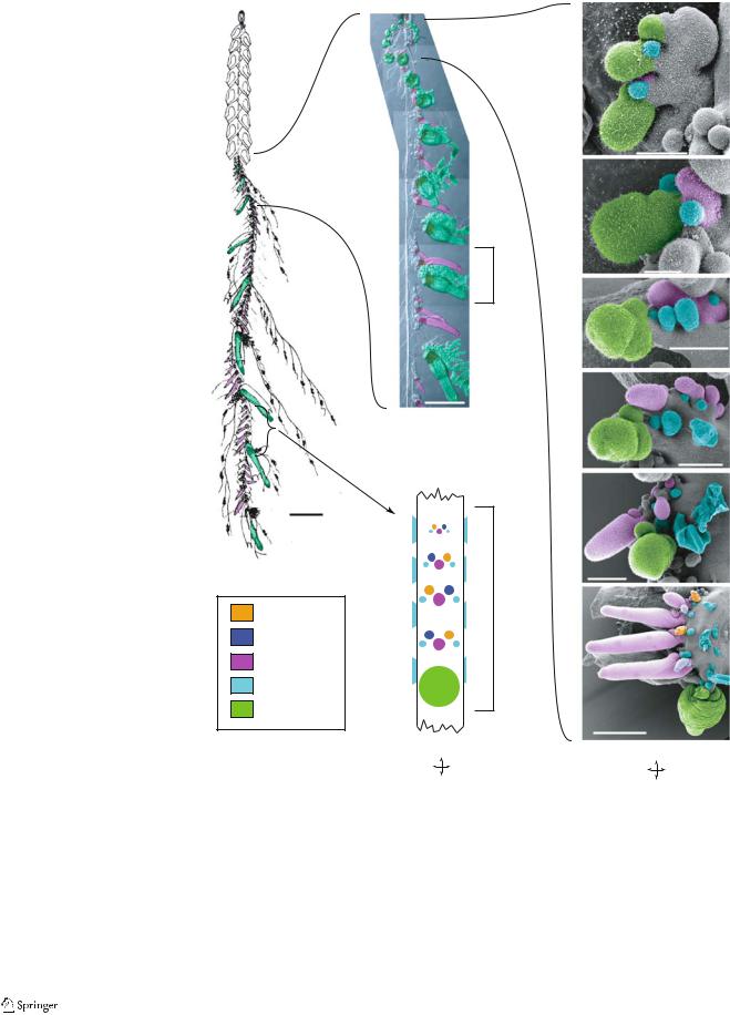
Fig. 5 Nanomia bijuga. False coloring indicates zooid identity. Anterior faces the top of the a page in all panes. a Mature
colony (adapted from Kawamura 1911). The stem is twisted, but the view is generally lateral.
b Photographic collage showing the growth zone and young cormidia of an anesthetized living specimen. The stem is twisted such that the youngest cormidia are shown from their left side, and the older (posterior) cormidia are shown from their right side. c Schematic of a single mature cormidium. Ventral view. The lateral bracts are not shown in their actual position along the stem relative to the other zooids. d–i SEMs of the youngest cormidia, shown from their left side in an ontogenetic sequence (from anterior to posterior). The largest bracts have been removed, leaving only their lamellae. d The three youngest cormidia at the tip of the horn; all other panes show only one cormidium. D Dorsal; L left; R right; V ventral
1 cm
Female
Male
Palpon
Bract
Gastrozooid
phylogeny of Dunn et al. (2005), was prepared for SEM. The siphosomal growth zone was extremely dense, with many tightly packed cormidia. There was a pronounced horn, and the young cormidia close to the tip were regular in organization and appeared to arise by probud subdivision (data not shown). The budding sequence and mature organization of cormidia were not determined.
b
d
50 µm
e
Cormidium |
50 µm |
|
|
|
f |
100 µm
1 mm |
g |
100 µm
c
h
100 µm
Cormidium |
i |
|
|
500 µm |
A |
A |
R L |
V D |
P |
P |
Forskalia formosa
SEM of the siphosomal growth zone of F. formosa indicates that the zooids arise by probud subdivision (not shown), although we did not describe the exact budding sequence. The mature cormidia consist of a posterior gastrozooid, a palpon, and a gonodendron bearing several gonopalpons and gonophores (Fig. 6). This is consistent with previous