

Cell Biology Protocols
.pdfList of Contributors
Judie B. Alimonti
Special Pathogens Program, National
Microbiology Laboratory, H2380, 1015
Arlington Ave. Winnipeg, Manitoba,
Canada, R3E 3R2
Genevieve´ Almouzni
Institut Curie, CNRS, UMR 218, Section Recherche, 26 rue Ulm, F-75248 Paris 05, France
Alicia Alonso
Universidad del Pais Vasco, EHU, CSIC, Unidad Biofis, Aptdo 644, E-48080, Spain
Susan L. Bane
Department of Chemistry, SUNY,
Binghamton, NY 13902-6016, USA
Julie Benesova
Lehrstuhl fur¨ Biochemie der Pflanzen, Ruhr-Universitat¨ Bochum, D-447780 Bochum, Germany
Eric Bertrand
Novartis Pharma AG, CH-4002,
Switzerland
Stephanie Boggasch
Institut fur¨ Allgemeine Botanik des Johannes-Gutenberg-Universitat,¨ Mullerweg¨ 6, D-55099 Mainz, Germany
Igor Bronstein
BBSRC Institute for Animal Health, High Street, Crompton RG20 7NN, UK
William J. Brown
Biochemistry, Molecular and Cell
Biology Sections, Cornell University,
Ithaca, NY 14853, USA
Koert N.J. Burger
Department of Biochemical Physiology,
Institute of Biomembranes, Room W210,
Padualaan 8, 3584 CH Utrecht, The
Netherlands
K. Chambers
Biochemistry, Molecular and Cell
Biology Sections, Cornell University,
Ithaca, NY 14853, USA
C. Yan Cheng
Population Council, Center of Biomedical
Research, 1230 York Avenue, New York
NY 10021, USA
Richard Chi
Department of Biological Science, Florida
State University, Tallahassee, FL
32306-4370, USA
Anton I.P.M. de Kroon
Department Biochemistry of Membranes, Centre for Biomembranes and Lipid Enzymology, Institute of Biomembranes, Padualaan 8, 3584 CH Utrecht, The Netherlands
Ben de Kruijff
Department Biochemistry of Membranes, Centre for Biomembranes and Lipid Enzymology, Institute of Biomembranes, Padualaan 8, 3584 CH Utrecht, The Netherlands
Daniela S. Dimitrova
Center for Single Molecule Biophysics and Department of Microbiology, 304 Sherman Hall, SUNY at Buffalo, Buffalo, NY 14214, USA

xiv LIST OF CONTRIBUTORS
A. Doody
Biochemistry, Molecular and Cell
Biology Sections, Cornell University,
Ithaca, NY 14853, USA
H. Dariush Fahimi
Institute of Anatomy and Cell Biology,
University of Heidelberg, INF 307,
Neuenheimer Feld 307, D-69120
Heidelberg, Germany
Paul G. Fitzgerald
Department of Cell Biology and Human
Anatomy, School of Medicine, 1 Shields
Avenue, Davis, CA 95616-8643, USA
Roland Foisner
Department of Molecular Cell Biology,
Institute of Medical Biochemistry, Vienna
Biocenter, University of Vienna, Dr. Bohr
Gasse 9/3, A-1030 Vienna, Austria
Barbara Gajkowska
The Laboratory of Cell Ultrastructure,
Polish Academy of Sciences, Warsaw,
Poland
Ya-sheng Gao
Department of Pathology, Duke
University Medical Center, Box No.
3020, Rm 225, Jones Bldg, Durham, NC
27, USA
Robert Gniadecki
University of Copenhagen, Bispebjerg
Hospital, Department of Dermatology
D92, Bispebjerg Bakke 23, DK-2400
Copenhagen NV, Denmark
Felix´ M. Goni˜
Universidad del Pa´ıs Vasco, EHU, CSIC, Unidad Biofis, Aptdo 644, E-48080, Spain
John Graham
JG Research Consultancy, 34 Meadway,
Upton Wirral CH49 6IQ, UK
Arnold H. Greenberg†
University of Manitoba, Department of
Medical Microbiology, 539-730 William
Avenue, Winnipeg, MB, R3E OV9,
Canada
†deceased
J. Robin Harris
Institute of Zoology, University of Mainz,
D-55099 Mainz, Germany
John F. Hess
Department of Cell Biology and Human
Anatomy, School of Medicine, 1 Shields
Avenue, Davis, CA 95616-8643, USA
Martin Hetzer
Molecular and Cell Biology Laboratory, The Salk Institute for Biological Studies, 10010 North Torrey Pines Road, La Jolla, CA 92037, USA
Matthew K. Higgins
MRC Laboratory of Molecular Biology,
Hills Road, Cambridge CB2 2QH, UK
Shin-ichi Hisanaga
Department of Biology, Tokyo
Metropolitan University, Graduate School
of Science, Hachioji, Tokyo 1920397,
Japan
David F. Holmes
Wellcome Trust Centre for Cell Matrix Research, School of Biological Sciences, University of Manchester, Stopford Building, Oxford Road, Manchester MI3 9PT, UK
Karl E. Kadler
Wellcome Trust Centre for Cell Matrix Research, School of Biological Sciences, University of Manchester, Stopford Building, Oxford Road, Manchester MI3 9PT, UK
Thomas C.S. Keller
Department of Biological Sciences,
Florida State University, Tallahassee, FL
32306-4370, USA
Helmut Kirchhoff
Institut fur¨ Botanik, Westfalische¨ Wilhelms-Universitat¨ Munster,¨ Schlossplatz 2, D-48149 Munster,¨ Germany
Doris Kirschner
Institut Carie, 26 rue d’Ulm, 75248 Paris Cedex 05, France
Barbara Korbei
Department of Molecular Cell Biology,
Institute of Medical Biochemistry, Vienna
Biocenter, University of Vienna, Dr. Bohr
Gasse 9/3, A-1030 Vienna, Austria
Marina Kriajevska
University of Leicester, Clinical Sciences
Unit, Leicester General Hospital,
Gwendolen Road, Leicester LE5
4PW, UK
Sven-T. Liffers
Lehrstuhl fur¨ Biochemie der Pflanzen, Ruhr-Universitat¨ Bochum, D-447780 Bochum, Germany
Yuechueng Liu
Department of Pathology, University of
Oklahoma Health Services Center,
Oklahoma City, OK 73104, USA
Eugene Lukanidin
Danish Center Society, Institute of Cancer
Biology, Department of Molecular Cancer
Biology, Strandblvd 49, 4-3, DK-2100
Copenhagen, Denmark
Ian G. Mills
Dept. of Neurobiology, MRC Laboratory
of Molecular Biology, Hills Road,
Cambridge CB2 2QH, UK
Nathaniel G.N. Milton
Department of Molecular Pathology &
Clinical Biochemistry, Royal Free
Hospital Campus, Rowland Hill Street,
London NW3 2PF, UK
Dolores D. Mruk
Population Council, Center of Biomedical
Research, 1230 York Avenue, New York,
NY10021, USA
Luis Eduardo Soares Netto
Departamento de Biologia, Instituto de Biociencias,ˆ Universidade de Sao˜ Paulo, Rua do Matao,˜ 277; Sala 327, Cidade Universitaria,´ CEP 05508-900, Sao˜ Paulo-SP, Brazil
LIST OF CONTRIBUTORS |
xv |
Jeffrey A. Nickerson
Department of Cell Biology, School of
Medicine, University of Massachusetts,
55 Lake Avenue N., Worcester, MA
01655, USA
Minnie O’Farrell
Department of Biological Sciences,
University of Essex, Wivenhoe Park,
Colchester CO4 3SQ, UK
Jacques Paiement
Departement´ de Pathologie et Biologie Cellulaire, Universite´ de Montreal´ N-813, Pavilion Principal, 2900 Edouard-Montpetit, Montreal,´ Quebec´ H3T 1J4, Canada
Harald Paulsen
Institut fur¨ Allgemeine Botanik der Johannes-Gutenberg-Universitat,¨ Mullerweg¨ 6, D-55099 Mainz, Germany
Brian J. Peter
McMahon Laboratory, Neurobiology
Division, MRC-LMB, Hills Road,
Cambridge CB2 2QH, UK
Reiner Peters
Institut fur¨ Medizinische Physik und Biophysik, Universitat¨ Munster,¨ Robert-Koch-Straße 31, D-48149 Munster,¨ Germany
Anuradha Pradhan
Department of Pathology, University of
Oklahoma Health Services Center,
Oklahoma City, OK 73104, USA
David Rickwood
Department of Biological Sciences,
University of Essex, Colchester, UK
Matthias Rogner¨
Lehrstuhl fur¨ Biochemie der Pflanzen,
Ruhr-Universitat¨ Bochum, D-447780
Bochum, Germany
T. Sasaki
Department of Biology, Tokyo
Metropolitan University, Graduate School
of Science, Hachioji, Tokyo 1920364,
Japan
xvi LIST OF CONTRIBUTORS
Rutger W.H.M. Staffhorst
Department Biochemistry of Membranes, Centre for Biomembranes and Lipid Enzymology, Institute of Biomembranes, Padualaan 8, 3584 CH Utrecht, The Netherlands
Elizabeth Sztul
Department of Cell Biology, University of Alabama, McCullum Bldg, Rm 668, 1530 S. 3rd Avenue, Birmingham, AL 35294, USA
Jun Tan
The Roskamp Institute, University of
South Florida, 3515 E. Fletcher Avenue,
Tampa, FL 33613, USA
Meinolf Thiemann
Graffinity Pharmaceuticals AG, Im
Neuenheimer Feld 518-519, D-69120
Heidelberg, Germany
Terrence Town
Yale University School of Medicine and
Howard Hughes Medical Institute, 310
Cedar St., PO Box 208011, New Haven,
CT 06520-8011, USA
Kenji Ueda´
Department of Biology, Tokyo
Metropolitan University, Graduate School
of Science, Hachioji, Tokyo 1920364,
Japan
Jean Underwood
Department of Cell Biology, University of
Massachusetts Medical School, 55 Lake
Avenue, Worcester, MA 01655, USA
Ana-Victoria Villar
Universidad del Pa´ıs Vasco, EHU, CSIC, Unidad Biofis, Aptdo 644, E-48080, Spain
John C. Voss
Department of Biological Chemistry,
School of Medicine, 1 Shields Avenue,
Davis, CA 95616-8643, USA
Stefan Wagner
Department of Cell Biology, University of
Massachusetts Medical School, 55 Lake
Avenue, Worcester, MA 01655, USA
Ivan Walev
Institute for Medical Microbiology and Hygiene, University of Mainz, Hochhaus Augustusplatz, D-55131 Mainz, Germany
Tobias C. Walther
EMBL, Gene Expression Programme,
Meyerhofstrasse 1, 69117 Heidelberg,
Germany
Anne Wilson
Woodbine Terrace, Stanton, Ashbourne
Derbyshire DE6 2DA
F.-Xabier Contreras
Universidad del Pais Vasco, EHU, CSIC, Unidad Biofis, Aptdo 644,
E-48080, Spain
Jinnan Xiao
Department of Pathology, University of
Oklahoma Health Services Center,
Oklahoma City, OK 73104, USA
Chunhong Yang
Institut fur¨ Allgemeine Botanik der Johannes-Gutenberg-Universitat,¨ Mullerweg¨ 6, D-55099 Mainz, Germany
Robin Young
Departement´ de Pathologie et Biologie Cellulaire, Universite´ de Montreal´ N-813, Pavilion Principal, 2900 Edouard-Montpetit, Montreal,´ Quebec´ H3T 1J4, Canada

1
Basic Light Microscopy
Minnie O’Farrell
Protocol 1.1 Setting up the microscope for bright field microscopy |
7 |
|
Protocol 1.2 Setting Kohler¨ illumination |
8 |
|
Protocol 1.3 |
Focusing procedure |
9 |
Protocol 1.4 |
Setting up the microscope for phase contrast microscopy |
11 |
Protocol 1.5 |
Setting up the microscope for epifluorescence |
14 |
Protocol 1.6 |
Poly-L-lysine coating |
18 |
Introduction
Light microscopy is an indispensable technique for cell and molecular biologists to study cellular structures and biological processes in both living and fixed cells. This chapter provides an overview of light microscopy, describes the important parts of the microscope and goes on to explain how to set up a standard research microscope for bright field and phase contrast microscopy. There is also a short section on confocal microscopy. More comprehensive descriptions of the different forms of light microscopy are found elsewhere [1–4].
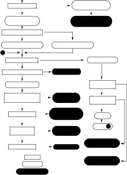
Microscopes are instruments that produce an enlarged image of a specimen. The eyepieces and the objectives are the main components of the magnification system of the microscope, the product of the magnification of the objective lens and the ocular lens give the total magnification of the microscope. The visibility of the magnified specimen depends on contrast and resolution. Contrast is the difference in light intensity between an object and its background. Some biological samples contain coloured compounds, for example pigmented animal cells and chlorophyll-containing chloroplasts in plant cells, but most biological samples are colourless and have to be fixed and stained before observation [5]. Such stained specimens are observed using bright field microscopy. Other kinds of microscope systems are available to enhance contrast in living samples; these include phase contrast, dark field, differential interference contrast (DIC) and fluorescence microscopy (Table 1.1). The flow chart in Figure 1.1 will help in the selection of the appropriate microscopic observation method.
Cell Biology Protocols. Edited by J. Robin Harris, John Graham, David Rickwood2006 John Wiley & Sons, Ltd. ISBN: 0-470-84758-1
2 |
BASIC LIGHT MICROSCOPY |
|
|
|
|
|
|
|
Table 1.1 Techniques for producing contrast in light microscopy |
|
|||
|
|
|
|
|
|
|
Type |
Mechanism |
Requirements |
Fixed |
Live |
Appearance |
|
|
|
|
|
cells |
cells |
|
|
|
|
|
|
|
|
Bright field |
Absorption of |
Any light microscope; |
Yes |
No |
Coloured image |
|
|
|
visible light |
range of |
|
|
depending on |
|
|
following |
histochemical |
|
|
stains |
|
|
staining of |
stains |
|
|
|
|
|
specimen |
|
|
|
|
Phase contrast |
Variations in |
Phase objective and |
Yes |
Yes |
Many shades of |
|
|
|
refractive index |
phase condenser |
|
|
grey |
|
|
within specimen |
|
|
|
|
Dark field |
Scattered light |
Dark field stop in |
Yes |
Yes |
Bright objects |
|
|
|
|
condenser |
|
|
against dark |
|
|
|
|
|
|
background |
Differential |
Gradient of |
Special objective lens |
Yes |
Yes |
3D effect |
|
|
interference |
refractive index |
|
|
|
|
|
contrast |
|
|
|
|
|
Fluorescence |
Excitation and |
An excitation light |
Yes |
Yes |
Bright colours |
|
|
|
emission of |
source and |
|
|
against a |
|
|
light by |
appropriate filters |
|
|
dark |
|
|
fluorophore |
for emission; range |
|
|
background |
|
|
|
of fluorescent |
|
|
|
|
|
|
probes including |
|
|
|
|
|
|
naturally |
|
|
|
|
|
|
fluorescent proteins |
|
|
|
|
|
|
|
|
|
|
The resolution of the optical system, that is the ability to distinguish objects separated by small distances, determines the degree of detail observable. The limit of resolution of the light microscope is about 0.2 µm. Enlarging the image too much results in ’empty magnification’ and the quality of the image deteriorates. The limits of resolution are determined by the quality of the objective and the condenser.
Key components of the compound microscope
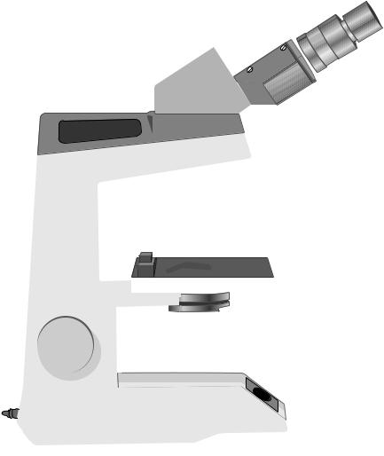
The eyepieces, body tube, nosepiece and objectives are part of the magnification system of the microscope. The condenser, condenser-iris diaphragm, filters, field iris diaphragm and light source are the parts that compose the illumination system of the microscope. To use a microscope properly, and to get the most out of it, it is important to understand the purpose and function of each of the microscope’s components (Figure 1.2).
The body and lamp
The binocular body, the arm and the base form the frame of the microscope. This provides the stability and holds the optical and other components rigid and in place. The lamp is in the base of the body; its brightness is controlled by an on/off switch and a rheostat control knob. Just above the lamp is a collector lens with a field diaphragm to control the area of illumination. The field diaphragm also aids focusing and centring of the illumination.
|
|
|
KEY COMPONENTS OF THE COMPOUND MICROSCOPE |
3 |
|||
START |
|
|
|
|
|
||
|
|
|
>> a cell |
Use hand lens or |
|
||
How small is it? |
|
|
|||||
|
|
|
dissecting microscope |
|
|||
|
|
|
|
|
|
||
|
≤ a cell |
|
|
|
|
|
|
|
|
|
|
|
|
||
|
|
|
|
|
|
||
Use a compound |
|
|
Bright field, Dark field |
|
|||
microscope |
|
|
Epi-illumination |
|
|||
|
|
|
|
|
|
||
|
|
|
|
|
|
|
|
What is it mounted on? |
Petri dish, etc. |
|
|
|
|||
|
Slide |
|
|
|
|||
|
|
|
|
|
|
||
|
|
|
|
|
|
||
Use an upright microscope |
Use an inverted microscope |
|
|||||
X
Is it thin (< 50 m)?
Yes
Is it fluorescently labelled?
No
Use transillumination
Is it coloured or densely contrasted or stained?
No
Use epi-illumination
Yes Epi-fluorescence
|
|
Is it reflective? |
Yes |
||
|
|
e.g. gold, silver |
|
||
Yes |
Bright field |
|
No |
Yes |
|
|
|||||
|
|||||
Is it fluorescently |
|||||
|
Phase contrast |
||||
|
labelled? |
|
|||
|
|
|
|||
|
|
No |
Yes |
Phase contrast |
|
No |
||||||
|
|
|
||||||||||
|
|
|
|
|
|
|
||||||
Is it transparent? |
|
Nomarski |
|
|
||||||||
|
|
|
|
Section |
||||||||
|
|
|
|
|||||||||
|
|
|
|
|
|
|
|
|
Autofluorescence |
|||
|
|
No |
Yes |
Go to X |
||||||||
|
|
Dark field |
||||||||||
|
|
|||||||||||
Is it reflective? |
||||||||||||
|
|
|||||||||||
e.g. gold, silver |
|
|
|
|
|
Reflected light |
|
|
||||
|
|
No |
Yes |
|
Epi-fluorescence |
|||||||
|
|
|
||||||||||
|
|
|
||||||||||
Is it birefringent? |
|
|
Polarized light |
± confocal imaging |
||||||||
|
|
|
|
|
|
|
||||||
|
|
|
|
|
|
|
|
|
|
|
|
|
|
|
Question |
|
|
|
|
Reflected light |
|||||
|
|
|
|
|
|
|
|
|
|
|||
KEY |
|
Conclusion |
|
±confocal imaging |
||||||||
|
|
|
|
|||||||||
|
Specific technique |
|
|
|
||||||||
|
|
|
|
|
|
|
|
|
|
|
|
|
Figure 1.1 Flow chart for selection of observation methods. Reproduced from Rawlins (1992) Light Microscopy, Fig 1.1 Bios Scientific Publishers, Oxford
The condenser
The condenser provides a bright, even illumination of the specimen for a wide range of magnifications. The condenser can be focused and light transmission regulated by the condenser-iris diaphragm; correctly used these will optimize resolution, contrast and depth of field. Modified condensers are required for contrasting techniques such as phase contrast and differential interference contrast.
4 BASIC LIGHT MICROSCOPY
Eyepiece 
Binocular body
|
|
|
Body lock screw |
|
|
|
Focusing reverse |
Arm |
|
|
nosepiece |
|
|
|
|
|
|
|
Infinity corrected |
|
|
|
objectives |
|
|
|
Graduated |
|
|
|
mechanical stage |
|
|
|
Condenser |
|
|
|
Condenser control lever |
|
|
|
Condenser |
|
|
|
adjustment screw |
|
|
|
Collector lens with field diaphragm lever |
|
|
|
Field diaphragm lever |
|
|
|
Rheostat control knob |
|
|
|
Base |
Coarse |
Fine |
On/off |
Condenser rack |
and pinion knob |
|||
adjustment knob |
adjustment knob |
switch |
|
Figure 1.2 The parts of the compound light microscope
The stage and focus mechanism
The specimen, usually on a slide, is held in place by a sprung arm on the mechanical stage. The stage can be moved in the x and y planes and mounted vernier scales can be used to locate sites of interest on the coverslip/slide. The course (outer) and fine (inner) focus adjustment knobs alter the level of the stage with respect to the objective.
The objective
The objectives lenses are mounted on a revolving nosepiece which allows for easy changes between magnification and also facilitates the maintenance of focus when the

|
|
KEY COMPONENTS OF THE COMPOUND MICROSCOPE |
5 |
|||||
|
Table 1.2 Properties of some objective lenses |
|
|
|
||||
|
|
|
|
|
|
|||
Magnification of |
Focal |
NA |
Working |
Diameter |
Depth of |
|||
objective |
length (mm) |
|
distance (mm) |
of field (mm) |
field (µm) |
|||
|
|
|
|
|
|
|
|
|
10 |
16 |
0.20–0.30 |
4 |
–8 |
1 |
–2 |
|
c. 10 |
40 |
4 |
0.65–0.85 |
0.2 |
–0.6 |
0.25 |
–0.50 |
|
1–2 |
100 (oil) |
2 |
1.20–1.30 |
0.11 |
–0.16 |
0.1 |
–0.2 |
|
0.5 |
|
|
|
|
|
|
|
|
|
different objectives are moved into position, giving parfocality. The objective lens of the microscope is the major component responsible for the magnification and resolution of the image; it is perhaps the single most important element of the microscope.
Basically, an objective consists of a set of lens elements that form an image of an object at a distinct distance beyond the objective; it collects light from every specimen point and forms a real intermediate image in the eyepiece focal plane. Besides collecting light from the specimen and ‘magnifying’ the latter, the objective contains lenses that correct the aberrations created as light passes through the collecting lens system. The ability to collect light and, therefore, to resolve detail is termed the numerical aperture (NA) of the objective. The limit of resolution is determined by the wavelength of light used (λ) and the NA, the light-gathering capacity of the objective:
Resolution = 0.61 × wavelength of light source(λ) numerical aperture (NA)
A dry objective cannot have an NA greater than 1 but an immersion medium, for example oil, can increase the NA beyond 1 (Table 1.2)
In selecting an objective for a given purpose it is useful to know certain figures. These are (1) the magnification, (2) the focal length, (3) numerical aperture, (4) the working distance, (5) the diameter of the field of view and (6) the depth of field. Average values for commonly used objectives are shown in Table 1.2
The working distance is the clearance between the lowest point of the objective and the upper surface of the coverslip. The depth of field is the range of distances over which objects can still appear reasonably sharp. The most important factor in deciding this quantity is the NA.
The objective lenses bear a number of inscriptions including the type, the magnification and the NA (Figure 1.3). Achromat, Plan Achromat and Plan Apochromat are the names of objectives of increasing quality. The Achromat lenses are colour corrected for two wavelengths (red, 656 nm and blue, 486 nm) and are corrected for spherical aberration in the green (546 nm). The Apochromats have been further corrected to give the best colour reproducibility. The Plan designation refers to correction for flatness of field across the whole image. The inscription on the objective lens shown in Figure 1.3 is, for example, 40/0.65 and 160/0.17. These figures indicate the initial magnification ×40, numerical aperture 0.65, for use with microscopes with a mechanical tube length of 160 mm and with a coverslip 0.17 mm thick.
The eyepieces
The real, intermediate image formed by the objective is observed and further magnified by means of an eyepiece. They usually have a magnifying power of 10× but can range

6 BASIC LIGHT MICROSCOPY
Figure 1.3 An objective lens showing the specifications engraved on the metal body tube
from 4× to 30×. Eyepieces over 12.5×, however, depending on the objective used, may result in ‘empty magnification’. Apart from its magnification, an eyepiece is characterized by its field of view number. With the aid of this number it is possible to calculate the diameter of the field covered in the specimen plane. The field of view number of the eyepiece divided by the magnification of the objective gives the diameter of the actual field of view in millimetres.
There are also eyepieces specially designed for spectacle wearers. They are usually marked with a diagram of a pair of glasses. The interpupillary distance can be altered in most binocular microscopes.
Techniques of microscopy
Bright field microscopy
Bright field microscopy is probably the most widely used form of microscopy and is used mainly for fixed and stained specimens. For optimal resolution the microscope should be aligned correctly and one of the most important alignments is setting up Kohler¨ illumination. This provides bright and even illumination over the specimen and allows for the control of contrast and depth of field.