

Cell Biology Protocols
.pdf6
In Vitro Techniques
Edited by J. Robin Harris
Nuclear components
Protocol 6.1 |
Nucleosome assembly coupled to DNA repair synthesis |
|
|
using a human cell free system |
204 |
Protocol 6.2 |
Single labelling of nascent DNA with halogenated |
|
|
thymidine analogues |
210 |
Protocol 6.3 |
Double labelling of DNA with different halogenated |
|
|
thymidine analogues |
214 |
Protocol 6.4 |
Simultaneous immunostaining of proteins and |
|
|
halogen-dU-substituted DNA |
217 |
Protocol 6.5 |
Uncovering the nuclear matrix in cultured cells |
220 |
Protocol 6.6 |
Nuclear matrix–lamin interactions: in vitro blot overlay |
|
|
assay |
228 |
Protocol 6.7 |
Nuclear matrix–lamin interactions: in vitro nuclear |
|
|
reassembly assay |
230 |
Protocol 6.8 |
Preparation of Xenopus laevis egg extracts and |
|
|
immunodepletion |
234 |
Protocol 6.9 |
Nuclear assembly in vitro and immunofluorescence |
237 |
Protocol 6.10 |
Nucleocytoplasmic transport measurements using |
|
|
isolated Xenopus oocyte nuclei |
240 |
Protocol 6.11 |
Transport measurements in microarrays of nuclear |
|
|
envelope patches by optical single transporter recording |
244 |
Cells and membrane systems
Protocol 6.12 |
Cell permeabilization with Streptolysin O |
248 |
Protocol 6.13 |
Nanocapsules: a new vehicle for intracellular delivery of |
|
|
drugs |
250 |
Protocol 6.14 |
A rapid screen for determination of the protective role |
|
|
of antioxidant proteins in yeast |
255 |
Protocol 6.15 |
In vitro assessment of neuronal apoptosis |
259 |
Cell Biology Protocols. Edited by J. Robin Harris, John Graham, David Rickwood2006 John Wiley & Sons, Ltd. ISBN: 0-470-84758-1
202IN VITRO TECHNIQUES
Protocol 6.16 The mitochondrial permeability transition: PT and
m loss determined in cells or isolated mitochondria |
|
with confocal laser imaging |
265 |
Protocol 6.17 The mitochondrial permeability transition: measuring |
|
PT and m loss in isolated mitochondria with Rh123 |
|
in a fluorometer |
268 |
Protocol 6.18 The mitochondrial permeability transition: measuring |
|
PT and m loss in cells and isolated mitochondria on |
|
the FACS |
270 |
Protocol 6.19 Measuring cytochrome c release in isolated |
|
mitochondria by Western blot analysis |
271 |
Protocol 6.20 Protein import into isolated mitochondria |
272 |
Protocol 6.21 Formation of ternary SNARE complexes in vitro |
274 |
Protocol 6.22 In vitro reconstitution of liver |
|
endoplasmic reticulum |
277 |
Protocol 6.23 Asymmetric incorporation of glycolipids into |
|
membranes and detection of lipid flip-flop movement |
280 |
Protocol 6.24 Purification of clathrin-coated vesicles from rat brains |
286 |
Protocol 6.25 Reconstitution of endocytic intermediates on a lipid |
|
monolayer |
288 |
Protocol 6.26 Golgi membrane tubule formation |
293 |
Protocol 6.27 Tight junction assembly |
296 |
Protocol 6.28 Reconstitution of the major light-harvesting chlorophyll |
|
a/b complex into liposomes |
300 |
Protocol 6.29 Reconstitution of photosystem 2 into liposomes |
305 |
Protocol 6.30 Golgi–vimentin interaction in vitro and in vivo |
307 |
Cytoskeletal and fibrillar systems
Protocol 6.31 Microtubule peroxisome interaction |
313 |
|
Protocol 6.32 Detection of cytomatrix proteins by immunogold |
|
|
|
embedment-free electron microscopy |
317 |
Protocol 6.33 Tubulin assembly induced by taxol and other |
|
|
|
microtubule assembly promoters |
326 |
Protocol 6.34 Vimentin production, purification, assembly and study |
|
|
|
by EPR |
331 |
Protocol 6.35 |
Neurofilament assembly |
337 |
Protocol 6.36 α-Synuclein fibril formation induced by tubulin |
342 |
|
Protocol 6.37 Amyloid-β fibril formation in vitro |
345 |
|
Protocol 6.38 |
Soluble Aβ1 – 42 peptide induces tau |
|
|
hyperphosphorylation in vitro |
348 |
Protocol 6.39 |
Anti-sense peptides |
353 |
Protocol 6.40 Interactions between amyloid-ß and enzymes |
359 |
|
Protocol 6.41 |
Amyloid-ß phosphorylation |
364 |
Protocol 6.42 Smitin–myosin II coassembly arrays in vitro |
369 |
|
Protocol 6.43 Assembly/disassembly of myosin filaments in the |
|
|
|
presence of EF-hand calcium-binding protein S100A4 |
372 |
|
in vitro |
|
Protocol 6.44 |
Collagen fibril assembly in vitro |
375 |
INTRODUCTION 203
Introduction
Modern cell biology is increasing moving onwards from the intact cell or component isolated directly from the cell, to a consideration of the experimental manipulation and reassembly of such components.
This chapter dealing with in vitro techniques in cell biology includes a range of recent methods that loosely fall within the sphere of experimental reconstitution/assembly procedures. The topics included integrate well with those within the other sections of the book and often rely upon both EM and advanced light microscopical techniques for assessment of what is achieved or produced experimentally.
Several assays for nuclear components, cellular and membrane systems are presented, together with the assembly of cytoskeletal proteins. In addition, protocols relating to in vitro collagen fibrillogenesis, amyloid-β fibrillogenesis, amyloid-β-enzyme interaction and amyloid-β phosphorylation are also included.
Because of the diversity of this material, most protocols (or a small group of protocols) includes a brief introduction, its own list of references and sometimes examples of typical data. It is hoped that these protocols will enable the book to indicate a way ahead for cell biology, as an addition to the well-used classical procedures dealing with cells and subcellular components presented in the other chapters of the book.

PROTOCOL 6.1
Nucleosome assembly coupled to DNA repair synthesis using a human cell free system
Genevieve` Almouzni and Doris Kirschner
Introduction
The assembly of nucleosomes onto DNA, in two steps, involves the loading of a H3–H4 histone tetramer followed by the subsequent addition of two H2A–H2B histone dimers. In vivo, histone deposition is promoted by histone chaperones, among which the evolutionary conserved three-subunit complex called the chromatin assembly factor 1 (CAF-1) is the only one that links nucleosome formation to DNA synthesis.
Here we describe an assay to monitor nucleosome assembly coupled to DNA repair in a human cell free system. The nucleotide excision repair (NER) pathway used in this assay repairs most of the UVphotoproducts (mainly cyclobutane pyrimidine dimers and 6–4 photoproducts) can be reproduced in vitro [4, 6].
A cell free extract (cytosolic extract) derived from Hela cells is thus used for its ability to support NER reactions on a damaged DNA template. This extract, however, cannot ensure the assembly into chromatin of the repaired DNA molecules. It can be complemented using either a nuclear extract as a crude source of assembly factors or using purified recombinant assembly factors such as CAF-1 [1, 3].
Reagents
Agarose (Ultra Pure, Sigma)
Ammonium acetate (5 M)
100 mM ATP (Pharmacia) stored at −80 ◦C (each aliquot is used only once)
Bacculovirus produced recombinant CAF- 1 4 ng/µl [5]
Creatine phosphokinase (Boehringer) 2.5 mg/ml in H2O 1
Cytosolic extracts derived from HeLa cells [2] 2 3
Ethidium bromide, 10 mg/ml (GIBCO BRL)
Glycogen, 20 mg/ml (Roche), store at −20 ◦C
Loading buffer 5×: 0.42% bromophenol blue, 50% glycerol
Nuclear extracts from HeLa (4C Biotech/ Belgium or [2])
αP32 dCTP, 3000 Ci/mmol (ICN) |
|
Phenol : chloroform : isoamyl |
alcohol |
(25 : 24 : 1) (GIBCO BRL) |
|
Proteinase K, 20 mg/ml (Roche), aliquots stored at −20 ◦C
5× Reaction buffer: 25 mM MgCl2, 200 mM Hepes-KOH (pH 7.8), 2.5 mM dithiothreitol (DTT; Sigma), 200 mM phosphocreatine (di-Tris salt, Sigma) 1
RNase A (Roche) 10 mg/ml in 0.01 M sodium acetate (pH 5.2), heat for 15 min at 100 ◦C and adjust the pH with 1 vol
of Tris-HCL (pH 7.4), aliquots stored at −20 ◦C
Stop-mix: 30 mM EDTA, 0.7% SDS
TAE 50×: 242 g Tris base, 57.1 ml glacial acetic acid, 37.2 g Na2EDTA, (pH 8.0), adjust to a final volume of 1 l with deionized water (or 50× TAE from Bio Media)
TE: 10 mM Tris-HCl (pH 8.0), 1 mM EDTA (pH 8.0)
UV treated Bluescript plasmid (pBS) DNA (50 ng/µl) 4 [1]
Equipment
Eppendorf thermomixer 5436
Eppendorf tubes (1.5 ml, siliconized) (precooled on ice)
Gelbox (35 × 20 cm, Biorad)
Germicidal lamp with a 254 nm peak (Philips)
Microfuge (e.g. Eppendorf centrifuge 5451)
3 MM Paper (Whatman)
Phosphoimager (Storm 860–Molecular Dynamics), and Image Quant 5.2 software (or films and standard film developer)
Power supply (Pharmacia Biotech, EPS 3500)
UV transilluminator (BIORAD Gel Doc 2000) equipped with a camera
Vacuum gel dryer (slab gel Dryer, SGD 2000, SAVANT)
Waterbath (e.g. polystat, Bioblock Scientific)
Procedure
A standard reaction contains 150 ng of plasmid DNA, 4 200–400 µg of cytosolic extract, 5 mM MgCl2, 40 mM HepesKOH (pH 7.8), 0.5 mM DTT, 40 mM phosphocreatine, 4 mM ATP, 2.5 µg creatine phosphokinase and 5 µCi α32P dCTP in 25 µl.
PROTOCOL 6.1 |
205 |
1.First, to prepare the DNA-mix, in a precooled Eppendorf tube, assemble on ice the following reagents (prepare
a proportionally larger mix for more than one reaction). 1
DNA-mix (amounts for one reaction): 3 µl DNA (50 ng/µl), 5 µl 5× reac-
tion buffer, 1 µl ATP, 1 µl creatine kinase, 2.5 µl dilution buffer 3
2.For each reaction, distribute 1 µl of HeLa nuclear extract to precooled Eppendorf tubes on ice (or a protein complex to test for nucleosome assembly or buffer controls). In an assay designed for an antibody inhibition strategy, you can add the antibodies
or the appropriate control serum at this step. 5
3.Add 10 µl of the cytosolic extract (as source of histones and repair factors) to each tube except to the DNA only control in which buffer is added instead. To preserve optimal activity of cytosolic extracts, defrost the extract
just prior to use, work quickly and discard any remaining material. 2
4.Add α32P dCTP to the prepared DNAmix (0.5 µl for 150 ng DNA), homogenize gently by pipetting up and down.
5. Start the reaction by adding 13 µl of DNA-mix containing the labelled precursor to each reaction tube, and immediately transfer to a preheated water bath, or to a thermo-mixer at 37 ◦C for a 3 h incubation, see note 6 and ref. 1.
6.Stop the reaction with 50 µl of stopmix and 25 µl of H2O.
7.Add 5 µl of RNase A (at 2 mg/ml) and incubate 30 min at 37 ◦C.
8.Add 2 µl of proteinase K (at 20 mg/ml) and incubate 30 min at 37 ◦C. 7
9.Add 110 µl of phenol : chloroform : isoamyl alcohol (25 : 24 : 1) and vortex
206 |
IN VITRO TECHNIQUES |
|
each tube at least 10 s. Centrifuge |
|
10 min at 14 000g in the Eppendorf |
|
centrifuge at room temperature and |
|
recover 90 µl of the aqueous phase in |
|
a fresh Eppendorf tube; do not take |
|
any of the interphase. |
10. |
Precipitate the DNA by adding of |
|
90 µl ammonium acetate (5 M), 1 µl |
|
of glycogen (20 mg/ml) and 300 µl (2 |
|
vol) of ice-cold ethanol, leave 30 min |
|
at −20 ◦C. |
11. |
Centrifuge 30–45 min at 14 000g at |
|
4 ◦C, recover the DNA pellet, wash |
with 500 µl of cold 70% ethanol, centrifuge for 5–10 min at 14 000 g at 4 ◦C. Remove the supernatant carefully and air-dry the pellet.
12.Dissolve the pellet in 8 µl of TE buffer and add 2 µl of 5× loading dye.
13.Prepare a 1% agarose gel in 1× TAE without ethidium bromide (EtBr). 8
9
14.Load the samples and migrate at 1.5 V/ cm for 20 h at 4 ◦C overnight, for optimal resolution of the topoisomers. 7
15.Soak the gel in EtBr staining solution
(1µg/ml), rinse for 30 min in water at room temperature, and place the gel on a UV transilluminator system equipped with a camera to visualize the migration pattern of the total DNA and make your image acquisition.
16.Transfer the gel onto a 3 MM Whatman paper of equal size, cover with
saran wrap and dry for 2 h at 80 ◦C in a vacuum gel dryer. Expose the covered and dried gel to a Phosphoimager screen to visualize the labelled DNA (repaired), if not available use an X- ray film. Quantification of the topoisomer distribution is carried out on both the labelled DNA (repaired) and the total DNA by densitometric scanning of the images. 10
Notes
1 Thawing and freezing of the 5× reaction buffer alter the concentration of DTT, phosphocreatine and dNTPs. The buffer is prepared in advance, aliquoted and stored at −80 ◦C, use only once.
2 Cytosolic and nuclear extracts are prepared from unsynchronized adherent HeLa cells, under cooling conditions (4 ◦C), for a detailed protocol see [2]. The protein concentration is determined by Bradford assay and usually ranges between 10 and 20 mg/ml [2]. Small aliquots of 50–100 µl frozen in liquid nitrogen are stored at −80 ◦C to avoid waste.
3 The cytosolic extract used in this assay, which contains only trace amounts of p60 and p150, needs to be complemented with the nuclear extract or bacculovirus recombinant CAF-1 to ensure an efficient chromatin assembly coupled to DNA repair synthesis [1]. For each extract, optimal conditions for both chromatin assembly and DNA repair synthesis have to be adjusted (protein/DNA ratio and ionic conditions). For example, the composition of the dilution buffer for commercial (4C) HeLa nuclear extract is: 20 mM Hepes (pH 7.8), 100 mM KCl, 0.2 mM EDTA, 0.2 mM PMSF, 0.5 mM DTT and 20% glycerol. This has to be taken into account.
4 Plasmid DNA is treated with germicidal UV light (Philips) with 254 nm peak and a dose of 500 J/m2. The general estimation of DNA damage produced by 100 J/m2 is roughly 1 pyrimidine dimer photoproduct per 1000 bp (the ratio is about 0.75 cyclobutane pyrimidine and 0.25 of 6-4 photo product) [1, 5].
PROTOCOL 6.1 |
207 |
NE CAF-1 PI anti-p60 anti-p150
Topoisomer distribution of labelled DNA
Topoisomer distribution of total
DNA
1
− |
+ |
− |
+ |
+ |
+ |
− |
− |
+ |
− |
− |
− |
− |
− |
− |
+ |
− |
− |
− |
− |
− |
− |
+ |
− |
− |
− |
− |
− |
− |
+ |
−Ir/II
−I
−Ir/II
−I
2 |
3 |
4 |
5 |
6 |
7 |
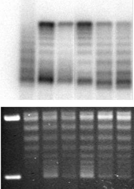
Figure 6.1 Chromatin assembly coupled to nucleotide excision repair synthesis mediated by CAF-1. UV-damaged DNA substrate was incubated in cytosolic extract (CE) competent for NER and complemented with nuclear extracts (NE) or recombinant CAF-1 to ensure the nucleosome assembly reaction. Lane 1 shows a DNA-only control (supercoiled DNA molecules) and lane 2 the DNA after incubation with cytosolic extract, which promotes the relaxation of the DNA and NER, which is visualized by the incorporation of radiolabelled nucleotides. Subsequent supercoiling of the repaired DNA is dependent upon addition of CAF-1, either as nuclear extract (lane 3) or as recombinant complex (lane 4). In this assay the CAF-1 dependent nucleosome formation was largely impaired by the addition of 1 µl of polyclonal antibodies directed respectively against the p60 subunit (lane 6) or the p150 subunit (lane 7) of CAF-1 and compared with a pre-immune (PI) serum (lane 5). Total DNA is visualized after ethidium bromide staining (lower panel) and radiolabel incorporation due to repair synthesis (labelled DNA) is visualized on a phosphoimager screen of the dried gel (upper panel). Relaxed/nicked circular DNA (Ir/II) and supercoiled DNA (I) are indicated
5 When antibodies are used as in Figure 6.1, the quantity added has to be adjusted for each serum or clone.
6 Nucleotide excision repair as followed by the DNA synthesis and chromatin
assembly reactions is usually completed after 3 h. Although specific UV-dependent nucleotide incorporation can be detected already after 15 min, the chromatin assembly reaction is a slower process [1].
208 IN VITRO TECHNIQUES
7 To monitor chromatin assembly, the supercoiling assay makes use of the topological properties of closed circular molecules. In the extracts, which contain topoisomerase activity, the deposition of nucleosomes induces topological stress that is relieved progressively. Therefore, after deproteinization, accumulation of topoisomers with an increasing number of negative supercoils can be used as an indication of the effectiveness of the assembly reaction. The repaired molecules (labelled DNA) are compared to the bulk DNA (total DNA) to evaluate a differential effect.
8 The concentration of the agarose gel is adjusted according to the size of the plasmid used. As a guideline, use 1% agarose gels for plasmids of 3–4 kb and 0.8% gels for plasmids of 6–7 kb in size.
9 It is crucial to run the supercoiling gel in the absence of EtBr. This intercalating agent would interfere with the analysis of the topological state of the molecules.
10 The radiolabelled DNA corresponds to incorporation of nucleotides during repair synthesis (one of the steps in NER) and can be analysed quantitatively and qualitatively using the Image Quant 5.2 software. Adapt the exposure time depending on the repair efficiency of the extract used. A higher proportion of supercoiled molecules in the radiolabelled DNA as compared to total DNA is indicative of a preferential assembly of repaired molecules,
reflecting a link between repair synthesis and nucleosome assembly.
References
1.Gaillard, P. H., Roche, D. M. and Almouzni, G. A. (1999) Meth. Mol. Biol., 119, 231–243.
2.Martini, E., Roche, D. M., Marheineke, K., Verreault, A., and Almouzni, G. A. (1998) Recruitment of phosphorylated chromatin assembly factor 1 to chromatin after UV irradiation of human cells. J. Cell Biol., 143,
563–575.
3. Mello, J. A., Sillje, H. H., Roche, D. M., Kirschner, D. B., Nigg, E. A. and Almouzni, G. A. (2002) Human Asf1 and CAF-1 interact and synergize in a repair-coupled nucleosome assembly pathway. EMBO Rep. 3(4), 329–334.
4.Moggs, J. G. and Almouzni, G. A. (1999) Assays for chromatin remodeling during DNA repair. Meth. Enzymol., 304, 333–351.
5.Verreault, A., Kaufman, P. D., Kobayashi, R. and Stillman, B. (1996) Nucleosome assembly
by a complex of CAF-1 and acetylated histones H3/H4. Cell, 87, 95–104.
6. Wood, R. D., Biggerstaff, M. and Shivji, M. K. K. (1995) Detection and measurement of nucleotide excision repair synthesis by mammalian cell extracts in vitro. Methods: A Companion to Methods Enzymol., 7, 163–175.
Acknowledgements
We thank Alain Verreault for the generous gift of CAF-1, Jill Mello for advice and Catherine Green for reading and critical comments. D.B.K. was supported by the EEC-RTN (to G.A.), the team of G.A. is supported by the Ligue Contre le Cancer, Euratom, EU-RTN and LCR of CEA.

PROTOCOLS 6.2–6.4
Immunocytochemical studies of DNA replication in mammalian nuclei
Daniela Dimitrova
Introduction
Indirect immunofluorescent staining of newly synthesized DNA and/or proteins of the replication machinery has emerged as a powerful technique for identification of proliferating cells [1] and for cancer diag-
nosis or prognosis [2, 3], as well as for studies of cell cycle kinetics [1, 4], intra-S- phase checkpoint control [5], organization of nuclear DNA replication sites [6, 7], establishment and execution of the temporal program for chromosomal DNA replication during the S-phase [8].

PROTOCOL 6.2
Single labelling of nascent DNA with halogenated thymidine analogues
Reagents
Phosphate-buffered saline (PBS): 8 g NaCl, 0.2 g KCl, 1.44 g Na2HPO4 and 0.24 g KH2PO4 per litre at pH 7.2
PBS-T: PBS containing 0.5% (v/v) Tween 20 (Sigma)
Halogenated nucleosides: stock solutions of 10 mg/ml of 5-bromo-2 -deoxyuri- dine (BrdU, Sigma), 5-chloro-2 -deoxy- uridine (CldU, Sigma), or 5-iodo-2 - deoxyuridine (IdU, Sigma) are prepared in cell culture medium (without serum) at pH 7.2. Store frozen at −20 ◦C in small aliquots
Antibodies: (A) Mouse anti-BrdU (No. 347580; Becton Dickinson) is used for detection of BrdU, CldU and IdU [1].
(B) Rat anti-BrdU (MAS250b; HarlanSera lab) is used for detection of BrdU and CldU [1]. (C) Secondary antibodies: FITC-conjugated donkey anti-rat IgG (No. 712-095-153; Jackson ImmunoResearch Laboratories) or FITC-conjugated donkey anti-mouse IgG (No. 715-095- 151; Jackson ImmunoResearch Laboratories) are used to detect the rat or mouse anti-BrdU antibodies, respectively
Blocking buffer: to suppress non-specific binding of antibodies to coverslips or to cellular material, antibody dilutions are made in PBS containing 2% bovine serum albumin and either 10% normal donkey serum, or 10% fetal bovine
serum. Supplement with NaN3 at 1 mM final concentration and store at 4 ◦C
DNA staining dye: a stock solution of 1 mg/ml of 4 ,6-diamidino-2-phenylin- dole (DAPI; Sigma) is prepared in ddH2O and stored frozen in small aliquots at −20 ◦C. A working solution of 10 µg/ml can be kept at 4 ◦C
Materials and equipment
Sterile glass coverslips
Cell culture dishes and medium
A pair of fine sharp forceps
Dark humid incubation chamber Plain glass slides (3 × 1 )
Mounting medium for fluorescent microscopy, e.g. Vectashield (Vector Laboratories)
Epifluorescent microscope
Procedure
1.Grow cells directly on glass coverslips 1 submerged in medium inside cell culture dishes. 2 Multi-well dishes can be used if cell cultures need to be grown in separate chambers. Alternatively, several coverslips can be placed within a single dish. Make sure that the coverslips do not overlap.
2.Add the nucleotide precursor of choice (i.e. BrdU, CldU or IdU) to the culture medium to a final concentration of