

Challenges in Colorectal Cancer, Second Edition
Edited by John H. Scholefield etc. Copyright © 2006 by Blackwell Publishing Ltd
.............................................................................................................................................................................
14: Surgery for metastatic disease in colorectal cancer
Timothy G. John and Myrddin Rees
.............................................................................................................................................................................
Introduction
The liver is the most common site of metastasis in patients with colorectal cancer. Following hematogenous dissemination through the portal venous system, the liver is thought to present as the first and only target site for overt metastatic spread in 30–40% of cases [1]. Up to 25% of colorectal cancer patients present with synchronous liver metastases at the time of diagnosis, and approximately 50% of all patients who undergo radical resection for primary colorectal cancer may be affected by isolated metastatic disease confined to the liver, usually within the first 2 years [2–4]. It is now recognized that for selected patients with colorectal liver metastases, hepatic resectional surgery is unique in offering the prospect of cure and has become a paradigm in the management of some such patients with “advanced” disease.
The proportion of patients with colorectal liver metastases eligible for hepatic resection with curative intent has been estimated at 20–30% [5]. While this may vary between centers and depends on prevailing selection policies, the general adoption of less restrictive selection criteria continues to broaden the limits of resectability as further advances in hepatobiliary surgical practice and multimodality treatments become established. Conversely, colorectal liver metastases do not usually cause symptoms and no role for palliative hepatic resection has been established. Also, the benefits of essentially palliative surgical therapies such as implantation devices for hepatic arterial chemotherapy remain unclear and shall not be considered further herein.
213
214 C H A P T E R 14
..............................................................................................................................................................................
The evidence for hepatic resection
It is recognized that the majority of patients with colorectal liver metastases left untreated will die from carcinomatosis within 9–12 months and that survival at 5 years is exceptional [5]. Although significant advances in treatment with systemic chemotherapy have been achieved in recent years, conventional non-surgical treatment alone has shown only modest survival benefits with no significant survival beyond 3 years [6]. In contrast, hepatic resection offers the prospect of long-term disease-free survival and may potentially be curative. It is therefore very unlikely that randomized controlled trials of liver resection in patients with colorectal metastases will ever be conducted because of ethical considerations in pursuing comparative data on non-surgical treatment in patients with resectable disease.
Evidence for the effectiveness of hepatic resection must, by necessity, be based on published case series and reviews, while available control data is historical. Wagner and colleagues [7] reported that, left untreated, patients with potentially resectable, unilobar disease had a median survival of less than 2 years. Similarly, Wood and co-workers [8] cited a median survival of only 17 months in 15 untreated patients with solitary colorectal liver metatases.
A recent evidence-based cost-effectiveness analysis was conducted by Beard and colleagues to address any uncertainty among UK healthcare purchasers regarding the role of liver resection for colorectal liver metastases [6]. From the very large body of literature reporting the results of hepatic metastasectomy, they considered 19 independent case series representing distinct cohorts of at least 100 patients and cited overall 5-year survival rates of 21–44%. A modeled health economic evaluation also supported hepatic resection as highly cost effective with significant marginal survival benefits compared with “conventional” non-surgical treatments in the contemporary UK healthcare setting [6]. Hepatic resection has thus come of age and is established as “best practice” for selected patients with colorectal liver metastases.
Indications for hepatic resection
The traditional view that liver resection yields best results in young fit patients with small solitary colorectal liver metastases has evolved to accept a much wider definition of resectability. However, there is no doubt that resection of solitary colorectal liver metastases has been associated with beneficial
S U R G E R Y F O R M E T A S T A T I C D I S E A S E I N C R C |
215 |
.............................................................................................................................................................................. |
|
5-year survival rates of 30–47% in several large series [4,9–11], while those highly selected patients with metastases that are solitary, small (<4 cm), and metachronous may fare even better [12].
The concept of the clinical risk score (CRS), as a basis for patient selection for surgery and for subgroup analysis for clinical studies, has been developed and popularized by Fong and co-workers [13]. The CRS is calculated assigning one point for each of five criteria, all independent predictors of poor long-term outcome [4,9,10], and include lymph-node positive primarytumor, disease-free interval (<12 months), number of hepatic tumors (>1), serum carcino embryonic antigen level (>200 ng/ml), and size of largest hepatic tumor (>5 cm diameter). Thus, “low risk” patients with CRS 0–2 were found to have better outcomes (47% 5-year survival/median survival of 56 months) compared with those with CRS 3–4 (24% 5-year survival/median survival of 32 months) [13].
Broadly similar findings were reported on 1818 patients in a retrospective multicenter study by the French Association of Surgery [14], although no significant difference in outcomes was evident between those undergoing unilobar and bilobar hepatic resections as long as radical resection margins were achieved.
Although liver resection may still be expected to offer the best chance of prolonged survival, and the opportunity should certainly not be denied those in this group, it may be that the management of such “high risk” patients can be modified. This may involve a “test of time,” with or without further systemic chemotherapy, additional imaging such as positron emission tomography-computed tomography (PET-CT) and/or staging laparoscopy and/or more aggressive treatment with adjuvant systemic chemotherapy following liver resection.
In straightforward terms, liver resection is indicated in any patient with colorectal liver metastases where the procedure can be performed safely and effectively, preserving adequate hepatic function and achieving a radical tumor-free margin. These criteria may be satisfied irrespective of a variety of seemingly unfavorable tumor-related factors such as bilobar distribution, multiple metastases, larger size, synchronous presentation, unfavorable primary pathology and, in some cases, extrahepatic disease.
Extrahepatic disease
Five subgroups of patients with extrahepatic disease can be identified – lung metastases, hilar lymph node metastases, peritoneal carcinomatosis,
216 C H A P T E R 14
..............................................................................................................................................................................
locoregional primary colorectal cancer recurrence (including retroperitoneal nodes), and a miscellaneous group comprising those with metastases to the ovaries, adrenals, body wall, and other extra-abdominal sites. A slightly separate group comprises those colorectal liver metastases extending locally to invade adjacent viscera such as the diaphragm or right adrenal in which en bloc resection is usually performed.
It is now widely accepted that colorectal pulmonary metastases suitable for resection (or ablation) should not necessarily be regarded a contraindication to liver resection as 5-year survival rates up to 50% have been reported following sequential liver and lung resections in patients with resectable hepatic and pulmonary colorectal metastases [4,15–17].
Also, the traditional view that intra-abdominal extrahepatic tumor represents an absolute contraindication to resection in patients with otherwise potentially resectable colorectal liver metastases has been challenged. Elias and colleagues [18] reported simultaneous hepatic and extrahepatic resections in 111 (30%) out of 376 patients for overall 3- and 5-year survival rates of 38 and 20%, respectively. They observe that these results surpass those associated historically with a non-surgical/chemotherapy-based approach [5] and suggest that the impact of cytoreductive surgery in combination with host immune factors and chemotherapy on a systemic disease process, rather than one which is purely regionalized, may explain these surprisingly good outcomes. Nevertheless, patients with more than five colorectal liver metastases in whom extrahepatic disease was discovered incidentally at laparotomy fared particularly badly and it seems that this particular pattern of disease continues to contraindicate attempts at resectional surgery. Also, poorer outcomes were observed in patients with peritoneal carcinomatosis and in those with extrahepatic disease at multiple sites [18].
While it is generally agreed that there is no rationale to support systematic routine en bloc hilar lymphadenectomy, anecdotal success stories support regional lymphadenectomy in selected individuals with limited hilar lymphatic involvement in favorable circumstances [18].
Synchronous colorectal liver metastases and the timing of liver resection
Decisions regarding the timing of hepatic resection relative to both primary colorectal resection and subsequent systemic chemotherapy require careful consideration for those with potentially resectable synchronous hepatic metastases. It is certainly feasible to perform simultaneous bowel resection
S U R G E R Y F O R M E T A S T A T I C D I S E A S E I N C R C |
217 |
.............................................................................................................................................................................. |
|
(with anastamosis) and hepatic metastasectomy safely [19]. However, there are good reasons for preferring separate staged operations in the majority of cases. A staged approach to hepatic resection allows proper interval liverspecific imaging, may improve surgical access through a more appropriate abdominal incision and avoids the risk of complications, such as anastamotic leakage, which may be poorly tolerated in the patient also recovering from a major hepatic resection. Planned limited hepatic resections for previously defined accessible small metastases are the main exception.
Decisions regarding the appropriateness of adjuvant chemotherapy vs hepatic resection as the next step following radical primary colorectal cancer resection will depend on individual patients’ circumstances. This mandates discussion between the multidisciplinary colorectal and hepatobiliary teams at an early stage, as hepatic metastasectomy may be preferred before empirical chemotherapy. In many patients, an attempt at complete tumor cytoreduction before chemotherapy may offer the best chance of cure. Also, a “window of opportunity” for hepatic resection may be lost among the approximately 50% of patients who fail to respond to chemotherapy, where metastases encroach upon vital vascular structures with potentially tight resection margins.
Conversely, systemic chemotherapy as a prelude to surgery may be beneficial to other patients. This particularly applies to those with unfavorable primary pathology and an increased risk of locoregional recurrence (such as perforated primary tumor or extensive regional malignant lymphadenopathy). Also, patients in whom the resectability of liver lesions is considered questionable, particularly those with multiple bilobar disease, or where evidence for extrahepatic disease is indeterminate, may benefit from systemic chemotherapy followed by re-staging investigations, typically after 3 months.
In this way, the concept of the “test of time” is well established as an appropriate strategy for improving patient selection for liver resection. Concerns regarding the possibility of metastases themselves metastasising during the period of observation (to the lungs, intrahepatic satellite nodules, and hilar lymph nodes especially) must be offset by the advantages of avoiding inappropriate surgery in those patients in whom further (unresectable) metastatic disease declares itself during this interval. This was studied by Lambert et al. [20] who identified no survival disadvantage following such interval reevaluation. Indeed, unnecessary surgery was avoided in approximately one-third of their patients due to the appearance of distant or additional metastases.
218 C H A P T E R 14
..............................................................................................................................................................................
Furthermore, for patients with synchronous and potentially resectable colorectal liver metastases, the importance of a response to neoadjuvant chemotherapy has recently been highlighted in retrospective studies. Allen et al. [21] observed significant improvements in outcome following liver resection in patients who had responded to neoadjuvant chemotherapy compared with those who had received none. Similarly, Adam and colleagues reported a substantial benefit following tumor control with neoadjuvant chemotherapy as a prelude to resection in those with multiple (≥4) colorectal liver metastases [22]. Chemotherapy non-responders fared significantly worse following curative liver resection leading to suggestions that “escape” from chemotherapy may represent a relative contraindication to metastasectomy.
The results of randomized controlled trials (such as the EORTC study) are needed to define the exact role of neoadjuvant chemotherapy before performing liver resection in patients with synchronous colorectal liver metastases and to address concerns that routine “pre-treatment” in patients with resectable disease might compromise the chance of cure.
Staging investigations
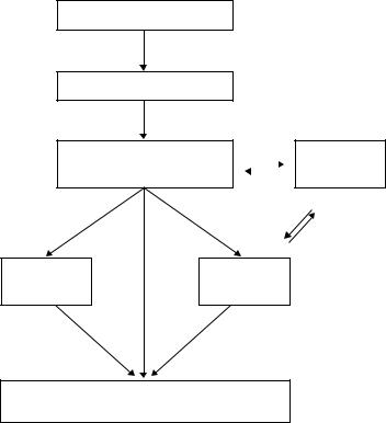
Although the role of routine intensive follow-up after curative primary colorectal cancer resection remains controversial, there is no doubt that a proportion of patients found to have liver metastases will benefit from prompt and appropriate treatment [23]. Interval CT (computed tomography) scanning and serial serum carcinoembryonic antigen (CEA) estimations are probably the “best buy” and have superseded the more basic and less sensitive traditional approach of clinical examination, liver function testing, and transbdominal ultrasonography. Ideally, chest CT is performed in search of pulmonary metastases, and colonoscopy (or barium enema or CT pneumocolon) is performed to confirm a “clean colon” having excluded local recurrence or metachronous primary disease. Thus, CT chest, abdomen, and pelvis will typically have been performed in the patient referred for consideration of liver resection. Further liver-specific imaging using more refined CT and/or MRI (magnetic resonance imaging) techniques in liaison with the hepatobiliary surgical team are the mainstay of the staging algorithm and are usually performed before final decisions regarding resectability can be made (Fig. 14.1).
The highly sensitive, but invasive, technique of CT angioportography (CTAP) has now been superseded by thin slice (≤5 mm collimation) portal
S U R G E R Y F O R M E T A S T A T I C D I S E A S E I N C R C |
219 |
.............................................................................................................................................................................. |
|
LFTs/CEA/USS/colonoscopy
CT (abdomen, pelvis, thorax)
Portal-phase spiral CT abdomen |
|
|
|
Test of time |
Liver-specific MRI |
|
|
|
|
|
|
|
|
|
|
|
|
|
Laparoscopy |
PET-CT |
|
+/– LapUS |
||
|
Laparotomy/IOUS +/– hepatic resection
Fig. 14.1 Investigative/staging algorithm for patients with liver metastases following radical resection of primary colorectal cancer.
venous-phase spiral CT, or MRI performed with intravenous gadolinium and liver-specific contrast agents, to maximize tumor conspicuity and diagnostic specificity. It is important that these staging investigations are performed before the commencement of systemic chemotherapy. Tumor imaging characteristics may change significantly during chemotherapy and good quality baseline imaging may be especially important where a good response to chemotherapy results in disappearance of metastases.
The importance of restraint in the biopsy of potentially resectable liver metastases should be emphasised (whether by radiological-guided needle biopsy, or surgical wedge biopsy during primary bowel resection). Although a histological diagnosis may be desirable in the palliative management of patients with unresectable disease, it is almost always unnecessary for diagnosis in those under consideration for liver resection, and it carries the well-documented risk of malignant needle-track seeding [24] and is
220 C H A P T E R 14
..............................................................................................................................................................................
associated with a significant reduction in long-term survival even when following curative liver resection [25]. In the authors’ experience, the rate of inadvertent liver resection for benign disease in patients not biopsied in this way was 1.2% which is considered acceptable within the context of modern, low morbidity surgery.
Staging laparoscopy remains an option in the investigation of some patients with colorectal liver metastases although its exact role continues to be debated. Interest in the technique as a means of improving patient selection arose following concerns regarding the apparent fallibility of conventional cross-sectional imaging in understaging disease in as many as 21% of patients [26]. The negative effects of such unnecessary operations are self-evident and include physical (postoperative pain, immunosupression, potential complications), psychological (anxiety, false hope), health economic factors, and delays in commencing palliative chemotherapy. While it seems likely that the yield of laparoscopy is too low to justify as a routine procedure, it may nevertheless be useful in selected “high-risk” patients with multiple bilobar metastases, unfavorable primary pathology and/or indeterminate imaging. The authors’ experience with selective laparoscopic staging following modern staging investigations indicates a 21% yield in detecting factors precluding curative resection, with a resectability rate of 88% in the remainder. However, it seems likely the technique will continue to be practiced only on a selective basis, with problem solving in high-risk patients increasingly deferred to the novel technique of PET-CT (Fig. 14.1).
Positron emission tomography with the glucose analog (18F) fluoro-2- deoxy-D-glucose images tumors based on their increased uptake of glucose and represents a significant advance in the investigation of patients with potentially resectable colorectal liver metastases. Meta-analysis of PET scanning in the detection of recurrent colorectal cancer has reported an overall sensitivity and specificity of 97 and 76%, respectively, and occult intrahepatic and extrahepatic metastases may be apparent in approximately 25% of patients previously studied with standard investigations [27]. Recently, Fernandez and co-workers [28] reported a superior 5-year overall survival rate of 58% following hepatic resection for colorectal liver metastases in 100 patients, all of whom had been selected on the basis of favorable PET scans. They suggest that improved patient selection with flurodeoxyglucosepositron emission tomography (FDG-PET) may have been primarily responsible for defining a new cohort of patients with a substantially improved prognosis following hepatic resection.
However, where less liberal access to PET scanning is available, its role may remain restricted to problem solving, confirming, or refuting the
S U R G E R Y F O R M E T A S T A T I C D I S E A S E I N C R C |
221 |
.............................................................................................................................................................................. |
|
presence of extrahepatic intra-abdominal disease in patients with indeterminate imaging in particular (Fig. 14.1). Recognized pitfalls associated with PET scanning include poor sensitivity in detecting smaller liver metastases in patients on chemotherapy. The fusion of contrast-enhanced CT and PET scanning (PET-CT) represents an important refinement in technique. Selzner et al. [29] recently reported improved diagnostic sensitivity for PETCT compared with conventional contrast-enhanced CT in the detection of locoregional recurrence and other extrahepatic disease, as well as the detection of intrahepatic recurrence during follow-up in the aftermath of hepatic resection.
Technical considerations in hepatic resection
The majority of liver resections are performed via upper abdominal incisions without resort to thoracotomy. Fixed costal margin retraction and careful mobilization of the liver by division of its retroperitoneal ligamentous attachments usually provide adequate access. Extensive adhesiolysis may be required following previous colorectal resection and a thorough inspection, palpation, and intraoperative ultrasound examination of the liver and extrahepatic tissues are performed before arriving at a final decision regarding resectability. Many hepatobiliary surgeons regard intraoperative ultrasonography as indispensable, and it has been estimated that it alone may be responsible for changing the operative plan in a proportion of patients [30].
The aims of hepatic resection are to achieve radical oncological (RO) clearance with tumor-free resection margins on the one hand, while preserving sufficient functioning hepatic parenchyma to avert postoperative hepatic failure on the other. Resections must preserve vital inflow (hepatic artery and portal vein) and outflow (hepatic vein and bile duct) structures, and an understanding of the hepatic segmental anatomy and intrahepatic vascular “watersheds” is fundamental. Couinaud’s seminal classification of the hepatic segmental anatomy [31] has been updated and the nomenclature standardized by the IHPBA Brisbane 2000 committee [32]. In this way, the vascular inflow to “hemilivers,” “sections,” and/or “segments” may be controlled selectively by extrahepatic dissection, or intrahepatically following parenchymal transection, and it forms the basis for precise resections “à la carte.” Liver resections are classified as major (≥3 hepatic segments), minor (<3 hepatic segments), or atypical wedge resections.
Although the traditional view held that tumor-free margins of ≥1 cm should be achieved to minimize the risk of oncological relapse, it is now
222 C H A P T E R 14
..............................................................................................................................................................................
recognized that the width of negative surgical margins does not necessarily affect the risk of recurrence and that RO resections may be achieved with very tight resection margins contingent on the preservation of the tumor pseudocapsule [33–37]. Similarly, although major hepatectomies sacrifice larger volumes of liver, perhaps ensuring generous margins and removing occult intrahepatic metastases or satellite lesions, a trend toward “tailored,” segment-orientated, parenchyma-sparing resections has emerged in recent years with the belief that this does not disadvantage patients oncologically and may facilitate re-resection in the event of intrahepatic recurrence.
Substantial falls in the perioperative mortality rates associated with major liver resection for colorectal liver metastases to 1–4% have been documented in recent years [6,33]. Perioperative blood loss requiring transfusion has been identified as the dominant risk factor for adverse outcome after liver resection [38], and the importance of minimizing blood loss during the dissection, parenchymal transection, and revascularization phases of hepatic resection has led to the concept of bloodless major liver surgery [33]. In this regard, the practice of low central venous pressure (0–4 cm H2O) anesthesia is critical in minimizing hepatic venous bleeding. Also, meticulous parenchymal transection technique using technology such as the cavitron ultrasonic surgical aspirator (CUSA Ex, Valleylab Inc., Amersham, Bucks, UK), and argon beam coagulation, have helped achieve a mean operative blood loss of 360 ml during hepatic resection. Indeed, the practice of perioperative blood transfusion is now regarded as exceptional in the authors’ practice [33] and by many others.
The liver’s unique regenerative properties, functional reserve, and tolerance of extended warm ischaemia permit extensive parenchymal resections to be performed utilizing intermittent portal triad inflow clamping (the “Pringle maneuver”), another important technique in the pursuit of bloodless liver surgery. However, more advanced clamping techniques such as hepatic vascular exclusion (portal triad occlusion plus suprahepatic and infrahepatic inferior vena cava (IVC) clamping) have been associated with increased hemodynamic intolerance and postoperative morbidity [39] and tend to be restricted to more complex resections involving the hepatic veins and IVC.
Liver related morbidity, such as bile leaks, hemorrhage, and mild hepatic insufficiency, as well as relatively minor complications such as right-sided pleural effusion, occur in 10–15% of patients and are usually managed successfully by conservative means.
S U R G E R Y F O R M E T A S T A T I C D I S E A S E I N C R C |
223 |
.............................................................................................................................................................................. |
|
Liver failure is now recognized as the main mode of postoperative death and is directly related to both the extent of resection and the presence of background liver disease. Estimation of an acceptable residual functioning liver volume, and the prediction of hepatic dysfunction following resection, can be difficult. Although objective tests such as CT volumetry and indocyanine green clearance studies are available, in practice it is generally accepted that a subjective estimate of approximately one-third the standard liver volume or the equivalent of a minimum of two normal liver segments is usually sufficient.
In this regard, a growing concern which merits consideration is the risk of post chemotherapy hepatotoxicity and its impact on the ability to perform major liver resection safely, specifically with the newer chemotherapeutic agents such as irinotecan and oxaliplatin. Fernandez and colleagues [40] recently reported that treatment with irinotecan and/or oxaliplatin, especially in obese patients, presented a risk for the development of severe steatohepatitis, and highlighted the concomitant risk of liver failure following major liver resection.
Subsequent suggestions included consideration of preoperative liver biopsy in patients considered at risk and delay of chemotherapy until after liver resection where possible. The authors’ own experience with liver resection for colorectal metastases following neoadjuvant chemotherapy of all types identifies no measurable excess morbidity or mortality in such patients. However, there appears to be evidence of an increased risk of complications when the duration of chemotherapy is prolonged beyond 3 months. This underlines the importance of the early involvement of specialist hepatobiliary surgeons in the multidisciplinary management of patients with metastatic colorectal cancer.
Follow up and hepatic re-resection
Following radical liver resection for colorectal metastases, up to 60% of patients may subsequently develop recurrent disease. Of these, approximately 20–30% may have metastases isolated to the liver and potentially amenable to hepatic re-resection. Repeat hepatic resection can present a daunting technical challenge, and the oncological rationale for re-resection of recurrent colorectal liver metastases may seem counterintuitive. Indeed, such concerns have stimulated interest in alternative minimal access techniques such as percutaneous radiofrequency ablation (RFA) [41].
224 C H A P T E R 14
..............................................................................................................................................................................
Nevertheless, favorable accounts of hepatic re-resection for patients with recurrent colorectal liver metastases have reported 5-year survival rates of 26–41% [42–44], and it seems appropriate to treat such patients in the same way as those first presenting with colorectal liver metastases.
Despite the technical demands presented by dense intra-abdominal adhesions and variations in hepatobiliary anatomy in the hypertrophied regenerated liver remnant, concerns regarding excess morbidity and mortality associated with repeat hepatic resection have not been borne out. In the authors’ own experience of 71 repeat hepatic resections in 66 patients with recurrent colorectal liver metastases, there were no postoperative deaths and the low morbidity rate of 11% compared favorably with that experienced following index hepatectomy [45]. Furthermore, beneficial 1-, 3-, and 5-year actuarial survival rates of 94, 68, and 44% were observed following repeat hepatic resection which exceeded those of all patients following a first hepatectomy for colorectal liver metastases.
It therefore seems appropriate to follow-up patients who have undergone hepatic resection for colorectal liver metastases. This is usually performed for a period of 5 years using CT of the chest and liver and serial serum CEA estimations in an attempt to identify those patients who may benefit from further intervention.
Laparoscopic liver resection
Though feasible, laparoscopic liver resection for patients with colorectal liver metastases is controversial and remains at an early stage of development. Thus far, most surgeons have focused on the more accessible small lesions in hepatic segments 2/3 and the caudal aspect of segments 4, 5, and 6. More adventurous procedures including right hepatectomy have been performed in highly selected cases and usually mandate a handassisted technique. While effective strategies for dealing with the risks of intraoperative hemorrhage, gas embolism, and bile leaks continue to evolve, the immediate benefits compared with conventional open surgery remain unclear.
Extending the limits of resectability
In recent years, the development of novel strategies and advanced techniques has dramatically extended the boundaries of resectability, permitting radical liver resections to be performed in patients who would formerly
S U R G E R Y F O R M E T A S T A T I C D I S E A S E I N C R C |
225 |
.............................................................................................................................................................................. |
|
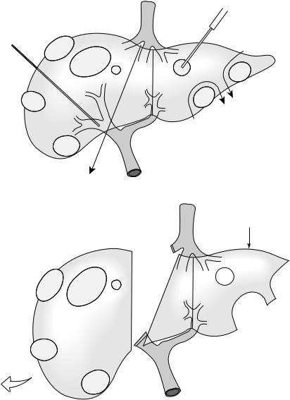
have been regarded as unresectable. It is important to emphasize that it is often a combination of the following techniques which are required to tackle colorectal liver metastases which have reached an advanced stage [36] (Fig. 14.2).
Neoadjuvant chemotherapy
The concept of downstaging neoadjuvant chemotherapy for otherwise unresectable colorectal liver metastases, first introduced 10 years ago [46], has reproducibly been shown to offer the chance of curative liver resection to 16–23% of such patients with reported 5-years survival rates of up to 40% [36].
Interestingly, the possibility that patients presenting with colorectal liver metastases initially deemed unresectable might be downstaged by oxaliplatin-based chemotherapy to become resectable comprised the sole recommendation for the provision of oxaliplatin-based chemotherapy in the UK National Institute for Clinical Excellence (NICE) 2002 guidance.
However, the success of downstaging chemotherapy in achieving a dramatic response, typically in patients with initially unresectable or indeterminate multiple bilateral metastases, can result in the disappearance of lesions from the imaging study. Thus, the dilemma presented in treating definitively the “missing metastasis” is increasingly encountered and reemphasizes the importance of pursuing good quality baseline staging investigations before commencing chemotherapy. Intuitively, “disappeared” metastases might be expected to eventually reappear as only a small minority of resected lesions demonstrate complete necrosis at histopathology [36]. Ideally, liver resections should target the parenchyma harboring the original lesion, but intraoperative localization can be problematic. Referral of such patients to the hepatobiliary team is therefore recommended before a “complete response” is necessarily achieved by their colleagues in Medical Oncology. Elias and co-workers [47] recently reported a subgroup of 11 patients identified as having “missing metastases,” none of which could be identified or resected, and eight (73%) of which remained quiescent during a median 31-month follow-up period.
Portal vein embolization
Some patients may be denied a technically feasible liver resection because of concerns that an insufficient future remnant liver volume may risk severe
226 C H A P T E R 14
..............................................................................................................................................................................
A |
Percutaneous/intraoperative |
|
RFA/microwave |
Percutaneous |
|
right PVE |
|
2/3
4
Atypical wedge resections
|
Portal |
Right hepatectomy |
vein |
(segments 5–8) |
|
B |
Tumor-free |
|
|
|
liver remnant |
2/3
4
Fig. 14.2 Combined advanced techniques used to extend the limits of resectability to patients with multiple bilobar colorectal liver metastases. A. Percutaneous right portal vein embolization (PVE), staged or simultaneous ablation and wedge resection of contralateral lesions with subsequent right hemihepatectomy (hepatic segments 5–8). B. The aims of this approach are to achieve a viable tumor-free functional hepatic remnant (in this example based on an hypertrophied left hemiliver [hepatic segments 2–4]).
S U R G E R Y F O R M E T A S T A T I C D I S E A S E I N C R C |
227 |
.............................................................................................................................................................................. |
|
postoperative hepatic failure. Preoperative percutaneous selective portal vein embolization (PVE) may be performed in an attempt to induce hypertrophy of the future liver remnant (estimated volume ≤25%) and appears to be especially useful where there are concerns regarding background liver disease or following prolonged chemotherapy (estimated liver remnant volume ≤40%). Typically, right PVE is performed with a view to right hepatectomy or extended right hepatectomy a month later in the patient with small hepatic segment 2/3 volume, and subsequent 5-year survival rates have been reported as comparable to those resected without PVE [48]. Such has been the acceptance of PVE in this role that the prospect of randomized trials of its efficacy is no longer regarded as ethical.
Interestingly, there is evidence that right PVE is not beneficial for patients with normal liver in whom a straightforward right hepatectomy is planned [49], nor is embolization of segment 4 in addition to right PVE thought to be necessary [50]. Disadvantages associated with the PVE/hepatic resection approach include a local and systemic inflammatory response which is usually mild, a slightly higher perioperative mortality rate, the possibility that liver resection will not be achieved following PVE, and the rapid growth of occult metastases found out with the embolization zone [48].
Staged liver resections
Two-stage liver resections involve a preliminary non-curative procedure followed by a second resection of residual hepatic metastases. This aggressive surgical approach may facilitate compensatory hypertrophy of a tumor-free residual liver remnant, with or without interim PVE, in patients with multiple bilobar metastases. Subsequent major resection of the contralateral embolized liver has been reported to yield long-term survival rates similar to those associated with initially resectable patients [51].
Combinations of resection and ablative techniques
Percutaneous interstitial ablative techniques include cryotherapy, ethanol injection, laser hyperthermia, microwave thermotherapy, and RFA whose dominant role remains the palliative treatment of surgically unfit patients or those with unresectable disease. These procedures performed intraoperatively, particularly RFA, have been used to treat deep-seated
228 C H A P T E R 14
..............................................................................................................................................................................
metastases within the future liver remnant in combination with major hepatectomy, staged resections, chemotherapy, and/or PVE to extend further the boundaries of resectability in an attempt to offer the best chance of cure to patients in an otherwise hopeless situation [52] (Fig. 14.2).
Conclusions
Liver resection offers the only chance of long-term survival, or cure, to selected patients with colorectal liver metastases. Major hepatic resection can be performed with negligible mortality in well-staged patients by experienced hepatobiliary surgical teams paying meticulous attention to bloodless surgery and the balance between hepatic functional reserve and oncological clearance. The basic risks compare favorably with those of other elective abdominal procedures such that perioperative mortality will become redundant as the main endpoint in favor of alternative measures such as quality of life and cost-effectiveness.
Intensive follow-up identifies patients with recurrence suitable for reresection which may be performed safely and with expectations not dissimilar to those of the index liver resection. The substantial improvements in long-term survival offered by liver resection for colorectal liver metastases may have attained a plateau, perhaps because the indications for resection have widened as hepatobiliary specialists continue to embrace increasing numbers of ever more difficult and advanced cases. However, it may be that a phase has been reached where surgical ingenuity alone is unlikely to significantly improve further the resectability or survival rates. Rather, the combination of hepatic resectional surgery with more efficient adjuvant and neoadjuvant therapy and/or interventional ablation techniques may offer the hope of further progress.
For patients treated with more powerful forms of chemotherapy, the timing of referral for hepatic resection may be critical. Some patients with synchronous disease may benefit from immediate hepatic resection and the results of current trials of neoadjuvant chemotherapy may help address this. For others, potentially disadvantageous effects of prolonged chemotherapy on hepatic functional reserve and precise tumor localization justify early involvement of hepatobiliary specialists in decision making. Indeed, such have the limits of resectability been extended that each and every fit patient with colorectal liver metastases, even those with apparently irresectable disease, deserves assessment by a hepatobiliary surgical team.
S U R G E R Y F O R M E T A S T A T I C D I S E A S E I N C R C |
229 |
.............................................................................................................................................................................. |
|
References
1Weiss E, Grundmann L, Torhorst J et al. Haematogenous metastatic patterns in colonic carcinoma: an analysis of 1541
necropsies. J Pathol 1986; 150: 195–203.
2Scheele J, Stangl R, Altendorf-Hofman A. Hepatic metastases from colorectal cancer: impact of surgical resection on the natural history. Br J Surg 1990; 77:
1241–6.
3Sugarbaker PH. Surgical decision making for large bowel cancer metastatic to the
liver. Radiology 1990; 174: 621–6.
4Scheele J, Stangl R, Altendorf-Hofman A, Paul M. Resection of colorectal liver metastases. World J Surg 1995; 19: 59–71.
5Stangl R, Altendorf-Hofman A, Charnley R, Scheele J. Factors influencing the natural history of colorectal liver
metastases. Lancet 1994; 343: 1405–10.
6Beard SM, Holmes M, Price C, Majeed AW. Hepatic resection for colorectal liver metastases: a cost-effectiveness analysis. Ann Surg
2000; 232: 763–76.
7Wagner J, Adson M, Van Heerdan J, Ilstrup D. The natural history of hepatic metastases from colorectal cancer. A comparison with resective treatment.
Ann Surg 1984; 199: 502–8.
8Wood CB, Gillis CR, Blumgart LH. A retrospctive study of the natural history
of patients with liver metastases from colorectal cancer. Clin Oncol 1976; 2: 285–8.
9Rosen CB, Nagorney DM, Taswell HF et al. Perioperative blood transfusion and determinants of survival after liver resection for metastatic colorectal carcinoma. Ann Surg 1992; 216:
493–505.
10Fong Y, Cohen AM, Fortner J et al. Liver resection for colorectal metastases. J Clin Oncol 1997; 15: 938–46.
11Taylor M, Forster J, Langer B et al. A study of prognostic factors for hepatic resection for colorectal liver metastases. Am J Surg 1997; 173: 467–71.
12Nuzzo G, Giuliante F, Giovannini I et al. Resection of hepatic metastases
from colorectal cancer.
Hepatogastroenterology 1997; 44: 751–9.
13Fong Y, Fortner J, Sun RL et al. Clinical score for predicting recurrence after hepatic resection for metastatic colorectal cancer: analysis of 1001 consecutive cases. Ann Surg 1999; 230: 309–18.
14Nordlinger B, Jaeck D, Guiguet M et al. Surgical resection of hepatic metastases. Multicentric retrospective study by the French Association of Surgery. In: Nordlinger B, Jaeck D, eds. Treatment of Hepatic Metastases of Colorectal Cancer. Paris: Springer-Verlag, 1992: 129–61.
15McAfee MK, Allen MS, Trastek VF. Colorectal lung metastases: results of surgical excision. Ann Thorac Surg 1992; 53: 780–6.
16Murata S, Moriya Y, Akasu T et al. Resection of both hepatic and pulmonary metastases in patients with colorectal carcinoma. Cancer 1998; 83: 1086–93.
17Ike H, Shimada H, Togo S et al. Sequential resection of lung metastasis following partial hepatectomy for colorectal cancer. Br J Surg 2002; 89: 1164–8.
18Elias D, Ouellet J-F, Bellon N et al. Extrahepatic disease does not contraindicate hepatectomy for colorectal liver metastases. Br J Surg 2003; 90: 567–74.
19Elias D, Detroz B, Lasser P et al. Is simultaneous hepatectomy and intestinal anastamosis safe? Am J Surg 1995; 169: 254–60.
20Lambert LA, Colacchio TA, Barth RJ. Interval hepatic resection of colorectal metastases improves patient selection. Br J Surg 2000; 135: 473–80.
21Allen PJ, Kemeny N, Jarnagin W et al. Importance of response to neoadjuvant chemotherapy in patients undergoing resection of synchronous colorectal liver metastases. J Gastrointest Surg 2003; 7: 109–15.
22Adam R, Pascal G, Castaing D et al. Tumor progression while on chemotherapy: a contraindication to liver
230 C H A P T E R 14
..............................................................................................................................................................................
resection for multiple colorectal metastases? Ann Surg 2004; 240: 1052–64.
23Jeffrey GM, Hickey BE, Hider P. Follow-up strategies for patients treated for non-metastatic colorectal cancer.
Cochrane Database Syst Rev
2002.
24John TG, Garden OJ. Needle track seeding of primary and secondary liver carcinoma after percutaneous liver biopsy. HPB Surg 1993; 6: 199–204.
25Jones OM, Rees M, John TG et al. Biopsy of resectable colorectal liver metastases causes tumour dissemination and adversely affects survival after liver resection. Br J Surg 2005; 92: 1165–8.
26Jarnagin WR, Fong Y, Ky A et al. Liver resection for metastatic colorectal cancer: assessing the risk of occult irresectable disease. J Am Coll Surg 1999; 188: 33–42.
27Huebner RH, Park KC, Shepherd JE
et al. A meta-analysis of the literature for whole-body FDG PET detection of recurrent colorectal cancer. J Nucl Med 2000; 41: 1177–89.
28Fernandez FG, Drebin JA, Linehan DC et al. Five-year survival after resection of hepatic metastases from colorectal cancer in patients screened by positron emission tomography with F-18 fluorodeoxyglucose (FDG-PET). Ann Surg 2004; 200: 438–50.
29Selzner M, Hany TF, Widbrett P et al. Does the novel PET/CT imaging modality impact on the treatment of patients with metastatic colorectal cancer of the liver. Ann Surg 2004; 240: 1027–36.
30Jarnagin WR, Bach AM, Winston CB et al. What is the yield of intraoperative ultrasonography during partial
hepatectomy for malignant disease? J Am Coll Surg 2001; 192: 577–83.
31Couinaud C. Le foie: études anatomiques et chirurgicales. Paris: Masson; 1957.
32Strasberg SM, Belghiti J, Clavien PA
et al. Terminology of liver anatomy and resections. HPB 2000; 2: 333–9.
33Rees M, Plant G, Wells J, Bygrave S. One hundred and fifty hepatic resections: the evolution of technique towards bloodless
surgery. Br J Surg 1996; 83: 1526–9.
34Rees M, Plant G, Bygrave S. Late results justify resection for multiple hepatic metastases from colorectal cancer. Br J Surg 1997; 84: 1136–40.
35Yamamoto J, Sugihara K, Kosuge T et al. Pathologic support for limited hepatectomy in the treatment of liver metastases from colorectal cancer. Ann Surg 1995; 221: 74–8.
36Adam R, Delvart V, Pascal G et al. Rescue surgery for unresectable colorectal liver metastases downstaged by chemotherapy. A model to predict long-term survival. Ann Surg 2004; 240: 644–58.
37Pawlik TM, Scoggins CR, Zorzi D et al. Effect of surgical margin status on survival and site of recurrence after hepatic resection for colorectal metastases. Ann Surg 2005; 241: 715–22.
38Kooby DA, Stockman J, Ben-Portat L et al. Influence of transfusions on perioperative and long-term outcome in patients following hepatic resection for colorectal metastases. Ann Surg 2003; 237: 860–70.
39Belghiti J, Noun R, Zante E et al. Portal triad clamping or hepatic vascular exclusion for major liver resection. A controlled study. Ann Surg 1996; 224: 155–61.
40Fernandez FG, Ritter J, Goodwin JW et al. Effect of steatohepatitis associated with irinotecan or oxaliplatin pretreatment on resectability of hepatic colorectal metastases. J Am Coll Surg 2004; 200: 845–53.
41Elias D, DeBaere T, Smayra T et al. Percutaneous radiofrequency thermoablation as an alternative to surgery for treatment of liver tumour recurrence after hepatectomy. Br J Surg 2002; 89: 752–6.
42Neeleman N, Andersson R. Repeated liver resection for recurrent liver cancer. Br J Surg 1996; 83: 893–901.
43Petrowsky H, Gonen M, Jarnagin W
et al. Second liver resections are safe and effective treatment for recurrent hepatic metastases from colorectal cancer: a
S U R G E R Y F O R M E T A S T A T I C D I S E A S E I N C R C |
231 |
.............................................................................................................................................................................. |
|
bi-institutional analysis. Ann Surg 2002; 235: 863–71.
44Adam R, Huguet E, Azoulay D et al. Hepatic resection after down-staging of unresectable hepatic colorectal metastases. Surg Oncol Clin N Am 2003; 12: 211–20.
45Shaw IM, Rees M, Welsh F et al. Repeat hepatic resection for recurrent colorectal liver metastases is associated with favourable long term survival. Br J Surg 2006; (in press).
46Bismuth H, Adam R, Lévi F et al. Resection of nonresectable liver metastases from colorectal cancer after neoadjuvant chemotherapy. Ann Surg 1996; 224: 509–22.
47Elias D, Youssef O, Sideris L et al. Evolution of missing colorectal liver metastases following inductive chemotherapy and hepatectomy. J Surg Oncol 2004; 86: 4–9.
48Elias D, Ouellet J-F, de Baère T et al. Preoperative selective portal vein embolization before hepatectomy for
liver metastases: long-term results and impact on survival. Surgery 2002; 131: 294–9.
49Farges O, Belghiti J, Kianmanesh R et al. Portal vein embolization before right hepatectomy: prospective clinical trial. Ann Surg 2003; 237: 208–17.
50Capussotti L, Muratore A, Ferrero A et al. Extension of right portal vein embolization to segment 4 portal
branches. Arch Surg 2005; 140: 1100–3.
51Jaeck D, Oussoultzoglou E, Greget M
et al. A two-stage hepatectomy procedure combined with portal vein embolization to achieve curative resection for initially unresectable multiple and bilobar colorectal liver metastases. Ann Surg 2004; 240: 1037–49.
52Elias D, Baton O, Sideris L et al. Hepatectomy plus intraoperative radiofrequency ablation and chemotherapy to treat technically unresectable multiple colorectal liver metastases. J Surg Oncol 2005; 90: 36–42.