

Challenges in Colorectal Cancer, Second Edition
Edited by John H. Scholefield etc. Copyright © 2006 by Blackwell Publishing Ltd
.............................................................................................................................................................................
4: MRI-directed rectal cancer surgery
Brendan Moran and John H. Scholefield
.............................................................................................................................................................................
Introduction
Rectal cancer is a common problem and accounts for approximately 30% of all colorectal malignancies. The treatment is predominantly surgical excision, with the addition of neoadjuvant therapy (preoperative radiotherapy or chemoradiotherapy) in selected cases. There is universal agreement that surgery alone can cure localized rectal cancer and now increasing evidence that neoadjuvant therapy may facilitate surgical excision in advanced disease. There is currently ongoing controversy as to whether all, or selected patients, should have neoadjuvant therapy. Major resectional rectal cancer surgery is both technically challenging and complex due to the relative inaccessibility within the confines of the bony pelvis, the difficulty in reconstruction with a high risk of leakage, and the particular problem of local recurrence within the pelvis.
Major surgery is associated with significant mortality and morbidity and is inappropriate for some cases. There is now a range of treatments, both surgical and non-surgical, for management of rectal cancer. For some patients with early tumors, local excision alone may be curative, while some elderly, unfit, or those with very advanced disease may only benefit from symptom control, for example, luminal ablation techniques. Furthermore, novel chemotherapeutic and radiotherapy regimens can result in local symptom control in patients who either refuse or are unsuitable for major surgery.
With this range of treatment options, patient assessment, cancer staging, and selection for appropriate therapy is becoming crucial to the optimal management of rectal cancer. Despite the range of treatments, and ongoing controversy as to their merits in individual situations, there is general agreement that, for the majority of patients with rectal cancer, trans-abdominal resection is the optimal treatment.
46
M R I - D I R E C T E D R E C T A L C A N C E R S U R G E R Y |
47 |
.............................................................................................................................................................................. |
|
The main aims in rectal cancer surgery are to cure the patient and if possible preserve normal bowel, bladder, and sexual function. The main mechanisms available to achieve these aims encompass two key treatment modalities, namely surgical technique [1,2] and neoadjuvant therapy, both of which have been shown to reduce the local recurrence rates [3,4]. There is substantial evidence that optimal surgery, in the form of total mesorectal excision (TME), reduces local recurrence and improves survival [1–5]. With regards to preoperative radiotherapy, there is good quality evidence for a reduction in local recurrence from large randomized trials [3,6–8], though to date only one trial has reported improved survival with the addition of radiotherapy [8].
The key advances in rectal cancer have resulted from awareness of the problem of local recurrence, the risk factors influencing recurrence, and the strategies needed to reduce it. These factors when combined almost always translate to both better quality of life and improved overall survival in patients with rectal cancer.
Local recurrence of rectal cancer
Local recurrence after rectal cancer surgery is defined as disease in the pelvis, including recurrence at the site of the anastomosis and in the perineum [9]. Local recurrence is for the most part incurable and results in severe morbidity with debilitating symptoms of pelvic pain, ureteric obstruction, intestinal and urinary tract fistulation, and poor bowel and urinary function. Palliative treatment has limited success. Increasingly local recurrence is being recognized as a failure of complete removal of tumor at the primary surgical procedure. Thus, in reality, local “recurrence” may, in many cases, represent persistent and progressive disease rather than true recurrence. There are a number of predictors of risk of local recurrence following surgery for rectal cancer, including size of the primary [10] involvement of the circumferential resection margin (CRM) [11], distal location of the tumor [10–12], extramural vascular invasion [13,14], tumor differentiation [13,14], nodal status [11,13,14], extent of extramural spread [11,13,14], and peritoneal involvement by tumor [12,13,15]. Practically all of these features with a high risk of local recurrence are associated with both locally advanced tumors and frequently, in addition, metastatic disease at presentation. These risk factors for local recurrence are also highly predictive of disseminated distant recurrence following surgery. Traditionally, the majority of these risk factors came to light either at
48 C H A P T E R 4
..............................................................................................................................................................................
surgery or, more commonly, at histopathological assessment of the excised specimen.
Current topical issues are whether some or all of these factors can be predicted preoperatively and if so, whether there are adjunctive treatments that can improve the outcome in high-risk patients?
Reported local recurrence rates vary from 2.6 to 32% and are probably most influenced by surgical technique [16,17]. The lowest recurrence rates and best survivals have been consistently reported with TME [1,2,5,16–18]. It is now well documented that low local recurrence rates translate into better overall survival with increasing evidence that the optimal treatment of rectal cancer undoubtedly involves strategies to minimize local recurrence.
Local recurrence and the circumferential resection margin
Rectal cancer spreads by local extension, via the lymphatics and via the bloodstream. Lymphatic drainage is associated with the vascular pedicle and is generally addressed by a combination of TME and high ligation of the inferior mesenteric artery. Local spread of a rectal cancer in the confines of the narrow pelvis results in a risk of involvement of the CRM. Of all the risk factors for local recurrence of rectal cancer, involvement of the CRM appears to be the main determinant of risk. A number of reports, initially from Quirke’s group, have shown that a positive CRM, defined as tumor within 1 mm of the edge of the resected specimen, and depth of extramural invasion are independent predictors of local recurrence and poor prognosis [10,11]. These observations are supported by recent reports from large studies, such as the Norwegian [19] and Dutch [20] studies.
While the CRM may be involved directly by tumor, CRM involvement may also be due to metastatic nodal disease. There is debate as to whether involved nodes, in their own right, increase local recurrence even if not directly involving the CRM. Jatzko et al. [21] reported that patients with positive lymph nodes had a higher risk of local recurrence. However, others found that lymph node involvement was not associated with higher local recurrence, attributed to the beneficial effects of TME [19,22]. Recently Cecil et al. [23] reported their experience in patients with lymph node positive rectal cancer with little impact on local recurrence rates which they attributed to optimal CRM clearance by TME.
While other risk factors, such as vascular invasion, differentiation, and so on are undoubtedly major determinants of long-term survival, the single main factor that can be manipulated by treatment is the CRM.
M R I - D I R E C T E D R E C T A L C A N C E R S U R G E R Y |
49 |
.............................................................................................................................................................................. |
|
Surgical technique and total mesorectal excision
Total mesorectal excision involves a number of steps, which aim to maximize circumferential clearance. TME surgery has been reported to reduce the rate of local recurrence from 30–40 to 5–15% [17,24]. A paper from Stockholm has been the first report of a direct benefit in a whole population following a video-based surgical training program [4]. Local recurrence was significantly lower in the TME group compared with historical controls from the Stockholm randomized controlled trials of preoperative radiotherapy (Stockholm I and II groups) (6 vs 15% and 14% respectively) as was cancer-related death (9 vs 15% and 16% respectively) [4]. The CRM positive rate was 4% – the lowest-reported incidence so far in any published series. They have recently updated their results at 5 years with significant survival benefits in patients who had TME surgery [25].
Evidence for preoperative radiotherapy
There is ongoing debate as to the role of preoperative radiotherapy in rectal cancer. However, there is now evidence that radiotherapy, particularly in the preoperative setting results in a reduction of local recurrence. The advantages and disadvantages of preoperative vs postoperative radiotherapy have been extensively discussed in the medical literature. The main disadvantage of preoperative treatment is the risk of overtreating some patients due to staging inaccuracies and thus exposing patients to unnecessary risks. Postoperative radiotherapy can be more appropriately targeted with the benefit of histopathology but has major tolerance problems, with acute toxicity, in patients who have just undergone major surgery. Pahlman and Glimelius [26] reported the results of, until recently, the only randomized trial to address this issue. There was a significantly lower recurrence rate in the preoperative arm (12 vs 21%) which may have been partly attributable to a delay of 6 weeks or more in over 50% of the patients randomized to postoperative radiotherapy [26].
The recently reported German Rectal Cancer Study Group of a randomized trial of preoperative vs postoperative chemoradiotherapy for rectal cancer is a major addition to this ongoing debate [27]. In total, 799 patients staged preoperatively as T3 or T4 tumors were randomized. Staging was by endorectal ultrasound and Sauer et al. found that 18% of the patients randomized to postoperative treatment had been overstaged compared to the pathology of the resected specimen. These 18% had been considered to
50 C H A P T E R 4
..............................................................................................................................................................................
have tumor penetration through the bowel wall (T3 or T4 disease) or nodal involvement but were subsequently found to have stage T1, or T2, lymph node negative disease. This is not surprising as the accuracy of endorectal ultrasonography is reported to be 67–93% for the assessment of rectal wall penetration and 62–83% for determination of nodal status [28].
The overall 5-year survival rates in the German Rectal Cancer Trial were similar (67 and 74%). The local recurrence was 6% for patients assigned to preoperative chemoradiotherapy vs 13% in the postoperative treatment group (p = 0.006).
Much of the background evidence in support of preoperative radiotherapy comes from the Swedish and Stockholm Rectal Cancer Trials [6–8]. The Swedish Rectal Cancer Trial showed a relative survival benefit of 21%, with an increase in the 5-year survival from 48 to 58% and a reduction in local recurrence from 27 to 11% [8]. A meta-analysis in 2000 of the then published randomized controlled trials concluded that in patients with resectable rectal cancer, preoperative radiotherapy significantly improved overall and cancer-specific survival compared with surgery alone, though these benefits were mainly attributable to the Swedish trial results [29].
It is now generally agreed that preoperative radiotherapy can reduce local recurrence rates to approximately half that in surgery alone. Therefore in situations with a high local recurrence rates, routine usage might be acceptable. However, with local recurrence rates of less than 10%, there is no data demonstrating a beneficial effect with the routine addition of radiotherapy [30]. In the Norwegian report [19], only 5% of patients with an uninvolved CRM developed local recurrence, so radiotherapy would have overtreated 97% of the patients with clear margins if all patients had routine preoperative radiotherapy.
The Dutch Colorectal Cancer Group attempted to address the role of preoperative radiotherapy when surgery was standardized to TME for rectal cancer [3]. The entry criteria included only mobile rectal cancer. With addition of radiotherapy, local recurrence was reduced from 8.2 to 2.4% after a median follow-up of 2 years in 1784 patients who underwent a macroscopically complete resection [3]. However, the surgery alone arm had high local recurrence rates for mobile tumors suggesting inadequate surgery in a significant proportion. The Dutch trial had attempted to standardize the surgery and provide quality control measures through workshops and live-video demonstrations. Despite this “standardization” of surgery, the involved margin rate in what were considered mobile tumors was 18.3%.
M R I - D I R E C T E D R E C T A L C A N C E R S U R G E R Y |
51 |
.............................................................................................................................................................................. |
|
Nevertheless, this is an important trial which has shown a reduction in local recurrence at 2 years with the updated results confirming a similar degree of reduction of local recurrence with no difference in overall survival (unpublished results). As practically all patients with local recurrence die from the disease, it seems surprising that 5-year survival is similar. This suggests that radiotherapy may either have lethal side-effects or, more likely, that the reduction in local recurrence is offset by death from systemic metastases in a group predestined to die of disease. Nevertheless, if radiotherapy was without harm, a reduction in local recurrence would still be worthwhile. Unfortunately there are risks, as well as inconvenience and costs, associated with radiotherapy.
Complications of preoperative radiotherapy
Preoperative radiotherapy is associated with toxicity, early postoperative complications, and long-term side effects. Early complications include perineal wound breakdown, diarrhoea, proctitis, urinary tract infection, small bowel obstruction, leucopenia, and venous thrombosis.
Radiotherapy has been shown to have adverse effects on anal function with negative effects on the function and integrity of a coloanal anastomosis with, or without, formation of a colonic pouch [31].
In the recently reported German Rectal Cancer Study [27], Grade 3 or 4 acute toxicity occurred in 27% with preoperative chemoradiotherapy vs 40% in the postoperative group (p = 0.001); the corresponding rates of long-term toxic effects were 14 and 24%, respectively (p = 0.01).
Downstaging and downsizing rectal cancer with neoadjuvant therapy
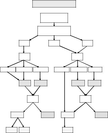
Rectal cancer staging has traditionally been a histopathological analysis of the excised specimen and the “Dukes’ staging” system described initially by Cuthbert Dukes [32] specifically in rectal cancer, and subsequently applied to colon cancer, is still in widespread use. However, the TNM classification system has advantages and is now considered the optimal universal staging system for colorectal cancer (Fig. 4.1).
The particular advantage of the TNM system, as applied to rectal cancer, is the “T” aspect whereby the depth of penetration within the bowel wall is a major determinant of the prognosis, particularly with regard to local recurrence in the lowest tumors. The rectum has been arbitrarily divided into three parts with the lower rectum 0–6 cm, the middle rectum 7–11 cm,
52 C H A P T E R 4
..............................................................................................................................................................................
Rectal cancer staging
Adenocarcinoma of rectum with lower edge <=15 cm from anal verge
Digital rectal examination (awake patient)
Rigid sigmoidoscopy
Distance in cm from anal verge
|
0–6 cm |
7–11 cm |
12–15 cm |
|||
|
MRI |
|
|
MRI |
|
|
Clear CRM |
Threatened CRM |
Involved CRM |
Clear CRM |
Threatened CRM |
Involved CRM |
|
“T1/T2” |
“T3” |
“T4” |
“T1/T2” |
“T3” |
“T4” |
|
|
Consider |
RT or CRT |
|
Consider |
RT or CRT |
|
|
RT/CRT |
|
RT/CRT |
|||
|
|
|
|
|||
|
Repeat |
|
Repeat |
|||
|
MRI |
|
|
MRI |
|
|
Surgery |
Palliative |
Surgery |
Palliative |
|||
treatment |
treatment |
|||||
|
|
|
|
|||
Extended |
AR |
|
Mainly AR |
|
||
APER |
|
|
||||
|
|
|
|
|
||
Fig. 4.1 Rectal cancer staging.
and the upper rectum 12–15 cm from the anal verge. There is a debate as to whether a T3 middle or upper rectal cancer (that is, above 7 cm) is particularly at high risk of local recurrence, providing surgery is adequate and that a TME has resulted in an intact cover of mesorectal fat and fascia.
However in the lower rectum a T3 tumor has, by definition, gone through the wall of the bowel and commonly will have an involved margin at the
M R I - D I R E C T E D R E C T A L C A N C E R S U R G E R Y |
53 |
.............................................................................................................................................................................. |
|
level of the sphincter complex unless the rectum is excised en bloc with the sphincter. One of the advantages of the TNM system is the ability to extrapolate this staging system to a preoperative staging system which may be helpful in planning treatment. Assessing the factors most likely to lead to local recurrence the detectable features might theoretically include the local extent of the tumor (T stage) and the nodal status (N stage). Obviously the M stage is of great prognostic importance, though local symptoms from a rectal cancer may override the presence of metastatic disease in proceeding to major resectional surgery.
Endoanal ultrasound appears to be particularly good at estimating the T stage [28]. An ultrasound staging system has been developed which is analogous to the pathological TNM system with a prefix “u” denoting that this is an ultrasound rather than a pathological staging. Correlations with pathology indicate that accuracy of ultrasound, particularly in the T staging, though less so in the N staging are accurate [28].
Endoluminal ultrasound (EUS) is particularly helpful in selecting patients who may be suitable for local excision, generally agreed to be T1 tumors. However the main determinant of local recurrence is undoubtedly a positive CRM and EUS is poor at assessment of the CRM.
In an attempt to improve the imaging of rectal cancer, endoluminal magnetic resonance imaging (MRI) has been assessed with good results from specialist centers [33]. However the very tumors most in need of accurate staging, in particular stenotic tumors, or low painful lesions, are unable to be staged by endoluminal techniques. Fortunately developments in phasedarray surface coil MRI now allow accurate local assessment of most patients with rectal cancer [34,35].
While the TNM staging is important, a key aspect of the local staging of rectal cancer is the relationship of the tumor, or its metastases, to the CRM. MRI has been particularly useful in the ability to visualize the CRM and to accurately predict either involved, threatened, or clear margins and thus direct the treatment strategy [34,35]. A patient with an obviously involved margin on MRI should be considered for preoperative neoadjuvant therapy to reduce margin involvement at subsequent surgery by “downstaging” and perhaps “downsizing” the tumor. All patients with a threatened margin (which really includes most very low tumors) should also be considered for preoperative treatment, whereas patients with clear margins can be treated by optimal surgery alone.
Increasingly neoadjuvant therapy includes a combination of chemotherapy and radiotherapy. Frykholm et al. [36] published a small randomized
54 C H A P T E R 4
..............................................................................................................................................................................
controlled trial in 2001 whereby patients with fixed rectal carcinomas were randomized to preoperative radiotherapy or chemo-radiotherapy (CRT). This trial showed a significant improvement in resectability and reduction in local failure with the use of CRT [36]. With preoperative irradiation of clinically mobile lesions, pathological complete response (PCR) rates of 10–20% have been reported, and with preoperative chemoradiation, higher PCR rates of 30–35% have been reported [37].
The delay following neoadjuvant treatment may be important. The 25 cGy in five fractions (short-course radiotherapy) is usually combined with surgery within 1 week with minimal downstaging or downsizing. A trial is ongoing in Stockholm assessing whether a delay of 6–8 weeks after shortcourse radiotherapy might result in similar downstaging and downsizing to conventional long-course radiotherapy which has always included an interval of 6–8 weeks between completion of radiotherapy and surgery. Personal anecdotal experience of a small number of patients who had an unplanned delay suggests that a 4–6 week interval can result in effective responses, including PCR in some cases.
There has been debate as to the optimal delay even in long-course regimens. The Lyon R90-01 trial reported that long-course radiotherapy with a delay of 6–8 weeks for surgery results in a significantly better tumor response and pathological downstaging of rectal cancer compared to an interval of 3 weeks [38]. In the same trial, there was a non-significant increase in the number of sphincter preserving operations in the long-interval group compared with the short-interval group (76 vs 68% respectively) [38].
The very low rectal cancer – APER vs ultralow AR
Cancers of the distal rectum are difficult to stage, have high local recurrence rates, and exist with particular problems in sphincter preservation. Abdominoperineal excision of the rectum (APER) was long considered the standard treatment of tumors within 6 cm from the anal verge. However, it is increasingly accepted that in some low cases “ultra” low anterior resection is possible without compromising oncological safety, as pathological assessment of rectal cancer has shown that spread is mainly circumferential and not distal. Most surgeons consider a distal margin of 1–2 cm safe, as distal intramural spread rarely exceeds 1 cm [39–41].
The optimal surgical treatment of low rectal cancer remains controversial with an absence of randomized trials comparing APER and restorative
M R I - D I R E C T E D R E C T A L C A N C E R S U R G E R Y |
55 |
.............................................................................................................................................................................. |
|
resection. It is generally accepted that patients with low tumors have a high CRM positive rate, particularly in those who have an APER [2,3,17]. This may be due to the mesorectum tapering out completely below the levator sling accounting for a higher chance of the tumor spreading to perirectal tissues. Therefore, there is a higher risk of tumor involvement of the CRM. In addition, cancers of the low rectum present with more of the adverse risk factors prognostically significant for local recurrence, including lymphatic and vascular invasion, perineural invasion, and positive nodal disease [42].
In a recent report of a consecutive series of 683 rectal cancer operations, 45% of patients had cancers in the lower rectum. Of the patients who had a curative low anterior resection (LAR) for tumors below 6 cm, the 5-year local recurrence rate was 7% and systemic recurrence rate was 27%, compared with 17 and 27% in patients who had curative APER [12]. Local recurrence after APER tends to be higher than for LAR in most series comparing rectal cancers of all stages, with a range of 10–33% from a review by Dehni et al. [43]. This is in contrast with a local recurrence rate of 4–8% for anterior resection in all stages of rectal cancer, using TME techniques [1,2,17].
Inferior cancer cure rates with APER may be due to less precision in the excision of the surrounding tissues and thus a definable dissection plane when compared with TME for higher tumors. It may be that adjuvant radiotherapy should be considered for all low rectal tumors. To avoid involved CRM margins in an abdominoperineal excision (APE) specimen, it has recently been recommended that abdominal dissection should stop at the pelvic floor and perineal dissection should encompass the tumor with a wide resection margin, taking a wide cuff of levators above the level of puborectalis, in a “cyclindrical fashion” [44]. Additionally, the higher rates of positive margins and local recurrence with APER may be due to the alternative lymphatic drainage of low rectal cancers through the internal iliac lymphatic vessels.
MRI can predict a clear margin and histopathological T staging
There is recent data which suggests that a CRM at risk of tumor involvement can be reliably seen at the preoperative MRI scan, which correlates with the postsurgery histological specimen of the rectal tumor [33–35]. Recent unpublished data from the prospective, multicentered MRI and Rectal Cancer European Equivalence Study (MERCURY) confirms accurate prediction of both the T staging and CRM clearance of 1 mm of the resection margin. When the CRM was predicted free of tumor and
56 C H A P T E R 4
..............................................................................................................................................................................
the patient had surgery alone a histologically clear CRM was achieved in 91%. Furthermore the extramural depth of penetration was accurately predicted to within 0.5 mm in 95% of 295 patients who had surgery alone. Thus it is possible to predict tumors preoperatively into T3a (extramural tumor extension less than 5 mm) and T3b (extramural tumor extension greater than 5 mm) subgroups and thus consider the use of neoadjuvant treatment. Vascular invasion and lymph nodes can also be documented, though sensitivity and specificity for involved nodes remains problematic.
Previous studies reported a varying accuracy for T staging between 67 and 83% with a considerable inter-observer variability [33].
However, it has been shown that the distance of tumor to the CRM is the most powerful predictor of local recurrence and not the T stage. In a large series of magnetic resonance (MR) evaluation of CRM, there was higher accuracy for predicting tumor-free resection margins (95%) than for the prediction of T stage [35].
Conclusions
Though radiotherapy may reduce local recurrence rates, its associated toxicity, early complications, long-term side effects, and costs suggest that it is best reserved for patients at high risk of recurrence. Nicholls and Hall [16] have identified two groups of tumors, locally not-extensive and locally extensive. The former group includes those tumors which are confined to the bowel wall or with less than 5 mm penetration into the extra-rectal tissues, where the survival is high and recurrence is low. This includes T1, T2, and less extensive T3, and Dukes’ A and less extensive B tumors. Tumors that have extended more than this (T3 and T4) would be locally extensive with a high risk of local recurrence and poor survival. The same subdivision of T3 tumors into T3a (up to 5 mm tumor invasion outside the muscularis propria) and T3b (more than 5 mm) was made by Merkel et al. [45] who noted a locoregional recurrence rate at 5 years of 10.4% for pT3a and 26.3% for pT3b tumors, and a 5-year survival of 85.4% for pT3a and 54.1% for pT3b. The use of MRI is the most promising modality to select out cases where the surgical resection margin is threatened, with consideration of preoperative radiotherapy in this group.
In summary, the optimal treatment for rectal cancer is complete surgical excision with selective use of preoperative neoadjuvant therapy. The outcomes can be evaluated from local recurrence rates and overall survival. The
M R I - D I R E C T E D R E C T A L C A N C E R S U R G E R Y |
57 |
.............................................................................................................................................................................. |
|
management of rectal cancer increasingly encompasses a treatment strategy based on the CRM. Personal experience suggests that most patients presenting with rectal cancer have clear margins and can be adequately treated by surgery alone [22,23].
In patients with involved or threatened margins, downstaging by neoadjuvant therapy and TME surgery are the major factors in reduction of local recurrence. MRI can accurately assess both the CRM and the extent of extramural tumor extension, can predict areas of surgical difficulty, and can in some cases predict nodal involvement and extramural vascular invasion. This information, together with a permanent objective map of the height and extent of the tumor can direct optimal surgery with a resultant low local recurrence rate, a high rate of restoration of normal function, and good survival rates.
References
1Enker WE. Total mesorectal excision – the new golden standard of surgery for rectal cancer. Ann Med 1997;
29: 127–33.
2Heald RJ, Moran BJ, Ryall RD. et al. Rectal cancer: the Basingstoke experience of total mesorectal excision, 1978–97.
Arch Surg 1998; 133: 894–9.
3Kapiteijn E, Marijnen CAM, Nagtegaal ID et al. Pre-operative radiotherapy combined with total mesorectal excision for resectable rectal cancer. N Engl J
Med 2001; 345: 638–46.
4Martling AL, Holm T, Rutqvist LE et al. Effect of a surgical training programme on outcome of rectal cancer in the County of Stockholm. Stockholm Colorectal Cancer Study Group, Basingstoke Bowel Cancer Research Project. Lancet 2000;
356: 93–6.
5Kapiteijn E, Putter H, van de Velde CJ. Impact of the introduction and training of total mesorectal excision on recurrence and survival in rectal cancer in The Netherlands. Br J Surg 2002;
89: 1142–9.
6Cedermark B, Johansson H, Rutqvist LE, Wilking N. The Stockholm I trial of preoperative short term radiotherapy in
operable rectal carcinoma. A prospective randomized trial. Stockholm Colorectal Cancer Study Group. Cancer 1995;
75: 2269–75.
7Randomized study on preoperative radiotherapy in rectal carcinoma. Stockholm Colorectal Cancer Study
Group. Ann Surg Oncol 1996; 3: 423–30.
8Improved survival with preoperative radiotherapy in resectable rectal cancer. Swedish Rectal Cancer Trial. N Engl J
Med 1997; 336: 980–7.
9Marsh PJ, James RD, Schofield PF. Definition of local recurrence after
surgery for rectal carcinoma. Br J Surg 1995; 82: 465–8.
10Birbeck KF, Macklin CP, Tiffin NJ
et al. Rates of circumferential resection margin involvement vary between surgeons and predict outcomes in rectal cancer surgery. Ann Surg 2002; 235: 449–57.
11Quirke P, Durdey P, Dixon MF, Williams NS. Local recurrence of rectal adenocarcinoma due to inadequate surgical resection. Histopathological study of lateral tumour spread and surgical excision. Lancet 1986;
2: 996–9.
58 C H A P T E R 4
..............................................................................................................................................................................
12Croxford MA, Salerno G, Watson, M et al. Colorectal 23–28. Br J Surg 2004;
91:63–5.
13Hermanek P. Current aspects of a new staging classification of colorectal cancer and its clinical consequences. Chirurg 1989; 60: 1–7.
14Hermanek P. International documentation system for colorectal cancer – reporting pathological findings.
Verh Dtsch Ges Pathol 1991;
75:386–8.
15Shepherd NA, Baxter KJ, Love SB. Influence of local peritoneal involvement on pelvic recurrence and prognosis in rectal cancer. J Clin Pathol 1995;
48:849–55.
16Nicholls RJ, Hall C. Treatment of non-disseminated cancer of the lower rectum. Br J Surg 1996; 83: 15–18.
17Abulafi AM, Williams NS. Local recurrence of colorectal cancer: the problems, mechanisms, management and adjuant therapy. Br J Surg 1994;
81:7–1911.
18Scholefield JH. How does surgical technique affect outcome in rectal cancer? J Gastroenterol 2000;
35:126–9.
19Wibe A, Rendedal PR, Svensson E et al. Prognostic significance of the circumferential resection margin following total mesorectal excision for rectal cancer. Br J Surg 2002;
89:327–34.
20Nagtegaal ID, Marijnen CA, Kranenbarg EK et al. Circumferential margin involvement is still an important predictor of local recurrence in rectal carcinoma: not 1 mm but 2 mm is the limit. Am J Surg Pathol 2002;
26:350–7.
21Jatzko GR, Jagoditsch M, Lisborg PH
et al. Long-term results of radical surgery for rectal cancer: multivariate analysis of prognostic factors influencing survival and local recurrence. Eur J Surg Oncol 1999; 25: 284–91.
22Simunovic M, Sexton R, Moran BJ, Heald RJ. Optimal pre-operative assessment and surgery for rectal cancer may greatly limit the need for
radiotherapy. Br J Surg 2003; 90: 999–1003.
23Cecil TD, Sexton R, Moran BJ, Heald RJ. Total mesorectal excision results in low local recurrence rates in
lymph node positive disease. Dis Colon Rectum 2004; 47: 1145–9.
24Heriot AG, Grundy A, Kumar D. Preoperative staging of rectal carcinoma. Br J Surg 1999; 86: 17–28.
25Martling AL, Holm T, Rutqvist LE et al. Impact of a surgical training programme on rectal cancer outcome in Stockholm.
Br J Surg 2004; 992: 225–9.
26Pahlman L, Glimelius B. Preor postoperative radiotherapy in rectal cancer and rectosigmoid carcinoma. Report from a randomised multicenter trial. Ann Surg 1990; 211: 187–95.
27Sauer R, Becker H, Hohenberger W et al. Preoperative vs postoperative chemoradiotherapy for rectal cancer.
N Engl J Med 2004; 351: 1731–40.
28Pijl MEJ, Chaoui AS, Wahl RL,
van Oostayen JA. Radiology of colorectal cancer. Eur J Cancer 2002; 38: 887–98.
29Camma C, Giunta M, Fiorica F et al. Preoperative radiotherapy for resectable rectal cancer: a meta-analysis. JAMA 2000; 284: 1008–15.
30Ross A, Rusnak C, Weinerman B et al. Recurrence and survival after surgical management of rectal cancer. Am J Surg 1999; 177: 392–5.
31Da Silva GM, Berho M, Wexner SD et al. Histologic analysis of the irradiated anal sphincter. Dis Colon Rectum 2003; 46:
1492–7.
32Dukes CE. The classification of cancer of the rectum. J Pathol Bacteriol 1932;
35: 323–32.
33Blomqvist L, Holm T, Rubio C, Hindmarsh T. Rectal tumours – MR imaging with endorectal and/or phased-array coils, and histopathological staging on giant sections. A comparative study. Acta Radiol 1997; 38: 437–44.
34Brown G, Radcliffe AG, Newcombe RG et al. Preoperative assessment of prognostic factors in rectal cancer using high-resolution magnetic resonance imaging. Br J Surg 2003; 90: 355–64.
M R I - D I R E C T E D R E C T A L C A N C E R S U R G E R Y |
59 |
.............................................................................................................................................................................. |
|
35Beets-Tan RG. MRI in rectal cancer: the T stage and circumferential resection margin. Colorectal Dis 2003; 5: 392–5.
36Frykholm GJ, Pahlman L, Glimelius B. Combined chemoand radiotherapy vs radiotherapy alone in the treatment of primary, nonresectable adenocarcinoma of the rectum. Int J Radiat Oncol Biol Phys 2001; 50: 427–34.
37Sebag-Montefiore D. Treatment of T4 tumours: the role of radiotherapy.
Colorectal Dis 2003; 5: 432–5.
38Gerard JP, Chapet O, Nemoz C et al. Improved sphincter preservation in low rectal cancer with high-dose preoperative radiotherapy: the Lyon R96-02 randomized trial. J Clin Oncol 2004; 22: 2404–9.
39Williams NS, Dixon MF, Johnston D. Reappraisal of the 5 cm rule of distal excision for carcinoma of the rectum: a study of distal intramural spread and of patients’ survival. Br J Surg 1983;
70: 150–4.
40Madsen PM, Christiansen J. Distal intramural spread of rectal carcinomas. Dis Colon Rectum 1986; 29: 279–82.
41Moore HG, Riedel E, Minsky BD et al. Adequacy of 1-cm distal margin after restorative rectal cancer resection with sharp mesorectal excision and preoperative combined-modality therapy. Ann Surg Oncol 2003;
10: 80–5.
42Enker WE, Thaler HT, Cranor ML, Polyak T. Total mesorectal excision in the operative treatment of carcinoma of the rectum. J Am Coll Surg 1995; 181: 335–46.
43Dehni N, McFadden N, McNamara DA et al. Oncologic results following abdominoperineal resection for adenocarcinoma of the low rectum.
Dis Colon Rectum 2003; 46: 867–74; discussion 874.
44Marr R, Birbeck K, Garvican J et al. The modern abdomino-perineal excision: the next challenge after total mesorectal excision. Ann Surg 2005; 242:
74–82.
45Merkel S, Mansman U, Siassi M et al. The prognostic inhomogeneity in pT3 rectal carcinomas. Int J Colorectal Dis 2001; 16: 298–304.
Challenges in Colorectal Cancer, Second Edition
Edited by John H. Scholefield etc. Copyright © 2006 by Blackwell Publishing Ltd
.............................................................................................................................................................................
5: Minimally invasive surgery – where are we?
Laparoscopic surgery for cancer of the colon and rectum
Pierre J. Guillou
.............................................................................................................................................................................
In appropriately selected patients who are operated upon by experienced surgeons laparoscopic surgery for colorectal cancer may be the new gold standard.
Myriam J. Curet [1]
Introduction
The concept of successfully undertaking resection of cancer of the colon and rectum by laparoscopic techniques has been propounded for almost a decade and a half and yet laparoscopic or laparoscopically assisted resections have not been widely accepted by the surgical community. The reasons for the concerns surrounding minimal access laparoscopic techniques for colorectal cancer have been thoroughly rehearsed and are well recognized – concerns relating to adequacy of resection margins, local recurrence, survival, atypical patterns of recurrence such as port site and cerebral recurrences, and cost-effectiveness given the skill levels and time required to perform the operations. The feasibility of using laparoscopic techniques to resect potentially curable colorectal cancer was established within a few years after the advent of the laparoscopic revolution in the late eighties and a number of enthusiastic surgeons have carried the banner for laparoscopic colorectal surgery despite the absence of data from large randomized controlled trials (RCTs) [2–5].
In a systematic review published in 2001 [6] it was concluded that the evidence base in favor of laparoscopic-assisted resection of colorectal malignancies was inadequate to determine the procedure’s safety and efficacy and that a randomized controlled clinical trial should be conducted.
The results of such RCTs are now beginning to emerge and it does seem an appropriate time to take stock of the current position. Interestingly those
60
L A P A R O S C O P I C S U R G E R Y F O R C R C |
61 |
.............................................................................................................................................................................. |
|
trials which have been presented have left us with new questions which will require answers in the next few years before laparoscopic surgery for colorectal cancer will, as suggested by Curet, truly be regarded as the new gold standard.
Currently available RCTs
The recent publication of the results of several large-scale RCTs has enabled some conclusions to be drawn and the ones which have provided the most information are as follows:
1 The Barcelona trial is a single-center trial of laparoscopic vs open surgery for surgically curable colon cancer only excluding the transverse colon. Between 1993 and 1998, 219 patients were randomized and the preliminary short-term end points in the first 51 were published in 1995 [7] and the survival data for the whole trial were published in 2002 [8].
2 The Milan trial is a single-center trial of laparoscopic vs open surgery for cancer of the colon and rectum which recruited 269 patients. The short-term outcomes and immunological end-points were reported in 2002 [9].
3 The COST (Clinical Outcomes of Surgical Therapy) study group is a multicenter randomized trial of laparoscopic-assisted vs conventional open surgery for colon cancer, again excluding the transverse colon, initiated by the American National Institute of Health in which 872 patients were randomized. The short-term quality of life outcomes for this trial were published in 2002 [10] and the survival and recurrence data in 2004 [11].
4 The COLOR (Colon Carcinoma Laparoscopic or Open Resection) is a multicenter RCT of laparoscopic vs open surgery for colon cancer conducted in Europe which accrued its target of 1200 patients in 2002 [12] and shortterm outcomes published in 2005 [13].
5 The Hong Kong trial is a single center RCT of laparoscopic vs open resection of rectosigmoid cancers which randomized 403 patients whose probabilities of survival at 5 years was published in 2004 [14]. This trial continues to recruit patients with rectal cancer only.
6 The CLASICC (Conventional vs Laparoscopic-Assisted Surgery in Colorectal Cancer) trial is a UK Medical Research Council-sponsored trial of laparoscopic vs open surgery in patients with cancer of the colon and rectum, excluding the transverse colon. A unique attribute of this study was the fact that in each center the resection specimen was treated by an identical technique which permitted the pathologist not only to record the local stage of the tumor but also the adequacy of the resection performed by the

62 C H A P T E R 5
..............................................................................................................................................................................
corresponding surgeon [15,16]. The pathology of each resection specimen was centrally reviewed by an expert colorectal pathologist and the stage and adequacy of resection confirmed. Recruitment commenced in 1996 and by 2002 had randomized 794 patients in a ratio of 2 : 1 laparoscopic-assisted to open surgery. The short-term end points of this trial were published in 2005 [17].
7 The Singapore trial is a trial of laparoscopic vs open surgery for colonic cancer randomized according to the CLASICC trial protocol but with the intention of including 200 patients into each arm of the trial. As yet, no clinical outcome results have been reported but the immunological end points were presented with 118 patients randomized to each arm [18].
Have the concerns been addressed?
Adequacy of excision
The first and perhaps most important of the concerns which have been raised against laparoscopic colorectal cancer surgery was whether or not those surgeons who practised the approach were performing resections which were as radical as those undertaken by conventional open approaches. Reports from single-center and multicenter large retrospective series [4,3,19] suggested that the pathological margins of the resected specimens were not less extensive than those removed by conventional open surgery but it could be argued that these reports were written by “enthusiasts” and the general applicability of this conclusion was questioned (Table 5.1). In general however the RCTs appear to have confirmed this conclusion that the numbers of lymph nodes and the longitudinal and circumferential resection margins appear to
Table 5.1 Resection margins and adequacy of excision.
|
Barcelona |
Milan |
COST |
COLOR |
Hong Kong |
CLASICC |
|
|
|
|
|
|
|
Proximal margin (cm) |
|
|
|
|
|
|
Lap |
— |
— |
13 |
— |
— |
11.0 |
Open |
— |
— |
12 |
— |
— |
10.5 |
Distal margin (cm) |
|
|
|
|
|
|
Lap |
— |
— |
10 |
— |
4.5 |
8.0 |
Open |
— |
— |
11 |
— |
4.5 |
8.0 |
L. node yield (No) |
|
|
|
|
|
|
Lap |
11.1 |
14.8 |
12 |
10 |
11.1 |
12 |
Open |
11.1 |
14.5 |
12 |
10 |
12.1 |
13.5 |
|
|
|
|
|
|
|
L A P A R O S C O P I C S U R G E R Y F O R C R C |
63 |
.............................................................................................................................................................................. |
|
be no different between the two arms. However, the CLASICC trial did highlight that in those patients undergoing sphincter-preserving surgery for rectal cancer, there was a higher (but statistically nonsignificant) incidence of positive circumferential resection margins in those undergoing laparoscopic surgery than in those who had open surgery, though paradoxically the frequency with which total mesorectal excision was performed for rectal cancer was significantly higher in the laparoscopic than in the open group (77 and 66% respectively). Whether the positive resection margins in the laparoscopic group has clinical implications in terms of local recurrence remains to be seen but the long-term recurrence rates in this trial are under analysis at the time of writing. For other colonic resections and abdominoperineal excisions of the rectum (APERs) for rectal cancer circumferential resection margin positivity rates were identical suggesting that the long-term outcomes should be no different. In summary, therefore, the pathological data thus far confirm that there is no difference in resection margins between the laparoscopic approach and the open operation, but pending the analysis of clinical results in terms of local recurrences and survival some caution is required before the laparoscopic approach can be widely adopted for sphincter-preserving resections of rectal cancers.
Atypical recurrence rates
The next most disturbing aspect of laparoscopic-assisted surgery for colorectal cancer which deterred many surgeons from adopting the approach relates to the possibility that it is associated with atypical patterns of recurrence such as cerebral metastases [20], peritoneal metastases [21], and port-site metastases [22]. The controversy over port-site metastases was hugely amplified by a letter to the Lancet in 1994 [23] which described three subcutaneous port-site metastases in 14 patients who underwent laparoscopic colon cancer resections. This worrisome complication has rather faded from the horizon in recent years, possibly because of repeated warnings from experienced laparoscopic colorectal surgeons about not grasping the segment of bowel bearing the tumor during any phase of the operation. In the Barcelona trial, at 5 years of follow-up, there was just one port site recurrence in the laparoscopic arm and no wound recurrences in the open arm. Peritoneal seedlings recurred in three of 106 patients in the laparoscopic arm and in five of 102 patients in the open arm. No port site or wound recurrences were observed in the Hong Kong trial and local or peritoneal recurrences were recorded in 6.6 and 4.4% of the laparoscopic and
64 C H A P T E R 5
..............................................................................................................................................................................
open arms respectively. In the Milan trial, at 1 year of follow-up, there were no port site recurrences and only one wound recurrence in the open group. Patterns of recurrence have not yet been reported for the COST trial but they are in the process of being analyzed for the CLASICC trial. Of data reported from randomized trials at this stage therefore there has been one port site metastasis in 409 patients randomized to laparoscopic resection and one wound recurrence in 405 patients randomized to open surgery. At this stage therefore it would appear that the issue of port site metastases has been resolved but this does not absolve laparoscopic surgeons from their responsibility to be vigilant in their surgical technique. There are precautions which should be taken such as the use of retrieval bags in order to avoid implantation of tumor cells within the extraction site. At present there are few, if any, surgeons who would accomplish totally laparoscopic left-sided resection for large tumors by transanal retrieval of the specimen.
Are the proposed advantages of laparoscopic surgery actually realized?
All the reported trials identify that the operative complication rates are statistically identical between the laparoscopic and open arms of the trials (2 vs 4% in the COST trial, 7 vs 8% in the CLASICC trial, and 7 vs 2% in the Barcelona trial) (Table 5.2). The difficulty in relating to these data revolves around the interpretation of what constitutes an intraoperative complication but on balance there appears to be no statistically significant difference between laparoscopic surgery and open surgery, despite the fact that in many of the trials there is a small (statistically nonsignificant) incidence of complications which are specific to laparoscopic surgery such as bowel perforation or vascular injury which occur during induction of the pneumoperitoneum. Modern methods of pneumoperitoneum induction such as use of the Hasson technique of introducing the first laparoscopic port, followed by insertion of the remaining ports under direct internal vision, have diminished these complications. In all the trials the 30-day mortality is very low with a trend toward a slightly lower mortality in the laparoscopic arm.
All the nominated randomized clinical trials which have reported their short-term results appear to agree that with regard to the recovery of bowel function and length of hospital stay, the laparoscopic resection group enjoy statistically significant advantages over their counterparts who undergo open surgery. The magnitude of this advantage varies between the trials where such parameters as length of hospital stay may be influenced by local

L A P A R O S C O P I C S U R G E R Y F O R C R C |
65 |
.............................................................................................................................................................................. |
|
Table 5.2 Short-term end points (colon cancers only – intention to treat).
|
Barcelona |
Milan |
COST |
COLOR |
Hong Kong |
CLASICC |
|
|
|
|
|||
Operative complication rates (%) |
|
|
|
|||
Lap |
2 |
N/A |
4 |
N/A |
N/A |
7 |
Open |
7 |
N/A |
2 |
N/A |
N/A |
8 |
30-day complication (%) |
|
|
|
|
||
Lap |
8 |
20.6 |
19 |
21 |
22 |
26 |
Open |
30.8 |
38.3 |
19 |
20 |
8 |
27 |
30-day mortality (%) |
|
|
|
|
|
|
Lap |
1 |
1 |
<1 |
1 |
2.0 |
4 |
Open |
3 |
0 |
1 |
2 |
2.4 |
5 |
Time to recovery of bowel function (days) |
|
|
||||
Lap |
1.5 |
2.1 |
N/A |
3.6 |
4 |
5 |
Open |
3 |
3.3 |
N/A |
4.6 |
4.6 |
6 |
Hospital stay (days) |
|
|
|
|
|
|
Lap |
5.2 |
10.4 |
5 |
8.2 |
8.2 |
9 |
Open |
7.9 |
12.5 |
6 |
9.3 |
8.7 |
9 |
|
|
|
|
|
|
|
factors. What is impressive about the data in these trials is the remarkable consistency between them with regard to the magnitude of the differences. For example, hospital stay is always lower in the laparoscopic group by the order of only 1 to 2 days. Whilst this may have global implications for overall healthcare economics, when considering the total number of patients undergoing surgery for colorectal cancer within any healthcare system, it is difficult to imagine that this has a huge impact on the individual patient’s impression of their hospital journey. Similarly the lower pain-scores and lower consumption of analgesia may impress statistically minded clinicians but the magnitude of the difference to an individual patient may not be so obvious. Nevertheless, that these differences exist cannot be denied, but increasingly the question is being raised as to whether the same effects cannot be achieved by alternative means such as the multimodal fast track procedures pioneered by Henrik Kehlet and his colleagues [24]. In a recently reported randomized double-blinded trial of laparoscopic (30 patients) and open (30 patients) colonic resection in which both groups received fast track rehabilitation (optimized pain relief, early oral feeding, and mobilization with a view to hospital discharge at 48 h), there was no difference between the laparoscopic and open groups in terms of their short-term functional recovery [25]. If this finding is verified in larger trials then at least one rationale for laparoscopic
66 C H A P T E R 5
..............................................................................................................................................................................
surgery (i.e. benefits for the patient) is considerably undermined and the future of laparoscopic colorectal surgery may be determined by the potential differences in major morbidity and mortality and the economic considerations. However, multimodal postoperative rehabilitation programs are quite labor-intensive and the economic question will reside between the costs of laparoscopic-assisted surgery and that of such intensive programs.
As far as the important 1- and 3-month postoperative complications are concerned the majority of the randomized trials are universal in their findings that the laparoscopic approach results in either fewer or similar postoperative complication rates for events such as wound infections, other infections, deep venous thrombosis, pulmonary embolism, and anastomotic dehiscence. In the Barcelona, the Hong Kong, and the Madrid trial, the incidence of wound infection was lower in the laparoscopic arm, whereas it was identical in the CLASICC trial and was not reported in detail in the COST trial. The analysis of the CLASICC trial data suggested that chest infections were more common after the laparoscopic operation. This is under further analysis but it may be related to the prolonged operative times which has been universally recorded for laparoscopic surgery in all the trials in which short-term results have been reported. The magnitude of the difference in complication rates is therefore small or negligible and certainly should not in itself deter those who wish to undertake laparoscopic surgery for colorectal cancer.
Last but certainly not the least, the randomized trials have reported that the operative mortality in the laparoscopic arm is not greater than that in the open surgical group. Indeed, overall the 30-day mortality may even be lower in the laparoscopic arm.
In summary there is little to choose between laparoscopic and open surgery for colorectal cancer with regard to the intraoperative and immediate postoperative complications. However the CLASICC trial has identified a difference in postoperative complication rates in the group of patients who undergo attempted laparoscopic surgery but have to be converted to open surgery, irrespective of the reason for the conversion.
Does the laparoscopic approach provide healthcare economic and quality of life benefits?
This is perhaps the single most difficult question to answer from the available data. The reasons for this are that healthcare economic assessments are notoriously difficult to conduct and subsequently to compare between different countries. In the United States, assessment is complicated by the fact that the

L A P A R O S C O P I C S U R G E R Y F O R C R C |
67 |
.............................................................................................................................................................................. |
|
Table 5.3 Surgical details and estimated costs.
|
Barcelona |
Milan |
COST |
COLOR |
Hong Kong |
CLASICC |
|
|
|
|
|
|
|
Operative time (min) |
|
|
|
|
|
|
Lap |
118 |
222 |
150 |
202 |
189.9 |
180 |
Open |
142 |
177 |
95 |
170 |
144.2 |
135 |
Length of incision (cm) |
|
|
|
|
|
|
Lap |
— |
— |
6 |
— |
— |
10 |
Open |
— |
— |
18 |
— |
— |
22 |
Conversion rates (%) |
|
|
|
|
|
|
Lap |
16 |
5.1 |
21.0 |
17 |
23.2 |
25 |
Estimated costs |
|
|
|
|
|
|
Lap |
— |
$931 |
— |
— |
$2100 |
— |
|
|
higher than |
|
|
higher than |
|
|
|
open surgery |
|
|
open surgery |
|
Open |
— |
— |
— |
— |
— |
— |
|
|
|
|
|
|
|
analyses are based on health insurance charges rather than the actual costs (Table 5.3). A further complication has been the assessment of the costs of readmissions after the primary surgery, the costs of visits by community healthcare resources and General Practitioners. Perhaps the simplest assessments are the easiest to conduct and to accept as measures of the immediate comparative healthcare costs. Naturally these relate to the immediate costs of the operative procedure and the duration of hospital stay, but difficulties are encountered in determining the impact of complications and readmissions. Furthermore, the effects of transferring the costs of postoperative care from the hospital setting to the community currently do not benefit from models which can be employed to compare the overall cost effectiveness of laparoscopic surgery compared with open surgery and the value of any cost benefit which may accrue from this.
In the Hong Kong trial, the direct costs of the operations were determined from operative time, costs of disposable instruments, and standardized cost of hospital in-patient services. This calculation led to the conclusion that the laparoscopic procedures cost, on average, US$2100 more than the open group. Comparison of the healthcare costs of the subsequent complications and readmissions could not be quantified, but because these appeared to be equivalent across the two arms it is assumed that they were approximately equal. In the Madrid study the hospital costs of the laparoscopic operation were US$931 greater than that of the open procedure but this was offset
68 C H A P T E R 5
..............................................................................................................................................................................
by a saving of $840 per patient for their reduced hospital stay. The healthcare cost analyses for the COLOR, COST, and CLASICC trials are being analyzed and there are no data reported for the Barcelona trial. A detailed methodology for assessing the healthcare economic analyses of laparoscopic trials has been published and the data from these trials will require analysis along these lines [26].
As far as quality of life is concerned, analyses so far have been only for the short-term data from the COST [10] and CLASICC trials [17] and in essence there is no difference between laparoscopic and open surgery in the short term.
Oncological outcomes
Threeto five-year survival data have been published for the Barcelona, COST, and Hong Kong trials. For patients undergoing curative resection of colon cancer overall survival and disease-free survival are identical between the open and laparoscopic arms of the trials. Only in the Barcelona trial [8] was there a suggestion that laparoscopic surgery was independently associated with a reduced risk of tumor relapse and this was almost entirely due to the majority of the overall benefit being in patients with stage III disease. A similar effect in these patients was not observed in the COST or Hong Kong trials. This effect has been attributed to improved preservation of immunological responses as shown by some authors [27,28] but not by others [18,29]. The CLASICC and COLOR trials have already analyzed the 3-year survival data but these are as yet unpublished, as is a meta-analysis of the 3-year follow-up of all patients with colon cancer randomized in the COST, COLOR, and CLASICC trials. The published data so far, however, would seem to indicate that laparoscopic surgery is at least as effective from the oncological standpoint as conventional surgery for colon cancer.
New issues raised by data from randomized clinical trials
The RCTs published so far have posed several new questions for which answers are needed. These are given below.
Is the laparoscopic approach suitable for treating rectal cancer?
The evidence is now becoming firm that laparoscopic-assisted resections for colon cancer are at least as effective as conventional open surgery.
L A P A R O S C O P I C S U R G E R Y F O R C R C |
69 |
.............................................................................................................................................................................. |
|
The question as to whether or not the same is true for rectal cancer remains to be answered from randomized data despite enthusiastic single-series reports of its efficacy [30,31]. Moreover, there were initial claims that because it was easier to perform APER laparoscopically than anterior resection, the frequency with which sphincter-saving laparoscopic resection was performed was less than was acceptable in some of the earlier series. The only trials to systematically address rectal cancer are the CLASICC trial and the ongoing Hong Kong trial. In the CLASICC trial the APER rate was 25% in both arms and there were no differences in circumferential resection margins positivity (which was itself very low for all rectal cancers in the trial) in patients undergoing APER. For those undergoing laparoscopic anterior resection, however, 12% of resection specimens had positive resection margins compared with only 6% in the open group (not statistically significant). This was despite the fact that the laparoscopic group had a higher rate of total mesorectal excision (TME) than the open group. Time will tell whether or not this pathological finding translates into a clinical consequence in terms of local recurrence but at present until the long-term results are reported, laparoscopic assisted anterior resection should be employed with caution.
This observation that TME was performed more readily in the laparoscopic arm than in the open group may have other consequences. In 2002 the Singapore Group [32] raised the concern that bladder and sexual function might be more often impaired after laparoscopic resection of rectal cancers than it was after open resection. This was investigated using postal questionnaires delivered to patients with rectal cancer who participated in the CLASICC trial. This study did indeed identify that male sexual function tended to be worse after the laparoscopic approach [33]. This was independently related to the performance of TME. This may underline the difficulties involved in the low dissection around the autonomic nerves in the region of the anterolateral mesorectal fascia which is difficult to identify clearly and laparoscopically.
What are the effects of conversion?
Conversion rates from laparoscopic to open surgery vary between the trials and range from 17% in the COLOR trial, 21% for COST, 23.2% in Hong Kong, 25% for colon resections in CLASICC, and 34% for rectal resections. In 1995 Slim et al. [34] suggested that the morbidity may be higher after converted laparoscopic colorectal surgery. Therefore the outcomes for the converted patients in CLASICC were systematically examined and although
70 C H A P T E R 5
..............................................................................................................................................................................
not statistically significant, the mortality and postoperative complications in these patients were higher than for the successful laparoscopic group and randomized open group. This suggests either that the reasons for conversion were related to tumor and/or patient characteristics, which rendered the patient unsuitable for laparoscopic surgery, or that the laparoscopic surgery followed by open surgery compromised patient recovery (e.g. by excessively prolonging the operative time). If the former then it should be possible to identify the unsuitable patient preoperatively and further analysis of this group of patients is currently under way.
Implications for training
Both the COST and the CLASICC trials required that participating surgeons should have performed at least 20 laparoscopic-colorectal resections before they were allowed to participate [35]. In the United States only 48 institutions contributed patients with a corresponding figure of 32 surgeons in the CLASICC trial. The number of surgeons able to train in laparoscopic colorectal surgery is therefore limited on both sides of the Atlantic. This is not a trivial issue because it is clear in retrospect that defining the learning curve as 20 cases was a singular underestimation [36]. Analysis of conversion rates year- on-year for the CLASICC trial identified a fall in the conversion rate from 38% overall in the first year of the trial to 16% in the sixth year. The fact that complications and mortality tend to be higher after conversion underscores the need not only for appropriate preoperative selection but also for adequate training for these technically demanding laparoscopic procedures. Initiatives such as the Laparoscopic Colorectal Fellowship Scheme in the United Kingdom are clearly very much to be welcomed.
Conclusions
The answers to questions about laparoscopic surgery for colorectal cancer posed over a decade ago [37] are now beginning to emerge. At present it seems appropriate to say that laparoscopic-assisted surgery for colonic cancer possesses some short-term advantages and produces long-term oncological results which are at least as good as conventional open surgery. There seems no reason therefore not to recommend its use by appropriately trained surgeons. As far as rectal cancer is concerned the current data requires some amplification before its routine use can be unequivocally recommended. It is also worth noting that things have moved on technically
L A P A R O S C O P I C S U R G E R Y F O R C R C |
71 |
.............................................................................................................................................................................. |
|
since the current trials were designed and initiated, and the introduction of ultrasonic dissection and improved videolaparoscopic imaging are contributing to the ease with which laparoscopic colorectal surgery may be conducted. The experience and results generated in the current RCTs represent proof of principle and with further refinement it may not be too many years before laparoscopic surgery does become the new gold standard.
References
1 Curet MJ. Laparoscopic-assisted resection of colorectal carcinoma. Lancet 2005; 365: 1666–8.
2 Phillips EH, Franklin M, Carroll BJ et al. Laparoscopic colectomy. Ann Surg 1992; 216: 703–7.
3Falk PM, Beart RW, Wexner SD, Thorson AG. Laparoscopic colectomy: a critical appraisal. Dis Colon Rectum
1993; 36: 28–34.
4Monson JRT, Darzi A, Carey PD, Guillou PJ. Prospective evaluation of
laparoscopic-assisted colectomy in an unselected group of patients. Lancet 1992; 340: 831–3.
5Milsom JW, Bohm B, Hammerhofer KA et al. A randomized trial comparing laparoscopic vs conventional techniques in colorectal cancer surgery: a preliminary report. J Am Coll Surg 1998;
187: 46–54.
6 Chapman AE, Levitt MD, Hewett P et al. Laparoscopic-assisted resection of colorectal malignancies. A systematic review. Ann Surg 2001; 234: 590.
7Lacy AM, Garcia-Valdecasas JC, Piqué JM et al. Short-term outcome analysis of a randomized study of laparoscopic vs open colectomy for colon cancer. Surg Endosc 1995; 9: 1101–5.
8Lacy AM, Garcia-Valdecasas JC, Delgado S et al. Laparoscopic-assisted colectomy vs open colectomy for treatment of non-metastatic colon cancer: a randomised trial. Lancet 2002; 359: 2224–9.
9Braga M, Vignali A, Gianotti L et al. Laparoscopic vs open colorectal surgery. A randomized trial on short-term outcome. Ann Surg 2002; 236: 759–67.
10Weeks JC, Nelson H, Gelber S et al. Short-term quality of life outcomes following laparoscopic-assisted colectomy vs open colectomy for colon cancer. A randomized trial. JAMA 2002; 287: 321–8.
11The COST Study Group. A comparison of laparoscopically-assisted and open colectomy for colon cancer. N Engl J Med 2004; 350: 2050–9.
12COLOR, Hazebrock EJ. A randomized clinical trial comparing laparoscopic and open resection for colon cancer. Surg Endosc 2002; 16: 949–54.
13COLOR Group. Laparoscopic surgery vs open surgery for colon cancer: short-term outcomes of a randomized trial. http://oncology.thelancet.com. 21st June 2005. Published online.
14Leung KL, Kwok SPY, Lam SCW et al. Laparoscopic resection of rectosigmoid carcinoma: prospective randomised trial. Lancet 2004; 363: 1187–92.
15Quirke P, Durdey P, Dixon MF, Williams NS. Local recurrence of rectal adenocarcinoma due to inadequate surgical resection. Lancet 1986; 328: 996–9.
16Adam IJ, Mohausdee MO, Martin IG et al. Role of circumferential resection margin in the local recurrence of rectal cancer. Lancet 1994; 344: 707–11.
17Guillou PJ, Quirke P, Thorpe H et al. Short-term endpoints of conventional vs laparoscopic-assisted surgery in patients with colorectal cancer (MRC CLASICC trial): multicenter randomised controlled trial. Lancet 2005; 365: 1718–26.
18Tang CL, Eu KW, Tai BC et al. Randomized clinical trial of the effect of
72 C H A P T E R 5
..............................................................................................................................................................................
open vs laparoscopically assisted colectomy on systemic immunity in patients with colorectal cancer. Br J Surg 2001; 88: 801–4.
19Fleshmann JW, Nelson H, Peter WR. Early results of laparoscopic surgery for colorectal cancer: retrospective analysis of 372 patients treated by COST study group. Dis Colon Rectum 1996; 39: 553–8.
20Lumley J, Stitz R, Stevenson A et al. Laparoscopic colorectal surgery for cancer. Intermediate to long-term outcomes. Dis Colon Rectum 2002; 45: 867–74.
21Ramos JM, Gupta S, Anthone GJ et al. Laparoscopy and colon cancer: is the port site at risk? A preliminary report. Arch Surg 1994; 129: 897–900.
22Cirrocco WC, Schwartzman A,
Golub RW. Abdominal wall recurrence after laparoscopic colectomy for colon cancer. Surgery 1994; 116: 563–5.
23Bereduo FJ, Kazemeier G, Bonjer HJ, Lange JF. Subcutaneous metastases after laparoscopic colectomy. Lancet 1994; 344: 58.
24Kehlet H, Wilmore DW. Multimodal strategies to improve surgical outcome. Am J Surg 2002; 183: 630–41.
25Basse L, Jakobsen DH, Bardram L et al. Functional recovery after open vs laparoscopic colonic resection. A randomized blinded study. Ann Surg 2005; 241: 416–23.
26Stead ML, Brown JM, Bosanquet N et al. Assessing the relative costs of standard open surgery and laparoscopic surgery in colorectal cancer is a randomised controlled trial in the United Kingdom.
Crit Rev Oncol Haematol 2000; 33: 99–104.
27Hewitt PM, Ip Sm, Kwok SPY et al. Laparoscopic-assisted vs open surgery for colorectal cancer: comparative study of immune effects. Dis Colon Rectum 1998; 41: 901–9.
28Ozawa A, Konishi F, Nagai H et al. Cytokine and hormonal responses in laparoscopic-assisted colectomy and open conventional open colectomy. Surg Today 2000; 30: 107–12.
29Klava A, Windsor A, Boylston AW et al. Monocyte activation after open and laparoscopic surgery. Br J Surg 1997; 84: 1152–6.
30Darzi A, Lewis C, Menzies-Gow N et al. Laparoscopic abdomino-perineal resection of the rectum. Surg Endosc 1995; 9: 414–17.
31Baker RP, White EE, Titu L et al. Does laparoscopic abdomino-perineal resection of the rectum compromise long-term survival. Dis Colon Rectum 2002; 45: 1481–5.
32Quah HM, Jayne DG, Eu KW, Seow-Choen F. Bladder and sexual dysfunction following laparoscopically assisted and conventional open mesorectal excision for cancer. Br J Surg 2002; 89: 1551–6.
33Jayne DG, Brown JM, Thorpe H et al. Bladder and sexual function following resection for rectal cancer in a randomized trial of laparoscopic versus open technique. Br J Surg 2005; 92: 1124–32.
34Slim K, Pezet D, Riff Y et al. High morbidity rate after converted laparoscopic colorectal surgery. Br J Surg 1995; 82: 1406–8.
35Simons AJ, Anthone GJ, Ortega AE et al. Laparoscopic-assisted colectomy learning curve. Dis Colon Rectum 1995; 38:
600–3.
36Reissman P, Cohen S, Weiss EG, Wexner SD. Laparoscopic colorectal surgery: ascending the learning curve.
World J Surg 1996; 20: 277–82.
37Guillou PJ. Laparoscopic surgery for disease of the colon and rectum – quo vadis? Surg Endosc 1994; 8:
669–71.
Challenges in Colorectal Cancer, Second Edition
Edited by John H. Scholefield etc. Copyright © 2006 by Blackwell Publishing Ltd
.............................................................................................................................................................................
6: Minimally invasive surgery – where are we?
Is there a role for TEM?
Theodore J. Saclarides
.............................................................................................................................................................................
Introduction
Transanal (local) excision of rectal adenomas and superficial rectal cancers is a sphincter-preserving means of addressing selected neoplasms that has less morbidity and faster recovery than transabdominal or trans-sacral approaches. Excision of adenomas is straightforward and technically easy with conventional instruments such as self-retaining or hand-held retractors. This is especially so when the lower edge of the lesion is within 5 cm of the dentate line. Exposure with these instruments can be problematic when the lesion is higher in the rectum or in obese patients and the surgeon may need to resort to a more invasive approach. Excision of recurrent adenomas may also be difficult with conventional instruments; fibrosis at the site will limit mobility of the lesion.
With respect to cancer, careful patient selection for transanal excision is essential to achieve recurrence and survival rates comparable to open resections. Transanal excision as definitive and sole treatment for cancer should be limited to lesions which, by ultrasound, are limited to the mucosa and submucosal and have moderate to well differentiation and no evidence of perineural or lymphovascular invasion. Negative lateral and deep margins are necessary in obtaining adequate locoregional control. Failure to meet these standards may predispose to higher recurrence rates because of persistent cancer within the mesorectum and the wound itself. Conventional transanal excision of rectal cancers has recently been closely scrutinized because of higher recurrence rates than might be achieved with radical surgery [1–3]. In fact, there has been skepticism that such treatment is not in the patient’s best interest.
Pioneered and developed by Professor Gerhard Buess (Tubingen, Germany), transanal endoscopic microsurgery (TEM) has emerged as an accepted means of transanally removing selected neoplasms [4–7].
73
74 C H A P T E R 6
..............................................................................................................................................................................
The highly specialized instrumentation was designed to circumvent the limitations posed by conventional transanal instruments, namely short reach and poor visibility. The experience gained in Europe, the United Kingdom, and the United States thus far has shown that TEM can be safely used and has few complications.
Transanal endoscopic microsurgery first received attention when minimally invasive surgery, specifically laparoscopy, began to stimulate interest in alternatives to open surgery. However, TEM has distinguished itself from laparoscopy in several ways. First, TEM has not changed the frequency with which local excision is performed for rectal cancer. Our institutional experience has shown that although the arrival of TEM was associated with an increase in the volume of rectal cancer referrals, the portion treated by TEM vs low anterior resection or abdominoperineal excision of the rectum (APER) has remained unchanged. Second, whereas exposure in laparoscopy can be facilitated by inserting another port to provide traction and countertraction, this is not possible with TEM, where the instruments are inserted and manipulated only in parallel. Improved exposure can only be achieved by repositioning the scope. This adds considerably to the technical difficulty. Third, the conditions treated with TEM, namely rectal cancers and adenomas, are encountered far less often than the wide variety of disorders treated laparoscopically, where the same instruments can be used for morbid obesity surgery, colectomy, fundoplication, or splenectomy. Finally, the lack of a specific procedure code for TEM, in contrast to laparoscopy, has discouraged its wide acceptance. These factors, combined with the cost of the equipment, will render TEM the domain of but a few surgeons.
Transanal endoscopic microsurgery utilization
Currently, there are approximately 600 TEM systems in use worldwide. As expected, it is most frequently used in the United Kingdom and Europe where approximately 110 units are present in Germany and 300 are in use in England and in the remaining areas of the continent. In Japan and Southeast Asia approximately 58 units are in use. In the United States, approximately 60 systems are functional; however, usage has been slow to catch on for the reasons mentioned above.
Indications
Virtually any adenoma regardless of size, location, and degree of circumferential involvement can be removed with TEM. As long as the lesion can
R O L E O F T E M I N M I N I M A L L Y I N V A S I V E S U R G E R Y |
75 |
.............................................................................................................................................................................. |
|
be reached and completely visualized with a standard rigid proctoscope, removal will likely be possible. If the lesion extends around the rectosigmoid junction and cannot be completely visualized, then perhaps TEM is not the best approach, although many lesions can be pulled downward into closer view and the procedure can still be successfully employed. Following removal of lesions which encompass 360◦ of the rectal circumference, intestinal continuity can be re-established with a hand-sewn end-to-end anastomosis performed through the scope.
Proper patient selection is extremely important when using TEM to remove cancers for cure. These selection criteria include superficial penetration (preoperative staging with endorectal ultrasound), well to moderatelywell differentiation, lack of lymphovascular invasion, lack of perineural invasion, and perhaps no mucinous component. If these criteria are not satisfied, the risk of lymph node metastases is increased and TEM should not be considered since extramural disease cannot be addressed by any of the currently available transanal techniques [8,9]. Large size (>3–4 cm) has been considered a relative contraindication for transanal excision of rectal cancers, simply because adequate exposure has been difficult to obtain with bi-valve retractors and other conventional equipment. TEM, however, circumnavigates the size issue since visualization and exposure of the entire lesion is less of a problem and complete removal with negative margins is more likely.
There are instances where transanal excision can be considered even though cure may not be possible. Palliative excision may be performed when diffuse systemic metastases are present; however, most primary tumors in these instances are bulky or stenotic and may not be amenable to transanal excision. If the patient is medically unfit to undergo radical surgery because of co-morbid diseases, TEM may be considered. If a patient refuses radical surgery or a colostomy when indicated, then transanal excision can be considered as an alternative either before or after radiation therapy and chemotherapy [7]. A large number of patients have not been treated in this fashion, and consequently direct comparison with open procedures is not possible. Further studies are needed to determine whether the oncologic results are acceptable.
It has been our practice to proceed directly with TEM resection of uT1 tumors; however, the patient should be informed that additional treatment may be required if final pathologic assessment of the lesion reveals that it is more deeply invasive. It is important to let a month or so elapse before considering radical surgery in order to let the wound contract and heal. Otherwise, the cavity could open during laparotomy and rectal mobilization.
76 C H A P T E R 6
..............................................................................................................................................................................
If the ultrasound reveals that the tumor has partially invaded the muscularis propria (uT2), and the lesion is small, we will still proceed with TEM and compare pathologic staging with sonographic staging. If ultrasound overstaged the lesion, TEM resection should be curative. If ultrasound showed that the tumor deeply penetrated into but not completely through the muscularis propria and, by proctoscopic examination, the tumor is large, we will proceed promptly with a radical resection of the tumor. If ultrasound reveals that the lesion is penetrating into the extrarectal fat or there are suspicious lymph nodes in the vicinity, we recommend preoperative chemoradiation over 6 weeks and then radical surgery after an additional 4–6 weeks have elapsed.
Equipment
The combined, multifunctional endosurgical unit is an essential component of the TEM setup. This unit simultaneously regulates four different functions, namely suction, irrigation, carbon dioxide (CO2) insufflation, and monitoring of intrarectal pressure. Suction removes fluid, blood, waste, and smoke. Irrigation helps to maintain a relatively clean operative field and can rinse the end of the scope. CO2 insufflation maintains distention of the rectum throughout the operation, since the TEM system is a closed unit. Flow can be increased to a rate of 6 L/min. Visibility is greatly enhanced by CO2 distention, and consequently excision of the lesion and closure of the wound is greatly facilitated. Intrarectal pressure is constantly monitored and the surgeon generally tries to maintain it between 12 and 15 cm of water. When the desired pressure is set at this level, suction and rate of CO2 insufflation are automatically adjusted to maintain that pressure. If additional suction is desired this can be manually regulated by the surgeon; however, the endosurgical unit will compensate and increase CO2 flow to maintain intrarectal pressure. If pressure does not rise or if the rectum does not distend, there is a leak in the system and the surgeon should systematically check his/her setup. This is probably the most frequently encountered problem that the surgeon must learn to successfully troubleshoot.
The operating rectoscopes are either beveled or straight faced and approximately 4 cm in diameter. If one is using a beveled scope, the beveled end must face down at the lesion, and consequently patients are positioned according to where the lesion is located along the anterior–posterior diameter of the rectum. For example, if a patient has an anteriorly located lesion, he or she should be placed in the prone position and the legs spread apart and
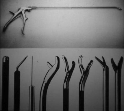
R O L E O F T E M I N M I N I M A L L Y I N V A S I V E S U R G E R Y |
77 |
.............................................................................................................................................................................. |
|
secured to long arm-boards placed under the mattress. When the foot of the table is dropped, the surgeon has unhindered access to the patient’s perineum and can sit immediately next to the patient. For a posteriorly located lesion, the patient is placed in the lithotomy position, and if the lesion is laterally located, the patient is placed in the appropriate lateral decubitus position with the hips flexed at 90◦. A straight, non-beveled rectoscope is also available and may be preferable for distal lesions. The surgeon’s end of the rectoscope is covered with a sealed locking-facepiece which has air-tight, sealed working ports through which are inserted the long-shafted instruments necessary for the dissection (Fig. 6.1). The suction catheter can be electrified; in this way a bleeding vessel can be coagulated while blood is simultaneously aspirated. Tissue graspers can also be electrified for control of a bleeding vessel. Vision is obtained either through a binocular stereoscopic eyepiece or an accessory scope which can enable video recording
Fig. 6.1 Long-shafted instruments needed for the dissection.

78 C H A P T E R 6
..............................................................................................................................................................................
Fig. 6.2 Completed TEM assembly.
and viewing by surgical assistants, students, and residents. The binocular eyepiece provides 6× magnification, has a 50◦ downward view and a 75◦ lateral field of view. In contrast, the accessory scope has a 40◦ downward view and a reduced lateral field of view. Because of this discrepancy, the image seen through the accessory scope and the video monitor is more narrow in its scope and it may appear as though the surgeon is going off the field. The complete assembly is shown in Fig. 6.2.
Preoperative consideration
A complete colonic evaluation with colonoscopy or air-contrast barium enema must be performed to rule out synchronous adenomas or cancer. For cancer, suitability for a transanal excision must be determined so that only those lesions with a low likelihood for nodal metastases are selected. In our practice, endorectal ultrasound is found to be excellent in providing accurate determination of depth of mucosal penetration; however, it is less so in detecting metastatic lymph nodes within the mesorectal fat. One may estimate the likelihood of nodal metastases based on the degree of mural penetration with which there is a direct correlation. For example, lesions which
R O L E O F T E M I N M I N I M A L L Y I N V A S I V E S U R G E R Y |
79 |
.............................................................................................................................................................................. |
|
are confined to the mucosa and submucosa metastasize to regional nodes in less than 5% of cases. For lesions which have penetrated partially into the muscularis propria, the incidence of nodal metastases is approximately 30%, and if the lesion has penetrated into the fat, the incidence of nodal metastases is 50%. The risk of spread to lymph nodes is also influenced by degree of tumor differentiation (poor differentiation confers an increased risk) and lymphatic invasion within the tumor itself [10]. Certainly if suspicious nodes are seen by ultrasound, this overrides the risk assessment based on degree of tumor penetration. However, in the absence of identifiable nodes, tumor penetration has clear significance.
Magnetic resonance imaging (MRI) is more widely used in Europe than transanal ultrasound as a means of staging rectal cancers. In a study comparing these two imaging modalities, Bianchi et al. examined 49 consecutive patients with a Pentax FG36UX ultrasound scanner. The first 28 patients were then studied using a linear body coil supplied with the MRI machine, and the next 21 patients were examined with a multichannel system of four coils. Patients who received preoperative radiation and chemotherapy were excluded from the study. Radical surgery was performed a mean 7.5 days from the time of staging, operative specimens were examined, and 931 lymph nodes were retrieved. Accuracies of T-staging were 70% for ultrasound, 43% for body coil MRI, and 71% for phased-array coil MRI (PA-MRI). No significant difference was noted between the techniques. The accuracies of N-staging were 63% for ultrasound, 64% for body-coil MRI, and 76% for PA-MRI. These differences were not significantly different [11].
Kim et al. studied 89 patients with ultrasound (in all), pelvic computed tomography (in 69), and MRI with an endorectal coil (in 73). Results were compared with histopathologic staging. For depth of invasion, accuracies were 81% for ultrasound, 65% for computed tomography (CT), and 81% for MRI. For staging regional lymph nodes, accuracies were 64% for ultrasound, 57% for CT, and 63% for MRI. The authors concluded that ultrasound and magnetic resonance had similar accuracies and were superior to conventional computed tomograms [12].
If a candidate for transanal excision is identified and the lesion is accessible with rigid proctoscopy, no further workup is needed. A CT scan is unnecessary and is not superior to ultrasound in determining the extent of local disease. Furthermore, the risk of distant disease within the liver or lungs is sufficiently low for superficial lesions that CT scans are not required. In contrast, for locally advanced cancers, that is, those that are large, fixed, deeply penetrating, or have already spread to nodes, CT scans have a useful
80 C H A P T E R 6
..............................................................................................................................................................................
role; that is, if unresectable disease within the liver or lungs is identified, radical surgery for the primary tumor may not be justified.
In preparation for TEM, a bowel cleansing with lavage solutions or cathartics is needed to ensure visibility and to lower the risk of infection if penetration into the peritoneal cavity should inadvertently occur. Oral, non-absorbable antibiotics may be given and intravenous antibiotics may be added as well. Informed consent should include possible laparotomy for lesions that pose a risk of entry into the peritoneum, that is, anterior tumors in women where the location of the anterior peritoneal reflection is unpredictable and may be quite low.
We perform TEM usually under general anesthesia; however, spinal or epidural anesthesia may be considered if the patient cannot tolerate general anesthesia. Patients are released from the hospital within 3–4 h following completion of the procedure. Proper consideration to coexisting morbid conditions that place the patient at higher risk for complications should be addressed and the appropriate precautions taken. Included are an assessment of cardiac, pulmonary, and deep venous thrombosis risk.
Technique
The patient is positioned according to the location of the tumor within the rectum. The buttocks and perineum are then washed with antiseptic solution, sterile drapes are placed, and the rectoscope is inserted up to the lesion under direct vision with manual insufflation of air. The scope is then secured to the operating room table and the facepiece is locked onto the end of the scope. Rubber sleeves and caps are placed onto the working ports on the facepiece. The long-shafted instruments are inserted and the tubing necessary for CO2 insufflation, saline irrigation, and pressure monitoring are connected. The binocular eyepiece and the accessory scope are inserted.
Dissection begins provided the bowel cleansing has been adequate, the entire lesion is visible and accessible, and intraluminal pressure can be maintained without signs of an air leak. If the entire lesion is not initially visible, one can still proceed with TEM provided it can be pulled down into view. The technique of excision will vary according to preoperative histology, suspicion that a “benign” lesion may harbor an occult cancer, and the location of the lesion within the rectum. Small adenomas may be removed by dissecting within the submucosal plane. This is especially appropriate for an anterior lesion in a woman where the anterior peritoneal reflection is unpredictable in
R O L E O F T E M I N M I N I M A L L Y I N V A S I V E S U R G E R Y |
81 |
.............................................................................................................................................................................. |
|
its location and a full thickness excision may be hazardous. A 5 mm margin of normal appearing mucosa is marked around the lesion, the mucosal edge lifted up with the tissue grasper, and the lesion excised without entering the muscularis propria. Larger adenomas may contain cancer; therefore such lesions are excised with a full-thickness technique facilitating assessment of depth of penetration if cancer is present. If the peritoneum is violated, it should be promptly repaired. This does not mandate immediate conversion to laparotomy; in fact, in the instances of peritoneal entry in our experience, the peritoneum was transanally repaired as a separate layer and TEM completed as planned. In one of the patients, intra-abdominal escape of CO2 caused a significant pneumoperitoneum which required needle decompression prior to extubation. Cancers are removed with a full thickness excision after a 1 cm margin of normal appearing mucosa has been marked around the lesion. The extrarectal fat is easy to identify and serves as a landmark to signify transmural penetration. Wounds are closed transversely with a running suture of long-lasting absorbable monofilament material with a tapered gastrointestinal needle (Fig. 6.3). After the lesion is excised, it should be fixed onto a cork board or Telfa paper to facilitate histologic examination of the deep and lateral margins.
Fig. 6.3 Closure of the wound following TEM excision: surgeon’s view.
82 C H A P T E R 6
..............................................................................................................................................................................
The following are technical pearls:
1 Short sutures are preferable. If the suture is too long, it is cumbersome and it may be impossible to pull the stitch taut within the confines of the scope. If the wound is large, closure requires several sutures.
2 Cross over of instruments should be avoided; the surgeon should manipulate them in parallel.
3 The needle should be passed from instrument to instrument. If the needle is dropped, it may retract out of the operative field and time may be wasted in frustrating attempts at needle retrieval.
4 Lesions located in the distal 5 cm of the rectum may be better addressed with conventional instruments. If the lower lip of the beveled scope falls out of the anus, CO2 will be lost and the operative field will collapse. Furthermore, one may encounter more bleeding just above the anus because of the hemorrhoidal veins. Alternatively, one can use the straight-ended scope that is not beveled at the end.
5 The most important aspect of the operation is patient positioning. The tumor should be located in the anterior–posterior plane during the office exam. The patient is then positioned accordingly so that the beveled end of the scope faces down at the lesion.
6 If there is an air leak, the operative field collapses and visibility is hampered. All tubing connections must be re-secured and the rubber tube and caps which seal the ports must be checked for cracks or tears which may occur secondary to normal wear and tear. The locking mechanisms which secure the scopes (accessory and main) and facepiece must be re-tightened. An adequate supply of CO2 with unrestricted flow should be verified if the rectum remains collapsed.
7 The operating rectoscope must be repositioned several times during the course of the dissection in order to keep the lesion in the center of the operative field.
Results
Following TEM, digital rectal examination and rigid proctosigmoidoscopy are performed at 3, 6, and 9 months. A follow-up colonoscopy should be performed at 1 year. For cancers, rectal ultrasound may be combined with proctosigmoidoscopy; however, caution should be exercised in interpreting the results. Tissue plans will be altered as a result of the excision. Consequently, no definitive decision should be made based upon the first examination. With serial ultrasound examinations, however, one can find
R O L E O F T E M I N M I N I M A L L Y I N V A S I V E S U R G E R Y |
83 |
.............................................................................................................................................................................. |
|
enlarging hypoechoic nodules either within the rectal wall or the rectal |
|
mesentery suggesting tumor recurrence. Such lesions can be biopsied using |
|
ultrasound-guided needle techniques. |
|
Complications are relatively infrequent and include entry into the peri- |
|
toneal cavity, conversion to laparotomy, bleeding, fecal soilage, fever, |
|
wound dehiscence, urinary dysfunction, and fistulas. In the author’s expe- |
|
rience, soilage has occurred in only 2% of patients and has usually been of |
|
short duration. |
|
Gerhard Buess has published extensively on his experience [4–6]. Of his |
|
first 186 TEMs, 137 were performed for adenomas and 49 for cancers. With |
|
respect to location in the rectum, 25, 47, and 18% of adenomas were located |
|
in the distal, mid, and upper rectum, respectively. Five percent recurred |
|
and were treated with endoscopic snare or cautery in the majority. Of the |
|
cancers, 18, 45, and 31% were located in the distal, mid, and upper rectum |
|
respectively. Most of the cancers (81%) were staged as pT1; recurrence |
|
was 4%. |
|
Some of the complications noted after TEM excision of both adenomas |
|
and cancers (n = 186) are listed below [6]: |
|
1 Bleeding (4) |
|
2 Rectovaginal fistula (2) |
|
3 Rectal stenosis (7) |
|
4 Peritoneal entry (1) |
|
5 Bladder dysfunction (5) |
|
6 Incontinence (1) |
|
At the 1995 annual meeting of the American Society of Colon and Rectal |
|
Surgeons, the centers in the United States presented their pooled initial |
|
data [7]. Of 153 TEM procedures, eight were converted to laparotomy |
|
because the lesion was not accessible in its entirety and in one case an inad- |
|
vertent entry into the peritoneum necessitated a laparotomy. Approximately |
|
half of the lesions were considered beyond reach of conventional instru- |
|
ments. Of the 82 adenomas, mean size was 4 cm. Recurrence rate following |
|
TEM was 11%, most were treated with cautery or snare excision, and repeat |
|
TEM was rarely required. Of the 54 cancers, mean size was 3 cm; there were |
|
30 T1, 15 T2, and 6 T3 lesions. Recurrence rate following TEM increased |
|
in relation to tumor depth: 10% for T1, 40% for T2, and 66% for T3 |
|
cancers. Overall complications were experienced infrequently; only three |
|
patients reported mild incontinence which was temporary in two. |
|
TEM has been utilized at Rush University Medical Center since 1991. |
|
From 1991 to June 2005, 260 cases have been performed; average patient |
|
84 C H A P T E R 6
..............................................................................................................................................................................
age was 65.7 years and the average distance from the dentate line was 7.4 cm. The mean estimated blood loss was approximately 24 cc and in only 12% of cases did the estimated blood loss exceed 50 cc. The mean operative time was 75 min; 17% of cases lasted less than 30 min and only 9% of cases lasted longer than 2 h. During this time period, 112 adenomas and 135 cancers were removed; the remainder consisted of gastrointestinal stroma tumors and carcinoids. For the adenomas, 65% were above 6 cm from the anus (21% above 10 cm), 57% were 2–4 cm in size (22% were >4 cm), and 93% took less than 2 h to perform (39% took <1 h). For the cancers, 62% were above 6 cm from the anus (23% above 10 cm), 52% were 2–4 cm in size (24% were >4 cm), and 86% took less than 2 h (38% took <1 h). Of the cancers, 26% were carcinoma in-situ, 47% were pT1, 23% pT2, and 4% were pT3.
The number of new rectal cancers treated by all means at Rush over the last 10 years has remained fairly stable at 30–45 cases per year and the use of TEM for cancer has remained constant as well. This reflects our policy of strict patient selection when using TEM for cure. We have used TEM to treat 53 patients with pathologic T1 cancers. Forty-six percent were located 5–10 cm from the anus, and 25% were greater than 10 cm. Forty-four percent of the cancers were between 2 and 4 cm in maximum diameter. Thirteen percent were larger than 4 cm. With a mean follow-up of 2.84 years, the recurrence rate has been 7.5%, occurring at 9 months, 15 months, 16 months, and 11 years. Of the recurrences, three were salvaged with radical surgery, and one was treated with simple fulguration because of advanced age. During this period of follow-up, there have been no cancer-related deaths and we attribute this in part to our close follow-up program as outlined above.
Registry and prospective series have shown similar morbidity rates compared with traditional local excision, comparable short lengths of stay, and low local recurrence rates [7,13,14]. Sengupta et al. [15] performed a metaanalysis that demonstrated recurrence rates from 4.2 to 25% for lesions excised by TEM. For pT1 lesions, recurrence rates have been reported from 0 to 12.5% (Table 6.1).
Comparison of TEM with radical surgery
A retrospective comparison of TEM and radical surgery was performed by Lee et al. [14]. In this study, neither group received adjuvant chemoradiation and there were no significant differences in age, gender, tumor location,

R O L E O F T E M I N M I N I M A L L Y I N V A S I V E S U R G E R Y |
85 |
.............................................................................................................................................................................. |
|
Table 6.1 Oncologic results for pT1 lesions excised with TEM.
|
|
Local recurrence |
5-year disease-free |
5-year survival |
References |
N |
% |
% |
% |
|
|
|
|
|
Lee, W [14] |
52 |
4.1 |
95.9 |
100 |
Buess, G [4] |
12 |
0 |
|
|
Langer, C [16] |
16 |
12.5 |
|
|
Winde, G [17] |
24 |
4.2 |
|
96.0 |
Demartines, N [18] |
9 |
8.3 |
|
|
Buess, G [6] |
25 |
4.0 |
|
|
Smith, L [7] |
30 |
10 |
|
|
Saclarides, TJ [13] |
53 |
7.5 |
100 |
|
|
|
|
|
|
or follow-up period between the two groups. Fifty-two patients had pT1 cancers removed by TEM, local recurrence was 4.1%, and 5-year diseasefree survival was 95.9%. Neither was statistically different than the results noted in the group undergoing radical surgery. Twenty-two patients had pT2 cancers removed by TEM, local recurrence was 19.5%, and 5-year disease-free survival was 80.5%. This local recurrence rate was higher than that noted in the radical surgery group (9.4%, p = 0.04); however, 5-year disease-free survival was not statistically different.
Winde et al. [17] prospectively randomized 52 patients with ultrasound stage uT1N0 rectal cancers into TEM or anterior resection groups. Patient and tumor demographics were similar. Local recurrence rates (4.2%) and 5-year survival rates (96%) did not differ significantly. Early postoperative mortality was zero. Significant differences were noted comparing time of hospitalization (10 days longer for anterior resection, p < 0.0001), blood loss (p < 0.001), operative time (45 min longer for anterior resection, p = 0.0021), and need for narcotic analgesia (3 times higher after anterior resection, p < 0.0001). Various early complications were noted in 21% of the TEM group and in 35% of the anterior resection group [17].
Conclusions
TEM is safe and there are few complications. Most patients can be treated on an outpatient basis. Virtually any adenoma can be removed with this technique, even large circumferential lesions that necessitate sleeve resections. Regarding cancers, strict selection criteria must be used to avoid compromising recurrence rates and survival. Transrectal ultrasound may help identify
86 C H A P T E R 6
..............................................................................................................................................................................
those lesions which are appropriate for TEM. If TEM is selected, close follow-up is necessary to detect cancer recurrences; if they occur, radical surgery is indicated. TEM is not superior to conventional instrumentation for removing lesions in the distal rectum; however, it has distinct advantages for excising lesions in the mid and upper rectum.
Because of the high cost of the equipment, the relative infrequency with which it is used, and the difficulty in mastering the technique, TEM will probably become the domain of only a few surgeons. Over the last several years at our institution, we have performed 25–30 cases each year (benign and malignant), and since its arrival in 1991, referrals for rectal cancer have increased steadily. However, in comparison to the portion of cancers treated with low anterior resection or APER, the relative percentage of cancers treated with TEM has remained stable. This reflects our policy of adhering to strict selection criteria. Having the technology at hand is not a license to use it inappropriately.
Local (transanal) excision of rectal tumors has been part of the surgeon’s armamentarium for over 100 years. All agree that benign disease can be safely addressed with this approach. However, debate has recently surfaced that cancers are best approached by more radical means. It is unlikely that randomized studies will ever be done comparing TEM with conventional transanal surgery. Comparison with radical surgery has been done in both a retrospective and prospective fashion, and the results suggest that TEM is a safe and effective means of treating pT1 cancers. Winde et al. [17] prospectively found operating time, blood loss, length of hospital stay, and analgesia requirements following TEM to be significantly less than for anterior resection. Local recurrence rates for pT1 tumors following APER are low (0 to 10%) with a 0% local recurrence rate reported in some series [19,20]. Overall 5-year survival rates for pT1 rectal cancers radically excised range from 78 to 100%. Local recurrence rates following conventional transanal excision without chemoradiation range from 0 to 33% for pT1 disease [15,21]. In our hands, local recurrence rate of pT1 lesions excised by TEM is 7.5%, and thus far we have not had any cancer-related deaths. In retrospect, the initial size and distance of pT1 lesions that did recur did not preclude safe removal by TEM, and it is unlikely that technical aspects played a role in these recurrences apart from one instance with a positive margin (patient refused further treatment).
In summary, therefore, TEM has emerged as another means of transanal excision of adenomas and selected cancers. These selection criteria have been embraced for decades, and if one assumes that they are appropriate for
R O L E O F T E M I N M I N I M A L L Y I N V A S I V E S U R G E R Y |
87 |
.............................................................................................................................................................................. |
|
distal lesions, then logic demands that these same criteria are appropriate for cancers in the mid and upper rectum as well. TEM, or any form of local excision, is not recommended as the sole form of therapy for pT2 cancers unless one is participating in a clinical trial investigating the role of chemotherapy and radiation either before or after excision. For pT1 cancers, a close surveillance program is essential in order to detect many of those which can be salvaged with radical surgery.
References
1Chorost MI, Petrelli NJ, McKenna M
et al. Local excision of rectal cancer. Am Surgeon 2001; 67: 774–9.
2Mellgren A, Sirivongs P,
Rothenberger DA et al. Is local excision adequate therapy for early rectal cancer? Dis Colon Rectum 2000; 43: 1064–74.
3 Graham RA, Garnsey L, Jessup JM. Local excision of rectal carcinoma. Am J Surg 1990; 160: 306–12.
4Buess G, Kipfmuller K, Ibald R et al. Clinical results of transanal endoscopic microsurgery. Surg Endosc 1988;
2: 245–50.
5Mentges B, Buess G. Transanal endoscopic microsurgery in the treatment
of rectal tumors. Perspect Colon Rectal Surg 1991; 4: 265–79.
6Buess G, Mentges B, Manncke K et al. Technique and results of transanal endoscopic microsurgery in early rectal
cancer. Am J Surg 1992; 163: 63–70.
7Smith LE, Ko ST, Saclarides T et al. Transanal endoscopic microsurgery: initial registry results. Dis Colon Rectum
1996; 39: 579–84.
8Taylor RH, Hay JH, Larsson SN. Transanal local excision of selected low
rectal cancers. Am J Surg 1998; 175: 360–3.
9Bleday R, Breen E, Jessup M et al. Prospective evaluation of local excision for small rectal cancers. Dis Colon
Rectum 1997; 40: 338–92.
10Saclarides TJ, Bhattacharyya AK, Britton-Kuzel C et al. Predicting lymph node metastases in rectal cancer. Dis Colon Rectum 1994; 37: 52–7.
11Bianchi PP, Ceriani C, Rottoli M et al. Endoscopic ultrasonography and magnetic resonance in preoperative staging of rectal cancer: comparison with histologic findings. J Gastrointest Surg 2005; 9: 1222–8.
12Kim NK, Kim MJ, Yun SH et al. Comparative study of transrectal ultrasonography, pelvic computerized tomography, and magnetic resonance imaging in preoperative staging of rectal cancer. Dis Colon Rectum 1999;
42: 770–5.
13Saclarides, TJ. Transanal endoscopic microsurgery: a single surgeon’s experience. Arch Surg 1998; 133: 595–9.
14Lee W, Lee D, Choi S et al. Transanal endoscopic microsurgery and radical surgery for T1 and T2 rectal cancer. Surg Endosc 2003; 17: 1283–7.
15Sengupta S, Tjandra JJ. Local excision of rectal cancer: what is the evidence? Dis Colon Rectum 2001; 44: 1345–61.
16Langer C, Markus P, Liersch T et al. Ultracision or high frequency knife in transanal endoscopic microsurgery (TEM)? Advantages of a new procedure. Surg Endosc 2001; 15: 513–17.
17Winde G, Nottberg H, Keller R et al. Surgical cure for early rectal carcinomas (T1): transanal endoscopic microsurgery vs anterior resection. Dis Colon Rectum 1996; 39: 969–76.
18Demartines N, vonFlue MO, Harder FH. Transanal endoscopic microsurgical excision of rectal tumors: indications and results. World J Surg 2001; 25: 870–5.
19Willet CG, Lewandrowski K, Donnelly S et al. Are there patients with stage I rectal
88 C H A P T E R 6
..............................................................................................................................................................................
carcinoma at risk for failure after abdominoperineal resection? Cancer 1992; 69: 1651–5.
20Sticca RP, Rodriguez-Bigas M, Penetrante RB et al. Curative resection for stage I rectal cancer: natural history,
prognostic factors, and recurrence patterns. Cancer Invest 1996; 14: 491–7.
21Wagman RT, Minsky BD. Conservative management of rectal cancer with local excision and adjuvant therapy. Oncology 2001; 15: 513–19.