

Challenges in Colorectal Cancer, Second Edition
Edited by John H. Scholefield etc. Copyright © 2006 by Blackwell Publishing Ltd
.............................................................................................................................................................................
7: What is the best strategy for the management of hereditary colorectal cancer?
Seung-Yong Jeong, David Chessin, Susan Ritchie, John H. Scholefield, and José G. Guillem
.............................................................................................................................................................................
Introduction
Five to ten percent of all cases of colorectal cancer (CRC) are believed to have a hereditary component [1]. Because hereditary CRCs are due to germline mutations, these patients have clinical features distinct from sporadic CRC. Generally, these features include (1) early age-of-onset of cancer,
(2)frequent association with synchronous or metachronous tumors, and
(3)characteristic extraintestinal manifestations. Due to these differences, the management strategy for patients with hereditary CRC is quite different from that for sporadic CRC. Additionally, there are important screening and surveillance implications for family members. Our aim is to review the most common hereditary CRC syndromes, namely familial adenomatous polyposis (FAP) syndrome, hereditary nonpolyposis colorectal cancer (HNPCC) syndrome, Peutz-Jeghers syndrome (PJS), juvenile polyposis syndrome (JPS), and MYH polyposis syndrome, with an emphasis on management strategies (Table 7.1).
Familial adenomatous polyposis
Familial adenomatous polyposis is an autosomal dominant hereditary CRC syndrome caused by a germline mutation of the APC gene [1]. FAP occurs in one of 10,000 live births and accounts for 1% of all CRC [2,3]. The majority of patients have a family history of FAP, but 20–30% of cases arise from a de novo APC mutation [4].
Clinical features
The FAP is characterized by at least 100 adenomatous polyps in the colon and rectum, but an attenuated form with fewer polyps has been
89
Table 7.1 |
Hereditary CRC syndromes. |
|
|
|
|
|
|
|
|
|
|
|
|
|
|
|
|
|
|
|
|
|
Frequency |
Polyp |
|
|
CRC |
|
|
|
|
|
|
|
|
|
|
|
|
|
Inheritance |
Causative gene |
(% of total CRC) |
Location |
Prevalence |
Number |
Risk |
Mean age |
|
|
|
|
|
|
|
|
|
|
|
FAP |
AD |
APC |
1 |
Colorectum |
100% |
>1000 |
100% |
39 |
|
HNPCC |
AD |
hMLH1, hMSH2, |
2–6 |
Colorectum |
20–40% |
1–10 |
80% |
45 |
|
|
|
hPMS1, hPMS2, |
|
|
|
|
|
|
|
|
|
hMSH6 |
|
|
|
|
|
|
|
PJS |
AD |
STK11 |
0.1 |
Small intestine |
>90% |
10–100 |
20% |
46 |
|
JPS |
AD |
SMAD4, BMPR1A |
0.1 |
Colorectum |
>90% |
3–200 |
10–38% |
34 |
|
MYH |
AR |
MYH |
1–3 (?) |
Colorectum |
? |
3–100 |
? |
50 |
|
polyposis |
|
|
|
|
|
|
|
|
|
|
|
|
|
|
|
|
|
|
|
FAP, familial adenomatous polyposis; HNPCC, hereditary non-polyposis colorectal cancer; PJS, Peutz-Jeghers syndrome; JPS, juvenile polyposis syndrome; AD, autosomal dominant; AR, autosomal recessive; CRC, colorectal cancer;? denotes that the values are still not clearly defined.
7 R E T P A H C 90
..............................................................................................................................................................................
M A N A G E M E N T O F H E R E D I T A R Y C R C |
91 |
.............................................................................................................................................................................. |
|
described [5,6]. Attenuated FAP (AFAP) is a variant characterized by fewer colorectal polyps (<5–100), later age of onset of polyps and cancer, infrequent rectal involvement, and a more proximal colonic distribution than classic FAP [5,6].
Colorectal cancer develops in nearly all FAP individuals with their colon and rectum in situ by age 40–50 [1]. In addition to colorectal polyps and CRC, patients with FAP also develop characteristic extracolonic manifestations. These include benign lesions of the stomach (adenoma and fundic gland retention polyps), small bowel (adenoma), skin (lipoma, fibroma, sebaceous, and epidermoid cysts), bone (osteoma), retina (congenital hypertrophy of the retinal pigment epithelium), teeth (supernumerary teeth), and soft tissue (desmoid) [7]. Extracolonic malignancies also develop at increased incidence in patients with FAP. These include cancers of the liver (hepatoblastoma), stomach, duodenum, pancreas, thyroid, biliary tract, and brain [8–11]. As increasing numbers of patients with FAP have been treated with prophylactic proctocolectomy, mortality from CRC has decreased. However, patients with FAP continue to have a reduced life expectancy after colectomy, largely due to mortality from desmoid tumors or upper gastrointestinal (GI) cancers [12].
Genetics and genetic testing
The FAP is caused by germline mutation of the APC gene on chromosome 5q21 [1]. APC is a tumor suppressor gene that inhibits the Wnt signaling pathway [13]. The key component in this pathway is β-catenin, which activates the transcription of growth-regulatory genes. The APC gene product targets β-catenin for degradation. However, when APC is mutated excess β-catenin is translocated to the nucleus, with resultant decreased regulation of cell growth [13].
Currently, several tests are available to test for an APC germline mutation, including sequencing of the entire gene, a combination of conformation strand gel electrophoresis (CSGE) and protein truncation test (PTT), PTT alone, and linkage analysis. Sequencing of the entire gene is the most sensitive test (95% mutation detection rate), but it is the most expensive. The mutation detection rate of the combination of CSGE and PTT, and PTT alone is 80–90%, and 70–80%, respectively. Linkage analysis can be performed when more than one affected family member is available and has a 95–99% accuracy [14]. As with all genetic testing, preand post-test counseling is strongly recommended [6].
92 C H A P T E R 7
..............................................................................................................................................................................
Attenuated FAP is also due to a germline mutation in the APC gene that is inherited in an autosomal dominant fashion. However, it has been suggested that the mutated locus in AFAP is further toward the 5 and 3 ends of the APC gene than that reported for classic FAP [6].
Screening and surveillance
At-risk individuals should begin screening for FAP at age 10–12 [14]. The screening tests available for FAP include genetic testing for a mutation of the APC gene and endoscopy. Flexible sigmoidoscopy has been regarded as the procedure of choice for FAP screening [14]. However, it may be replaced by a genetic test when an APC mutation has been documented in an individual’s family. In addition, colonoscopy may be used when screening is performed at an older age or when there is suspicion for AFAP [14].
If an individual has multiple polyps or a positive genetic test, referral of all first-degree relatives for genetic counseling is recommended. Patients with FAP should be followed by annual colonoscopic examinations until the time of colectomy. In addition, upper GI endoscopy should be performed beginning at age 21 and repeated at 6-month intervals or longer depending on the polyp burden [15]. Screening guidelines for the other extracolonic cancers associated with FAP are less well defined.
Management
Because 5% of untreated patients with FAP develop colorectal cancer by the age of 20 years [16], prophylactic surgery is advised shortly after FAP is diagnosed. However, the timing and type of prophylactic surgery in patients younger than age 20 are often influenced by educational, developmental, and self-image concerns. When polyps are small (<6 mm) and there is no evidence of dysplasia, cancer, or symptoms, prophylactic surgery may be delayed until after high school [16].
The optimal surgical management of FAP should include (1) removal of all at-risk colorectal mucosa, and (2) maintenance of anorectal function. Current surgical options for FAP include total proctocolectomy (TPC) with an ileal pouch-anal anastomosis (IPAA), total colectomy and ileorectal anastomosis (IRA), and TPC with ileostomy.
Total proctocolectomy with IPAA is the operation of choice for most patients with FAP, as it nearly eliminates the risk of CRC and maintains peranal fecal evacuation. However, in a selected subset of individuals, a total
M A N A G E M E N T O F H E R E D I T A R Y C R C |
93 |
.............................................................................................................................................................................. |
|
colectomy with IRA and close endoscopic surveillance is an option, as IRA is associated with better functional results and lower perioperative morbidity [16]. Patients unable or unwilling to have aggressive postoperative surveillance are not suitable for IRA. TPC with permanent ileostomy results in a permanent stoma. However, it may be necessary when patients present with a distal rectal cancer, where a sphincter-preserving procedure may compromise oncologic results.
Patients with AFAP are also at increased risk for the development of CRC. However, given their lower polyp burden and potential for rectal sparing, management may be tailored to the individual. In very selected patients with very few adenomas, colonoscopic polypectomy and subsequent close endoscopic surveillance may be acceptable in patients not willing to undergo a prophylactic colectomy. When resection is required, a total colectomy with IRA may be appropriate in patients with AFAP who have rectal sparing [6].
Hereditary non-polyposis colorectal cancer
Hereditary non-polyposis colorectal cancer is an autosomal dominant hereditary CRC syndrome with an 80% penetrance, caused by mutations in one of several DNA mismatch-repair (MMR) genes [1]. It is the most common hereditary CRC syndrome and accounts for 2–6% of all CRC.
Clinical features
Hereditary non-polyposis colorectal cancer is characterized by early age-of- onset cancers in multiple family members. CRC is the most common tumor in HNPCC patients, but extracolonic cancers of the endometrium, small bowel, urinary tract, stomach, biliary tract, ovary, pancreas, and brain also can occur. CRC in HNPCC differs from sporadic CRC in that there is an increased incidence of synchronous and/or metachronous tumors and a predilection for the proximal colon [1]. CRC in HNPCC more frequently has mucinous or poorly differentiated histology and peritumoral lymphocytic infiltration [17]. In addition, patients with HNPCC have improved survival when compared to patients with sporadic CRC of similar stage [18]. Adenomas in HNPCC differ from sporadic adenomas in that there is an increased incidence of villous architecture, high-grade dysplasia, and morphologically flat lesions [19,20]. In addition, malignant transformation in adenomas in patients with HNPCC may be more rapid than sporadic adenomas [21]. In 1990, the International Collaborative Group on HNPCC

94 C H A P T E R 7
..............................................................................................................................................................................
Table 7.2 Revised Amsterdam criteria for diagnosis of HNPCC.
There are at least three relatives diagnosed with an HNPCC-associated cancer (colorectal, endometrial, small bowel, ureter, or renal pelvis but not including stomach, ovary, brain, bladder, or skin)
One affected person is a first-degree relative of the other two At least two successive generations are affected
At least one person was diagnosed before the age of 50 years Familial adenomatous polyposis has been excluded
Tumors have been verified by pathologic examination
Source: Vasen HF, Watson P, Mecklin JP, Lynch HT. Gastroenterology 1999; 116: 1453–6.
developed the Amsterdam criteria (revised in 1999), which defines HNPCC by clinical findings and family history (Table 7.2) [22].
Genetics and genetic testing
Hereditary non-polyposis colorectal cancer is caused by a germline mutation in DNA MMR genes, which play a critical role in repairing mismatched nucleotides pairs during DNA replication [1]. The majority (more than 95%) of germline mutations have been found in the hMLH1 and hMSH2 genes, whereas mutations in hMSH6, hPMS1, and hPMS2 have been reported, but are rare [23,24].
Tumors due to MMR gene mutations exhibit a unique molecular phenotype termed microsatellite instability (MSI), which is characterized by accumulation of single nucleotide mutations and alterations in the length of simple repetitive sequences found throughout the genome. Over 90% of tumors in HNPCC exhibit MSI [25].
Initial screening for HNPCC may be performed with MSI testing or immunohistochemistry (IHC) for MMR proteins. MSI is identified using DNA extracted from tumor tissue. In 1997, the National Cancer Institute Workshop on HNPCC recommended a panel of five microsatellite markers for determination of MSI [26]. The tumor is defined as MSI-high when two or more markers display instability.
Selection of patients for testing for HNPCC is a major clinical issue. To date, the most well-accepted guidelines for MSI testing are the Bethesda Guidelines, which were developed in 1996 and revised in 2002 (Table 7.3) [27]. IHC for MMR proteins may also be used to screen for HNPCC. It has lower sensitivity than MSI testing, especially for inherited hMLH1 mutations [28]. However, it has distinct advantages over MSI testing in

M A N A G E M E N T O F H E R E D I T A R Y C R C |
95 |
.............................................................................................................................................................................. |
|
Table 7.3 Revised Bethesda guidelines for testing of colorectal tumors for MSI.
Colorectal tumors should be tested for MSI in the following situations: 1 CRC diagnosed in a patient less than 50 years of age
2 Presence of synchronous, metachronous, or other HNPCC-associated tumors, regardless of age
3 CRC with MSI-H histology in a patient less than 60 years of age
4 CRC diagnosed in one or more first-degree relatives with an HNPCC-related tumor, with one of the cancers diagnosed before 50 years of age
5 CRC diagnosed in two or more firstor second-degree relatives with HNPCC-related tumors,† regardless of age
Includes tumors of the colorectum, endometrium, stomach, ovary, pancreas, ureter, renal pelvis, biliary tract, brain, sebaceous glands, and small bowel.
† Includes presence of tumor infiltrating lymphocytes, Crohns-like lymphocytic reaction, mucinous histology, signet ring cell differentiation, or medullary growth pattern. Source: Umar A, Boland CR, Terdiman JP. J Natl Cancer Inst 2004; 96: 261–8.
terms of simplicity and availability. If it is used to complement MSI testing, IHC may reduce the cases to be tested by MSI and simplify the subsequent steps for mutation analysis. This approach may further decrease the cost for analysis and turnaround time. An algorithm for diagnosing HNPCC is provided in Fig. 7.1.
Management
HNPCC with verified germline mutation
Total colectomy with IRA may be offered to patients with a verified germline mutation in a MMR gene, due to the high risk of metachronous CRC [29]. Following total colectomy, close surveillance of the rectal remnant is required [30]. TPC with IPAA may be an option for patients who present with rectal cancer and sparing of the sphincter complex. In the highly unusual case of sphincter involvement with rectal cancer and synchronous colon polyps, a TPC and end ileostomy may be indicated.
Management of a known MMR gene mutation carrier with adenomas but no CRC is controversial. Prophylactic colectomy should be considered in an individual with multiple, high-grade adenomas or when colonoscopic follow-up is not possible. In mutation carriers without any clinical features of HNPCC, there is an 80% lifetime risk of CRC, as well as other extracolonic tumors. Cancer surveillance, including colonoscopy and extracolonic screening, should be conducted in these patients. Currently,
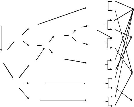
Indications
•Amsterdam II criteria
•Modified Bethesda criteria
Family mutation known
|
|
Normal |
Clinically |
|
expression |
affected family member |
|
|
|
Family |
IHC for |
|
mutation |
MMR proteins* |
|
unknown |
Loss of |
|
|
|
|
|
expression |
• Clinical assessment |
|
of one or more |
|
of MMR protein |
|
• Genetic counselling |
|
|
|
|
|
|
Family |
|
|
mutation |
|
|
unknown |
|
|
but with available |
|
|
affected member |
|
Clinically not |
Family |
|
affected, |
mutation |
|
but at-risk |
unknown |
|
family member |
and no available |
|
|
affected member |
|
Family mutation known
MSI-H
MSI testing
MSI-L or MSS
|
(+) |
Genetic testing for |
|
specific site |
(–) |
of MMR gene |
|
Germline |
(+) |
|
|
genetic testing |
|
for MMR genes† |
(–) |
|
|
|
(+) |
Very high index |
|
of suspicions for |
(–) |
HNPCC |
|
Genetic testing |
(+) |
|
|
for specific |
|
MMR gene |
|
|
(–) |
Consider |
(+) |
|
|
germline testing |
|
for MMR genes |
(–) |
|
|
Genetic test for |
(+) |
|
|
specific site |
|
of MMR gene |
(–) |
|
HNPCC
High risk for HNPCC
Sporadic case or low risk for HNPCC
Inconclusive
* Initially for MLH1 and MSH2 protein. If these are negative, then MSH6 and PMS2 can be pursued.
† Initially for hMLH1 and hMSH2 genes. If these are negative, then hPMS1, hPMS2, and hMSH6 can be pursued. When clinically indicated, additional mutational analysis, including searching for a large deletion or changing the method of mutation detection, can be added.
Fig. 7.1 Algorithm for identification of hereditary non-polyposis CRC.
7 R E T P A H C 96
..............................................................................................................................................................................
M A N A G E M E N T O F H E R E D I T A R Y C R C |
97 |
.............................................................................................................................................................................. |
|
some suggest prophylactic colectomy in patients with a MMR gene mutation, but no clinical features of HNPCC [30,31]. However this is controversial as the lifetime risk for developing CRC is 80% and not 100%.
Currently, there is insufficient evidence to definitively recommend prophylactic hysterectomy and oophorectomy in female patients with HNPCC. However, when women with HNPCC have surgery for CRC they may be offered these procedures, particularly if they are postmenopausal [30].
High risk for HNPCC
Patients at high risk for HNPCC include individuals with clinical suspicion of HNPCC who have (1) CRC with positive IHC or MSI testing but no verified germline mutation, and (2) CRC but negative IHC, MSI, and germline testing. Individuals with CRC and clinical suspicion for HNPCC should be managed as those with HNPCC, even when genetic testing is negative.
Non-informative genetic testings
Individuals in a HNPCC-kindred group who have negative genetic testing may be managed as the average risk population. Individuals in a HNPCCkindred group with non-informative (no mutation identified) genetic testing should be managed according to the guidelines for HNPCC [32]. However, in these cases, the clinician should be aware of the potential for laboratory errors.
Surveillance
Germline mutation carriers and patients with the clinical diagnosis of HNPCC should undergo colonoscopic screening at intervals of 1–2 years beginning at age 20–25 years, or 10 years earlier than the youngest CRC diagnosis in the family [1,33]. Since HNPCC patients are also at risk for extracolonic tumors, screening may include abdominal sonography and urine cytology every 1–2 years. Female gene carriers should undergo screening for endometrial and ovarian cancer, which may include annual gynecological exams, transvaginal sonography, endometrial aspiration, and serum CA-125 levels beginning at age 25–35 years.
Peutz-Jeghers syndrome (PJS)
Peutz-Jeghers syndrome is an autosomal dominant harmartomatous polyposis syndrome caused by a germline mutation in the STK11 (LKB1) gene,
98 C H A P T E R 7
..............................................................................................................................................................................
which encodes for a multifunctional serine–threonine kinase [34,35]. It occurs in 1 in 120,000–200,000 live births [36,37].
Clinical features
Peutz-Jeghers syndrome is characterized by GI hamartomatous polyps and melanin hyperpigmentation of the lips, buccal mucosa, and digits [38]. The polyps are located mainly in the small intestine but may be found in the stomach and colorectum. PJS polyps may cause abdominal pain, intussusception, bleeding, or anemia [38]. A clinical diagnosis of PJS can be made if ≥2 Peutz-Jeghers polyps are found in the GI tract or if one Peutz-Jeghers polyp is found in association with classic pigmentation or a family history of the syndrome [39]. PJS is associated with an increased risk of colorectal (20%), gastric (5%), small bowel, pancreas, breast, ovary, lung, cervix, uterus, and testis cancer [40].
Genetics and genetic testing
Peutz-Jeghers syndrome is caused by a germline mutation in the STK11 (LKB1) gene that is located on chromosome 19p13.3 [34,35], which has been documented in 18–63% of cases [41–43]. Genetic testing for PJS is performed by DNA sequencing of the STK11 gene. Individuals with PJS and their first-degree family members are candidates for genetic testing.
Management and surveillance
Currently, prophylactic colectomy is not recommended for patients with PJS. However, surveillance for CRC and extracolonic cancer is important. Most experts recommend colonoscopy at intervals of 2–3 years beginning during the late teenage years, with upper GI endoscopy and small-bowel radiography at intervals of 2–3 years and annual hemoglobin level beginning at age 10 [38]. Surveillance for pancreatic cancer with endoscopic or transabdominal ultrasonography, or CT scan may begin at 30 years of age and be repeated every 1–2 years. Mammography at intervals of 2–3 years, annual clinical breast and pelvic exam, annual PAP smear, and annual pelvic ultrasound may begin at age 20–25 years in females. Because male patients with PJS are at risk for testicular tumors, annual clinical testicular exam beginning at age 10 is recommended.
M A N A G E M E N T O F H E R E D I T A R Y C R C |
99 |
.............................................................................................................................................................................. |
|
Although most clinicians recommend endoscopic polypectomy when technically feasible, surgery is indicated in certain circumstances. Indications include symptoms (obstruction, intussusception, and bleeding) and large (≥15 mm) or adenomatous polyps that cannot be removed endoscopically [16]. During surgery, as much of the small bowel and colon as is feasible should be cleared of polyps, either by enterotomy or intraoperative endoscopic resection [38].
Juvenile polyposis syndrome (JPS)
Juvenile polyposis syndrome is an autosomal dominant GI hamartomatous polyposis syndrome associated with an increased risk of CRC and upper GI cancer. The incidence is one per 100,000 live births [44]. Juvenile polyps are defined histologically as hamartomas with dilated mucus-filled cysts and hyperplastic stroma [45].
Clinical features
Juvenile polyposis syndrome is diagnosed clinically using the following criteria: (1) more than 3–10 juvenile polyps in the colorectum, (2) juvenile polyps throughout the GI tract, or (3) juvenile polyps (any number) with a family history of JPS [46]. Polyps in JPS are most common in the colorectum but may be found throughout the GI tract. The number of juvenile polyps ranges from 50 to 200 [45], with symptoms usually associated with increasing polyp size. The most common symptom is chronic anemia, followed by acute GI bleeding, rectal prolapse of the polyp, protein-losing enteropathy, and intussusception with or without obstruction [47]. JPS is associated with a 10–38% lifetime risk of CRC and a 15–21% lifetime risk of gastric and duodenal cancer [48,49]. Malignant tumors appear to arise from adenomatous components present in some juvenile polyps and usually occur after the fourth decade [49].
Genetics and genetic testing
Juvenile polyposis syndrome has been associated with germline mutations of two genes; SMAD4 (DPC4) located on chromosome 18q21 and BMPR1A on chromosome 10q21–22 [50,51]. SMAD4 encodes for a protein which is an intracellular regulator of transforming growth factor (TGF)-β, and mediates growth inhibitory signals from the cell surface to the nucleus. BMPR1A
100 C H A P T E R 7
..............................................................................................................................................................................
mediates intracellular signaling through SMAD4 and is a member of the TGF-β superfamily. Genetic testing involves DNA sequencing for mutations in these two genes and detects the genetic etiology of approximately 35–50% of cases [52,53]. Candidates for genetic testing are individuals who meet the clinical criteria for JPS and first-degree relatives of individuals with a SMAD4 or BMPR1A mutation.
Management
Patients with GI bleeding, anemia, diarrhea, protein-losing enteropathy, or with high polyp burden may be treated surgically. Operative options include TPC with IPAA or total colectomy with IRA. Regardless of the surgery performed, patients require endoscopic surveillance due to the high rate of polyp formation in the remaining rectum or pouch [54]. In asymptomatic patients, surveillance colonoscopy with removal of all detectable polyps is recommended every 1–2 years [14]. First-degree relatives of patients with JPS should be screened with colonoscopy at 3-year intervals beginning at age 12 [32].
MYH polyposis
The MYH polyposis, first described in 2002, is an autosomal recessive colonic polyposis syndrome which is associated with biallelic mutations in the MYH gene [55]. It may account for up to 40% of patients with an FAP or AFAP phenotype in whom a germline mutation in the APC gene has not been detected [55–58].
Clinical features
The mean age of diagnosis of polyposis and cancer are 46 and 50 years, respectively [57,59]. Clinical features of MYH polyposis are similar to AFAP. However, MYH polyposis may be differentiated by its recessive pattern of inheritance.
Genetics and genetic testing
The MYH gene is involved in the base excision DNA repair process by removing adenine nucleotides misincorporated into DNA opposite guanine or oxoguanine [55]. Genetic testing for MYH polyposis is by sequencing
M A N A G E M E N T O F H E R E D I T A R Y C R C |
101 |
.............................................................................................................................................................................. |
|
and should be considered in patients with clinically suspected FAP or AFAP without a demonstrable germline APC mutation who have a family history compatible with recessive inheritance.
Management and surveillance
There are no established guidelines for management and surveillance of MYH polyposis. However, total colectomy with IRA may be considered in patients where germline mutations in both MYH alleles have been documented and in whom multiple adenomatous polyps have been detected. Endoscopic surveillance of the remaining rectum should be performed following IRA [44].
Average- (low) and moderate-risk groups
Those individuals at “average or low risk” for CRC (the majority of the population) are those with no family history of CRC. These individuals have a 1 in 35 lifetime risk of developing bowel cancer. Those with a weak family history – one first-degree relative (FDR) under 45 years or two over 70 years have a 1 in 17 lifetime risk of developing. Individuals with two first-degree relatives (e.g. 2 FDRs average age 50–60 years; see Table 7.4) may meet the criteria for the moderate-risk group. They have a lifetime risk of 1 in 12 or greater but do not fulfill the high-risk criteria.
There are differences in recommendations for screening in these groups between the United Kingdom and the United States, which will be discussed further.
The management of those individuals in families with an identified Cancer Predisposition Syndrome leading to a significantly high risk of colorectal cancer has been discussed in detail above. It is important to identify those at highest risk in order to allocate limited endoscopic resources appropriately. However, it has been estimated that in 20% or more of individuals with colorectal cancer, there may be a genetic component. Less than 5% of these will be attributable to the syndromes described so far.
In the vast majority of colorectal cancers (>95%), the genetic predisposition is much less clearcut. The majority are likely to result from mutations in frequent alleles of low penetrance, acting either alone or in combination. Included in this may be functional polymorphisms in genes responsible for DNA repair, the metabolism of carcinogens, or the anti-tumor immune response. The complex interaction of these within individuals and with

102 C H A P T E R 7
..............................................................................................................................................................................
Table 7.4 Inherited risk of CRC with screening recommendations based on risk assessment.
Risk group |
Family history |
Action |
|
|
|
|
|
Low risk |
1 |
FDR > 45 years |
No screening |
|
2 |
FDR > 70 years |
No screening |
Low to moderate risk |
2 |
FDR average 60–70 years |
Single colonoscopy at 55 years |
Moderate risk |
1 |
FDR < 45 years |
Colonoscopy every 5 years from |
|
|
|
5 years prior to age of index case |
|
2 |
FDR average 50–60 years |
As above + refer for genetic testing |
High to moderate risk |
2 |
FDR < 50 years |
Colonoscopy every 3–5 years |
|
|
|
beginning at 35 years + refer for |
|
|
|
genetic testing |
|
3 |
FDR (Amsterdam negative) |
As above + gyne screening |
High risk |
3 |
FDR (Amsterdam positive) |
Colonoscopy every 2–3 years |
|
|
|
beginning at 30 years |
|
FAP |
Annual sigmoidoscopy/genetic |
|
|
|
|
screening |
|
|
|
|
FDR – first-degree relative.
the environment is likely to lead to a widely variable phenotype both within and between families.
Determining the influence of such alleles at a population level presents a major challenge and makes individual risk-determination extremely complex. With the continuing new identification of more susceptibility alleles for different cancers provided by the human genome project, it is possible that in the future, using DNA chip technology, there may come a time when simultaneous assay for multiple susceptibility alleles may allow more individual, accurate risk-estimation and targeted-screening protocols.
At the present time, practice in the United Kingdom varies but most centers use empiric risk-data and varying guidelines in order to offer these individuals some level of surveillance. Recommendations for regular endoscopic screening as advocated in the United States are not feasible in the United Kingdom due to a lack of endoscopic manpower and facilities. Whether regular surveillance is necessarily required in the low-risk group is also debatable on a cost/benefit analysis.
Moderate-risk group
The moderate-risk group (Table 7.4) is significantly larger than the high-risk group and consists of those who have more than one affected relative (or one
M A N A G E M E N T O F H E R E D I T A R Y C R C |
103 |
.............................................................................................................................................................................. |
|
under 45 years of age) but do not fulfill the Amsterdam criteria (Table 7.1). The risk assessment and subsequent recommendation for screening depends on the number of affected relatives, how closely related they are, and the age of onset (Table 7.2).
Application of these criteria depends on a detailed family history but this may often be uncertain or incomplete. It is good practice to obtain consent and request the records of living relatives or obtain confirmation from the relevant Cancer Registry about those who are deceased. Many centers see this as an essential part of the process because in up to 15% of cases the diagnosis reported by the relative turns out to be incorrect, thus compromising the risk assessment and recommendation for lifelong screening [60]. It is therefore essential that appropriately trained staff are available to triage all referrals of those with a family history of CRC in order that they are assigned to the appropriate risk category.
This service is best placed in the screening units where surveillance can be arranged if necessary and the procedures as well as the risks and benefits are fully explained. There should be strong links with the Regional Genetics Service so that families with identifiable cancer predisposition syndromes can be referred for consideration of genetic-mutation analysis. Ideally, a database should be established to ensure that patients obtain their screening examinations and that the results are recorded so that the efficacy of screening can be reviewed. For those in whom polyps are identified, there should be mechanisms to alter their follow-up and amend their risk category where appropriate.
Genetic testing?
At present, no informative molecular-genetic tests are available in this group but there is an argument for storing DNA from an affected individual in the family. This may allow identification of other possibly lower-penetrance susceptibility genes in the future or testing may become indicated if the family history should change and fulfill the Amsterdam criteria for HNPCC. Also, in some small families with young-age-onset CRC, where the likelihood of a genetic predisposition is suspected (but the number of individuals in the family is too small to fulfill the high-risk criteria for HNPCC), it may be worth looking at the tumor tissue for evidence of microsatellite instability or carrying out immunohistochemistry studies for measurement of gene expression. If these point to a high likelihood of an inherited cancer then the family would be reassigned to a higher-risk group and mutation
104 C H A P T E R 7
..............................................................................................................................................................................
analysis for HNPCC is justified. However, currently there is limited availability of such techniques due to lack of financial resources for genetic testing.
Management and surveillance
After the risk level has been established, most centers in the United Kingdom offer colonoscopy (or barium enema) every 5 years to these moderate riskindividuals once a normal colon has been demonstrated (Table 7.2). Ages for commencing endoscopic screening, 35–40 years or 5 years before age of onset in youngest family member, and ceasing (>75 years) are controversial and should be discussed with the individual after taking into account any comorbidities. The reasoning behind this approach is that the adenoma–carcinoma sequence is likely to take more than 10 years (unlike the high-risk group) and that early lesions can be removed simply and progression to cancer prevented. The downside of this approach is that there is a high volume of repeat endoscopies which in some centers cannot be achieved.
An alternative approach adopted by some centers is a baseline colonoscopy at 35–40 years or at first contact (whichever is the later) and a further one at 55 years [61]. If adenomatous polyps are confirmed at either of these screening episodes, then appropriate adenoma surveillance should be arranged. This approach has gained support because it requires less endoscopic follow-up.
The rationale for having the two assessments at 35 and 55 years is as follows:
1 Full colonic evaluation at 35–40 years aims to identify those (very few) people who might have HNPCC but no significant family history (a new mutation). It also reassures those concerned about waiting until 55 years. However, the likelihood of identifying a polyp in the 35–39 years age group is only 2% and the likelihood of detecting a cancer is only 1 : 1660.
2 The benefit of full colonic evaluation at 55 years in this moderate-risk group is perhaps easier to justify as the proportion of people in this age group with a polyp is 17–21% and 1 : 181 people will have a large-bowel cancer detected. The reduction in cancer incidence would be appreciable since those identified as having polyps would be entered into an ongoing surveillance program.
However, this approach can understandably sometimes cause concern in families because of the 20-year “gap” between screening episodes.
M A N A G E M E N T O F H E R E D I T A R Y C R C |
105 |
.............................................................................................................................................................................. |
|
Patient pressure sometimes leads to a further examination between these intervals.
In the United States the moderate group is defined slightly differently in that having one relative affected with colorectal cancer under the age of 60 years (as opposed to 45 years in the UK) or 2 relatives at any age (as opposed to average age <70 years in the UK) puts you in this category. The recommendation would be a Total Colorectal Examination (colonoscopy or Double Contrast Barium Enema (DCBE) with Flexible Sigmoidoscopy (FS)) every 5 years in the first group and every 10 years in the second group starting at age 40 or 10 years before the youngest familial CRC.
Evaluation
Randomized controlled trials of screening strategies for family history are highly unlikely because of the numbers required and the accepted benefit of detecting polyps at an early stage; therefore it is important to continue to audit the outcomes of the screening protocols in place including total number of referrals, adenoma, and carcinoma prevalence in those recommended screeningand surveillance-related morbidity/mortality.
The financial costs and feasibility of providing the recommended screening should also be carefully considered, for example, appropriately trained staff, endoscopy costs, treatment of the complications of surveillance, and so on. Inevitably there will be variations from one center to another depending on resources. The production of national guidelines by National Institute for Clinical Excellence (NICE) over the next few years may set clearer standards.
Average- (low) risk group
This group includes those with no family history (who are at the population risk of 1 in 35) and those with one relative affected with CRC over 45 years or two close relatives affected over 70 years (1 in 17). None of these individuals would currently be considered at high enough risk to warrant screening by colonoscopy and no genetic investigations would be indicated or possible at present.
However, it should be remembered that even in those with a weak family history, their risk is slightly raised above the population and it is worth counseling them about diet and lifestyle measures to minimize this. Also they should be aware of any change in bowel habit or symptoms which
106 C H A P T E R 7
..............................................................................................................................................................................
may suggest a problem and report this at an early stage to their family doctor.
Additionally, it should be borne in mind that family histories are dynamic and that people should report any potentially relevant new diagnoses in the family as it may alter the risk assessment.
Management
Ideally this group should be managed in primary care but GPs and their support staff will require education and training in taking family histories and making a preliminary risk assessment. Any family where the situation is borderline or unclear should be referred to an assessment unit so that anxious family members can receive appropriate advice and reassurance.
Population screening
Colorectal cancer is a common condition with a known pre-malignant lesion (adenoma). The incidence of adenomatous polyps in the colon increases with age, and although these polyps can be identified in up to 20% of the population, most of these are small and unlikely to undergo malignant change.
The best available evidence suggests that only 10% of 1 cm adenomas undergo malignant change after 10 years [62]. The vast majority (90%) of adenomas can be removed at colonoscopy. Therefore, there is great potential for reducing the mortality from this disease by detecting adenomas and early cancers by screening asymptomatic individuals.
The single greatest risk factor for the development of CRC is advancing age as over 90% of CRCs occur in the over 60 age group. Due to the estimated 10 year timescale for the adenoma–carcinoma sequence, most experts agree that screening should target those over 50 years of age.
Population screening for bowel cancer will be introduced for the over 50s in the United Kingdom starting in 2006 (coordinated through Primary Care) and of course all individuals over 50 would be eligible for this. Fecal occult blood (FOB) testing will be used as a first-line screening tool and there is increasingly compelling evidence to show that such programs can save lives at a cost similar to that of the existing breast screening program [63–65].
A single FS is a potentially promising alternative to FOB testing, but conclusive data will not be available for a few years [66]. For this to be possible
M A N A G E M E N T O F H E R E D I T A R Y C R C |
107 |
.............................................................................................................................................................................. |
|
in the United Kingdom there would need to be a considerable investment in endoscopy facilities and expertise. Currently, this service is already stretched beyond capacity in many centers.
In the United States, the recommendation for those over 50 years who do not fit either the moderate-or high-risk criteria varies from:
Either FOB or Fecal Immunochemical Testing (FIT) – every year Or Flexible Sigmoidoscopy (FS) – every 5 years
Or Double Contrast Barium Enema (DCBE) – every 5 years Or Colonoscopy – every 10 years [67–69].
Summary
There are clearly a large number of people who have an increased lifetime risk of CRC which is difficult to quantify. Triaging procedures and risk assessment will vary from center to center and from one country to another. However, it is important to aim toward a consistent approach to avoid confusion and make sure that the allocation of resources is done according to risk and the results of screening are audited.
Population screening will soon be available for the over 50s in the United Kingdom and should address the concerns of those in the lower-risk groups. In the United States there are a number of options for these individuals and presumably this will depend to some extent on where they live and what health care services are available to them.
Genetic testing will not provide the answer for the majority of these lowand moderate-risk patients in the short term and indeed resources are extremely limited even for the higher-risk groups currently in the United Kingdom. In the future we may be able to tailor screening on a more individual basis as the relevance of an individual’s lifestyle, genetic profile, and therefore susceptibility becomes more apparent.
References
1Lynch HT, de la Chapelle A. Hereditary colorectal cancer. N Engl J Med 2003;
348: 919–32.
2Campbell WJ, Spence RA, Parks TG. Familial adenomatous polyposis. Br J
Surg 1994; 81: 1722–33.
3Jarvinen HJ. Epidemiology of familial adenomatous polyposis
in Finland: impact of family screening on the colorectal cancer rate and survival. Gut 1992;
33: 357–60.
4Rustin RB, Jagelman DG, McGannon E et al. Spontaneous mutation in familial adenomatous polyposis. Dis Colon Rectum 1990; 33: 52–5.
108 C H A P T E R 7
..............................................................................................................................................................................
5Lynch HT, Smyrk T, McGinn T et al. Attenuated familial adenomatous polyposis (AFAP). A phenotypically and
genotypically distinctive variant of FAP. Cancer 1995; 76: 2427–33.
6Hernegger GS, Moore HG, Guillem JG. Attenuated familial adenomatous polyposis: an evolving and poorly understood entity. Dis Colon Rectum
2002; 45: 127–34; discussion 134–6.
7Jagelman DG. Extra-colonic manifestations of familial adenomatous polyposis. Oncology (Huntingt) 1991;
5: 23–7; discussion 31–6.
8Giardiello FM, Offerhaus GJ, Krush AJ et al. Risk of hepatoblastoma in familial adenomatous polyposis. J Pediatr 1991; 119: 766–8.
9Giardiello FM, Offerhaus GJ, Lee DH et al. Increased risk of thyroid and pancreatic carcinoma in familial
adenomatous polyposis. Gut 1993; 34: 1394–6.
10Hamilton SR, Liu B, Parsons RE et al. The molecular basis of Turcot’s syndrome. N Engl J Med 1995;
332:839–47.
11Debinski HS, Spigelman AD, Hatfield A et al. Upper intestinal surveillance in familial adenomatous polyposis. Eur J Cancer 1995; 31A: 1149–53.
12Nugent KP, Spigelman AD, Phillips RK. Life expectancy after colectomy and ileorectal anastomosis for familial adenomatous polyposis. Dis Colon Rectum 1993; 36: 1059–62.
13Chung DC. The genetic basis of colorectal cancer: insights into critical pathways of tumorigenesis.
Gastroenterology 2000;
119:854–65.
14Grady WM. Genetic testing for high-risk colon cancer patients. Gastroenterology 2003; 124: 1574–94.
15Giardiello FM, Brensinger JD, Petersen GM. AGA technical review on hereditary colorectal cancer and
genetic testing. Gastroenterology 2001;
121:198–213.
16Guillem JG, Smith AJ, Calle JP, Ruo L. Gastrointestinal polyposis
syndromes. Curr Probl Surg 1999; 36: 217–323.
17Gryfe R, Kim H, Hsieh ET et al. Tumor microsatellite instability and clinical outcome in young patients with colorectal cancer. N Engl J Med 2000;
342:69–77.
18Myrhoj T, Bisgaard ML, Bernstein I et al. Hereditary non-polyposis colorectal cancer: clinical features and survival. Results from the Danish HNPCC register. Scand J Gastroenterol 1997;
32:572–6.
19Jass JR. Colorectal adenomas in surgical specimens from subjects with hereditary non-polyposis colorectal cancer. Histopathology 1995; 27: 263–7.
20Watanabe T, Muto T, Sawada T, Miyaki M. Flat adenoma as a precursor of colorectal carcinoma in hereditary nonpolyposis colorectal carcinoma. Cancer 1996; 77: 627–34.
21De Jong AE, Morreau H,
Van Puijenbroek M et al. The role of mismatch repair gene defects in the development of adenomas in patients with HNPCC. Gastroenterology
2004; 126: 42–8.
22Vasen HF, Watson P, Mecklin JP, Lynch HT. New clinical criteria for hereditary nonpolyposis
colorectal cancer (HNPCC, Lynch syndrome) proposed by the International Collaborative group on HNPCC.
Gastroenterology 1999;
116:1453–6.
23Peltomaki P, Vasen HF. Mutations predisposing to hereditary nonpolyposis colorectal cancer: database and results of a collaborative study. The International Collaborative Group on Hereditary Nonpolyposis Colorectal Cancer.
Gastroenterology 1997;
113:1146–58.
24Peterlongo P, Nafa K, Lerman GS et al. MSH6 germline mutations are rare in colorectal cancer families. Int J Cancer 2003; 107: 571–9.
25Liu B, Parsons R, Papadopoulos N et al. Analysis of mismatch repair genes in hereditary non-polyposis colorectal
M A N A G E M E N T O F H E R E D I T A R Y C R C |
109 |
.............................................................................................................................................................................. |
|
cancer patients. Nat Med 1996; 2: 169–74.
26Boland CR, Thibodeau SN, Hamilton SR et al. A National Cancer Institute Workshop on Microsatellite Instability for cancer detection and familial predisposition: development of international criteria for the determination of microsatellite instability in colorectal cancer. Cancer Res 1998;
58:5248–57.
27Umar A, Boland CR, Terdiman JP et al. Revised Bethesda Guidelines for hereditary nonpolyposis colorectal cancer (Lynch syndrome) and microsatellite instability. J Natl
Cancer Inst 2004; 96: 261–8.
28Shia J, Ellis NA, Klimstra DS. The utility of immunohistochemical detection of DNA mismatch repair gene proteins.
Virchows Arch 2004; 445: 431–41.
29Vasen HF, Nagengast FM, Khan PM. Interval cancers in hereditary non-polyposis colorectal cancer (Lynch syndrome). Lancet 1995;
345:1183–4.
30Church J, Simmang C. Practice parameters for the treatment of patients with dominantly inherited colorectal cancer (familial adenomatous polyposis and hereditary nonpolyposis colorectal cancer). Dis Colon Rectum 2003;
46:1001–12.
31Lynch HT. Is there a role for prophylactic subtotal colectomy among hereditary nonpolyposis colorectal cancer germline mutation carriers? Dis Colon Rectum 1996; 39: 109–10.
32Half EE, Bresalier RS. Clinical management of hereditary
colorectal cancer syndromes. Curr Opin Gastroenterol 2004; 20: 32–42.
33Burke W, Petersen G, Lynch P et al. Recommendations for follow-up care of individuals with an inherited
predisposition to cancer. I. Hereditary nonpolyposis colon cancer. Cancer Genetics Studies Consortium. JAMA 1997; 277: 915–9.
34Jenne DE, Reimann H, Nezu J et al. Peutz-Jeghers syndrome is caused by
mutations in a novel serine threonine kinase. Nat Genet 1998; 18: 38–43.
35Hemminki A, Markie D, Tomlinson I et al. A serine/threonine kinase gene defective in Peutz-Jeghers syndrome. Nature 1998; 391: 184–7.
36Boardman LA. Heritable colorectal cancer syndromes: recognition
and preventive management.
Gastroenterol Clin North Am 2002;
31:1107–31.
37McGarrity TJ, Kulin HE, Zaino RJ. Peutz-Jeghers syndrome.
Am J Gastroenterol 2000; 95: 596–604.
38Chessin DB M, AJ, Guillem JG. Peutz-Jeghers syndrome, 1st edn. Sao Paulo: Lemar-Livraria Editoria Marina; 2005.
39Tomlinson IP, Houlston RS. Peutz-Jeghers syndrome. J Med Genet 1997; 34: 1007–11.
40Giardiello FM, Brensinger JD, Tersmette AC et al. Very high risk of cancer in familial Peutz-Jeghers syndrome. Gastroenterology 2000;
119:1447–53.
41Jiang CY, Esufali S, Berk T et al. STK11/LKB1 germline mutations are not identified in most Peutz-Jeghers syndrome patients. Clin Genet 1999;
56:136–41.
42Boardman LA, Couch FJ, Burgart LJ et al. Genetic heterogeneity in Peutz-Jeghers syndrome. Hum Mutat 2000; 16: 23–30.
43Westerman AM, Entius MM, Boor PP et al. Novel mutations in the LKB1/STK11 gene in Dutch Peutz-Jeghers families. Hum Mutat 1999; 13: 476–81.
44Jarvinen HJ. Hereditary cancer: guidelines in clinical practice. Colorectal cancer genetics. Ann Oncol 2004;
15:iv127–31.
45Jass JR, Williams CB, Bussey HJ, Morson BC. Juvenile polyposis – a precancerous condition. Histopathology 1988; 13: 619–30.
46Giardiello FM, Hamilton SR, Kern SE et al. Colorectal neoplasia in juvenile
110 C H A P T E R 7
..............................................................................................................................................................................
polyposis or juvenile polyps. Arch Dis Child 1991; 66: 971–5.
47Coburn MC, Pricolo VE, DeLuca FG, Bland KI. Malignant
potential in intestinal juvenile polyposis syndromes. Ann Surg Oncol 1995;
2:386–91.
48Desai DC, Murday V, Phillips RK et al. A survey of phenotypic features in juvenile polyposis. J Med Genet 1998;
35:476–81.
49Howe JR, Mitros FA, Summers RW. The risk of gastrointestinal carcinoma in familial juvenile polyposis. Ann Surg Oncol 1998; 5: 751–6.
50Howe JR, Roth S, Ringold JC et al. Mutations in the SMAD4/DPC4 gene in juvenile polyposis. Science 1998;
280:1086–8.
51Howe JR, Bair JL, Sayed MG et al. Germline mutations of the gene encoding bone morphogenetic protein receptor 1A in juvenile polyposis. Nat Genet 2001;
28:184–7.
52Sayed MG, Ahmed AF, Ringold JR et al. Germline SMAD4 or BMPR1A mutations and phenotype of juvenile polyposis. Ann Surg Oncol 2002;
9:901–6.
53Solomon CH, Pho LN, Burt RW. Current status of genetic testing for colorectal cancer susceptibility.
Oncology (Huntingt) 2002; 16: 161–71; discussion 176, 179–80.
54Oncel M, Church JM, Remzi FH, Fazio VW. Colonic surgery in patients
with juvenile polyposis syndrome: a case series. Dis Colon Rectum 2005; 48: 49–55; discussion 55–6.
55Al-Tassan N, Chmiel NH, Maynard J et al. Inherited variants of
MYH associated with somatic G:C–>T:A mutations in colorectal tumors. Nat Genet 2002;
30:227–32.
56Isidro G, Laranjeira F, Pires A et al. Germline MUTYH (MYH) mutations in Portuguese individuals with multiple colorectal adenomas. Hum Mutat 2004;
24:353–4.
57Kairupan CF, Meldrum CJ,
Crooks R et al. Mutation analysis of the
MYH gene in an Australian series of colorectal polyposis patients with or without germline APC mutations. Int J Cancer 2005; 116: 73–7.
58Sieber OM, Lipton L, Crabtree M et al. Multiple colorectal adenomas, classic adenomatous polyposis, and germ-line mutations in MYH. N Engl J Med 2003;
348:791–9.
59Sampson JR, Dolwani S, Jones S et al. Autosomal recessive colorectal adenomatous polyposis due to inherited mutations of MYH. Lancet 2003;
362:39–41.
60Fiona Douglas, Lindsay C O’Dair, Marion Robinson et al. The Accuracy of diagnoses as reported in families with cancer: a retrospective study. J Med Genet 1999; 36: 309–12.
61Malcolm G Dunlop. Guidance on large bowel surveillance for people with two first degree relatives with colorectal cancer or one first degree relative diagnosed with colorectal cancer under 45 years. Gut 2002; 51: v17–v20.
62Stryker SJ, Wolff BG, Culp CE et al. Natural history of untreated colonic polyps. Gastroenterology 1987;
93:1009–13.
63Hardcastle JD, Chamberlain JO, Robinson MH et al. Randomised controlled trial of faecal occult blood screening in colorectal cancer. Lancet 1996; 348: 1472–7.
64Mandel JS, Bond JH, Church TR et al. Reducing mortality from colorectal cancer by screening for Faecal occult blood. N Engl J Med 1993;
328:1365–71.
65Winawer SJ, Schottenfield D, Felhinger BJ. Colorectal cancer screening. J Natl Cancer Inst 1991; 83: 243–53.
66UK Flexible Sigmoidoscopy Screening Trial Investigation. Single flexible sigmoidoscopy screening to prevent colorectal cancer: baseline finding of a UK multicentre randomised trial. Lancet 2002; 359: 1291–300.
67Winawer S, Fletcher R, Rex D et al. Colorectal cancer screening and surveillance: clinical guidelines and rationale – update based on new
M A N A G E M E N T O F H E R E D I T A R Y C R C |
111 |
.............................................................................................................................................................................. |
|
evidence. Gastroenterology 2003; 124: 544–60.
68Smith RA, von Eschenbach AC,
Wender R et al. American Cancer Society guidelines for the early detection of cancer: update of early detection guidelines for prostate, colorectal, and endometrial cancers. Also: update 2001 –
testing for early lung cancer detection. CA Cancer J Clin 2001; 51: 38–75.
69Simmang CL, Senatore P, Lowry A et al. Practice parameters for detection of colorectal neoplasms. The Standards Committee, the American Society of Colon and Rectal Surgeons. Dis Colon Rectum 1999; 42: 1123–9.