

Challenges in Colorectal Cancer, Second Edition
Edited by John H. Scholefield etc. Copyright © 2006 by Blackwell Publishing Ltd
.............................................................................................................................................................................
3: What can the pathologist tell the multidisciplinary team about rectal cancer resection?
Phil Quirke
.............................................................................................................................................................................
The pathologist is an essential member of the multidisciplinary team. Their skills and advice change clinical management and can improve team performance for the benefit of the patient. To do this they need the support of the team in ensuring that adequate time and resources are available to them.
Pathologists can play an important role in the prediction of prognosis, the need for further therapy, and audit of the quality of surgery and radiology. In return, the patient and team members should expect high-quality pathology.
Staging
The United Kingdom changed from reporting using Dukes’ staging to TNM version 5 (Tumor, Nodal status and presence of Metastases V5) in 1997 with the publication of the Royal College of Pathologists minimum dataset for reporting colorectal cancer [1]. This was an important change, bringing UK pathologists into line with international colleagues. The minimum dataset itself also fulfills several functions. First, it sets standards, second, it is an aide mémoire, third, it is a concise summary of key features, and finally, it can be entered onto audit databases or returned to cancer registries. The first revision will appear in 2005. This document aims to use the best available evidence to guide practice and yet be simple enough to be used in all sizes of hospital.
The TNM staging is an improvement over Dukes’ in that it allows different modalities of staging and these are denoted with a prefix: “c” for clinical, “u” for ultrasound, “p” for pathology, and a further prefix “y” if neoadjuvant therapy has been given. Thus pathology staging post therapy
31
32 C H A P T E R 3
..............................................................................................................................................................................
is “yp.” It also includes a staging system for complete resection “R0,” microscopic involvement of a margin “R1,” and “R2” where macroscopic disease is left behind. The pathologist can also describe pathological features denoting high-risk states such as vascular invasion, for example, v1, if present. TNM V5 was a robust staging system that worked well in practice. TNM V6 has proved problematic. First, the R1 definition has not been revised to take account of the evidence from over 5000 patients [2–13] that 0–1 mm is the optimum definition of microscopic involvement of a surgical margin, but more importantly, major issues have arisen with its change of definition of lymph node and venous involvement, two key treatment factors. From a definition that was quantitative of tumor deposits ≥3 mm in diameter as nodal involvement and a standard definition of venous involvement, it has changed to describing lymph nodes as round structures and venous involvement as irregular nodules. The latter is an unusual approach as veins are round structures and early venous involvement will always be round. The evidence base for these changes were weak, based on retrospective single institution studies of relatively small numbers of cases. The weakness of these changes have been proven in a Cardiff study [14] where they demonstrated poor reproducibility with a kappa value of 0.36 and significant upstaging with 5/80 (6%) cases changing from N0 to N1. TNM V6 has thus undermined confidence in this system and also changed patient treatment in the absence of clinical trial evidence. In those countries adopting this version, it may also distort cancer registry data by increasing the pathological stage of TNM V6 cases vs previous years staged under TNM V5. After consultation, it has been agreed that the United Kingdom will remain on TNM V5 until further notice and likewise the national Belgium PROCARE project is also recommending TNM V5 rather than V6.
A number of features are very important for the pathologist to report. The presence of tumor deposits within lymph nodes or mesorectal deposits greater than 3 mm will lead to a tumor being designated as node positive and would be eligible for adjuvant chemotherapy. The current benefits of 5-fluorouracil (5-FU) appear to be around 7%. A combination of 5-FU and oxaliplatin [15] has been reported to increase disease-free survival in Dukes’ C cases by up to 5% over 5-FU alone. Failure to find involved lymph nodes leads to an increased risk of recurrence for patients and must be avoided. A median of 12 lymph nodes for every case is possible in routine practice and has been achieved in the Yorkshire region, and higher median node counts of 15 or more are achieved by specialist gastrointestinal (GI) pathologists. It is also important to describe peritoneal involvement and extramural
W H A T C A N T H E P A T H O L O G I S T T E L L ? |
33 |
.............................................................................................................................................................................. |
|
vascular invasion. These factors, alongside incomplete resection, are highrisk features for recurrent disease in node negative cases [16]. At a recent multidisciplinary National Cancer Research Institute in a colorectal cancer studies group 100% of the oncologists stated they would consider adjuvant therapy in such “high risk” Bs.
The pathologist needs to look carefully for an incomplete resection. This occurs most frequently in the rectum, but also occurs in the right colon. In the Conventional vs Laparoscopic Assisted Surgery in Colorectal Cancer (CLASICC) study [17] 13% of the right hemicolectomies showed involvement of the posterior cecal/ascending colon retroperitoneal surgical margin. Involvement of this margin has been reported by Warren [18] in 7% of 100 right hemicolectomies, but there are as yet no local recurrence or survival figures available. In 1072 cases of colon cancer in the Colon Carcinoma Laparoscopic or Open Resection (COLOR) study, where there was no specific pathological training or proforma asking this question, only 1.5% of cases were reported as involved [19].
Involvement of the rectal circumferential margin has now been assessed in over 5000 patients, many of them in clinical trials. These studies show a higher rate of local recurrence and a poorer survival for patients within 1 mm of the surgical margin [2–12]. It has been suggested that 2 mm should be used [13], but this study alone is recommending this and is based only on small numbers of patients in this group. Data on a further 1350 patients from MRC CR07 study and 300 patients in the MRC CLASICC study will become available in early 2006 and will confirm or refute this idea. The involvement of the circumferential resection margin (CRM) by tumor contained wholly within a lymph node but within 1 mm of the margin is also controversial. Unfortunately there are few cases reported [6]. Review of CR07 cases may help to decide this matter. At present patients with involved CRMs are being offered postoperative chemoradiotherapy or radiotherapy alone. The effect of such therapy is uncertain and CR07 should help in such decision making. By accurately reporting this margin the pathologist not only alerts the team to the possibility of local failure but also indirectly helps to provide feedback on the effectiveness of the team treatment decision.
Quality of surgery
In 1994 on a visit to Norway, I was privileged to dissect a Total Mesorectal Excision (TME) rectal cancer specimen removed by Professor Bill Heald. This specimen with its mesorectal bulk and smooth surfaces was clearly
34 C H A P T E R 3
..............................................................................................................................................................................
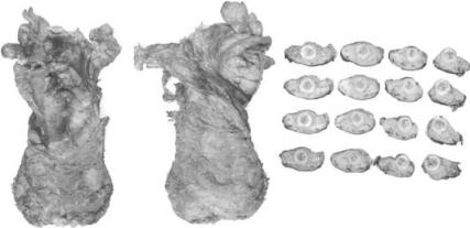
(a) (b) (c)
Fig. 3.1 Examples of a good mesorectal excision. (a) Anterior surface with Denovilliers fascia and (b) posterior surface with mesorectal fascia apparent. (c) Cross sections of the rectum showing a regular smooth surface covered in black ink.
superior to the specimens seen in my routine surgical pathology practice in the United Kingdom and also to many of the Norwegian specimens alongside it. Subsequently, we devised a quality grading system that was incorporated into the MRC CLASICC and MRC CR07 trials. It was also adopted into the Dutch mesorectal excision and short-course radiotherapy study [20]. This classification described the smoothness and bulk of the mesorectum and divided them into three groups. Subsequently, it was decided that the best way of describing the surgery was by the plane of the surgical dissection.
Mesorectal fascial plane. The mesorectum should be smooth with no violation of the fat, good bulk to the mesorectum anteriorly and posteriorly, and the distal margin should appear adequate with no coning near the tumor. No defect should be more than superficial or 5 mm deep (see Fig. 3.1).
Intramesorectal plane. Moderate bulk to mesorectum but irregularity of the mesorectal surface is present. Moderate coning of the specimen toward the distal margin. At no site is the muscularis propria visible with the exception of the area of insertion of levator muscles. Moderate irregularity of the CRM.
Muscularis propria plane. There will be areas of substantial loss of mesorectal tissue. Deep cuts and tears down onto the muscularis propria will be present. On cross-section there will be a very irregular CRM with

|
W H A T C A N T H E P A T H O L O G I S T T E L L ? |
35 |
.............................................................................................................................................................................. |
||
Distal |
Anterior |
|
R
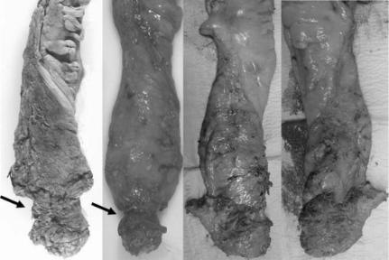
Distal
Fig. 3.2 Two resections where the dissection plane reaches onto the muscularis propria (arrows). These are poor resections.
little bulk to the mesorectal fat and the muscularis propria will form the CRM in places (see Fig. 3.2).
We are still awaiting the CR07 and CLASICC results, but a small study was performed within the Dutch trial. This showed that an involved CRM had the greatest effect but resections where the CRM reached down onto the muscularis propria had a higher rate of local recurrence and a poorer survival than resections where this did not happen [20]. So-called incomplete resections (muscularis propria plane) also had a CRM much closer to the tumor and a higher rate of CRM involvement.
Other features to note when describing the mesorectum are the anatomical variation between individuals. Some people have very small mesorectums whereas others are quite large. Thus the distance of extramural penetration of a tumor into the mesorectum may have very different implications in different people. The other feature of interest is the variation in shape of the mesorectum. Anteriorly, there is less tissue leading to a higher risk of CRM involvement. This is also the hardest area for the surgeon to dissect due to the poor visibility and difficult access in some pelvises. The distal part of the mesorectal dissection is the most arduous to undertake and frequently

36 C H A P T E R 3
..............................................................................................................................................................................
Distal
Distal
Distal
Fig. 3.3 Coning at the distal margin of three specimens leads to the tumor being very close to the CRM even though it is clear of the distal margin. The arrow shows the site of the lowest part of the tumor.
an excellent mesorectal dissection can be ruined by coning of the surgical margin as shown in two cases in Fig. 3.3.
Quality and the abdominoperineal dissection
In a recent study [12], we have demonstrated that the CRM is involved in 36.5% of abdominoperineal excisions of the rectum (APERs) compared to 22.3% of anterior resections (ARs). This was also seen in the MRC CLASICC study where 21% APERs showed margin involvement vs 10% ARs [17]. In the MERCURY study [21,22], 33% of APERs vs 13% of ARs below 6 cm showed CRM positivity; in the Dutch TME/RT [23] study, 29% APERs had margin involvement vs 13% of ARs; and in the Norwegian national
W H A T C A N T H E P A T H O L O G I S T T E L L ? |
37 |
.............................................................................................................................................................................. |
|
audit of curative excisions of rectal cancer, 12% APERs and 5% ARs had positive margins [11]. In series with the follow-up, the increased rate of margin positivity always equated with an increased rate of local recurrence and a poorer survival. Thus when pathologically assessing, APERs always look carefully for CRM positivity in the area of the low mesorectum and sphincter.
There is also a much higher rate of tumor perforation in APERs than in ARs; in the Dutch study 13.7% of APERs were perforated vs 2.5% ARs, and in the Norwegian study 16% APERs vs 4% ARs [24].
With this data it became apparent that there was a wide variation in the quality of the APER resections and a new quality classification was derived. This was similar to the mesorectal grading system in that it describes the surgical plane of dissection.
Levator plane. The surgical plane lies external to the levators with them being removed en bloc with the specimen. This creates a cylindrical specimen with the levators forming an extra protective layer on the sphincters.
Sphincteric plane. Either there are no levator muscles attached to the specimen or only a very small cuff and the resection margin is on the surface of the sphincters. The specimen has a waisted/apple core appearance.
Intrasphincteric/submucosal plane. The surgeon has inadvertently entered the sphincters or even deeper into the submucosa or perforated the specimen at any point.
It has been possible to review photographs of 271 of the APERs from the Dutch TME trial with interesting results. This review showed that in onethird of the cases the surgical margin was either in the sphincter muscle, the submucosa, or into the lumen with a perforation. In the other two-thirds the CRM was on the sphincter muscle. This plane of surgical dissection explains the high frequency of CRM involvement. This study also showed that the cylindrical radical APER of Holm from the Karolinska was not performed in Holland. This operation has the theoretical advantage of approaching the tumor from below, outside of the levator plane. This avoids the difficulties of the very low pelvic floor dissection from above, removes the risk of perforation by maintaining a cylindrical package around the tumor, increases the bulk of tissue around the tumor avoiding the waist seen on standard APERs, and, importantly, increases the ease of the anterior dissection as this occurs under excellent vision. It creates a very different shaped
38 C H A P T E R 3
..............................................................................................................................................................................
Fig. 3.4 The two specimens on the left are standard British APERs showing the typical waist (arrows) formed by the surgeon following the mesorectal fascial plane and coming down onto the sphincters. The two pictures on the right show the two sides of a Swedish APER performed by Mr T Holm from the Karolinska hospital. The projection is the coccyx that has also been removed en bloc.
specimen that should reduce the frequency of margin positivity at this site (see Fig. 3.4).
Reporting the tumor after neo-adjuvant therapy
Within the United Kingdom we are seeing an increasing usage of preoperative radiotherapy and chemotherapy. These are increasingly being used together and the number of chemotherapy agents used in combination is rising.
Short-course radiotherapy was popularized by the improvement in survival and reduction in local recurrence as seen in the Swedish rectal cancer studies [24–29] and the effect of halving the local recurrence was confirmed by the Dutch TME/RT trial [23]. Short-course 5 × 5 Gy radiotherapy is reported not to downstage tumors but is aimed at small micro-metastases within the mesorectum. There are, however, tumors that do show significant damage from such therapy, but the relative effect of this on patient outcome is not known.
W H A T C A N T H E P A T H O L O G I S T T E L L ? |
39 |
.............................................................................................................................................................................. |
|
Long-course radiotherapy over a period of 6 weeks can cause significant effects on the tumor, although complete response is unusual at between 0 and 8%. The addition of 5-FU to radiotherapy increases the complete response rate to 8% [30]. Combination chemotherapy of 5-FU with irinotecan or oxaliplatin has been reported to increase the complete response rate up to 10–30% but these are small phase II studies and definitive studies are awaited.
CRM and preoperative treatment
A major source of debate is the relative importance of achieving a clear CRM over a complete response. Recent results [31] on 150 patients strongly suggest that clear CRM is the key factor. After chemoradiotherapy with 5FU local recurrence occurred in 10% of negative CRM and 62% of positive CRM or R2 resections. Distant metastases occurred in 29% of negative CRM and 75% of positive CRM or R2 resections. Three-year overall survivals were 25 and 64% respectively for patients with and without a positive CRM. A multicenter audit of 650 patients confirmed these results [32] as did a study performed in Leeds [33]. This is important as the assessment of this margin is well described whereas the reporting of a complete response is highly variable. For the assessment of complete response we recommend using the criteria established for the dissection and reporting of complete response in the Capicitabine/Oxaliplatin, Radiotherapy and Excision (CORE) trial. The recommendations were to extensively sample the area of the tumor by taking a minimum of five blocks. If no tumor is found the whole of the area of the tumor should be embedded. If there is still no tumor then three levels should be cut on each block. If there is still no tumor then the case should be reported as a complete response. Since the efficacy of chemoradiotherapy regimens is frequently judged on complete response it is essential that such assessments are standardized across all trials.
It is also possible to grade the degree of regression of tumor after neoadjuvant therapy. The results from the preoperative radiochemotherapy vs postoperative radiochemotherapy study [34] support the use of regression grading. Unfortunately grading has become complicated by the reversal of the scoring system by different authors. They all use a modification of Mandard grading [35] first described in the esophagus, and for the CORE study we used the Dworak scoring [36] where grade 4 equals a complete response. This was also used in a preoperative 5-FU plus radiotherapy study
40 C H A P T E R 3
..............................................................................................................................................................................
by Rodel et al. [37] with complete loss of tumor cells and the presence of very few tumor cells (defined as difficult to find microscopically) leading to a 72% relapse-free survival vs 28% in the tumors showing less regression. Bouzourene et al. [38] also reported that the presence of rare tumor cells was associated with a similar disease-free survival of 75% vs 25–50% for the other groups. More recently the German group has reported regression grading to be a good predictor of outcome. However, the relative importance of the regression grade was not compared to margin positivity to see whether it was of additional value over the assessment of complete resection alone. Use of the description rather than a number should ease communication between different groups and studies.
Chemoradiotherapy response scoring
Dworak scoring used for the CORE study
Grade 0 No regression detectable.
Grade 1 Minimal regression: dominant tumor mass with obvious fibrosis and/or vasculopathy.
Grade 2 Moderate regression: dominantly fibrotic changes with few tumor cells or groups (easy to find).
Grade 3 Good regression: very few (difficult to find microscopically) tumor cells in fibrotic tissue with or without mucin.
Grade 4 Total regression: no tumor cells, only fibrotic mass or mucin.
Simplified scoring system for CORE study
Poor response – No regression to moderate regression: dominantly fibrotic changes with few tumor cells or groups (easy to find) (Dworak 0–2).
Excellent response – Good regression: very few (difficult to find microscopically) tumor cells in fibrotic tissue with or without mucin to total regression (Dworak 3–4).
High-risk rectal cancer for adjuvant chemotherapy
It is now well established that node positive colonic cancer patients benefit from adjuvant chemotherapy. The Quasar adjuvant chemotherapy study also suggests that rectal cancer patients benefit too. It is a proven fact that
W H A T C A N T H E P A T H O L O G I S T T E L L ? |
41 |
.............................................................................................................................................................................. |
|
the higher the number of lymph nodes found by the pathologist the higher the chance of finding a positive node [9]. The average number of lymph nodes retrieved in routine practice is still too low in many countries. In the United Kingdom the numbers of nodes found has been rising with an increase in a population-based study in Yorkshire from 8 to 13 in Dukes’ B and from 9 to 14 in Dukes’ C from 1995 to 2000 after the introduction of a proforma. Reviews of CR07 and the MRC CLASICC [17] studies have shown a respectable harvest of lymph nodes of a median of 13. Audits in Leeds and Gloucester report an average of 15 and 17 lymph nodes respectively. In Sweden local audits have revealed an average of 8 lymph nodes and the COLOR study, 10 nodes [19]. Many rectal cancer trials do not report the average number of lymph nodes found thus we do not know the true stage of the patients. Centers should strive for an average of 15 lymph nodes; indeed TNM suggests that a case with under 12 lymph nodes should not be staged but this dictat is widely ignored even in many European and US studies. Thus it is essential for the pathologist to retrieve as many nodes as possible. If a good node yield has been obtained and there is no evidence of metastatic spread, high-risk features should be reported. The nature of these features has been reported for colonic cancer [16] but is less well investigated in rectal cancer. High-risk features that should be reported are extramural vascular invasion, peritoneal involvement, perforation, and the distance the tumor has spread from the muscularis propria. Incomplete resection is important but may need local as well as systemic treatment depending on prior therapy.
Locally advanced and metastatic cancer
In some specialist centers the pathologist will be presented with en bloc resections or even extenterations. These represent a major challenge. The surgeon who performed the operation should be invited to demonstrate the anatomy and highlight any areas that he/she had concerns over. The presence of residual recurrent tumor, the completeness of excision, and which organs are involved are all important. The resection of liver metastases in patients with spread to the liver is increasingly common and in selected patients relatively good outcomes are achievable. How are these specimens reported? The surgeon wants to have each lesion confirmed as adenocarcinoma and to know whether excision is complete, whether there is spread through the liver capsule, whether there is venous invasion present, and the state of the surrounding liver. Occasionally, lung resections
42 C H A P T E R 3
..............................................................................................................................................................................
will be performed for metastatic colorectal cancer, especially in younger patients.
The pathologist and the radiologist
The use of Magnetic Resonance Imaging (MRI) has transformed the management of rectal cancer. The early work of Blomquist [38] and Brown [39] and confirmed by Beets Tan [40] and other centers [41] demonstrated the potential for this modality. The recent MERCURY [21,22] data has confirmed the accuracy of a well-performed MRI in predicting distance of extramural spread to within 1 mm and involvement of the CRM with an accuracy of 82%. Seeing the preoperative MRI can help the pathologist to dissect complicated specimens and to look for extramural vascular invasion and peritoneal involvement. The presentation of the macroscopic pathology slices at the MDT meeting can help the radiologist to improve their accuracy for the benefit of the patient and the team. This will become increasingly important if 3 Tesla scanners become routine instruments, increasing the resolution available to radiologists.
Conclusions
Thus the pathologist is an important member of the multidisciplinary team guiding their colleagues on the accuracy of the preoperative assessment of the MRI, the completeness of excision and the presence of residual disease, the pathological stage of the tumor, the quality of the excision, and in suggesting the change to a more radical perineal approach to low rectal cancer. We identify complete resection after chemoradiotherapy and surgery and determine the degree of regression and whether there has been a complete response. In Dukes’ stage B cancers (TNM Stage II) we can identify high-risk cases and guide adjuvant therapy.
References
1 Quirke P, Williams GT. The |
http://www.rcpath.org/ resources/pdf/ |
Royal College of Pathologists. |
colorectal cancer.pdf |
Minimum dataset for colorectal |
2 Quirke P, Durdey P, Dixon MF, |
cancer histopathology reports. |
Williams NS. Local recurrence of rectal |
London: The Royal |
adenocarcinoma is caused by inadequate |
College of Pathologists, 1998. |
surgical resection. Histopathological |
W H A T C A N T H E P A T H O L O G I S T T E L L ? |
43 |
.............................................................................................................................................................................. |
|
study of lateral tumour spread and surgical excision. Lancet 1986; 2: 996–9.
3Ng IO, Luk IS, Yuen ST et al. Surgical lateral clearance in resected rectal carcinomas. A multivariate analysis of clinicopathological features. Cancer
1993; 71: 1972–6.
4Adam IJ, Mohamdee MO, Martin IG et al. Role of circumferential margin
involvement in the local recurrence of rectal cancer. Lancet 1994; 344: 707–11.
5de Haas-Koch DF, Baeten CGMI, Jager JJ et al. Prognostic significance of radial margins of clearance in rectal
carcinoma. Br J Surg 1996; 83: 781–5.
6Birbeck KF, Macklin CP, Tiffin NJ et al. Rates of circumferential margin involvement vary between surgeons and predict outcomes in rectal cancer surgery.
Ann Surg 2002; 235: 449–57.
7Wibe A, Rendedal PR, Svensson E et al. on behalf of the Norwegian Rectal Cancer Group. Prognostic significance of the circumferential resection margin following total mesorectal excision
for rectal cancer. Br J Surg 2002; 89: 327–34.
8Nagetaal ID, Marijnen CAM, Kranenbarg EK et al. Circumferential margin involvement is still an important predictor of local recurrence in rectal carcinoma. Not 1 mm but 2 mm is the
limit. Am J Surg Pathol 2002; 26: 350–7.
9Maughan NJ, Morris E, Craig SC et al. Analysis of Northern and Yorkshire
Cancer Registry Data 1995–2001.
J Pathol 2003; 201: 18A.
10Martling A, Singnomklao T, Holm T et al. Prognostic significance of both surgical and pathological assessment of curative resection for rectal cancer. Br J Surg 2004; 91: 1040–5.
11Wibe A, Syse A, Andersen E et al. on behalf of the Norwegian Rectal Cancer Group. Oncological outcomes after total mesorectal excision for cure for cancer of the lower rectum: anterior resection vs abdominoperineal resection. Dis Colon Rectum 2004; 47: 48–58.
12Marr R, Birbeck K, Garvican J et al. The abdomino-perineal excision – the next
challenge after total mesorectal excision. Ann Surg July 2005; 242: 74–82.
13Nagtegaal ID, van de Velde CJH, Marijnen CAM et al. for the pathology review committee and the cooperative clinical investigators of the Dutch ColoRectal Cancer Group. Low rectal cancer; a call for a change of approach in abdominoperineal resection. J Clin Oncol 2005; 23: 9257–64.
14Howarth SM, Morgan JM, Williams GT. The new (6th edition) TNM classification of colorectal cancer – a stage too far. Gut 2004; 53: A21.
15Andre T, Bonni C, Mounedjii-Boudiaf L et al. Oxaliplatin, fluorouracil and leucovorin as adjuvant treatment for colon cancer. N Engl J Med 2004;
350:2343–51.
16Petersen VC, Baxter KJ, Love SB, Shepherd NA. Identification of objective pathological prognostic
determinants and models of prognosis in Dukes’ B colon cancer. Gut 2002;
51:65–9.
17Guillou P, Quirke P, Thorpe H et al. for the MRC CLASSICC Trial Group. Short-term endpoints of conventional versus laparoscopic assisted surgery in patients with colorectal cancer (MRC CLASICC trial): multicentre, randomised controlled trial. Lancet 2005;
365:1718–26.
18Bateman AC, Carr NJ, Warren BF. The retroperitoneal surface in distal caecal and proximal ascending colon carcinoma: the Cinderella surgical margin? J Clin Pathol 2005; 58: 426–8.
19Laparoscopic surgery versus open surgery for colon cancer: short-term outcomes of a randomized trial. The Colon Laparoscopic or Open Resection Study Group. Lancet Oncol 2005; 6: 477–84.
20Nagtegaal ID, van de Velde CJH,
van der Worp E et al. and the pathology review committee for the cooperative clinical investigators of the Dutch Colorectal Group. Macroscopic evaluation of rectal cancer resection specimen: clinical significance of the
44 C H A P T E R 3
..............................................................................................................................................................................
pathologist in quality control.
J Clin Oncol 2002; 20: 1729–1734.
21Daniels I and the MERCURY Study Group. Magnetic resonance imaging of low rectal cancers: a multicentre, multidisciplinary European study of 285 tumours located within 6 cm of the anal verge. Colorectal Dis 2004; 6: 1.
22The MERCURY study group. MRI predicts extramural tumour spread and circumferential resection margin status in patients with rectal cancer: Results of the MERCURY study. Br Med J submitted May 2005.
23Kapiteijn E, Marijnen CAM, Nagtegaal ID et al. for the Dutch Colorectal Cancer Group. NEJM 2001;
345:638–46.
24Eriksen MT, Wibe A, Syse A et al. Norwegian Rectal Cancer Group; Norwegian Gastrointestinal Cancer Group. Inadvertent perforation during rectal cancer resection in Norway. Br J Surg 2004; 91: 779.
25Cedermark B, Johansson H, Rutqvist LE, Wilking N. The Stockholm I trial of preoperative short term radiotherapy in operable rectal carcinoma. A prospective randomized trial. Stockholm Colorectal Cancer Study Group. Cancer 1995;
75:2269–75.
26Martling A, Holm T, Johansson H et al. The Stockholm II trial on preoperative radiotherapy in rectal carcinoma: long-term follow-up of a population-based study. Cancer 2001;
92:896–902.
27Stockholm Rectal Cancer Study Group. Preoperative short-term radiation therapy in operable rectal carcinoma. A prospective randomized trial. Cancer 1990; 60: 49–55.
28Stockholm Colorectal Cancer Study Group. Randomised study on preoperative radiotherapy in rectal carcinoma. Ann Surg Oncol 1996;
3:423–30.
29Sauer R, Becker H, Hohenberger W et al. for the German Rectal Cancer Study Group. Preoperative versus postoperative chemoradiotherapy for rectal cancer.
N Engl J Med 2004; 351: 1731–40.
30Mawdsley S, Glynne-Jones R, Grainger J et al. Can the histopathological assessment of the circumferential margin following pre-operative pelvic chemo-radiotherapy for T3/4 rectal cancer predict for three year disease free survival? Int J Radiat Oncol 2005; 63: 745–52.
31Sebag-Montefiore D, Glynne-Jones R, Mortensen N et al. Pooled analysis of outcome measures including the histopathological R0 resection rate after preoperative chemoradiation for locally advanced rectal cancer. Colorectal Dis 2005; 7: A20.
32Sebag-Montefiore D, Hingorani M, Cooper R, Chesser P. Circumferential resection margin status predicts outcome after pre-operative chemoradiation for locally advanced rectal cancer. http://www.asco.org/ac/ 1,003,12-002636-0018-0036- 00190010208.00.asp
33CORE study: Capicitabine/Oxaliplatin, Radiotherapy and excision protocol Study No. C8601. Sanofi-Synthelabs 2002 Appendix 7, Pathological techniques (P. Quirke).
34Mandard AM, Dalibard F, Mandard JC et al. Pathological assessment of tumour regression after preoperative chemoradiotherapy of esophageal carcinoma. Clinicopathological correlations. Cancer 1994;
73:2680–6.
35Dworak O, Keilholtz L, Hoffmann A. Pathological features of rectal cancer after preoperative radiochemotherapy.
Int J Colorectal Dis 1997;
12:19–23.
36Rodel C, Grabenbauer GG, Papadopoulos T et al. Apoptosis as a cellular predictor for histopathological response to neo-adjuvant radiochemotherapy in patients with rectal cancer. Int J Rad Oncol Biol Phys
2002; 52: 294–303.
37Bouzourene H, Bosman FT, Seelentag W et al. Importance of tumour regression assessment in predicting outcome in patients with locally advanced rectal carcinoma who are treated with
W H A T C A N T H E P A T H O L O G I S T T E L L ? |
45 |
.............................................................................................................................................................................. |
|
preoperative radiotherapy. Cancer 2002; 94: 1121–30.
38Blomquist L, Rubio C, Holm T et al. Rectal adenocarcinoma:assessment of tumour involvement of the lateral resection margin by MRI of the resected specimen. Br J Radiol 1999; 72: 18–23.
39Brown G, Richards CJ, Newcombe RG et al. Rectal carcinoma: thin-section MR imaging for staging in 28 patients. Radiology 1999; 211: 215–22.
40Beets-Tan RGH, Beets GL, Vliegen RFA et al. Accuracy of magnetic resonance imaging in prediction of tumour-free resection margin in rectal cancer surgery. Lancet 2001; 357: 497–504.
41Botterill ID, Blunt DM, Quirke P et al. Evaluation of the role of pre-operative magnetic resonance imaging in the management of rectal cancer.
Int J Colorectal Dis 2001; 3: 295–304.