

Essentials of Orthopedic Surgery, third edition / 09-The Elbow
.pdf9
The Elbow
MUSTAFA A. HAQUE
Although the elbow is rarely given top priority in teaching or training situations, its function is critical to upper extremity use. In looking at the arm as a unit, the tremendous range of motion of the shoulder can be thought of positioning the hand on the outer surface of a sphere. It is the flexion, extension, pronation, and supination of the elbow and forearm that allow positioning of the hand within that sphere, thus creating the ability to function throughout a huge volume of space surrounding a person. When elbow and forearm function are compromised by pain, injury, or loss of motion, significant disability can result. The goals of this chapter are to present the elbow’s functional anatomy, describe how to evaluate this region, and present an approach to diagnosis and treatment of common elbow problems.
Functional Anatomy
Skeletal
The elbow contains two distinct types of joints that allow hinge-type motion in the flexion–extension plane and rotatory motion in the pronation– supination plane. Its bony anatomy starts several centimeters proximal to the joint itself, as the humeral shaft divides and flares into medial and lateral columns that end in condyles (Fig. 9-1). The lateral condyle consists of the lateral epicondyle and the capitellum, a hemispherical structure that articulates with the proximal surface of the radial head. The medial column develops a broad outcropping called the medial epicondyle; laterally it is bridged to the capitellum by the trochlea, a spool-shaped articular segment that engages the proximal ulna with a high degree of congruity and constraint. The trochlea has a 300 degree arc of cartilage when viewed in the sagittal plane, allowing for the tremendous flexion–extension arc of the elbow while maintaining stability. The humeral columns and condyles create two fossae on the volar and dorsal aspects of the distal humerus. They are respectively called the coronoid and olecranon fossa; they allow
364

|
|
|
|
|
|
|
|
|
|
|
|
|
9. The Elbow |
365 |
|
Medial supracondylar |
|
|
|||||||||||
|
ridge |
|
|
|||||||||||
Lateral |
|
|
|
|
|
|
|
|
|
|
|
|
Olecranon |
|
supracondylar |
|
|
|
|
|
|||||||||
|
Coranoid |
|
||||||||||||
ridge |
|
fossa |
|
|
fossa |
|
||||||||
|
|
|
|
|||||||||||
Lateral |
|
|
|
Medial |
|
|
|
|
||||||
|
|
|
epicondyle |
|
|
Leteral |
|
|||||||
|
|
|
|
|
|
|
||||||||
epicondyle |
|
|
|
|
|
|
|
|
|
|
|
|
|
|
|
|
|
|
|
|
|
|
|
|
|
|
epicondyle |
||
Capitellum |
|
|
|
|
|
|
|
|
|
|
|
|
||
|
|
|
|
|
|
|
|
|
|
|
|
|
|
|
|
|
|
Trochlea |
|
|
|
|
|
|
Radial |
||||
|
|
|
|
Olecranon |
|
|
|
|||||||
|
|
|
|
|
|
|
|
|||||||
Radial head |
|
|
|
|
|
|
|
|
|
|
|
head |
||
|
|
|
|
|
|
|
|
|
|
|
|
|
||
|
|
|
|
|
|
|
|
|
|
|
|
Radial |
||
|
|
|
Coranoid |
|
|
|
|
|
||||||
|
|
|
|
|
|
|
|
|
|
|
neck |
|||
Radial neck |
|
|
process |
|||||||||||
Biceps |
|
|
|
|
|
|
|
|
|
|
|
|
Radius |
|
tuberosity |
|
|
|
|
|
|
|
|
|
|
|
Ulna |
|
|
|
Anterior View |
Posterior View |
|
|||||||||||
FIGURE 9-1. Anterior and posterior views of the elbow joint demonstrate normal skeletal anatomy, including the three articulations, including the ulnotrochlear joint, the radiocapitellar joint, and the proximal radioulnar joint.
for the coronoid and olecranon processes of the ulna to recess below the surface level of the humerus in extremes of flexion and extension.
The proximal ulna has a deep sigmoid notch, framed by the olecranon and coronoid processes, which cradles the trochlea. Radially, it has a lesser sigmoid notch, which articulates with the periphery of the radial head. Distally, it narrows to the tubular bone of the ulnar shaft. The radial head has a cup-shaped proximal surface articulating with the capitellum; its sides are covered with a 240 degree arc of articular cartilage, which interfaces with the lesser sigmoid notch and allows nearly 180 degree of pronation and supination. Distally, a prominent tuberosity is present on the radius for the attachment of the biceps.
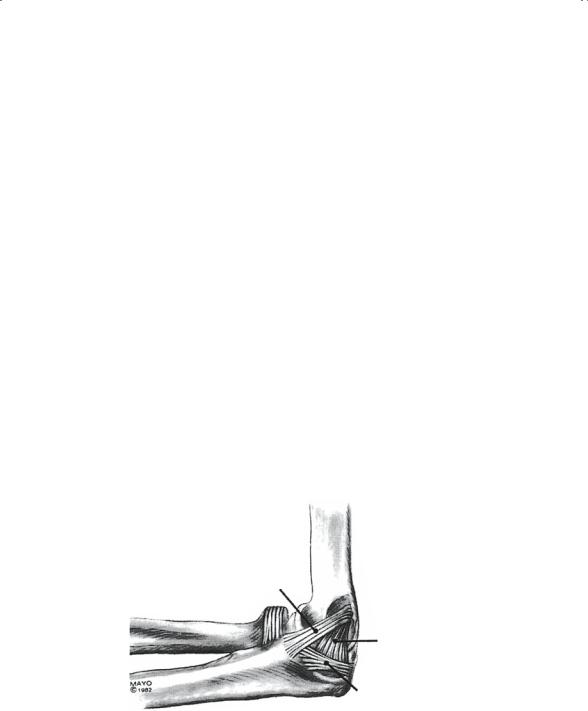
In contrast to the shoulder, whose stability is dependent on surrounding soft tissues, the elbow is highly constrained skeletally. It is further supplemented by two important ligament complexes medially and laterally. The medial ulnar collateral ligament has three segments; the most important for stability is the anterior bundle (Fig. 9-2). The lateral complex consists of the lateral ulnar collateral ligament, which originates on the lateral epicondyle and inserts on the ulna; the annular ligament, which surrounds
366 M.A. Haque
and stabilizes the radial head; and the radial collateral ligament, which extends from the lateral epicondyle to the annular ligament.
Anteriorly and posterior the elbow joint is lined by a single cell layer of synovium, which in turn is covered by a relatively thick fibrous capsule. In the olecranon and coronoid fossa, a fatty layer of tissue is present between the synovium and the capsule. This layer is of significance in radiographic evaluation of elbow trauma, in which intraarticular (intracapsular) effusion (fluid) or hemarthrosis (bleeding into the joint) causes capsular distension and displacement of these fat pads either anterior or posterior to their usual position (Fig. 9-3). Identification of these usually absent fat pads (particularly the posterior fat pad, which is usually deeply contained within the olecranon fossa) suggests joint injury or fracture.
Muscles
The muscles surrounding the elbow can be divided into five separate groups on the basis of location and function. The two groups that originate in the upper arm include the elbow flexor and extensor compartments. The flexor compartment is on the anterior surface and consists of the brachialis, which inserts on the coronoid process, and the biceps, which inserts primarily on the radial tuberosity to provide both flexion and supination. The extensor compartment of the elbow consists of the triceps, which inserts on the olecranon process to provide a powerful extension moment.
The three forearm muscle groups originating around the elbow include the mobile wad, the extensor compartment, and the flexor–pronator mass.
Anterior bundle
Posterior bundle
Transverse ligament
FIGURE 9-2. This sagittal view demonstrates the three bundles or bands of the normal medial collateral ligament. The anterior band is most important in elbow stability. (From Morrey BF (ed) The Elbow and Its Disorders, 2nd ed. Philadelphia: Saunders, 1993. Reprinted by permission of Mayo Foundation for Medical Education and Research. All rights reserved.)

|
|
|
|
|
|
|
|
|
9. The Elbow |
367 |
|
|
|
|
|||||||
|
|
Corpus humeri |
|
M. brachialis |
|
|||||
|
|
|
|
|
|
|
|
|
|
|
|
|
|
M. biceps brachii |
|
||||||
M. triceps brachii |
|
|
||||||||
|
|
|
|
|
|
|
|
|
Lig. anterius |
|
|
|
|
|
|
|
|
|
|
Fat pad |
|
|
|
Lig. posterius |
|
|
|
|||||
|
|
|
|
|
||||||
Synchondrosis |
|
epiphyseos |
|
M. pronator teres |
|
|||||
|
|
|
|
|
Trochlea |
|
|
|
||
|
|
|
|
|
|
N. medianus |
|
|||
|
|
|
|
|
|
|
|
|
|
|
|
|
|
|
|
|
|
|
|
A. et V. brachialis |
|
|
|
|
|
|
|
|
|
M. flexor digitorum sublimis |
||
Capsula articularis |
pars fibrosa |
|||||||||
|
|
|
|
|
|
|||||
pars synovialis |
Processus coronideus |
|||||||||
Bursa subtendinea olecrani |
|
|
M. flexor digitorum profundus |
|||||||
Cavum articulare |
|
|
Incisura semilunaris ulnae |
|||||||
|
|
|
|
|
|
|
|
|
Ulna |
|
|
|
|
|
|
Olecranon |
|
||||
|
|
|
|
|
|
|
||||
Synchondrosis |
epiphyseos ulnae |
|
|
|||||||
|
|
|
|
|
|
|
|
|
|
|
Bursa subcutanea olecrani
M. anconaeus
FIGURE 9-3. Sagittal illustration of the elbow joint demonstrates the normal skeletal and soft tissue anatomy. Note the presence of fat pads both anteriorly and posteriorly, directly outside the joint capsule. Intraarticular swelling can lead to displacement out of the olecranon (posterior) or coronoid (anterior) fossae, leading to the appearance of “positive fat pad sign(s)” on lateral X-rays. (From Morrey BF (ed) The Elbow and Its Disorders, 2nd ed. Philadelphia: Saunders, 1993. Reprinted by permission of Mayo Foundation for Medical Education and Research. All rights reserved.)
The mobile wad is an outcropping of three muscles arising from the lateral humerus and running on the radial aspect of the forearm. They are the brachioradialis, which inserts on the radial styloid and flexes the elbow in pronation, and the extensor carpi radialis longus and brevis, which insert on the index and middle metacarpal, respectively. They are the most important extensors of the wrist. The extensor compartment of the forearm has a common origin from the region of the lateral epicondyle and distally. It consists of all the finger extensors and the extensor carpi ulnaris. In addition, there is a muscle called the anconeus. This relatively small triangular structure originates on the lateral epicondyle and inserts on the lateral aspect of the olecranon. It is thought to assist with elbow extension. The flexor–pronator mass takes its origin from the medial epicondyle, the medial ulna, and the interosseous membrane. It consists of the muscles that flex the fingers and wrist as well as the pronator teres.
Neurovascular
In contrast to the deeper-seated neurovascular structures of other extremities, those about the elbow are both tightly concentrated and superficial,

368 M.A. Haque
making them uniquely vulnerable to both direct and indirect injury. Injuries or symptoms resulting from nerve involvement around the elbow make familiarization with normal neurovascular anatomy crucial.
Musculocutaneous Nerve
Continuing from the lateral cord of the brachial plexus and composed of fibers from the C5–C8 nerve roots, this nerve travels through (and innervates) the biceps and brachialis, terminating as the lateral antebrachial cutaneous nerve of the forearm.
Median Nerve
Arising from C5–T1 nerve roots, combined from the upper and lower cords, the median nerve travels along anterior to the brachialis muscle, enters the antecubital fossa, then passes medial to the biceps tendon and the brachial artery. It then passes through the pronator teres and gives off the anterior interosseous branch, which supplies motor innervation to the flexor pollicis longus, the index and middle flexor digitorum profundus, and the pronator quadratus. The remainder continues distally in the forearm under the flexor digitorum sublimis. Distally the median nerve provides motor and sensory innervation to part of the radial aspect of the hand.
Radial Nerve
Originating from C6–C8 nerve roots, the radial nerve is a continuation of the posterior cord, which travels in the radial groove of the humerus. It innervates the triceps, brachioradialis, and extensor carpi radialis longus and brevis muscles. In the antecubital fossa the nerve divides into a deep motor branch (posterior interosseous nerve) and a superficial sensory branch. The superficial branch continues underneath the brachioradialis to provide sensation to the dorsum of the radial aspect of the wrist and hand.
Ulnar Nerve
Derived from roots C8 and T1, the ulnar nerve continues from the medial cord of the brachial plexus along the arm until passing posteriorly through the intermuscular septum at the level of the midhumerus. It then travels through the cubital tunnel, where pathologic compression, traction, or irritation can occur. In the forearm, the ulnar nerve innervates the flexor carpi ulnaris and the ulnar half of the flexor digitorum profundus. Distally, it continues to provide motor function to many of the intrinsic hand muscles and sensation to the skin of the ulnar wrist and hand.
Brachial Artery
The brachial artery lies anterior to the medial aspect of the brachialis muscle, entering the antecubital space medial to the biceps tendon and

9. The Elbow |
369 |
lateral to the median nerve. At the level of the radial head, it divides into its terminal branches, the ulnar and radial arteries.
Evaluation of Elbow Problems
The evaluation of elbow problems relies on a thorough history, physical examination, and radiographic examination, supplemented by other pertinent tests when indicated.
History
Elbow problems can be divided into two major categories: (1) acute traumatic injuries, and (2) atraumatic problems, which tend to be more chronic. In a situation of acute trauma, a detailed history of the event must be obtained. The mechanism of injury including the position of the arm at the time, initial treatment, and subsequent symptoms are all very important in guiding further evaluation and management. It is also important to elicit a history of any prior injury or underlying symptoms in the elbow and forearm.
For nonacute elbow conditions, the most common complaint is pain, although stiffness or other mechanical symptoms such as locking, catching, or instability may accompany or become the primary problem. The examiner must try to define the complaint as completely and accurately as possible. Identify the onset of the symptoms, including the time frame before the examination and whether it was acute or insidious. Try to pinpoint the exact location of the symptoms and any zone to which it radiates. Characterize the nature of the pain: is it burning or radiating (nerve), or is it an aching related only to activity (tendonitis)? Does it hurt at rest or at night (tumor, infection)? Is it associated with any other symptoms, such as neck pain (referred pathology from the cervical spine) or wrist pain (distal radioulnar joint problem)?
What is the relationship of the patient’s activity to their symptoms? For example, in a throwing athlete, when during the pitch or throw does the pain occur? Medial elbow pain when the arm is in the “cocking position” suggests medial collateral ligament pathology, whereas medial pain during follow-through suggests involvement of the flexor pronator group.
Determine what treatments, if any, have helped. Has the patient had any cortisone injections? What type of other treatments (physical therapy, antiinflammatories, etc.) have they had, and with what effect? Has there been previous surgery?
The elbow is commonly involved (and sometimes one of the first joints affected) in inflammatory arthritides, so it is important to elicit a history of other joint complaints, known arthritis, and family history. Is there a history of skin problems (lupus, dermatitis, psoriasis) or gastrointestinal

370 M.A. Haque
problems (colitis)? Have there been any systemic symptoms of illness (malaise, fevers)?
Numbness, tingling, and weakness may be obvious clues to neurologic involvement, but sometimes nerve entrapment syndromes present with pain only. In addition to inquiring about tingling or numbness, ask about weakness or loss of dexterity.
Perhaps the most important part of the history is determining how the symptoms interfere with function, as this directs the treatment more than any other factor. For example, inability to flex the elbow completely is well tolerated by most patients, because we generally rely on an arc of 30 to 130 degrees for most activities of daily living. But in the patient with rheumatoid arthritis, for example, in whom shoulder motion is also compromised, elbow restriction may interfere with their ability to feed or clean themselves.
Physical Examination
The examination of the elbow begins with inspection, palpation, range-of- motion assessment, and evaluation for strength and neurovascular integrity. These features are then followed by special tests designed to evaluate specific conditions, based on a differential diagnosis from the history and initial tests. A thorough physical examination must also include a directed evaluation of the shoulder, wrist, and hand, and, when relevant, the cervical spine.
Inspection begins with careful observation of elbow use as soon as one begins interaction with the patient. Does the patient extend the elbow to shake hands with the examiner? Are there obvious adaptive maneuvers that the patient uses to avoid pain or compensate for functional loss? A more formal visual exam is then performed to look for presence of swelling, ecchymosis, atrophy, asymmetry, or masses. One should evaluate the “carrying angle” formed between the longitudinal axis of the humerus and the forearm, normally 10 to 15 degrees.
With the elbow flexed 90 degrees, note that the normal bony prominences (medial and lateral epicondyles and the olecranon) form an equilateral triangle. In dislocations, this normal relationship is distorted. Look for evidence of joint swelling laterally by inspection of the soft tissue triangle bordered by the radial head, olecranon tip, and lateral epicondyle.
Palpate for tenderness, soft tissue integrity, and crepitus. Include the anterior, medial, lateral, and posterior structures in an organized, systematic fashion. Be specific in trying to identify the exact area of tenderness. For example, lateral epicondylitis (lateral tennis elbow) causes focal tenderness over the lateral epicondyle. Tenderness more distally in the proximal forearm may instead suggest posterior interosseous nerve entrapment. Medial elbow pain may reflect medial epicondylitis (medial

9. The Elbow |
371 |
tennis elbow) if tender directly over the epicondyle. When more distal, it may be caused by medial ulnar collateral ligament (MCL) insufficiency. Palpate posteriorly over the olecranon fossa. Notice the presence of any bursae over the olecranon tip, occasionally containing fluid, palpable fibrous fragments, or both (olecranon bursitis). Palpate over the antecubital fossa for any defect in the biceps tendon attachment (distal biceps tendon rupture).
Check both active and passive motion, noting any difference between them. If passive motion is greater than active motion, consider pain, muscle, or nerve injury as possible causes. Patients tend to splint their elbow at 80 to 90 degrees following trauma because the capsule accommodates the maximum amount of fluid in this position. In the absence of trauma, pain on passive elbow motion suggests infection.
Note the location and timing of pain during motion. Discomfort at terminal extension is common in posterior olecranon impingement. Crepitus over the radiocapitellar joint during pronation/supination may indicate synovial or chondral pathology, degenerative changes, or radial neck fracture.
The extent of neurologic evaluation depends on the patient’s symptoms, but be familiar with sensory, motor, and reflex exam. Check for sensation to light touch in the distribution of the specific peripheral nerves. For the ulnar nerve, check the ulnar border of the little finger. For the median, use the radial border of the index finger. Check radial sensory function over dorsal thumb–index web space. The specific nerve roots have overlapping innervation, but in general, the lateral aspect of the deltoid is the C5 dermatome, the dorsal first web space is C6, the middle finger tip is C7, and the ulnar aspect of the forearm and arm is T1.
Strength testing depends on familiarity with the innervation of the various muscle groups. Elbow flexion relies on C5, whereas wrist extension is mainly C6. Elbow extension is from C7, which also provides finger extension and wrist flexion. Reflex testing is performed for the biceps (C5), brachioradialis (C6), and triceps (C7).
Vascular assessment includes palpation of the radial and ulnar arteries at the wrist and the brachial artery in the antecubital fossa.
Additional specific physical examination tests may be useful depending on the condition suspected. When considering medial epicondylitis, check for pain on wrist flexion or forearm pronation against resistance. Medial collateral ligament sprain or attenuation is determined by applying a valgus stress to the 15 to 30 degree flexed elbow, looking to reproduce pain or joint opening. Lateral epicondylitis can be assessed by eliciting pain with wrist extension or grip, whereas radial tunnel syndrome is implied by pain with resisted middle finger extension or forearm supination.
The Tinel’s sign is useful in assessment of nerve problems. Gently tapping over a nerve in the vicinity of suspected entrapment or pathology reproduces the symptoms, causing numbness, tingling, or pain in the nerve’s

372 M.A. Haque
distribution. During flexion and extension, the ulnar nerve may be “unstable” and can be felt subluxating or completely dislocating out of its groove posterior to the medial epicondyle in the cubital tunnel.
Radiographic Evaluation
Anteroposterior (AP) and lateral X-rays are the minimum views necessary to evaluate the elbow joint. Following trauma, additional views are sometimes helpful, including oblique and radial head views. Beyond this, the following special radiographic tests can be helpful.
Stress X-Rays
Stress views may be helpful in evaluating the patient with a suspected tear of the medial collateral ligament. This view is achieved through manual stress, during which the clinician applies a valgus stress to the elbow in an effort to open up the medial side. A difference in medial gapping of more than 2 mm between the affected and normal elbow is usually significant.
Computed Tomography/Magnetic Resonance Imaging Examination
Computed Tomography (CT) scans are effective in preoperative planning of complex elbow trauma, assessment of bony and joint deformity, and occasionally for evaluation of loose bodies of the elbow.
Magnetic Resonance Imaging (MRI) provides superior soft tissue imaging and allows visualization of marrow and vascularity changes in bone. Its current use about the elbow includes imaging occult fractures, tumors, infections, synovitis or other causes of joint effusion, and osteochondritis dissecans. It is occasionally useful in evaluating ligament disruptions, but it is usually unnecessary in evaluating medial or lateral epicondylitis and rarely helpful in nerve entrapment syndromes.
Technetium-99 Bone Scan
Technetium-99 injected intravenously is taken up in areas of increased vascularity. Although it is very sensitive, this test is not very specific, because increased blood flow can occur as a result of fracture, infection, tumor, or arthritis. In patients with heterotopic ossification, serial bone scans may help determine when the process has become quiescent enough to permit safe bone mass excision.
Electrodiagnostic Tests
Electromyography (EMG) and nerve conduction velocity (NCV) testing have definite indications in the patient with suspected nerve entrapment or injury. Such testing may indicate the site of the compression or injury.

9. The Elbow |
373 |
However, failure to demonstrate specific neurologic findings by electrodiagnostic testing does not rule out their presence. This problem is common in the workup of the patient with early ulnar nerve symptoms, or the patient with suspected radial tunnel syndrome, in whom such tests are commonly negative.
Arthroscopy
The techniques and procedures for arthroscopy of the elbow have developed more slowly than in other joints such as the knee, shoulder, or wrist. Because of the very tight concentration of nerves and blood vessels in the area, the depth of the joint capsule under the musculature, and the tight articular constraint, it can be difficult and involves more risk than arthroscopy at most other joints. Although it provides a minimally invasive means with which to visually inspect and, when necessary, to palpate the intraarticular structures, it is rarely used for diagnostic purposes alone.
Treatment of Elbow Problems
Treatment of elbow problems is algorithmic, dividing conditions into either traumatic or atraumatic cause (Figs. 9-4, 9-5). One general principle of treatment in the elbow is to minimize the time of immobilization. The elbow has a high propensity for developing contractures with immobilization, especially after fractures or dislocations. The resultant loss of motion can be disabling, and treatment for it can be prolonged and difficult. When necessary, splinting is usually done with the elbow in 90 degrees of flexion and neutral pronation to allow for maximal capsular volume and maintenance of the most useful arc of function.
With any significant elbow trauma, after appropriate initial treatment, one should carefully follow the patient’s neurovascular examination, as the multiple confined fascial compartments of the forearm leave patients vulnerable to compartment syndrome or other severe compromise.
Nonoperative Treatment
Rehabilitation
Rehabilitation, either through a patient self-guided program or by formal occupational or physical therapy, plays an important role in the treatment of elbow problems. The goals should include (1) reduction of pain and inflammation, (2) restoration of motion, (3) rebuilding strength, and (4) return to normal function and activity. These goals should be carefully monitored by the treating physician until the patient is discharged or alternative management is instituted.