

Essentials of Orthopedic Surgery, third edition / 04-Tumors of the Musculoskeletal System
.pdf4
Tumors of the Musculoskeletal System
MARTIN MALAWER and KRISTEN KELLAR-GRANEY
Both benign and malignant tumors (neoplasms) may arise from any mesenchymal soft tissue or bony tissue of the extremities, pelvis, shoulder girdle, or the axial skeleton. All tumors arise from one of the different histologic types of tissue that comprise the musculoskeletal system: bone (osteoid-forming tumors), cartilage (chondroid-forming tumors), and muscle and the fibrous connective tissue (soft tissue tumors). Only rarely do tumors arise from the arteries or nerves.
In general, most tumors are benign, but malignant tumors may also occur. Malignant mesenchymal tumors are termed sarcomas. Sarcomas rank among the least common malignant diagnoses. There are only 8,000 new cases (6,000 soft tissue and 2,000 bone) of sarcoma of the 1.2 million new patients who will be diagnosed with cancer each year in the United States.
Most bone sarcomas (osteosarcoma and Ewing’s sarcoma) occur during childhood or adolescence, in contrast to most malignancies (carcinomas), which occur in the later decades of life (more than 40 years of age). Soft tissue sarcomas tend to occur in young adults, and the risk of development increases with each decade of life. Bone tumors usually present with pain, in contrast to soft tissue tumors, which often present as a painless mass (usually greater than 5 cm in size).
Orthopedic surgeons are often the first physician to see these patients and are called upon to make a correct diagnosis and/or to determine if an individual should be referred to a specialist (orthopedic oncologist). To emphasize the rarity of these entities, the average orthopedic surgeon will see only one or two tumors every 5 to 10 years of practice.
A high degree of clinical suspicion, necessary for early diagnosis, is ever more important as fundamental changes in healthcare delivery alter patient access to specialists and to expensive imaging studies. Early detection, combined with proper techniques of diagnosis and treatment, can dramatically improve the chances of achieving functional limb salvage and survival. Continued progress in radiographic imaging, chemotherapy, radiation therapy, and biotechnology, coupled with a better understanding of the
106

4. Tumors of the Musculoskeletal System |
107 |
biologic behavior of mesenchymal neoplasms, have led to a rational basis of diagnosis, staging, and surgical treatment.
This chapter reviews the common benign as well as malignant tumors arising from bone and soft tissues of the extremities, shoulder girdle, and pelvis. The biologic and radiographic characteristics are emphasized, evaluation with the use of staging studies such as computed tomography (CT) and magnetic resonance imaging (MRI) is described, and the basic surgical procedures are presented. The biologic basis of tumor growth, staging, and radiographic determination are emphasized.
Natural History of Bone and Soft Tissue Tumors
Mesenchymal neoplasms have characteristic patterns of behavior and growth that distinguish them from other malignancies. These patterns form the basis of a staging system and provide a focus for current treatment strategies.
Biology and Growth
Spindle cell sarcomas form solid lesions through circumferential growth in which the periphery of each lesion is composed of the least mature cells. In contradistinction to benign lesions, which are surrounded by a true capsule composed of compressed normal cells, malignant tumors are generally enclosed by a pseudocapsule consisting of viable tumor cells and a fibrovascular zone of reactive tissue with a variable inflammatory component that interdigitates with the normal tissue adjacent and beyond the lesion. The thickness of the reactive zone varies with the degree of malignancy and histogenetic type.
High-grade sarcomas characteristically have a poorly defined reactive zone that may be locally invaded and destroyed by the tumor. In addition, tumor nodules not in continuity with the main tumor may be present in tissue that appears to be normal. Low-grade sarcomas rarely form tumor nodules beyond the reactive zone (Fig. 4-1).
Local anatomy influences the growth of sarcomas by setting natural barriers to extension. In general, bone sarcomas take the path of least resistance. The three mechanisms of growth and extension of bone tumors are compression of normal tissue, resorption of bone by reactive osteoclasts, and direct destruction of normal tissue. Benign tumors grow and expand by the first two mechanisms; direct tissue destruction is characteristic of malignant bone tumors. Most benign bone tumors are unicompartmental; they remain confined and may expand the bone in which they arise. Most malignant bone tumors are bicompartmental; they destroy the overlying cortex and push directly into the adjacent soft tissue. Soft tissue tumors may start in one compartment (intracompartmental) or between
108 M. Malawer and K. Kellar-Graney
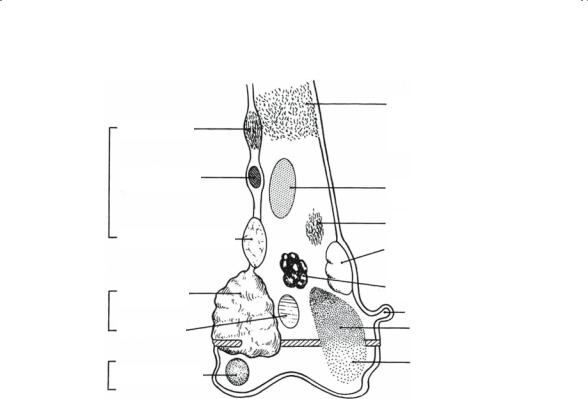
Diaphysis
Epiphysis Metaphysis
Adamantinoma
Osteoid osteoma
Chondromyxoid fibroma
Osteosarcoma
MFH
Enchondroma
Chondroblastoma
Round cell lesions: Ewing sarcoma Histiocytic lymphoma Myeloma
Fibrous dysphasia
Fibrosarcoma
Fibrous cortical defect
Bone cyst
Osteochondroma
Aneurysmal bone cyst
GCT
FIGURE 4-1. Schematic of typical locations of common bone tumors. The anatomic location, whether diaphyseal, metaphyseal, or epiphyseal, is one of the most important determinants of tumor type. Cortical, medullary, or eccentric location is also important. One should always pay special attention to the presence of matrix formation. (Source: Devita VT, Hellman S, Rosenberg SA. Cancer: Principles and Practice of Oncology, Fourth Edition. Lippincott Williams & Wilkins, 1993.)
compartments (extracompartmental). The determination of anatomic compartment involvement has become more important with the advent of limb preservation surgery. In general, most benign and malignant bone and soft tissue tumors grow like a ball, pushing normal tissue away as they expand.
The Five Basic Patterns of Sarcoma Behavior
On the basis of biologic considerations and natural history, all bone and soft tissue tumors, benign and malignant, may be classified into five categories, each of which shares certain clinical characteristics and radiographic patterns and requires similar surgical procedures. These five categories and the associated tumor behavior are as follows (Table 4-1):
1. Benign/latent: Lesions whose natural history is to grow slowly during normal growth of the individual and then to stop, with a tendency to heal

4. Tumors of the Musculoskeletal System |
109 |
spontaneously. They never become malignant and heal rapidly if treated by curettage (e.g., lipomas and unicameral bone cysts).
2.Benign/active: Lesions whose natural history is progressive growth. Excision leaves a reactive zone with some tumor (e.g., chondroblastoma of bone).
3.Benign/aggressive: Lesions that are locally aggressive but do not metastasize. Tumor extends through the capsule into the reactive zone. Local control can be obtained only by removing the lesion with a margin of normal tissue beyond the reactive zone (i.e., giant cell tumors of bone or fibromatosis of soft tissues).
4.Malignant, low-grade: Lesions that have a low potential to metastasize. Histologically, there is no true capsule but rather a pseudocapsule. Tumor nodules exist within the reactive zone but rarely beyond. Local control can be accomplished only by removal of all tumor and reactive tissue with a margin of normal bone. These lesions can be treated successfully by surgery alone; systemic therapy is not required (i.e., myxoid chondrosarcoma of bone and low-grade soft tissue sarcomas).
5.Malignant, high-grade: Lesions whose natural history is to grow rapidly and to metastasize early. Tumor nodules are usually found within and beyond the reactive zone and at some distance in the normal tissue. Surgery is necessary for local control, and systemic therapy is warranted to prevent metastasis (i.e., osteosarcoma, Ewing’s sarcoma of bone. and high-grade malignant fibrous histiocytoma of soft tissues).
Examples of bone and soft tissue tumors in each of these categories are described following and in Table 4.2.
Mechanism of Tumor Spread
In contrast to carcinomas, bone and soft tissue sarcomas disseminate almost exclusively through the blood (i.e., to the lungs and other bones). About 5% to 10% of soft tissue tumors spread through the lymphatic system to regional nodes. Hematogenous spread is manifested by pulmonary involvement in the early stages and by bony involvement in later stages. Bone metastasis occasionally is the first sign of dissemination.
TABLE 4-1. Behavioral classification of bone and soft tissue tumors.
Classification |
Bone |
Soft tissue |
Benign/latent |
Nonossifying fibroma |
Lipoma |
Benign/active |
Aneurysmal bone cyst |
Angiolipoma |
Benign/aggressive |
Giant cell tumor |
Aggressive fibromatosis |
Malignant/low grade |
Parosteal osteosarcoma |
Myxoid liposarcoma |
Malignant/high grade |
Classic osteosarcoma |
Malignant fibrous histiocytoma |
|
|
|
(Adapted from Enneking WF. Staging of musculoskeletal tumors. In: Enneking WF, Musculoskeletal Tumor Surgery, vol.1, New York: Churchill Livingstone; 1983:87–88.)

110 |
M. Malawer and K. Kellar-Graney |
|
|
TABLE 4-2. General classification of bone and soft-tissue tumors. |
|||
Histologic type |
Benign |
Malignant |
|
Hematopoietic (41.4%) |
|
Myeloma |
|
|
|
|
Reticulum cell sarcoma |
Chondrogenic (20.9%) |
Osteochondroma |
Primary chondrosarcoma |
|
|
|
Chondroma |
Secondary chondrosarcoma |
|
|
Chondroblastoma |
Dedifferentiated chondrosarcoma |
|
|
Chondromyxoid fibroma |
Mesenchymal chondrosarcoma |
Osteogenic (19.3%) |
Osteoid osteomas |
Osteosarcoma |
|
|
|
Benign osteoblastoma |
Parosteal osteosarcoma |
Unknown origin (9.8%) |
Giant cell tumor |
Ewing’s sarcoma |
|
|
|
|
Malignant giant cell tumor |
|
|
|
Adamantinoma |
Fibrogenic (3.8%) |
Fibrous histiocytoma |
Malignant fibrous histiocytoma |
|
|
|
Fibroma |
Fibrosarcoma |
|
|
Desmoplastic fibroma |
|
Notochordal (3.1%) |
|
Chordoma |
|
Vascular (1.6%) |
Hemangioma |
Hemangioendothelioma |
|
|
|
|
Hemangiopericytoma |
Lipogenic (0.5%) |
Lipoma |
Liposarcoma |
|
Neurogenic (0.5%) |
Neurilemmoma |
|
|
|
|
|
|
Source: From Sim FH, Bowman W, Chao E. Limb salvage surgery and reconstructive techniques. In: Sim FH (ed) Diagnosis and Treatment of Bone Tumors: A Team Approach. A Mayo Clinic Monograph. Thorofare, NJ: Slack, 1983.5
The histologic hallmark of malignant sarcomas is their potential to break through the pseudocapsule to form satellite lesions called skip metastases. A skip metastasis is a tumor nodule that is located within the same bone as the main tumor but is not contiguous to it. Transarticular skip metastases are located in the joint adjacent to the main tumor. Although relatively uncommon, skip metastases occur most frequently with high-grade sarcomas.
Sarcoma Tumor Staging
Selection and use of a prognostically significant staging system are fundamental both for the selection of appropriate treatment protocols and for the development of tumor registries necessary for basic research analysis. In 1980, the Musculoskeletal Tumor Society adopted a surgical staging system (SSS), proposed by Dr. William F. Enneking, for both bone and soft tissue sarcomas. This system is based upon the fact that mesenchymal sarcomas of bone and soft tissue behave alike, irrespective of histogenetic type. The SSS is encompasses the “GTM” classification: grade (G), location (T), and lymph node involvement and metastases
(M).

4. Tumors of the Musculoskeletal System |
111 |
1.Surgical grade. The letter G incorporates both the histologic grade of a lesion and clinical factors related to aggressiveness, such as growth rate. A low-grade tumor is rated G1 whereas a high-grade tumor is rated G2. Low-grade lesions are malignancies with small potential to metastasize.
2.Surgical site. The letter T represents anatomic site, either intracompartmental (T1) or extracompartmental (T2). A compartment is defined as “an anatomic structure of space bounded by natural barriers of tumor extension.” T1 lesions are easier to delineate clinically, surgically, and radiographically than T2 lesions and have a correspondingly higher chance of adequate removal without amputative procedures.
3.Lymph nodes and metastases. Local disease without evidence of metastasis is designated M0. When a bone or soft tissue sarcoma has metastasized (M1), the prognosis is extremely poor. Lymphatic spread is a sign of extensive dissemination. Regional lymphatic involvement is equated with distal metastases.
The SSS developed for surgical planning and assessment of bone sarcomas is summarized as follows:
Stage IA (GI, T1, M0) Low-grade intracompartmental lesion without metastasis.
Stage IB (GI, T2P, M0) Low-grade extracompartmental lesion without metastasis.
Stage IIA (G2, TI, M0) High-grade intracompartmental lesion without metastasis.
Stage IIB (G2, T2, M0) High-grade extracompartmental lesion without metastasis.
Stage IIIA (GI or G2, M1) Intracompartmental lesion, any grade, with metastasis.
Stage IIIB (GI or G2, T2, MI) Extracompartmental lesion, any grade, with metastasis.
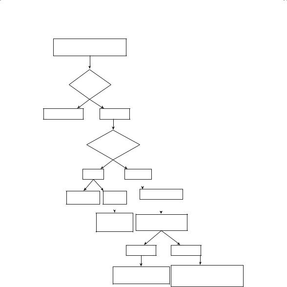
Decision-Making Process
In order to accurately assess the diagnosis, stage, and grade of a suspected bone tumor, rigid protocol should be followed to facilitate the decisionmaking process as to what staging studies are required and when a biopsy should be performed. Figure 4-2 entitled Evaluation of Suspected Bone Tumor succinctly describes the steps any clinician should follow for patients presenting with a bone tumor.
112 M. Malawer and K. Kellar-Graney
Evaluation of Suspected
Bone Tumor
Is the lesion neoplastic?
Non-neoplastic Neoplastic
Is this benign or malignant?
Benign |
|
|
Malignant |
|
|||
|
|
|
|
|
|
|
|
No treatment |
CT Scan |
|
Imaging Studies |
||||
Observe |
Biopsy |
|
|
|
|
||
|
|
|
|
||||
|
|
|
|
|
|
|
|
|
Surgery |
|
Perform CT-guided |
||||
Curettage vs. |
|||||||
|
needle biopsy |
||||||
|
Resection |
|
|
||||
|
|
|
|
|
|||
|
|
|
Low-grade |
High-grade |
|||
Resection or curettage |
Induction chemotherapy |
|
Limb-sparing surgery |
||
possible bone graft |
||
Postoperative chemotherapy |
||
|
FIGURE 4-2. Process taken when evaluating abnormal bone radiographs.
Clinical and Radiographic Evaluation, Staging, and
Biopsy
Staging of a bone or soft tissue tumor mass means determining the local extension (anatomy) of the tumor and any possible sites of distal dissemination. If the clinical examination or plain radiographs suggest an aggressive or malignant tumor, staging studies should be performed before biopsy (see Fig. 4.1). All radiographic studies are influenced by surgical manipula-
4. Tumors of the Musculoskeletal System |
113 |
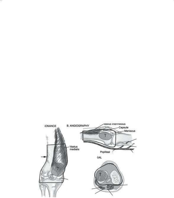
tion of the lesion, making interpretation more difficult. Bone scintigraphy, computed tomography or magnetic resonance imaging, and angiography are required to delineate local tumor extent, vascular displacement, and compartmental localization. Figure 4-3 demonstrates the distal femoral anatomic compartment as viewed by various radiographic modalities.
Radiographic Evaluation
X-Rays
Plain radiographs taken in perpendicular planes [anteroposterior (AP) and lateral] remain essential to the characterization and diagnosis of lesions involving the skeleton. Selection and interpretation of other imaging techniques is often guided by the radiographic properties of the lesion. Proper interpretation of a lesion seen on a radiograph can be summarized by answering “four questions” as proposed by Dr. William F. Enneking:
1.What are the anatomic location and extent of the lesion?
2.What is the lesion doing to the bone?
|
|
|
|
|
|
|
|
|
|
|
|
|
|
Vastus intermedius |
|
|
|
|
||||
A. MAGNETIC |
|
RESONANCE |
|
B. ANGIOGRAPHY |
|
|
|
|
|
|
|
|
|
|||||||||
|
|
|
|
|
|
|
|
|
|
Capsule |
|
|
|
|||||||||
|
|
|
|
|
|
|
|
|
|
|
||||||||||||
|
|
|
|
|
|
|
|
|
|
|
|
|
|
|
|
|
|
|
||||
IMAGING / BONE |
|
|
|
|
|
|
|
|
|
|
|
|
|
|
|
|
|
|||||
|
|
|
|
|
|
|
|
|
|
|
|
|
|
|
Meniscus |
|
||||||
SCINTIGRAPHY |
|
|
|
|
|
|
|
|
|
|
|
|
|
|
|
|
||||||
|
|
|
|
|
|
|
|
|
|
|
|
|
|
|
|
|
|
|
|
|
|
Anterior |
|
|
|
|
|
|
|
|
|
|
|
|
|
|
|
|
|
|
|
|
|
|
tibial a. |
|
|
|
|
|
|
|
|
|
|
|
|
|
|
|
|
|
|
|
|
|
|
|
|
|
|
|
|
|
|
Vastus |
|
|
|
|
|
|
|
|
|
|
|
|
|
Peroneal a |
|
|
|
|
|
|
|
|
medialis |
|
|
|
|
|
|
|
|
|
|
|
|
|
|
|
|
|
|
|
|
|
|
|
|
|
|
|
|
|
|
|
|
|
|
|
|
||
Bone |
|
|
|
|
|
|
|
|
|
|
|
Popliteal a. |
|
|
|
|
|
|
Posterior |
|||
|
|
|
|
|
|
|
|
|
|
|
|
|||||||||||
|
|
|
|
|
|
|
|
|
|
|
|
|
|
|
|
|
||||||
|
|
|
|
|
|
|
|
|
|
|
|
|
|
|
|
|
|
|
|
|||
|
|
|
C. COMPUTERIZED |
|
|
|
|
|
|
|
|
|
|
tibial a. |
||||||||
|
|
|
|
|
|
AXIAL |
|
|
|
|
|
|
|
|
||||||||
resection |
|
|
TOMOGRAPHY / |
|
|
|
|
|
|
|
|
|
|
|
|
|||||||
|
|
|
|
|
|
|
|
|
|
|
|
|
||||||||||
|
|
|
|
|
|
MAGNETIC RESONANCE |
|
|
|
|
|
|
|
|
|
|
||||||
|
|
|
|
|
|
IMAGING |
|
|
|
|
|
|
|
|
|
|
||||||
medial
capsule
Popliteal a. v.
Semimembranosus
Semitendinosus
Tibial nerve
FIGURE 4-3. Surgical schematic illustrations demonstrating the distal femoral anatomic compartment as viewed by various radiographic modalities. In this illustration, a tumor arising in the lateral femoral condyle is noted. The black outlines demonstrate the planned region of resection in relation to the critical anatomy. This type of preoperative surgical mapping is critical for a successful limb-sparing surgery. Preoperative imaging studies usually include CT, MRI, and bone scans. (Source: Devita VT, Hellman S, Rosenberg SA. Cancer, Principles and Practice of Oncology, 5th Edition. Lippincott, Williams & Wilkins, 1997.)

114 M. Malawer and K. Kellar-Graney
3.What is the bone doing to the lesion?
4.Are there any radiographic peculiarities of the lesion that give a hint as to its tissue type?
Distinction between benign, aggressive, and frankly malignant lesions can be made on the basis of this analysis. In general, the plain radiograph is the single most important study in determining the type of bone tumor.
Bone Scans
Bone scintigraphy is useful for evaluation of both bony and soft tissue tumors. Bone scans determine whether other bones are involved by tumor. The scans assist in determining metastatic disease, polyostotic involvement, intraosseous extension of tumor, and the relationship of the underlying bone to a primary soft tissue sarcoma. Malignant bone tumors may present with skeletal metastasis (1.6%). The initial flow phase correlates to the vascularity of the tumor. Recent studies using quantitative techniques and the isotope thallium-201 have shown that the histologic tumor response to chemotherapy can be predicted based upon comparison of preand posttreatment studies.
Computed Tomography
Computed tomography (CT) allows accurate determination of intraand extraosseous extension of skeletal neoplasms. CT accurately depicts the transverse anatomic relationship of a tumor to the surrounding structures. By varying window settings, one can study cortical bone, intramedullary space, adjacent muscles, and extraosseous soft tissue extension. The anatomic compartmental involvement by soft tissue sarcomas is easily determined. High-resolution CT scans (1-mm cuts) and two-dimensional or three-dimensional reconstruction can be extremely valuable in preoperative planning, particularly in the pelvis or spine. CT evaluation must be individualized to obtain the maximum benefit of image reconstruction. Close interaction between the surgeon and the radiologist facilitates accurate and effective imaging. CT is most useful for bony lesions.
Magnetic Resonance Imaging
Magnetic Resonance Imaging (MRI) provides valuable imaging of both bone and soft tissues in multiple planes. Excellent visualization of anatomic compartments, neurovascular bundles, and areas of reactive tissue allow for detailed preoperative planning. Although signal characteristics of any given mass on the traditional T1- or T2-weighted images (or on the more-recent fat-suppressed and gradient-echo images) can be diagnostic, distinction between benign or low-grade malignant lesions (such as lipomas

4. Tumors of the Musculoskeletal System |
115 |
versus well-differentiated liposarcomas) cannot be reliably made on the basis of MRI images alone.
Angiography
The arteriographic technique for bone and soft tissue lesions differs from that used for arterial disease. A minimum of two views (biplane) is necessary to determine the relation of the major vessels to the tumor. Extraosseous extension is easily demonstrated by angiography. As experience with limb-sparing procedures has increased, surgeons have become more aware of the need to determine the individual vascular patterns before resection; this is especially crucial for tumors of the proximal tibia, where vascular anomalies are common. The increasing preoperative use of intraarterial chemotherapy also has increased the need for accurate angiography. Reduction of vascularity following chemotherapy can be correlated to overall histologic response of the tumor. Preoperative embolization of highly vascular tumors before surgical resection can significantly reduce blood loss and intraoperative morbidity.
The combination of plain radiographs, bone scintigraphy, cross-sectional anatomic imaging (via CT or MRI), and longitudinal imaging of vascular supply (via angiography) allows the surgeon to develop a threedimensional construct of the local tumor area before resection and to formulate a detailed surgical approach for limb salvage.
Biopsy Considerations and Importance
The planning and technique of a biopsy is extremely important. A biopsy should be performed after the staging studies are obtained. If a resection is to be performed, it is crucial that the location of the biopsy be in line with the anticipated incision for the definitive procedure. Extreme care should be taken not to contaminate potential tissue planes or flaps that will compromise the management of the lesion. Improper biopsy technique often eliminates the opportunity for limb salvage. Mankin documented that 60% of referred patients had a major error in diagnosis and 18% had less than optimal treatment secondary to problems related to the biopsy.6
Core-Needle Biopsy
To minimize contamination and reduce patient morbidity, needle biopsy of soft tissue masses or of extraosseous components should be attempted before an incisional biopsy whenever possible. Needle or core biopsy of bone tumors often provides adequate specimen for diagnosis (Fig. 4-4). Cooperation between the radiologist and pathologist is vital to ensuring