

- •Contents
- •Preface
- •Introduction
- •The importance of cross-sectional anatomy
- •Orientation of sections and images
- •Notes on the atlas
- •References
- •Acknowledgements
- •Interpreting cross-sections: helpful hints for medical students
- •BRAIN
- •HEAD
- •NECK
- •THORAX
- •ABDOMEN
- •PELVIS
- •LOWER LIMB
- •UPPER LIMB
- •Index

LOWER LIMB
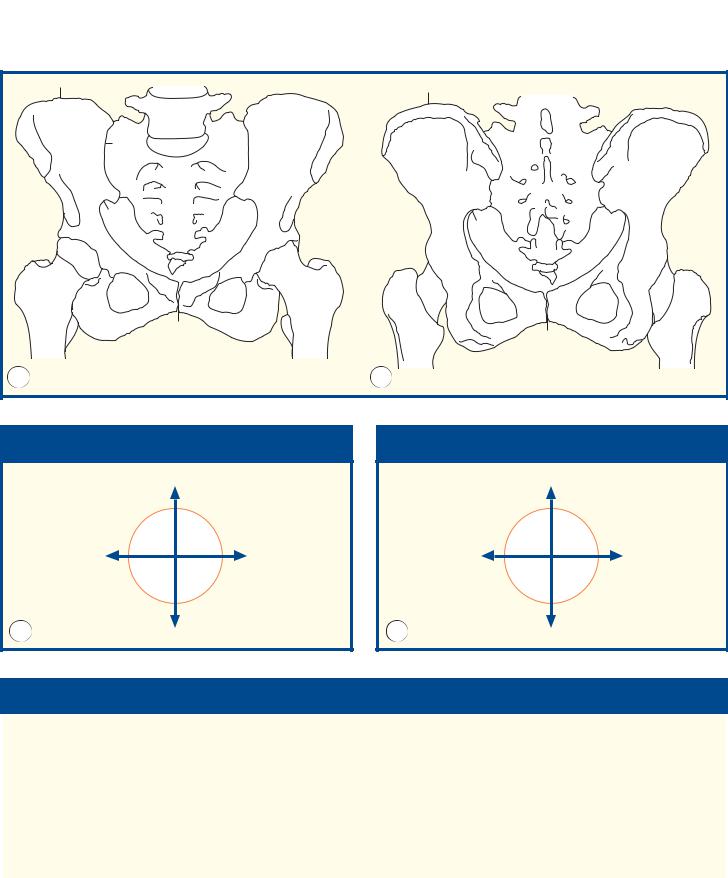
Hip left – Coronal section 1 – Female
7
8
5
96
|
|
|
4 |
1 |
|
|
|
3 |
2 |
|
|
|
|
|
|
|
|
11 |
|
|
|
|
12 |
|
|
|
|
16 |
|
|
|
10 |
|
|
|
|
|
|
14 |
|
31 |
|
13 |
17 |
|
|
|
||
|
|
|
|
|
|
11 |
|
15 |
18 |
29 |
|
|
13 |
|
12 |
|
14 |
|
|
|
|
|
||
|
28 |
30 |
25 |
|
|
|
|
|
|
|
|
|
|
20 |
27 |
26 |
|
|
19 |
|
|
|
|
|
|
24 |
|
23 |
21 |
|
|
|
|
|
|
|
|
22 |
|
1 |
Gluteus medius |
17 |
Iliofemoral ligament |
2 |
Superior gluteal |
18 |
Greater trochanter |
|
neurovascular bundle |
19 |
Shaft of femur |
3 |
Gluteus minimus |
20 |
Iliotibial tract |
4 |
Ilium |
21 |
Vastus lateralis |
5 |
Iliacus |
22 |
Vastus medialis |
6 |
Psoas major |
23 |
Profunda femoris artery |
7 |
Femoral nerve |
24 |
Profunda femoris vein |
8 |
External iliac artery |
25 |
Iliopsoas tendon |
9 |
External iliac vein |
26 |
Adductor longus |
10 Head of femur |
27 |
Ischiopubic ramus |
|
11 Rim of acetabulum |
28 |
Obturator externus |
|
12 Acetabular labrum |
29 |
Obturator internus |
|
13 Zona orbicularis of capsule |
30 |
Medial circumflex femoral |
|
14 Capsule of hip joint |
|
artery and vein |
|
15 Neck of femur |
31 |
Ligament of head of femur |
|
16 Articular cartilage |
|
(ligamentum teres) |
|
208

Hip left – Coronal section 1 – Female
LOWER LIMB
■ Section level
View
■ Orientation
Superior
■ Notes
This coronal section through the hip illustrates the ‘ball-and-socket’ arrangement of the joint. This socket is much deeper and the ball much rounder than at the shoulder. Stability is an important function here. The two powerful abductors of the hip – gluteus medius (1) and minimus (3) – have their own neurovascular bundle (the superior gluteal nerve, artery and vein), and these can be seen between the two sheets of muscle (2).
The ligament of the head of the femur, the ligamentum teres (31), is the important source of blood supply to the femoral head in the fetus and infant. It transmits the acetabular branch of the obturator artery. It becomes obliterated during early childhood, when periosteal vessels are of key importance before vessels traverse the epiphyseal plate. The blood supply to the femoral head remains of importance throughout life: avascular necrosis has many causes. The zona orbicularis of the capsule of the hip joint (13) transmits vessels from the lateral and medial circumflex femoral branches of the deep femoral artery (profunda femoris) to the head and neck of the femur (10). A subcapital fracture of the femoral head thus deprives the head of its blood supply and often leads to avascular necrosis.
Medial 
 Lateral
Inferior
4 |
|
3 |
1 |
11
12
10
31
15
 20 28
20 28 
19 |
209 |
|
Coronal magnetic resonance image (MRI)

LOWER LIMB Selected images – Pelvic girdle
3D computed tomogram (CT)
A
3D computed tomogram (CT)
B
210
Selected images – Pelvic girdle |
|
|
LOWER LIMB |
|
|
|
|
|
|
9 |
|
|
|
9 |
|
|
|
|
|
|
|
|
|
|
|
|
|
|
|
|
1 |
|
|
|
2 |
|
|
|
|
|
|
|
|
|
|
8 |
|
7 |
3 |
|
|
|
|
|
|
|
|
|
3 |
|
||
10 |
|
|
5 |
8 |
|
|
||
|
|
|
|
|
||||
|
|
4 |
|
|
|
|||
|
|
|
|
|
|
|
|
|
|
11 |
|
26 |
|
|
26 |
|
|
|
|
12 |
|
|
|
|
|
|
|
|
|
|
|
|
|
|
|
|
20 |
|
6 |
12 |
|
|
|
|
|
24 |
|
16 |
15 |
|
|
||
21 |
|
|
|
|
|
|||
|
|
|
21 |
|
16 |
|
||
|
|
13 |
27 |
18 |
|
|
||
|
|
|
|
18 |
||||
|
|
|
17 |
23 |
13 |
27 |
||
|
|
|
|
|
||||
|
22 |
14 |
|
19 |
22 |
14 |
17 |
19 |
25 |
|
|
|
|
|
|||
|
|
|
|
|
|
|
|
|
|
|
|
|
|
25 |
|
|
|
A |
|
|
|
|
B |
|
|
|
■ Orientation |
|
■ Orientation |
||||||
|
|
|
|
Superior |
|
|
|
Superior |
|
Right |
|
Left |
|
Left |
|
Right |
|
A |
|
|
|
Inferior |
B |
|
|
Inferior |
|
|
|
|
|
|
|
||
■ Notes
Surface-shaded three-dimensional volume-rendered |
general principles of the pelvic girdle well. Note how |
|
||||||
CT images. Because bone attenuates the X-ray beam |
the femoral head (two-thirds of a hemisphere) is |
|
||||||
so much, its CT attenuation value (around +1000 HU) |
much better contained within the acetabular fossa |
|
||||||
is much greater than that of the surrounding soft |
than the humeral head, thereby providing stability at |
|
||||||
tissues. Thus, the bones can be ‘extracted’, with no |
the expense of mobility. The obliquity of the |
|
||||||
overlying artefacts, to provide information equivalent |
acetabulum means that the femoral head can just be |
|
||||||
to that from a cadaveric skeleton. |
|
|
seen on the anterior view, but not posteriorly. |
|
||||
|
These two views, anterior and posterior, show the |
|
|
|
|
|
||
|
|
|
|
|
|
|
||
|
|
|
|
|
|
|||
1 |
Body of fifth lumbar vertebra |
8 Ilium |
|
19 |
Pubic symphysis |
|
||
2 |
Spinous process of fifth lumbar |
9 Iliac crest |
|
20 |
Head of femur |
|
||
|
vertebra |
10 |
Anterior superior iliac spine |
21 Greater trochanter |
|
|||
3 |
Intervertebral disc between |
11 |
Anterior inferior iliac spine |
22 Lesser trochanter |
|
|||
|
fifth lumbar vertebra and first |
12 |
Acetabulum |
|
23 |
Intertrochanteric crest |
|
|
|
segment of sacrum |
13 |
Ischium |
|
24 |
Neck of femur |
|
|
4 |
Promontory of sacrum |
14 |
Ischial tuberosity |
25 Shaft of femur |
|
|||
5 |
Upper surface of latter part of |
15 |
Ischial spine |
|
26 |
Greater sciatic notch |
|
|
|
sacrum (ala) |
16 |
Superior pubic ramus |
27 Obturator foramen |
|
|||
6 |
Coccyx |
17 |
Inferior pubic ramus |
|
|
|
|
|
7 |
Sacroiliac joint |
18 |
Body of pubic bone |
|
|
|
211 |
|
|
|
|
|
|
|
|
|
|
|
|
|
|
|
|
|
|
|
LOWER LIMB
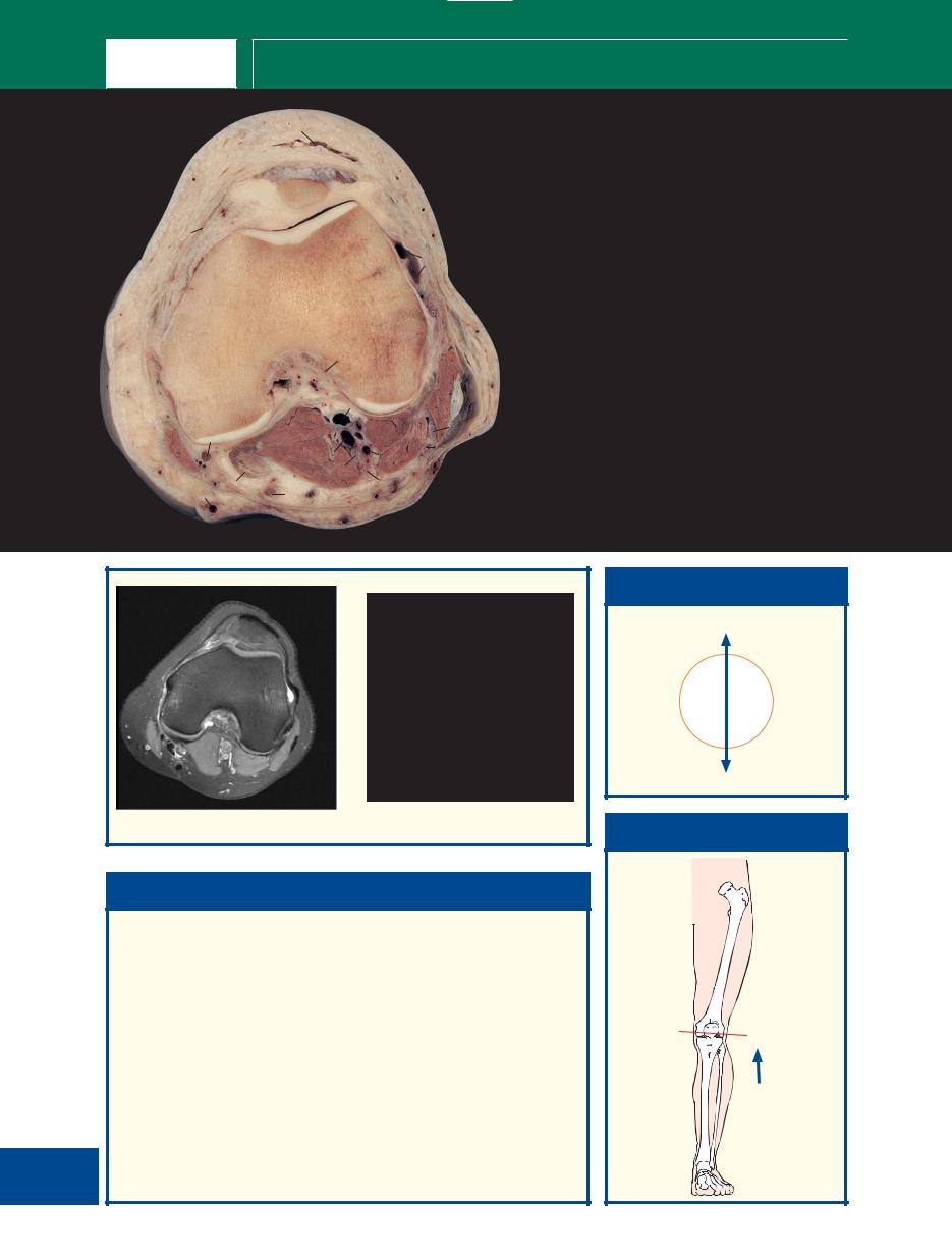
Thigh left – Axial section 1 – Male
|
|
|
|
|
|
1 |
Rectus femoris |
12 |
Semimembranosus |
|
|
|
|
1 |
2 |
2 |
Vastus lateralis |
13 |
Fascia lata (deep |
|
|
|
|
3 |
3 |
Vastus intermedius |
|
fascia of thigh) |
|
|
|
|
|
|
4 |
Femur |
14 |
Great saphenous |
|
|
|
|
|
|
|
||||
|
|
23 |
|
|
|
5 |
Lateral |
|
vein |
|
|
|
4 |
|
|
intermuscular |
15 |
Gracilis |
|
|
|
|
|
|
|
||||
|
|
|
|
|
|
|
septum |
16 |
Adductor magnus |
22 |
|
19 |
|
18 |
|
6 |
Iliotibial tract |
17 |
Adductor longus |
|
|
|
|
||||||
|
|
17 20 |
|
|
|
7 |
Biceps femoris – |
18 |
Profunda femoris |
|
21 |
|
|
|
|
short head |
|
artery |
|
|
|
|
7 |
5 |
|
|
|||
|
|
|
|
8 |
Sciatic nerve |
19 |
Saphenous nerve |
||
|
|
|
|
|
|
9 |
Biceps femoris – |
20 |
Femoral vein |
14 |
|
|
|
|
|
|
long head |
21 |
Femoral artery |
|
|
|
|
6 |
10 Semitendinosus |
22 |
Sartorius |
||
|
|
|
16 |
8 |
|||||
15 |
|
|
9 |
11 Posterior cutaneous |
23 |
Vastus medialis |
|||
|
|
|
|||||||
|
|
|
|
|
nerve of thigh |
|
|
||
|
|
|
|
|
|
|
|
|
|
|
|
|
|
10 |
|
|
|
|
|
12
11
13
|
|
1 |
|
23 |
|
3 |
2 |
20/21 |
4 |
|
|
|
|
||
22 |
18 |
|
|
14 |
17 |
|
|
|
|
|
16 |
|
|
15 |
|
|
8 |
6 |
|
|
|
|
9 |
|
Axial computed tomogram (CT)
■ Notes
|
|
This section passes through the upper third of the thigh and provides a |
|
|
useful view of the three muscular compartments of the thigh: |
|
|
• The anterior compartment, containing quadriceps femoris, made |
|
|
up of the vasti (2, 3, 23) and rectus femoris (1), supplied by the |
|
|
femoral nerve. |
|
|
• The adductor compartment, containing the three adductors (of |
|
|
which only adductor magnus (16) and adductor longus (17) are |
|
|
present at this level, brevis having already found insertion into the |
|
|
femoral shaft), together with gracilis (15). These muscles are |
|
|
supplied by the obturator nerve; in addition, adductor magnus |
|
|
receives innervation from the sciatic nerve. |
|
|
• The posterior compartment contains the hamstrings, the biceps |
|
|
with its long (9) and short heads (7), semitendinosus (10) and |
212 |
|
semimembranosus (12), all supplied by the sciatic nerve. |
|
Sartorius (22) lies in a separate fascial sheath. |
|
|
|
|
|
|
|
■ Orientation
Anterior
Medial 
 Lateral
Posterior
■ Section level
View

Thigh left – Axial section 2 – Male
LOWER LIMB
|
|
|
1 |
|
|
1 |
Rectus femoris |
14 |
Great saphenous |
|
|
|
|
|
2 |
Vastus lateralis |
|
vein |
|
|
|
|
|
|
2 |
|
|||
|
|
|
|
|
3 |
Vastus intermedius |
15 |
Gracilis |
|
|
|
|
|
|
|
||||
|
|
|
|
3 |
|
4 |
Femur |
16 |
Adductor magnus – |
|
|
23 |
|
|
|
5 |
Lateral |
|
medial part |
|
|
|
|
4 |
|
|
intermuscular |
17 |
Adductor magnus – |
|
|
|
|
|
|
|
septum |
|
lateral part |
|
|
20 |
18 |
|
|
6 |
Iliotibial tract |
18 |
Profunda femoris |
|
|
|
|
|
7 |
Biceps femoris – |
|
artery |
|
|
|
|
|
|
|
|
|||
22 |
21 |
19 |
17 |
5 |
|
|
short head |
19 |
Saphenous nerve |
|
|
|
|||||||
|
|
|
|
|
8 |
Sciatic nerve |
20 |
Superficial femoral |
|
|
|
|
|
7 |
|
||||
|
|
16 |
|
|
9 |
Biceps femoris – |
|
vein |
|
|
|
8 |
|
|
6 |
long head |
21 |
Superficial femoral |
|
|
|
|
|
|
|||||
15 |
|
|
|
9 |
|
10 Semitendinosus |
|
artery |
|
|
12 |
|
|
11 Posterior cutaneous |
22 |
Sartorius |
|||
|
|
|
|
|
|
nerve of thigh |
23 |
Vastus medialis |
|
14 |
|
|
|
|
|
|
|||
|
|
|
|
|
12 Semimembranosus |
|
|
||
|
|
|
|
|
|
|
|
||
|
|
|
10 |
|
|
13 Fascia lata (deep |
|
|
|
|
|
|
|
|
|
|
fascia of thigh) |
|
|
13
11
|
|
|
1 |
|
|
|
|
22 |
|
3 |
|
|
|
23 |
|
|
|
|
15 |
|
4 |
|
2 |
|
|
|
|
|
|
|
16 |
17 |
|
|
5 |
|
|
|
|
||
|
12 |
10 |
|
9 |
8 |
|
|
|
|||
|
|
|
|
||
|
|
|
|
|
|
Axial magnetic resonance image (MRI)
■ Notes
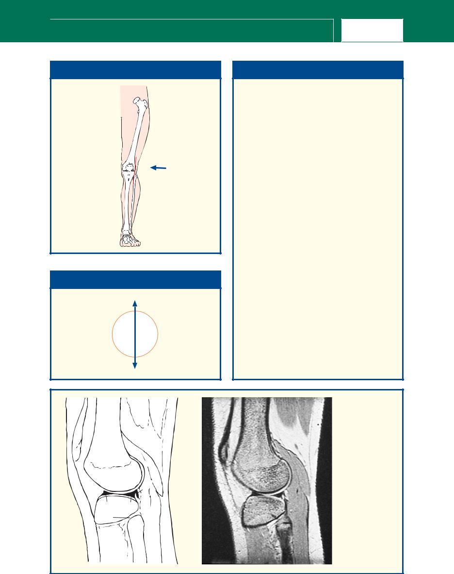
This section passes through the mid-shaft of the femur (4). Note that at this level, adductor magnus is dividing into two sections. Its lateral part (17), which arises from the ischial ramus, forms a broad aponeurosis, which inserts along the linea aspera along the posterior border of the femoral shaft (4). The medial part (16), which arises mainly from the ischial tuberosity, descends almost vertically to a tendinous attachment to the adductor tubercle of the medial condyle of the femur. Between the two parts distally is the osseo-aponeurotic adductor hiatus, which admits the femoral vessels to the popliteal fossa.
Being a composite muscle, adductor magnus also has a composite nerve supply; the medial part is innervated by the tibial division of the sciatic nerve (8) and the lateral part by the obturator nerve.
■ Orientation
Anterior
Medial 
 Lateral
Posterior
■ Section level
View
213

LOWER LIMB
Thigh left – Axial section 3 – Male
|
|
|
1 |
|
|
|
|
|
|
|
|
|
|
1 |
Rectus femoris |
12 |
Semimembranosus |
|
|
22 |
2 |
|
2 |
Vastus intermedius |
13 |
Fascia lata (deep |
|
|
|
|
|
3 |
Femur |
|
fascia of thigh) |
|
|
|
|
|
4 |
Vastus lateralis |
14 |
Gracilis |
|
|
20 |
3 |
|
5 |
Lateral |
15 |
Great saphenous |
21 |
|
|
|
|
intermuscular |
|
vein |
|
|
19 |
|
|
4 |
septum |
16 |
Adductor magnus |
|
|
|
|
|
|
||||
|
|
17 |
18 |
|
|
|||
|
16 |
|
6 |
Biceps femoris – |
17 |
Superficial femoral |
||
|
|
|
|
|||||
15 |
|
|
|
|
|
short head |
|
vein |
|
|
|
|
7 |
Biceps femoris – |
18 |
Profunda femoris |
|
14 |
|
|
|
5 |
||||
|
|
6 |
|
long head |
|
artery and vein |
||
|
|
|
|
|
|
|||
|
|
|
|
8 |
Iliotibial tract |
19 |
Saphenous nerve |
|
|
|
11 |
|
|
||||
|
|
|
|
9 |
Posterior cutaneous |
20 |
Superficial femoral |
|
|
|
12 |
|
|
|
nerve of thigh |
|
artery |
|
|
|
7 |
|
10 Semitendinosus |
21 |
Sartorius |
|
13 |
|
|
8 |
11 Sciatic nerve |
22 |
Vastus medialis |
||
|
|
|
||||||
|
10 |
|
|
|
|
|
||
|
|
|
|
|
|
|
|
|
|
|
9 |
|
|
|
|
|
|
|
|
1 |
|
22 |
2 |
|
4 |
|
|
|
|
|
|
3 |
|
21 |
|
|
16 |
5 |
|
14 |
6 |
|
12 |
7 |
|
|
10 |
|
|
|
Axial magnetic resonance image (MRI)
■ Notes
This section transects the lower third of the thigh. This and the previous two sections demonstrate the anatomy of the adductor, or subsartorial, canal (Hunter’s canal). This is formed as a triangular aponeurotic tunnel, which leads from the femoral triangle above to the popliteal fossa below, via the hiatus in adductor magnus. The canal lies between sartorius (21) anteromedially, adductor longus and, more distally, adductor magnus (16) posteriorly and vastus medialis (22) anterolaterally. Its contents are the femoral artery (20) and vein (17), the saphenous nerve (19) and the nerve to vastus medialis until this enters and supplies this muscle.
John Hunter (1728–93) described ligation of the femoral artery within this canal in the treatment of popliteal aneurysm, and his name is now used to describe the canal.
214
■ Orientation
Anterior
Medial 
 Lateral
Posterior
■ Section level
View

Knee left – Axial section 1 – Male
LOWER LIMB
|
|
1 |
|
|
|
|
|
|
|
|
2 |
|
|
|
|
|
|
|
|
3 |
|
|
1 |
Prepatellar bursa |
11 |
Tibial nerve |
|
20 |
|
|
2 |
Tendon of |
12 |
Popliteal vein |
|
|
4 |
|
5 |
|||||
|
|
|
|
quadriceps femoris |
13 |
Popliteal artery |
||
|
6 |
|
|
6 |
3 |
Patella |
14 |
Semimembranosus |
|
|
|
|
|
4 |
Articular cartilage of |
15 |
Semitendinosus |
|
|
7 |
|
|
|
patella |
16 |
Gracilis tendon |
|
|
|
|
|
5 |
Lateral patellar |
17 |
Sartorius |
|
|
|
|
|
|
retinaculum |
18 |
Great saphenous |
|
|
|
|
|
6 |
Capsule of knee |
|
vein |
|
19 |
|
8 |
|
|
joint |
19 |
Gastrocnemius |
|
13 |
12 |
9 |
7 |
Femur |
20 |
Tendon of vastus |
|
|
|
8 |
Plantaris origin |
|
medialis |
|||
|
|
|
|
|
|
|||
18 |
17 |
|
|
|
9 |
Biceps femoris |
|
|
|
|
|
10 Common fibular |
21 |
Vastus medialis |
|||
16 |
|
|
|
|||||
|
11 |
10 |
|
|||||
|
|
|
(peroneal) nerve |
|
|
|||
|
14 |
|
|
|
|
|||
|
15 |
|
|
|
|
|
|
|
|
|
|
|
3 |
|
|
|
|
7 |
|
|
|
19 |
|
17 |
16 |
|
13 |
9 |
18 |
|
14 |
12 |
|
|
|
|
|
|
|
15 |
|
|
|
Axial magnetic resonance image (MRI)
■ Notes
This section passes through the upper part of the patella (3) and the femur just as this widens into its condyles (7). Note how the lateral portion of the patella (3) has a larger and flatter articular surface than the medial surface. This, together with the low insertion of vastus medialis (20) into the medial side of the patella, helps to prevent lateral dislocation of the patella. The exact alignment of the patellar depends on the relative contributions of the vasti muscles via their tendons (medial and lateral retincacula).
The sciatic nerve has now divided into the common fibular (peroneal) nerve (10) and tibial nerve (11); the latter is usually about twice the size of the former. Division usually takes place just proximal to the knee, but the sciatic nerve may divide anywhere along its course. Indeed, its division may take place at the sciatic plexus, when the common fibular (peroneal) nerve usually pierces the piriformis muscle in the greater sciatic foramen and the tibial division emerges caudal to this muscle.
■ Orientation
Anterior
Medial 
 Lateral
Posterior
■ Section level
View
215
LOWER LIMB
Knee left – Axial section 2 – Male
|
|
1 |
|
|
1 |
Prepatellar bursa |
14 |
Sural |
|
|
|
|
|
||||
|
|
2 |
|
|
2 |
Ligamentum |
|
communicating |
|
|
3 |
|
|
|
patellae |
|
nerve |
|
|
4 |
|
3 |
Patella |
15 |
Gastrocnemius – |
|
|
|
|
|
|||||
6 |
|
|
|
|
4 |
Lateral patellar |
|
lateral head |
|
5 |
|
|
|
retinaculum |
16 |
Plantaris |
|
|
|
|
|
7 |
5 |
Articular cartilage of |
17 |
Small saphenous |
|
|
|
|
|
femur |
|
vein – termination |
|
|
|
|
|
|
|
|
||
|
|
|
|
|
6 |
Medial patellar |
18 |
Sural nerve |
|
|
|
|
|
|
retinaculum |
19 |
Tibial nerve |
|
|
9 |
|
|
7 |
Capsule of knee |
20 |
Popliteal vein |
|
|
|
|
|
joint |
21 |
Popliteal artery |
|
|
|
11 |
8 |
|
8 |
Lateral condyle of |
22 |
Gastrocnemius – |
|
|
|
|
|
femur |
|
medial head |
|
10 |
|
|
|
12 |
|
|
||
|
|
|
9 |
Intercondylar fossa |
23 |
Semitendinosus |
||
|
|
21 |
|
|
||||
|
|
|
|
10 Medial condyle of |
|
tendon |
||
|
|
|
|
13 |
|
|||
26 |
|
|
16 |
|
femur |
24 |
Semimembranosus |
|
|
22 |
15 |
14 |
11 Anterior cruciate |
|
tendon |
||
27 |
|
|
||||||
|
20 |
17 |
|
|
ligament |
25 |
Great saphenous |
|
|
|
19 |
18 |
|
|
|||
|
24 |
|
12 Biceps femoris |
|
vein |
|||
25 |
23 |
|
|
|||||
|
|
13 Common fibular |
26 |
Gracilis tendon |
||||
|
|
|
||||||
|
|
|
|
|
||||
|
|
|
|
|
|
(peroneal) nerve |
27 |
Sartorius |
6
2
5
4
26 |
10 |
|
|
|
|
|
8 |
27 |
|
21 |
|
|
|
|
|
|
22 |
20 |
12 |
|
|
15 |
|
|
|
|
|
|
23 |
|
|
Axial magnetic resonance image (MRI)
■ Notes
This section passes through the distal extremity of the patella (3) and the femoral condyles (8, 10).
The anterior cruciate ligament (11) arises from the intercondylar fossa (9) of the femur laterally and slightly more proximally than the posterior cruciate ligament, whose attachment is seen better in the next cadaveric section. The anterior cruciate ligament passes downwards and forwards laterally to the posterior cruciate ligament, to attach to the anterior intercondylar area of the tibia.
The small saphenous vein (17), which will be seen in later sections as it lies in the superficial fascia of the back of the calf, has here pierced the deep fascia of the popliteal fossa and is about to drain into the popliteal vein (20). On the magnetic resonance images, these veins are joining.
216
■ Orientation
Anterior
Medial 
 Lateral
Posterior
■ Section level
View

Knee left – Axial section 3 – Male
LOWER LIMB
1
2
3
5 |
4 |
8 |
|
|
6 |
|
|
7 |
|
|
|
|
|
|
|
|
|
9 |
|
|
17 |
|
16 |
10 |
|
27 |
|
11 |
|||
23 |
18 |
||||
|
|
|
|||
26 |
|
|
|
||
19 |
15 |
|
|
||
|
|
12 |
|||
25 |
|
|
|||
|
|
|
|||
|
|
|
|
||
24 |
22 |
20 |
14 |
13 |
|
|
|
|
|||
|
|
21 |
|
|
1 |
Infrapatellar bursa |
14 |
Gastrocnemius lateral |
2 |
Ligamentum patellae |
|
head |
3 |
Infrapatellar fat pad |
15 |
Plantaris |
4 |
Lateral patellar |
16 |
Popliteus |
|
retinaculum |
17 |
Posterior cruciate |
5 |
Medial patellar |
|
ligament |
|
retinaculum |
18 |
Popliteal artery |
6 |
Sliver of cartilage over |
19 |
Popliteal vein |
|
lateral condyle of tibia |
20 |
Tibial nerve |
7 |
Medial condyle of |
21 |
Small saphenous vein |
|
tibia |
22 |
Gastrocnemius – |
8 |
Medial collateral |
|
medial head |
|
ligament |
23 |
Semimembranosus |
9 |
Lateral meniscus |
|
tendon |
10 Lateral collateral |
24 |
Semitendinosus |
|
|
ligament |
|
tendon |
11 Tendon of biceps |
25 |
Great saphenous vein |
|
|
femoris |
26 |
Gracilis tendon |
12 Common fibular |
27 |
Sartorius tendon |
|
|
(peroneal) nerve |
|
|
13 Lateral cutaneous |
28 |
Semimembranosus |
|
|
nerve of calf |
|
bursa |
|
|
|
|
|
5 |
|
2 |
|
4 |
26 |
7 |
23 |
|
6 |
|
|
|
27 |
|
17 |
|
19 18 |
|
|
|
|
25 |
28 |
11 |
|
||
22 |
|
|
24 |
|
|
|
14 |
|
|
|
Axial magnetic resonance image (MRI)
■ Notes
This section passes through the tibial condyles (6, 7). The posterior cruciate ligament (17) is here finding attachment to the posterior intercondylar area of the proximal articular surface of the tibia.
The popliteus tendon (16), which inserts on to the femur in a depression immediately distal to the lateral epicondyle, passes between the lateral meniscus (9) and the lateral collateral ligament (10) of the knee. In contrast, the medial collateral ligament (8) is applied closely to the medial meniscus, which lies just proximal to this plane of section. This tethering of the medial meniscus probably accounts for the much higher incidence of tears of the medial compared with the lateral meniscus.
The semimembranosus bursa contains a trace of fluid on the magnetic resonance image (28). It can enlarge greatly to form a popliteal cyst (a misnomer).
■ Orientation
Anterior
Medial 
 Lateral
Posterior
■ Section level
View
217

LOWER LIMB
Knee left – Coronal section 1 – Male
1 |
3 |
|
|
|
2 |
4
5
8
|
7 |
|
6 |
|
|
|
|
|
|
9 |
10 |
|
|
|
|
|
|
|
11 |
|
12 |
13 |
14 |
|
|
||
21 |
|
|
15 |
17 |
|
16 |
24 |
18 |
19 |
23
20
22
|
1 |
Vastus medialis |
16 |
Lateral condyle (plateau) of tibia |
|
2 |
Shaft of femur |
17 |
Medial condyle (plateau) of tibia |
|
3 |
Vastus lateralis |
18 |
Tibia |
|
4 |
Fascia lata |
19 |
Extensor digitorum longus |
|
5 |
Superior lateral genicular artery |
20 |
Tibialis anterior |
|
6 |
Lateral condyle of femur |
21 |
Medial collateral ligament |
|
7 |
Medial condyle of femur |
22 |
Popliteus (most medial fibres) |
|
8 |
Adductor tubercle of femur |
23 |
Tendon of gracilis |
|
9 |
Posterior cruciate ligament |
24 |
Tendon of sartorius |
|
10 Anterior cruciate ligament |
|
|
|
|
11 Lateral meniscus |
25 |
Popliteus tendon |
|
|
12 Medial meniscus |
26 |
Lateral collateral ligament |
|
|
13 Medial intercondylar eminence/tubercle |
27 |
Head of fibula |
|
|
|
(also known as spine) |
28 |
Great saphenous vein |
218 |
14 Lateral intercondylar eminence/tubercle |
29 |
Medial gastrocnemius |
|
|
(also known as spine) |
30 |
Biceps femoris |
|
|
|
|||
|
15 Articular cartilage |
|
|
|

Knee left – Coronal section 1 – Male
LOWER LIMB
■ Section level |
■ Notes
The posterior cruciate ligament (9) lies on the medial side of the anterior cruciate ligament (10). The former prevents posterior sliding movement of the tibia on the femur, while the latter prevents anterior displacement and resists torsional movement at the knee joint. They may be torn in violent torsional injury of the knee especially in the flexed position, when the collateral ligaments (21) are less tense.
It can be seen that the menisci (11, 12) do little to deepen the concavity of the knee joint on either side. They do act, however, as ‘shock absorbers’ at the knee, for example when jumping from a height.
Note that the medial collateral ligament is continuous with the medial meniscus, whereas the lateral collateral ligament is discontinuous with the lateral meniscus. This contributes to the medial meniscus being more static and being injured more commonly; the lateral meniscus is more mobile.
■ Orientation
Proximal
Medial 
 Lateral
Distal
|
|
|
|
|
|
|
|
|
|
|
|
1 |
|
|
3 |
|
|
|
|
|
|
|
|
|
|
|
|
|
|
|
|
|
|
|
|
29 |
|
30 |
|
|
|
|
|
|
|
|
|
2 |
|
4 |
|
|
|
|
|
|
|
|
|
|
|
|
|
|
|
||
24 |
7 |
10 |
6 |
|
|
|
|
9 |
10 |
11 |
|
7 |
9 |
10 |
25 |
|
|
21 |
9 |
|
7 |
6 |
|
8 |
|
||||||||
28 |
|
|
|
|
|
12 |
|
|
|
|||||||
|
|
12 |
|
|
21 |
|
|
|
26 |
|||||||
|
12 |
|
|
11 |
|
|
|
13 |
14 |
11 |
||||||
|
|
|
26 |
|
|
|
|
16 |
|
|
|
|
||||
|
17 |
|
16 |
|
|
|
|
|
|
17 |
|
|
16 |
|
||
|
|
|
|
|
|
17 |
|
24 |
|
|
|
|||||
|
|
|
|
|
|
|
|
|
|
|
||||||
|
|
18 |
|
27 |
|
|
21 |
|
18 |
|
28 |
|
18 |
27 |
|
|
A |
|
|
|
|
|
C |
|
|
||||||||
29 |
|
B |
|
|
|
|
|
|||||||||
|
|
|
|
|
|
|
|
|
|
|
|
|||||
|
|
|
|
|
|
|
|
|
|
|
|
|
||||
|
|
|
|
|
|
|
|
|
|
|
|
|
|
|
|
|
A |
|
B |
|
C |
219 |
|
|
|
|
|
|
|
|
|
|
|
Coronal magnetic resonance images (MRIs)

LOWER LIMB
Knee left – Sagittal section 1 – Female
5 |
1 |
4 |
3 |
|
2 |
6
7
|
|
8 |
|
|
|
|
9 |
|
12 |
|
|
|
11 |
|
13 |
|
|
|
10 |
|
|
|
|
|
|
|
|
14 |
|
|
|
|
|
|
|
|
|
15 |
|
|
|
|
17 |
16 |
16 |
21 |
|
|
|
|
|||
|
18 |
|
20 |
|
|
|
19 |
|
|
|
|
|
|
|
|
|
|
|
24 |
|
22 |
|
|
|
|
23 |
|
||
|
|
|
31 |
|
25 |
|
|
|
|
|
|
|
|
|
|
27 |
26 |
|
|
32 |
29 |
|
|
|
|
30 |
28 |
|
|
|
|
|
|
|
|
1 |
Superficial fascia |
|
17 |
Infrapatellar pad of fat extending into |
|
2 |
Deep fascia |
|
|
infrapatellar fold |
|
3 |
Biceps femoris |
|
18 |
Ligamentum patellae |
|
4 |
Vastus intermedius |
|
19 |
Lateral condyle (plateau) of tibia |
|
5 |
Vastus lateralis |
|
20 |
Tendon of popliteus |
|
6 |
Tendon of quadriceps femoris |
|
21 |
Plantaris |
|
7 |
Suprapatellar bursa |
|
22 |
Superior tibiofibular joint |
|
8 |
Lateral superior geniculate artery and |
23 |
Head of fibula |
||
|
vein |
|
24 |
Infrapatellar bursa |
|
9 |
Patella |
|
25 |
Gastrocnemius lateral |
|
10 Prepatellar bursa |
|
26 |
Soleus |
|
|
11 Lateral condyle of femur |
|
27 |
Neck of fibula |
||
12 Fibrous capsule of knee joint |
|
28 |
Shaft of fibula |
||
13 Common fibular (peroneal) nerve |
29 |
Anterior tibial artery and vein |
|||
14 Lateral cutaneous nerve of calf |
|
30 |
Interosseous membrane |
||
15 Articular cartilage |
|
31 |
Tibialis posterior |
||
16 Lateral meniscus |
|
32 |
Tibialis anterior |
||
220
Knee left – Sagittal section 1 – Female
LOWER LIMB
■ Section level |
■ Notes |
View
■ Orientation
Proximal
Anterior 
 Posterior
Distal
The prepatellar bursa (10) and infrapatellar bursa (24) are both subcutaneous. Either may become inflamed by continual kneeling, which produces a traumatic bursitis. A prepatellar bursa comes into contact with the ground on scrubbing the floor (hence ‘housemaid’s knee’), while the infrapatellar bursa does so when kneeling to pray (hence ‘clergyman’s knee’).
The communication of the suprapatellar bursa (7) with the main synovial cavity of the knee is demonstrated well. It extends a hand’s breadth superior to the border of the patella (9) and lies posterior to the quadriceps tendon (6). It becomes distended when there is an effusion into the knee joint. A puncture wound within a hand’s breadth of the superior border of the patella must always be suspected of having penetrated the knee joint. Failure to do so may result in septic arthritis of the knee.
Plantaris (21) is absent in about ten per cent of subjects. Very rarely, it has two heads.
The tendon of popliteus (20) is connected to the lateral meniscus (16). It may thus retract and protect the mobile lateral meniscus during lateral rotation of the femur in flexion of the knee joint, protecting the meniscus from being crushed between the femoral and tibial condyles during this movement.
The superior tibiofibular joint (22) is a plane synovial joint, in contrast to the fibrous inferior tibiofibular joint.
The lateral meniscus is of even thickness throughout. Thus, a lateral sagittal slice creates a bowtie appearance to this portion of the lateral meniscus.
Sagittal magnetic 3 resonance image
(MRI)
6
 9
9
11 |
25 |
|
20 |
16 16
19
22
23
27 26
32/31
28 |
221 |
|
|
LOWER LIMB
Knee left – Sagittal section 2 – Female
1
54
3 2
|
|
|
6 |
|
10 |
9 |
|
|
|
|
11 |
12 |
|
|
|
|
|
|
|
|
8 |
|
|
|
13 |
|
|
7 |
6 |
|
|
|
22 |
|
|
|
|
|
|
14 |
15 |
|
|
|
|
|
|
|
|
|
|
|
|
23 |
16 |
19 |
18 |
20 |
21 |
|
6 |
|||
|
18 |
|
|
|
17 |
|
|
|
|
|
|
|
24 |
|
|
29 |
|
||
|
|
|
||
|
30 |
|
|
|
|
|
|
28 |
|
|
|
|
|
25 |
|
|
|
|
6 |
|
|
31 |
|
26 |
|
|
|
|
27 |
|
1 |
Semitendinosus |
17 |
Ligamentum patellae |
|
2 |
Semimembranosus |
18 |
Medial meniscus |
|
3 |
Sciatic nerve |
19 |
Anterior cruciate ligament |
|
4 |
Vastus intermedius |
20 |
Posterior cruciate ligament |
|
5 |
Rectus femoris |
21 |
Fibrous capsule of knee joint |
|
6 |
Popliteal vein |
22 |
Superficial fascia |
|
7 |
Popliteal artery |
23 |
Deep fascia |
|
8 |
Popliteal surface of femur |
24 |
Gastrocnemius |
|
9 |
Shaft of femur |
25 |
Tendon of plantaris |
|
10 Tendon of quadriceps femoris |
26 |
Soleus |
|
|
11 Suprapatellar bursa |
27 |
Tibial nerve |
|
|
12 Popliteal pad of fat |
28 |
Popliteus |
|
|
13 Patella |
29 |
Proximal end of tibia |
|
|
14 Prepatellar bursa |
30 |
Tibial tuberosity |
|
222 |
15 Articular cartilage |
31 |
Shaft of tibia |
|
16 Infrapatellar pad of fat (Hoffa) |
|
|
||
|
|
extending into infrapatellar fold |
32 |
Transverse intermeniscal ligament |

Knee left – Sagittal section 2 – Female
LOWER LIMB
|
|
|
|
2 |
|
|
|
1 |
|
|
|
|
10 |
|
2 |
||
|
|
|
|
|
|
|||
|
|
|
|
|
|
|
||
10 |
|
9 |
|
|
|
|
|
9 |
|
|
7 |
|
|
|
|
||
|
|
|
|
13 |
|
20 |
||
|
|
|
|
|
|
|||
13 |
|
|
|
|
|
|||
|
|
6 |
|
|
|
|
||
|
|
|
|
|
|
|
|
|
16 |
|
19 |
|
|
|
16 |
24 |
|
|
7 |
|
|
|
|
|||
|
24 |
|
|
|
|
|||
17 |
32 |
18 |
17 |
|
19 |
|||
|
|
29 |
||||||
20 |
|
|
||||||
|
|
|
|
|
|
|
||
|
|
|
29 |
25 |
|
|
|
30 |
|
|
|
28 |
|
|
|
|
|
|
|
|
|
|
|
|
28 |
|
|
|
|
|
|
|
|
|
|
|
|
|
|
|
|
|
|
26 |
A |
|
|
|
26 |
B |
|
|
|
|
31 |
|
|
31 |
||||
|
|
|
|
|
|
|
|
|
A |
B |
Sagittal magnetic resonance images (MRIs)
■ Section level
View
■ Orientation
Proximal
Anterior 
 Posterior
Distal
■ Notes
The relationships in the popliteal fossa comprise the |
Both gastrocnemius (24) and soleus (26) contain |
tibial nerve (27) most superficially, the popliteal vein |
large veins, an important component of the calf |
(6) and then, more deeply, the popliteal artery (7). |
pump mechanism in venous return from the lower |
The valves in the vein are shown well. It is within |
limb. Note also the density of the deep fascia (23), |
these large veins that postoperative (or post- |
which assists the pumping action of the muscles. |
immobilization) thrombosis of the deep veins of the |
Note that with the knee in the extended position, |
lower limb usually commences. |
the anterior cruciate ligament is taut and straight; |
The fossa contains a large amount of fat (12) as |
there is less tension on the posterior cruciate, which |
well as the rather insignificant popliteal lymph nodes, |
appears curved in that position. The cruciate |
usually five or six in number. Note the composition of |
ligaments take their names (anterior and posterior) |
the floor of the popliteal fossa comprises superiorly |
from the site of attachment to the tibia. The anterior |
the popliteal surface of the femur (8), the capsule of |
cruciate passes lateral to the posterior ligament. |
the knee joint (21) and finally popliteus (28). |
|
223

LOWER LIMB
Knee left – Sagittal section 3 – Female
3
2
1
4
1
5
6
|
8 |
|
|
7 |
9 |
|
|
|
|
14 |
10 |
13 |
13 |
11 |
|
|
|
|
15 |
12 |
5
1 |
Semimembranosus |
9 Medial head/tendon of |
|
2 |
Adductor magnus |
|
gastrocnemius |
3 |
Femoral artery |
10 |
Tendon of |
4 |
Vastus medialis |
|
semitendinosus |
5 |
Medial gastrocnemius |
11 |
Superficial fascia |
6 |
Suprapatellar bursa |
12 |
Deep fascia |
7 |
Medial condyle of |
13 |
Medial meniscus |
|
femur |
14 |
Articular cartilage |
8 |
Fibrous capsule of knee |
15 |
Medial condyle |
|
joint |
|
(plateau of tibia) |
224

Knee left – Sagittal section 3 – Female
LOWER LIMB
■ Section level
View
■ Orientation
Proximal
Anterior 
 Posterior
Distal
■ Notes
The femoral artery (3) passes through the hiatus in adductor magnus (2) to become the popliteal artery about two-thirds of the distance along a line that joins the femoral pulse at the groin, with the adductor tubercle on the medial condyle of the femur.
The posterior third of the medial meniscus is usually a little thicker than the mid and anterior thirds, in contrast to the lateral meniscus, which is of constant thickness around its circumference. Furthermore, the posterior third frequently undergoes myxoid change during early middle age; thus, this part of the medial meniscus often appears rather heterogeneous in consistency.
This section shows the possible consequence of a fracture of the shaft of the femur at its lower extremity. The medial (9) and lateral heads of gastrocnemius tilt the otherwise unsupported distal femoral fragment posteriorly. This may well injure the popliteal vessels, lying immediately behind. (See also Sagittal section 2.)
4
1
10
9
7
13
13
15
Sagittal magnetic resonance image (MRI)
5
225

LOWER LIMB
Leg left – Axial section 1 – Male
|
|
|
|
|
|
|
|
|
1 |
Subcutaneous |
12 |
Gastrocnemius – |
|
|
|
|
|
|
|
|
|
|
surface of tibia |
|
lateral head |
|
|
|
4 |
|
|
|
|
|
2 |
Tibia |
13 |
Small saphenous |
|
|
|
|
5 |
|
|
|
3 |
Vertical ridge of |
|
vein |
|
|
|
|
|
|
|
|
|
|
||||
|
|
|
|
|
|
|
|
|
tibia |
14 |
Deep fascia of calf |
|
|
|
|
|
|
|
|
|
|
|
|||
1 |
|
|
|
|
|
|
|
|
4 |
Tibialis anterior |
15 |
Gastrocnemius – |
2 |
|
|
6 |
|
7 |
|
|
8 |
5 |
Extensor digitorum |
|
medial head |
|
|
|
|
|
|
|
|
|||||
|
|
|
|
|
|
|
|
|
longus |
16 |
Great saphenous |
|
|
|
|
24 |
|
|
9 |
|
|
||||
|
3 |
|
|
10 |
|
6 |
Interosseous |
|
vein |
|||
|
|
|
22 |
|
|
|
|
|||||
|
|
|
|
|
|
|
|
membrane |
17 |
Plantaris tendon |
||
23 |
|
|
|
|
|
|
|
|
||||
|
|
|
|
11 |
|
|
|
|
||||
|
|
|
|
|
|
|
7 |
Anterior tibial artery |
18 |
Soleus |
||
|
|
21 |
|
|
|
|
|
|||||
|
|
|
20 |
|
|
|
|
|
and vein, with deep |
19 |
Tibial nerve |
|
|
|
19 |
|
|
|
|
|
fibular (peroneal) |
20 |
Posterior tibial |
||
|
|
|
|
|
|
|
|
|||||
|
|
17 |
|
18 |
|
|
|
|
|
nerve |
|
artery |
16 |
|
|
|
|
12 |
|
|
8 |
Fibularis (peroneus) |
21 |
Posterior tibial vein |
|
|
|
|
|
|
|
|
||||||
|
|
|
|
|
|
|
|
|
|
longus |
22 |
Fibular (peroneal) |
15 |
|
|
|
|
|
|
|
|
9 |
Superficial fibular |
|
artery |
|
|
|
|
|
|
|
|
|
|
(peroneal) nerve |
23 |
Popliteus |
14 |
|
|
|
|
|
|
|
|
10 Fibula |
24 |
Tibialis posterior |
|
|
|
|
|
|
|
|
|
11 Medial crest of fibula |
|
|
||
|
|
|
|
|
13 |
|
|
|
|
|
||
|
|
|
|
|
|
|
|
|
|
|
|
|
|
|
4 |
|
|
2 |
5 |
|
|
23 |
24 |
8 |
14 |
10 |
|
|
|
|
|
|
16 |
18 |
19/20/21 |
|
15 |
12 |
|
|
|
|
|
Axial computed tomogram (CT)
■ Notes
This section traverses the proximal end of the tibial shaft (2) and the shaft of the fibula (10) immediately distal to the neck of the fibula.
At this level, the common fibular (peroneal) nerve, which sweeps around the neck of the fibula deep to fibularis (peroneus) longus (8), has divided into its superficial fibular (peroneal) (9) and deep fibular (peroneal) (7) branches. The superficial fibular (peroneal) nerve lies deep to fibularis (peroneus) longus. The deep fibular (peroneal) nerve passes obliquely forwards, deep to extensor digitorum longus (5), to descend with the anterior tibial vessels (7).
The tendon of plantaris (17) lies in a well-defined tissue plane between soleus (18) and gastrocnemius (12, 15). Fluid enters this plane following rupture of a semimembranosus bursa (Baker’s cyst).
■ Orientation
Anterior
Medial 
 Lateral
Posterior
■ Section level
View
226

Leg left – Axial section 2 – Male
LOWER LIMB
|
|
|
|
|
1 |
Subcutaneous |
12 |
Flexor hallucis |
|
|
|
|
|
|
border of tibia |
|
longus |
|
|
3 |
|
|
2 |
Tibia |
13 |
Deep fascia of calf |
|
|
|
|
3 |
Tibialis anterior |
14 |
Fibular (peroneal) |
|
|
1 |
|
|
|
||||
|
|
5 |
4 |
4 |
Superficial fibular |
|
artery, with venae |
|
|
2 |
|
|
|
|
(peroneal) nerve |
|
comitantes |
|
|
|
6 |
|
|
|
||
|
|
|
|
5 |
Extensor digitorum |
15 |
Tibial nerve |
|
|
|
8 |
7 |
|
||||
|
|
9 |
|
longus |
16 |
Venae comitantes of |
||
|
|
|
|
|||||
|
23 |
18 |
|
10 |
6 |
Extensor hallucis |
|
posterior tibial |
22 |
|
|
longus |
|
artery |
|||
|
11 |
|
|
|
||||
|
|
|
|
|
||||
|
17 |
16 14 |
|
7 |
Anterior tibial artery |
17 |
Posterior tibial |
|
|
|
|
||||||
|
|
12 |
|
|
and vein, with deep |
|
artery |
|
|
|
|
|
|
|
|||
|
|
|
|
|
fibular (peroneal) |
18 |
Tibialis posterior |
|
|
16 |
15 |
|
|
|
|||
|
|
|
|
nerve |
19 |
Soleus |
||
|
|
|
|
|
|
|||
|
|
|
|
|
8 |
Interosseous |
20 |
Plantaris tendon |
|
|
19 |
|
|
|
membrane |
21 |
Gastrocnemius |
|
|
|
|
9 |
Fibularis (peroneus) |
22 |
Great saphenous |
|
|
|
20 |
|
|
||||
|
|
|
|
|
brevis |
|
vein |
|
|
21 |
|
13 |
|
|
|
||
|
|
|
10 Fibularis (peroneus) |
23 |
Flexor digitorum |
|||
|
|
|
|
|
||||
|
|
|
|
|
|
longus |
|
longus |
|
|
|
|
|
11 Fibula |
|
|
|
|
|
3 |
|
|
2 |
6 |
5 |
|
23 |
18 |
9 |
|
|
||
|
|
|
|
|
15/16/17 |
1211 |
10 |
|
19 |
|
|
|
|
|
|
|
|
21 |
|
|
|
|
|
Axial computed tomogram (CT)
■ Notes
This section traverses the mid-calf. Note that the whole of the anteromedial aspect of the shaft of the tibia (1) is subcutaneous, covered only by skin, superficial fascia and periosteum, and crossed, in its lower part, only by the great saphenous vein (22) and saphenous nerve.
The neurovascular bundle of the anterior tibial vessels and deep fibular (peroneal) nerve (7), having descended first between extensor digitorum longus (5) and tibialis anterior (3), now runs between the latter and extensor hallucis longus (6), as this takes origin from the anterior aspect of the fibular shaft (11).
■ Orientation
Anterior
Medial 
 Lateral
Posterior
■ Section level
View |
227 |
|

LOWER LIMB
Ankle left – Axial section 1 – Male
1 |
|
|
|
|
|
|
|
3 |
1 |
Tibialis anterior tendon |
11 |
Fibula |
|
2 |
4 |
2 |
Anterior tibial artery, |
12 |
Fibularis (peroneus) |
|
|
||||||
|
|
with venae comitantes |
|
longus tendon |
||
|
|
|
|
|||
26 |
6 |
|
and deep fibular |
13 |
Fibularis (peroneus) |
|
5 |
|
(peroneal) nerve |
|
brevis |
||
|
|
|
||||
7 |
3 |
Extensor hallucis longus |
14 |
Sural nerve |
||
|
||||||
25 |
8 |
|
and tendon |
15 |
Small saphenous vein |
|
4 |
Extensor digitorum |
16 |
Tendo calcaneus |
|||
24 |
|
|
longus tendon |
|
(Achilles tendon) |
|
|
9 |
|
|
|||
|
5 |
Tibia |
17 |
Soleus |
||
|
10 |
|||||
|
6 |
Fibularis (peroneus) |
18 |
Plantaris tendon |
||
23 |
11 |
|||||
|
tertius |
19 |
Flexor hallucis longus |
|||
|
|
|||||
|
19 |
|
||||
|
7 |
Superficial fibular |
20 |
Tibial nerve |
||
|
|
|||||
|
|
12 |
(peroneal) nerve |
21 |
Posterior tibial vein |
|
|
|
|
||||
22 |
13 |
8 |
Perforating branch of |
22 |
Posterior tibial artery |
|
20 |
|
|
fibular (peroneal) artery |
23 |
Flexor digitorum longus |
|
21 |
|
9 |
Inferior tibiofibular |
|
and tendon |
|
|
|
|
||||
17 |
14 |
|
joint (interosseous |
24 |
Tibialis posterior tendon |
|
|
ligament) |
25 |
Great saphenous vein |
|||
18 |
15 |
10 Fibular (peroneal) artery |
26 |
Saphenous nerve |
||
16 |
|
|
|
|
||
|
|
|
|
|
||
|
|
1 |
|
|
|
3 |
4 |
|
|
|
|
24 |
5 |
|
6 |
|
|
||
23 |
|
11 |
12 |
20 |
19 |
|
|
21 |
13 |
|
|
|
|
||
22 |
|
|
|
16
14/15
Axial computed tomogram (CT)
■ Notes
This section passes immediately above the ankle joint at the level of the inferior tibiofibular joint (9). This is the only fibrous joint, apart from the skull sutures, and represents the thickened distal extremity of the interosseous membrane. (See also Axial section 2.)
At this level, gastrocnemius has already become tendinous (16), although soleus (17) still displays muscle fibres. A little more distally, this too will become tendinous and fuse into the tendo calcaneus (tendo Achilles tendon).
■ Orientation
Anterior
Medial 
 Lateral
Posterior
■ Section level
228
View

Ankle left – Axial section 2 – Male
LOWER LIMB
12
|
4 |
5 |
|
|
3 |
6 |
|
|
|
||
25 |
|
|
|
10 |
11 |
|
|
24 |
|
||
|
|
||
9 |
|
7 |
|
23 |
|
||
|
|
||
|
8 |
|
|
|
|
12 |
|
22 |
|
13 |
|
|
19 |
15 |
|
21 |
20 |
||
|
|||
|
|
14 |
18 |
16 |
|
|
|
17 |
1 |
Tibialis anterior tendon |
13 |
Fibularis (peroneus) |
2 |
Extensor hallucis longus |
|
longus |
|
and tendon |
14 |
Small saphenous vein |
3 |
Anterior tibial artery |
15 |
Sural nerve |
|
and venae comitantes, |
16 |
Fat |
|
with deep fibular |
17 |
Tendo calcaneus |
|
(peroneal) nerve |
18 |
Plantaris tendon |
4 |
Extensor digitorum |
19 |
Flexor hallucis longus |
|
tendon |
|
tendon |
5 |
Superficial fibular |
20 |
Tibial nerve |
|
(peroneal) nerve |
21 |
Posterior tibial artery, |
6 |
Fibularis (peroneus) |
|
with venae comitantes |
|
tertius and tendon |
22 |
Flexor digitorum longus |
7 |
Lateral malleolus |
|
tendon |
8 |
Inferior tibiofibular joint |
23 |
Tibialis posterior tendon |
9 |
Ankle joint |
24 |
Great saphenous vein |
10 Medial malleolus |
25 |
Saphenous nerve |
|
11Talus
12Fibularis (peroneus) brevis tendon
|
1 |
|
|
|
2 |
|
|
10 |
4 |
6 |
|
|
11 |
|
|
|
|
|
|
23 |
|
7 |
|
22 |
|
|
|
|
19 |
12 |
|
|
13 |
|
|
|
21/20 |
|
|
|
17 |
|
|
|
|
|
|
Axial computed tomogram (CT)
■ Notes
This section passes through the ankle joint (9) and the inferior tibiofibular joint (8). Note that this section illustrates the fibrous nature of the inferior tibiofibular joint.
Fibularis (peroneus) brevis (12) and fibularis (peroneus) longus (13) pass behind the lateral malleolus (7) of the fibula and will groove the bone a little more distally to form the malleolar fossa.
This section demonstrates the order of structures that pass behind the medial malleolus (10). These are, from the medial to the lateral side, the tendon of tibialis posterior (23), the tendon of flexor digitorum longus (22), the posterior tibial artery with its venae comitantes (21), the tibial nerve (20) and, most laterally, the tendon of flexor hallucis longus (19).
■ Orientation
Anterior
Medial 
 Lateral
Posterior
■ Section level
229
View

LOWER LIMB
Ankle left – Axial section 3 – Male
|
27 |
|
|
|
26 |
|
|
|
1 |
|
|
4 |
|
|
|
|
|
2 |
|
25 |
|
|
|
3 |
|
|
|
|
|
24 |
5 |
|
|
|
23 |
|
|
|
8 |
|
|
|
|
|
22 |
6 |
7 |
|
|
|
|
9 |
||
|
20 |
|
|
|
|
|
|
10 |
|
|
|
|
|
|
21 |
19 |
|
|
13 |
|
|
|
|
|
18 |
|
|
|
12 |
|
|
|
11 |
14 |
|
|
|
|
17
16
15
1 |
Extensor digitorum longus tendon |
17 |
Lateral plantar neurovascular |
2 |
Extensor digitorum brevis |
|
bundle |
3 |
Fibularis (peroneus) tertius tendon |
18 |
Medial plantar neurovascular |
4 |
Talocalcaneonavicular joint |
|
bundle |
|
(anterior talonavicular part) |
19 |
Flexor hallucis longus tendon |
5 |
Head of talus |
20 |
Sustentaculum tali |
6 |
Talocalcaneonavicular joint |
21 |
Flexor digitorum longus tendon |
|
(posterior part) |
22 |
Tibialis posterior tendon |
7 |
Interosseous talocalcanean |
23 |
Deltoid ligament of ankle |
|
ligament |
24 |
Great saphenous vein |
8 |
Sulcus tali (arrowed) |
25 |
Saphenous nerve |
9 |
Lateral process of talus |
26 |
Tibialis anterior tendon |
10 Talocalcanean (subtalar) joint |
27 |
Extensor hallucis longus tendon |
|
11 Calcaneus |
|
|
|
12 Capsule of talocalcanean joint |
28 |
Tibia |
|
13 Fibularis (peroneus) brevis tendon |
29 |
Medial malleolus |
|
14 Fibularis (peroneus) longus tendon |
30 |
Abductor hallucis |
|
15 Tendo Achilles |
31 |
Abductor digiti minimi |
|
16Quadratus plantae (flexor accessorius)
230

Ankle left – Axial section 3 – Male
LOWER LIMB
■ Section level |
|
26 |
27/1 |
|
28 |
29 |
|
|
3 |
22/21 |
5 |
|
|
18/17 |
13 |
|
|
19 |
11 |
|
|
16 |
14 |
|
|
30 |
31 |
|
|
Axial computed tomogram (CT) |
|
View |
|
■ Orientation |
|
Anterior |
|
Medial
 Lateral
Posterior
■ Notes |
|
|
|
|
|
|
|
|
|
|
|
|
|
|
|
This section passes through the head (5) and lateral |
concave posterior facet on the inferior surface of the |
|
|
process (9) of the talus and the calcaneus (11). The |
talus. The talocalcaneonavicular joint is complex. It is |
|
|
CT image is in a more coronal plane, and hence the |
formed by the rounded head of the talus (5), which |
|
|
tibia (28) is seen with its articulation with the talus |
fits into the concavity on the posterior aspect of the |
|
|
(5). |
navicular, the upper surface of the plantar |
|
|
The tendon of flexor hallucis longus (19) passes |
calcaneonavicular ligament (the spring ligament), |
|
|
behind the sustentaculum tali (20) and, more distally, |
which runs between the sustentaculum tali and the |
|
|
grooves its inferior aspect. The sulcus tali (8), with its |
inferior aspect of the navicular, and the anterior and |
|
|
corresponding sulcus calcanei, forms the sinus tarsi |
middle facets for the talus on the calcaneus. The |
|
|
and contains the strong interosseous talocalcanean |
anterior (4) and posterior (6) portions of this joint are |
|
|
ligament. |
shown. |
|
|
The talocalcanean joint (10), also termed the |
A considerable degree of inversion and eversion of |
|
|
subtalar joint, lies between the convex posterior facet |
the foot takes place at the talocalcanean and |
|
|
on the upper surface of the calcaneus and the |
talocalcaneonavicular joints. |
|
231 |
|
|
|
|
|
|
|
|
LOWER LIMB
Ankle left – Coronal section 1 – Female
1
4
2
3
7
6 |
5 |
8
9
10
11
23 |
|
12 |
|
|
|
|
|
|
21 |
13 |
|
|
|
|
|
22 |
|
14 |
15 |
|
|
|
|
20 |
|
24 |
|
|
|
|
16 |
19
17
18
1 |
Tibialis posterior |
14 |
Cuboid |
2 |
Inferior tibiofibular joint |
15 |
Tendon of fibularis |
3 |
Small saphenous vein |
|
(peroneus) brevis |
4 |
Tibia |
16 |
Tendon of fibularis |
5 |
Lateral malleolus of fibula |
|
(peroneus) longus |
6 |
Medial malleolus of tibia |
17 |
Abductor digiti minimi |
7 |
Ankle joint |
18 |
Plantar aponeurosis |
8 |
Body of talus (talar dome) |
19 |
Flexor digitorum brevis |
9 |
Lateral collateral ligament of |
20 |
Tendon of flexor digitorum |
|
ankle |
|
longus |
10 Talocalcanean interosseous |
21 |
Tendon of flexor hallucis |
|
|
ligament |
|
longus |
11 Deltoid ligament (medial |
22 |
Abductor hallucis |
|
|
collateral ligament) |
23 |
Tendon of tibialis posterior |
12 Body of calcaneus |
24 |
Quadratus plantae (flexor |
|
13 Calcaneocuboid joint |
|
accessories) |
|
232

Ankle left – Coronal section 1 – Female
LOWER LIMB
■ Section level
4
6
8
11 
22 |
24 |
12 |
|
|
19 17
Coronal magnetic resonance image (MRI)
View
■ Orientation
Proximal
Medial 
 Lateral
Distal
■ Notes
By convention, the articulation between the lower |
The hyaline cartilage and subchondral bone of the |
|
|
end of the fibula and the tibia is described as the |
talar dome (8) is commonly damaged by relatively |
|
|
inferior tibiofibular joint (2) and is stated to be the |
minor trauma. Loose fragments may break off and |
|
|
only fibrous joint apart from those pertaining to the |
cause symptoms. Cystic degenerative change may |
|
|
skull. In effect, this ‘joint’ represents the considerable |
follow in later life. |
|
|
thickening of the lowermost part of the interosseous |
In spite of the fact that the talocalcanean ligament |
|
|
membrane between the shafts of these two bones. |
(10) is thick and powerful, the major part of the |
|
|
The mortice joint of the ankle (7) is demonstrated |
movements of inversion and eversion of the foot take |
|
|
well. The lateral collateral ligament (9), especially its |
place at this joint. |
|
|
anterior talofibular component, is commonly injured. |
|
|
|
The plantar aponeurosis (18) is thick and tough. It |
|
|
|
adheres closely to flexor digitorum brevis (19). |
|
|
|
|
|
233 |
|
|
|
|
|
|
|
|
|

LOWER LIMB
Foot left – Sagittal section 1 – Male
12
3
4
5
6
10
7
11
13 |
12 |
89
|
|
|
15 |
14 |
|
33 |
|
31 |
34 |
16 |
|
|
|
|
|
|
|
|
32 |
29 |
30 |
35 |
17 |
|
|
|
|
||
|
|
|
|
|
|
|
|
28 |
26 |
25 |
|
|
|
27 |
|
||
|
|
|
|
|
19
20
18
21
24
22 23
1 |
Gastrocnemius |
13 |
Interosseous talocalcanean |
26 |
Abductor hallucis |
2 |
Soleus |
|
ligament |
27 |
Plantar aponeurosis |
3 |
Flexor digitorum longus |
14 |
Head of talus |
28 |
Dense subcutaneous fibrofatty |
4 |
Tibia |
15 |
Sustentaculum tali |
|
tissue |
5 |
Tendon of flexor hallucis longus |
16 |
Navicular |
29 |
Abductor digiti minimi |
|
(posterior relation to ankle joint |
17 |
Medial cuneiform |
30 |
Lateral plantar artery, vein and |
|
– see also 25) |
18 |
First metatarsal bone |
|
nerve |
6 |
Tendo calcaneus (Achilles |
19 |
Tributary of great saphenous |
31 |
Quadratus plantae (flexor |
|
tendon) |
|
vein |
|
accessories) |
7 |
Fat deep to tendo calcaneus |
20 |
Extensor hallucis longus |
32 |
Medial process of tuberosity of |
8 |
Bursa deep to tendo calcaneous |
21 |
Proximal phalanx of hallux |
|
calcaneus |
9 |
Medial tubercle of posterior |
22 |
Distal phalanx of hallux |
33 |
Calcaneus |
|
process of talus |
23 |
Nail bed |
34 |
Plantar calcaneonavicular |
10 Ankle joint |
24 |
Sesamoid bone |
|
(spring) ligament |
|
11 Body of talus |
25 |
Tendon of flexor hallucis longus |
35 |
Tendon of tibialis posterior |
|
12 Tendon of tibialis anterior |
|
(in foot – see also 5) |
|
|
|
234

Foot left – Sagittal section 1 – Male
LOWER LIMB
■ Section level
View
■ Orientation
Proximal
■ Notes
Flexor hallucis longus (5) is the immediate posterior relation of the ankle joint. It grooves the posterior aspect of the lower extremity of the tibia (4); then, distal to the capsule of the ankle joint (10), it grooves the posterior process of the talus between its medial (9) and lateral tubercle. The tendon (25) grooves a third bone as it passes beneath the sustentaculum tali of the calcaneus (15). Surprisingly, the flexor hallucis longus at this point is lateral to the flexor digitorum longus; they cross on the foot.
This section shows clearly the role of the plantar calcaneonavicular (or spring) ligament (34) as this passes from the sustentaculum tali (15) to the navicular (16). It supports the head of the talus (14). In standing, the weight of the body is borne on the medial (32) and lateral processes of the posterior tuberosity of the calcaneus behind, and on the heads of the metatarsals anteriorly. That of the first metatarsal, the hallux, bears two sesamoid bones (24), each within a tendon of flexor hallucis brevis. This section demonstrates well the dense subcutaneous fibrofatty tissue (28), which is developed particularly well over these two areas of contact of the foot with the ground on standing.
Posterior 
 Anterior
Distal
Sagittal magnetic resonance image (MRI)
|
|
4 |
|
5 |
12 |
|
11 |
|
|
|
|
|
|
13 |
6 |
|
14 |
15 16
33
34 17
18
28
27
235

LOWER LIMB
Foot left – Coronal section 1 – Male
9
8
610
|
|
|
|
7 |
|
|
|
|
|
|
|
|
|
11 |
|
|
|
|
|
1 |
2 |
3 |
|
|
|
|
|
|
|
|
|
4 |
|
|
|
|
|
|
15 |
19 |
26 |
|
5 |
|
|
|
|
|
|
|
|
||
|
|
|
|
|
21 |
|
|
|
|
|
|
|
|
20 |
23 |
24 |
|
|
|
|
|
18 |
|
|||
|
|
|
|
|
|
|
||
12 |
13 |
14 |
|
|
|
|
22 |
|
|
17 |
|
|
|
|
|||
|
|
|
|
|
|
|
||
|
|
|
25 |
|
|
|
|
|
|
|
|
|
|
|
|
|
|
|
|
|
|
16 |
|
|
|
|
1 |
First metatarsal |
15 |
Fibularis (peroneus) |
2 |
Second metatarsal |
|
longus tendon |
3 |
Third metatarsal |
16 |
Plantar aponeurosis |
4 |
Fourth metatarsal |
17 |
Flexor digitorum brevis |
5 |
Fifth metatarsal |
18 |
Flexor digitorum longus |
6 |
Medial cuneiform |
|
tendon |
7 |
Fragment of lateral |
19 |
Adductor hallucis (oblique |
|
cuneiform |
|
head) |
8 |
Extensor hallucis longus |
20 |
Second plantar |
|
tendon |
|
interosseous |
9 |
Extensor hallucis brevis |
21 |
Third plantar interosseous |
10 Extensor digitorum longus |
22 |
Flexor digiti minimi |
|
|
tendon |
23 |
Opponens digiti minimi |
11 Extensor digitorum brevis |
24 |
Abductor digiti minimi |
|
12 Abductor hallucis |
25 |
Medial plantar artery and |
|
13 Flexor hallucis brevis |
|
nerve |
|
14 Flexor hallucis longus |
26 |
Lateral plantar artery and |
|
|
tendon |
|
nerve |
236

Foot left – Coronal section 1 – Male
LOWER LIMB
■ Section level |
■ Notes |
|
|
This section of the lower limb passes through the |
|
|
forefoot and the bases of the metatarsal bones. It |
|
|
demonstrates the appearance of the transverse arch |
|
|
of the foot. |
|
|
The tendon of fibularis (peroneus) longus (15), |
|
|
having grooved the inferior aspect of the cuboid, |
|
|
passes forward and medially to insert into the |
|
|
inferolateral aspect of the medial cuneiform (6) and |
|
|
the base of the first metatarsal (1). The sling-like |
|
|
action of this tendon helps maintain the transverse |
|
|
arch. |
|
|
The medial plantar nerve (25) has a cutaneous |
|
|
distribution that closely resembles that of the |
|
|
median nerve of the hand – that is, the medial two- |
|
|
thirds of the sole of the foot and plantar aspects of |
|
|
the medial three and a half toes. Similarly, the |
|
View |
lateral plantar nerve supplies the lateral third of the |
|
skin of the sole and the plantar aspects of the later |
||
|
||
|
one and a half toes, similar to the distribution of the |
|
|
ulnar nerve to the palm of the hand and fingers. |
■ Orientation
Dorsal
Medial 
 Lateral
Planter
1 |
2 |
|
|
|
3 |
12 |
15 |
4 |
|
||
|
|
|
13 |
18 |
5 |
|
||
|
17 |
20/21 |
22/23/24
237
Coronal computed tomogram (CT)